alzheimer’s disease and non-demented high … · control nonagenarians: comparing and contrasting...
TRANSCRIPT

Alzheimer’s Disease and Non-Demented High PathologyControl Nonagenarians: Comparing and Contrasting theBiochemistry of Cognitively Successful AgingChera L. Maarouf1, Ian D. Daugs1, Tyler A. Kokjohn1,2, Douglas G. Walker3, Jesse M. Hunter1, Jane C.
Kruchowsky3, Randy Woltjer4, Jeffrey Kaye5, Eduardo M. Castano6, Marwan N. Sabbagh7, Thomas G.
Beach8, Alex E. Roher1*
1 The Longtine Center for Neurodegenerative Biochemistry, Banner Sun Health Research Institute, Sun City, Arizona, United States of America, 2 Department of
Microbiology, Midwestern University, Glendale, Arizona, United States of America, 3 Laboratory of Neuroinflammation, Banner Sun Health Research Institute, Sun City,
Arizona, United States of America, 4 Department of Pathology, Oregon Health & Science University, Portland, Oregon, United States of America, 5 Layton Aging and
Alzheimer’s Disease Center, Department of Neurology, Oregon Health and Science University, United States of America, 6 Fundacion Instituto Leloir, Buenos Aires,
Argentina, 7 Cleo Roberts Center for Clinical Research, Banner Sun Health Research Institute, Sun City, Arizona, United States of America, 8 Civin Laboratory for
Neuropathology, Banner Sun Health Research Institute, Sun City, Arizona, United States of America
Abstract
The amyloid cascade hypothesis provides an economical mechanistic explanation for Alzheimer’s disease (AD) dementiaand correlated neuropathology. However, some nonagenarian individuals (high pathology controls, HPC) remain cognitivelyintact while enduring high amyloid plaque loads for decades. If amyloid accumulation is the prime instigator ofneurotoxicity and dementia, specific protective mechanisms must enable these HPC to evade cognitive decline. Weevaluated the neuropathological and biochemical differences existing between non-demented (ND)-HPC and an age-matched cohort with AD dementia. The ND-HPC selected for our study were clinically assessed as ND and possessed highamyloid plaque burdens. ELISA and Western blot analyses were used to quantify a group of proteins related to APP/Ab/taumetabolism and other neurotrophic and inflammation-related molecules that have been found to be altered inneurodegenerative disorders and are pivotal to brain homeostasis and mental health. The molecules assumed to be criticalin AD dementia, such as soluble or insoluble Ab40, Ab42 and tau were quantified by ELISA. Interestingly, only Ab42demonstrated a significant increase in ND-HPC when compared to the AD group. The vascular amyloid load which was notused in the selection of cases, was on the average almost 2-fold greater in AD than the ND-HPC, suggesting that a higherdegree of microvascular dysfunction and perfusion compromise was present in the demented cohort. Neurofibrillarytangles were less frequent in the frontal cortices of ND-HPC. Biochemical findings included elevated vascular endothelialgrowth factor, apolipoprotein E and the neuroprotective factor S100B in ND-HPC, while anti-angiogenic pigment epitheliumderived factor levels were lower. The lack of clear Ab-related pathological/biochemical demarcation between AD and ND-HPC suggests that in addition to amyloid plaques other factors, such as neurofibrillary tangle density and vascular integrity,must play important roles in cognitive failure.
Citation: Maarouf CL, Daugs ID, Kokjohn TA, Walker DG, Hunter JM, et al. (2011) Alzheimer’s Disease and Non-Demented High Pathology Control Nonagenarians:Comparing and Contrasting the Biochemistry of Cognitively Successful Aging. PLoS ONE 6(11): e27291. doi:10.1371/journal.pone.0027291
Editor: Mark P. Mattson, National Institute on Aging Intramural Research Program, United States of America
Received August 18, 2011; Accepted October 13, 2011; Published November 7, 2011
Copyright: � 2011 Maarouf et al. This is an open-access article distributed under the terms of the Creative Commons Attribution License, which permitsunrestricted use, distribution, and reproduction in any medium, provided the original author and source are credited.
Funding: This study was supported by the National Institute on Aging grants R01 AG019795, R21 AG035078 (to AER) and R21 AG034409 (to DGW). The BrainDonation Program at Banner Sun Health Research Institute is supported by the National Institute on Aging (P30 AG19610 Arizona Alzheimer’s Disease CoreCenter), the Arizona Department of Health Services (contract 211002, Arizona Alzheimer’s Research Center), the Arizona Biomedical Research Commission(contracts 4001, 0011, 05-901 and 1001 to the Arizona Parkinson’s Disease Consortium) and the Michael J. Fox Foundation for Parkinson’s Research. The fundershad no role in study design, data collection and analysis, decision to publish or preparation of the manuscript.
Competing Interests: The authors have declared that no competing interests exist.
* E-mail: [email protected]
Introduction
The incidence of Alzheimer’s disease (AD) is increasing
worldwide and imposing enormous economic burdens [1].
Alzheimer’s disease is the most common form of dementia,
presently accounting for 5.5 million cases in the USA, a number
projected to double by the end of this decade. Unprecedented
advances in the biomedical field, hygiene and nutrition have
increased the average life expectancy with an impressive
exponential growth in the numbers of those surviving beyond 90
years, defined as the ‘‘oldest-old’’ [2–5]. The general health of
aging populations is an urgent issue in terms of the overwhelming
and mounting emotional burdens and expenses that the elderly
generate. Awareness and intervention are promptly needed given
the fact that AD dementia is reaching alarming proportions.
Worldwide, there will be 1.3 billion people over the age of 65 years
and the numbers of those over age 80 is predicted to increase by
233% by 2040 [6].
Alzheimer’s disease is defined by the profuse deposition of
amyloid-beta (Ab) peptides in amyloid plaques and walls of
cerebral vessels as well as by the accumulation of intracellular
neurofibrillary tangles (NFT). These lesions are accompanied by
PLoS ONE | www.plosone.org 1 November 2011 | Volume 6 | Issue 11 | e27291

synaptic depletion, neuronal demise, gliosis, demyelination and
severe brain atrophy. The ‘‘amyloid cascade hypothesis’’ suggests
that the production and profuse deposition of insoluble fibrillar
amyloid and of increased soluble oligomeric forms of Ab initiates a
series of events culminating in neuronal damage, cognitive
impairment and ultimately dementia [7,8]. This hypothesis is
supported by studies demonstrating higher probability of AD and
an increasing number of amyloid plaques and NFT with
increasing age [2,9,10]. The discovery of familial AD mutations
in the amyloid precursor protein (APP) coding region, and APP
processing genes presenilin-1 (PS-1) and presenilin-2 (PS-2) as well
as subsequent work in transgenic mouse models carrying human
APP and PS mutations also lent strong support to the amyloid
cascade hypothesis [11]. Based on these observations, a large
number of therapeutic interventions have been designed to
prevent the generation of Ab, reduce its deposition or remove
already existing amyloid plaques.
While the amyloid cascade hypothesis is the prevailing
mechanism to explain the pathogenesis of sporadic AD, amyloid
plaque density has not been shown to robustly correlate with either
AD diagnosis or as a measure of disease severity or progression
[10,12]. Levels of amyloid/Ab determined by 11C-Pittsburgh
compound B (PIB)-PET imaging, or altered plasma or CSF Abvalues have not yet been proven to adequately predict AD or
cognitive decline, without being combined with other biomarkers
of dementia such as tau [13–15]. As many as 30% of elderly
individuals with no cognitive impairment have positive PIB/
amyloid imaging signals [16,17]. Furthermore, a significant
proportion of elderly individuals exhibit sufficient plaque densities
warranting a neuropathology-based classification as probable AD,
yet were normal by cognitive assessments [10]. Some oldest-old
individuals are able to remain cognitively intact and endure high
amyloid plaque loads for years or even decades [17]. These
observations cast doubts on the amyloid cascade hypothesis as the
sole determinant of dementia and demand an explanation as to
why some elderly individuals harbor such high levels of amyloid
without cognitive impairment. If amyloid accumulation produces
neurotoxicity and dementia, protective mechanisms must be in
place to enable these individuals to evade cognitive decline. If, on
the other hand, the amyloid cascade mechanism of dementia
production is incorrect, why are amyloid plaques produced and
what purpose do they serve?
In an attempt to resolve these conundrums, we are initiating a
series of systematic studies to determine at the molecular level the
differences and similarities between AD and non-demented oldest-
old meeting the neuropathological criteria for AD. In the first of
these studies, we assessed the neuropathological differences
between these two groups. In addition, we quantified by ELISA
and Western blots a group of proteins related to APP/Ab/tau
metabolism and other neurotrophic and inflammatory-related
molecules that have been found to be altered in neurodegenerative
disorders and that are pivotal to brain homeostasis and mental
health.
Materials and Methods
Human subjectsBrain specimens were obtained from the Banner Sun Health
Research Institute (BSHRI) Brain and Body Donation Program
[18]. All cases were selected for advanced age (90 years and older)
and were neuropathologically classified as having ‘‘moderate’’ or
‘‘frequent’’ CERAD neuritic plaque scores. In addition, they were
free of other neurodegenerative disorders such as vascular
dementia, Parkinson’s disease, dementia with Lewy bodies,
frontotemporal dementia, hippocampal sclerosis, progressive
supranuclear palsy, dementia lacking distinctive histology, multiple
system atrophy, motor neuron disease with dementia and
corticobasal degeneration. Included in the study was a cohort of
8 individuals (cases 1–8) with a mean age of 92.8 years (range: 90–
100 years) that were clinically assessed as non-demented (ND) as
shown in Table 1. On neuropathological examination these cases
contained sufficient AD amyloid plaque and neurofibrillary tangle
density to meet at least NIA-Reagan ‘‘intermediate’’ neuropath-
ological criteria for AD, and hence were classified as non-
demented high pathology controls (ND-HPC). A second cohort of
6 demented individuals (cases 10–15), with a mean age of 94.2
years (range: 90–96 years) were confirmed by neuropathological
examination as having at least NIA-Reagan ‘‘intermediate’’ AD
and were free of other neuropathological diagnoses (Table 1; see
below for additional information). The NFT score and Braak stage
as well as the scores for cerebral amyloid angiopathy (CAA), white
matter rarefaction (WMR) and apolipoprotein E (ApoE) genotype
were not considered in the selection of these cases. The age,
gender distribution, postmortem interval (PMI), brain weight, last
Mini-Mental State Examination (MMSE) score, ApoE genotype,
total plaque score, total tangle score, Braak stage, total WMR
score and total CAA score of each individual in the ND-HPC and
AD groups are presented in Table 1.
Neuropathological evaluationBrain sections (40 mm thickness) were stained with Campbell-
Switzer, Thioflavine-S, Gallyas and hematoxylin and eosin (H&E)
to visualize amyloid deposits and NFT and the grade of WMR
(leukoaraiosis). The clinicopathological diagnosis of AD was
established by the presence of dementia and an NIA-Reagan
rating of at least ‘‘intermediate’’ in terms of neuritic plaque density
and Braak NFT stage [19]. Total plaque score for each brain was
obtained by estimating the density of all plaque types including
compact, neuritic, classical and diffuse revealed by Thioflavine-S
and Campbell Switzer silver stains. Plaque densities were
evaluated using the CERAD templates [20,21] as none, sparse,
moderate and frequent and reported numerically as 0, 1, 2 and 3,
respectively. Five regions were appraised: frontal, temporal,
parietal, hippocampal and entorhinal, to render a maximum
score of 15. The total NFT score was assessed in the same fashion
as described for the total plaque score, again using the published
CERAD templates for this purpose. The Braak stage (I-VI) was
estimated by the method described by Braak and Braak [22].
White matter rarefaction was evaluated in the frontal, temporal,
parietal and occipital lobes on one quarter of hemisphere sections
stained by H&E. The scores were none, mild (less than 25%
affected), moderate (25–50% affected) and severe (greater than
50% affected) and were converted into numeric scores of 0, 1, 2, 3,
yielding a maximum possible score of 12 [18]. The CAA score was
ranked in a similar fashion as none, mild, moderate and severe (0,
1, 2 and 3) estimated in the cortical areas of the frontal, temporal,
parietal and occipital lobes using Thioflavine-S staining. ApoE
genotypes were obtained using the technique of Hixson and
Vernier [23] on DNA isolated from cerebellar samples.
Ab, tau and a-synuclein ELISA quantificationAll steps were performed at 4uC. Gray matter and white matter
were dissected from frozen frontal lobe tissue (100 mg) and
homogenized in 6 volumes (600 ml) of 20 mM Tris-HCl, 5 mM
EDTA, pH 7.8, protease inhibitor cocktail (PIC, Roche Diagnos-
tics, Mannheim, Germany) with a Teflon tissue grinder. The
homogenate was centrifuged in a TLA 120.2 rotor (Beckman) for
20 min at 435,000 6 g. The Tris-HCl-soluble supernatant was
Biochemical Bases of Cognitively Successful Aging
PLoS ONE | www.plosone.org 2 November 2011 | Volume 6 | Issue 11 | e27291

collected and total protein measured with the Micro BCA protein
assay kit from Pierce (Rockford, IL). The remaining pellet was
dissolved in 600 ml of 90% glass distilled formic acid (GDFA) with
an electric grinder (Omni TH, Kennesaw, GA) and incubated for
1 h. The GDFA homogenates were then centrifuged at 435,0006g in a TLA 120.2 rotor for 20 min. The supernatant was collected
and dialyzed 3 times, 30 min each against deionized water then
twice for 1 h against 0.1 M ammonium bicarbonate and
lyophilized. The lyophilized material was reconstituted in 500 ml
5 M guanidine hydrochloride (GHCl), 50 mM Tris-HCl, pH 8.0,
PIC (Roche), shaken for 3 h, centrifuged at 435,000 6 g in a TLA
120.2 rotor for 20 min, the supernatant collected and total protein
determined with Pierce’s Micro BCA protein assay kit. Ab40,
Ab42, tau and a-synuclein were quantified with ELISA kits from
Invitrogen according the manufacturers’ instructions.
Tumor necrosis factor-a (TNF-a) ELISA quantificationAll steps were performed at 4uC and have been described in
detail [24]. Briefly, frozen frontal lobe gray matter (100 mg) was
homogenized in 1 ml 20 mM HEPES, 1.5 mM EDTA, pH 7.4,
PIC (Roche), centrifuged at 3000 6 g and the supernatant
centrifuged again at 40,000 6 g (TLA 120.2 rotor). The resulting
supernatant was submitted to Pierce’s Micro BCA protein assay
for total protein determination. A kit from PromoKine (Heidel-
berg, Germany) was used to quantify human TNF-a levels
following the manufacturer’s instructions.
CD200 ELISAFrozen frontal lobe gray matter samples were extracted in 5
volumes of RIPA buffer (25 mM Tris-HCl (pH 7.6), 150 mM
NaCl, 1% sodium deoxycholate, 0.1% SDS) containing Halt
proteinase and phosphatase inhibitor mixture (Thermo Scientific,
Pierce) for 30 min. The supernatant resulting from centrifugation
(18,000 6 g/30 min) was assayed for total protein concentration.
The CD200 ELISA used two monoclonal antibodies to CD200
(R&D Systems, Minneapolis, MN). The capture antibody was
used at 1 mg/ml and the biotinylated detection antibody was used
at 50 ng/ml. Samples were added to plates at 1.5 mg/well (100 ml)
diluted in phosphate buffered saline (PBS) containing 0.05%
Tween-20 (PBST) and 1% BSA (sample diluent). Plates were
blocked with sample diluents, the samples and standards incubated
on plates for 2 h at room temperature and the plates washed using
an automated plate washer. The detection antibody was added
and incubated for 2 h. After further washing, bound immune
complexes were detected by incubation in 1:200 dilution of
Streptavidin-HRP (R&D Systems). Plates were developed with
tetramethylbenzidine (TMB) ELISA substrate for 20 min and after
the reaction was terminated with 1 M sulfuric acid and the
absorbance of each well read by spectrophotometry at 450 nm. A
standard curve was constructed using values of diluted CD200
recombinant purified protein (R&D Systems). Amounts of CD200
protein in each sample were calculated from the standard curve.
Synaptophysin ELISA quantificationSynaptophysin protein concentrations were measured in each
sample using an ELISA. Gray matter samples (frozen frontal lobe)
were extracted in 10 volumes of RIPA buffer (Thermo Scientific),
centrifuged at 18,000 6 g for 30 min and the supernatants
adjusted to 15 mg/ml protein concentration. Plates were coated
with 1:1000 dilution of monoclonal antibody to synaptophysin
(SP-17 – Covance Research Products, Princeton, NJ) as capture
antibody. Samples were applied to ELISA plates along with
Table 1. Oldest-old BSHRI Study Subject Data.
ND-HPCExpiredage (y) Gender
PMI(h)
BrainWeight(g)
Last MMSEscore ApoE GT
Total plaquescore
Total tanglescore
Braakstage
Total WMRscore
Total CAAscore
1 91 M 3.0 1050 - 3/4 10.75 5 III 0 0
2 100 M 2.5 1160 29 3/3 14 8 IV 1 8
3 90 F 4.3 975 28 3/3 10.5 5 III 1 0
4 94 M 3.5 1100 27 3/3 15 10.5 IV 10 9
5 90 F 2.5 966 25 3/3 13.5 8 IV 2 1
6 92 M 3.2 1300 27 2/4 14 12 V 1 8
7 91 M 4.3 1150 29 2/3 14.5 8.5 IV 1 1
8 94 M 2.5 1050 29 3/3 15 12 IV 2 1
Mean 92.8 3.2 1094 27.7 13.4 8.6 2.3 3.5
ADExpiredage (y) Gender
PMI(h)
BrainWeight(g)
Last MMSEscore ApoE GT
Total plaquescore
Total tanglescore
Braakscore
Total WMRscore
Total CAAscore
10 95 F 3.2 1040 16 3/4 12.2 10 VI 0 8
11 90 M 14.0 1300 19 3/3 11 6.25 IV 7 2
12 96 F 3.0 1000 18 3/3 13.75 8 IV 1 6
13 96 F 3.0 900 5 2/3 10 15 VI 12 12
14 96 F 3.3 960 13 3/4 11.5 15 VI 10 9
15 92 F 2.8 900 13 4/4 14.5 15 VI 3 2
Mean 94.2 4.9 1017 14.0 12.2 11.5 5.5 6.5
ND-HPC = non-demented high pathology controls; AD = Alzheimer’s disease; y = years; M = male; F = female; PMI = postmortem interval; h = hours; g = grams;MMSE = mini-mental state examination; ApoE = apolipoprotein E; GT = genotype; WMR = white matter rarefaction; CAA = cerebral amyloid angiopathy.doi:10.1371/journal.pone.0027291.t001
Biochemical Bases of Cognitively Successful Aging
PLoS ONE | www.plosone.org 3 November 2011 | Volume 6 | Issue 11 | e27291

dilutions of recombinant synaptophysin standard (AbNova,
Taipei, Taiwan). Samples and standard were incubated overnight
at 4uC. Plates were washed and bound synaptophysin quantified
by sequential incubations with detection antibody (Millipore,
Temecula, CA), horseradish peroxidase (HRP) labeled anti-rabbit
immunoglobulin and TMB ELISA substrate. Reactions were
terminated after 30 min with 1 M sulfuric acid and absorbances
measured at 450 nm. The concentration of synaptophysin in each
sample was calculated by comparison with the standard curve.
GFAP ELISASample preparation was the same as for the CD200 ELISA
samples. The glial fibrillary acidic protein (GFAP) ELISA used a
pool of monoclonal antibodies to GFAP as capture antibodies
(0.25 mg/ml; BD Biosciences) and a rabbit polyclonal to GFAP as
detection antibody (1:10,000 dilution - DAKO). Samples were
added to plates at 15 ng/well (100 ml), diluted in PBST and 1%
skimmed milk (sample diluent). Plates were blocked with sample
diluent and the specimens and standards were incubated on plates
for 2 h at room temperature and washed using an automated
washer. The detection antibody was added and incubated for 2 h.
After further washing, bound immune complexes were detected by
incubation in 1:20,000 dilution of HRP labeled anti-rabbit
immunoglobulin (Pierce). Plates were developed with TMB ELISA
substrate and after the reaction was terminated with 1 M sulfuric
acid, the absorbance of each well read by spectrophotometry at
450 nm. A standard curve was constructed using values of diluted
GFAP purified protein (EMD Merck). Amounts of GFAP protein
in each sample were calculated from the standard curve.
ApoE ELISA quantificationA human ApoE ELISA was performed as previously described
[25]. ELISA plates were coated with goat anti-human ApoE
(1:2000 dilution in PBS, Millipore), washed 1X with PBST,
blocked (PBST, 1% BSA, 50 mM glycine, PIC (Roche)), then
washed 3X with PBST. Tris-soluble and GHCl-soluble gray
matter samples were prepared as described for Ab, tau and a-
synuclein ELISA quantification (see above) and were diluted
1:2500 in blocking buffer. RIPA and 5% SDS soluble samples
from BSHRI were prepared as described in the Western blot
analysis section (see below). RIPA and 5% SDS soluble samples
were diluted 1:5000 and 1:10,000 respectively in blocking buffer.
Samples (100 ml/well) were then added to the ELISA plates and
incubated overnight at 4uC. Plates were washed 4X with PBST
and incubated for 2 h at 4uC with 100 ml/well of detection
antibody (biotinylated goat anti-ApoE, 1:2000 dilution in blocking
buffer, Meridian Life Sciences). The plates were washed 4X with
PBST, incubated for 1 h at 4uC with 100 ml of Streptavidin–HRP
reporter (1:20000 dilution in PBST, Invitrogen), washed 4X with
PBST, incubated with 100 ml of TMB substrate for 8 min, stopped
with 100 ml of 1 N sulfuric acid and read at 450 nm.
Western blot analysisAll steps were performed at 4uC (except for 5% SDS
homogenates) and all materials were from Invitrogen and
chemicals from Sigma unless otherwise noted. One-hundred mg
of frozen gray matter from the frontal lobe was homogenized in
1 ml of RIPA buffer (Sigma) containing PIC and PhosSTOP
(phosphatase inhibitor cocktail, Roche) using an Omni TH electric
grinder. The samples were centrifuged at 14,0006g for 20 min in
a Beckman 22R centrifuge, the supernatant was recovered and
total protein determined with a Micro BCA protein assay (Pierce).
Alternatively, 100 mg of gray matter was homogenized in 1 ml of
5% SDS, 5 mM EDTA, 20 mM Tris-HCl, pH 7.8 with the Omni
TH electric grinder. The samples were centrifuged as described
above and Pierce’s Micro BCA protein assay kit used the measure
total protein. A total of 10 mg, 20 mg or 40 mg of total protein was
brought up to 15 ml with NuPage 2XLDS sample buffer, 50 mM
dithiothreitol (Sigma) then incubated for 10 min at 80uC. The
proteins were separated on 15 well 4–12% Bis-Tris gels with
NuPage 1XMES run buffer supplemented with NuPage antiox-
idant. The Kaleidoscope prestained marker (Bio-Rad, Hercules,
CA) was loaded onto each gel as a molecular weight standard. The
proteins were then transferred onto nitrocellulose membranes
(0.45 mm pore) with NuPage transfer buffer and 20% methanol
(Pharmco-Aaper). The membranes were blocked in 5% Quick-
Blocker (G-Biosciences) in PBS (EMD Chemicals, Gibbstown, NJ),
0.5% Tween 20. Primary and secondary antibodies were diluted
in the same blocking buffer. Table 2 lists the antibodies applied
for these experiments. SuperSignal WestPico Chemiluminescent
(Pierce) substrate, CL-Xpose film (Pierce) and Kodak GBX
developer and fixer were used to detect the proteins. To control
for any inadvertent differences in total protein loading, antibodies
were stripped from the membranes with RestoreTM Western Blot
Stripping Buffer (Pierce). After washing, the membranes were re-
blocked and re-probed with anti-mouse or anti-rabbit actin
antibody (Table 2). A GS-800 calibrated densitometer (Bio-
Rad) and Quantity One software (Bio-Rad) were used to scan and
analyze the films. The trace quantity feature in Quantity One was
used to appraise the density of each band and refers to the
measured area under each band’s intensity profile curve. The units
are in optical density (OD) x mm.
Results and Discussion
A large number of studies have compared the structural and
biochemical differences that exist between AD and ND control
individuals with minimal AD pathology. Our study represents an
initial exploration into the molecular and neuropathological
conditions that prevail in the oldest-old AD and ND-HPC, which
are of paramount importance in understanding the etiology of AD.
The study involved uncomplicated AD cases with overt dementia
and high amyloid plaque burdens. In a similar fashion we selected
as a control population, cases with high MMSE scores and a high
amyloid plaque load (ND-HPC). In both groups we allowed the
rest of the neuropathological AD lesions and genetics to behave as
unknown variables. In this investigation we are trying to address
some basic questions such as 1) Is AD dementia caused by
neurotoxic amyloid plaques and NFT? 2) Are plaques and NFT
the result of the aging process in the oldest-old? 3) Do the ND-
HPC have some specific increase or decrease molecular expression
that confers protection from dementia?
I. Human subjects and Neuropathology analysesThe average age of the oldest-old groups was 93 years for the
ND-HPC (n = 8) and 94 years for the AD individuals (n = 6). In
reference to gender distribution there was a preponderance of
males in the ND-HPC (6 males and 2 females) with the opposite in
the AD group (5 females and 1 male) (Table 1). The postmortem
interval was 3.2 h and 4.9 h for the ND-HPC and AD groups,
respectively. A total plaque numeric score was calculated which
accounted for the occurrence of all types of plaques (compact,
neuritic, classical and diffuse) and evaluated separately for each of
the four cerebral lobes. In addition, an overall neuritic plaque
density was calculated according to the CERAD guidelines as the
highest density achieved in any of the four lobes (Table 1). As the
oldest-old ND-HPC and AD subjects were selected for abundant
amyloid plaque loads, their total plaque scores were similar 13.4
Biochemical Bases of Cognitively Successful Aging
PLoS ONE | www.plosone.org 4 November 2011 | Volume 6 | Issue 11 | e27291

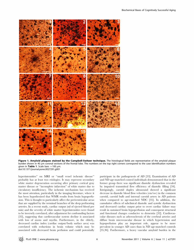
and 12.2 for ND-HPC and AD cases, respectively. The relative
plaque densities between these two cohorts are illustrated in
Figure 1. From a neuropathological point of view, the ND-HPC
and AD cases were purposefully selected for abundant amyloid
plaques. However, the load of vascular amyloid (total CAA score),
which was not used in the selection of cases, was on the average
almost 2-fold more abundant in AD than in the ND-HPC
(Table 1), suggesting that a higher degree of microvascular
dysfunction and perfusion compromise was present in the
demented cohort. The ApoE allelic distribution in the ND-HPC
and AD groups was: ApoE2 = 0.125, ApoE3 = 0.75 and
ApoE4 = 0.125, and ApoE2 = 0.08, ApoE3 = 0.58 and
ApoE4 = 0.33, respectively (Table 1).
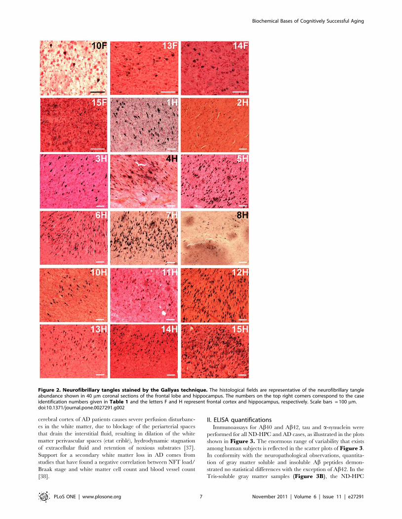
In reference to the NFT distribution, the AD group demon-
strated a more advanced Braak stage than the ND-HPC
(Table 1). Intriguingly, the ND-HPC showed no NFT in the
frontal lobe (data not shown), while in the AD group 4 out of 6
cases harbored NFT in this region (Figure 2, 10F, 13–15F).Appraisal of NFT in the hippocampus revealed moderate to
abundant levels in both the ND-HPC and AD groups, with the
exception of AD case # 11 and ND-HPC cases # 2 and # 8
(Figure 2, 1-8H, 10–15H) in which the NFT were scarce.
Disparities were likewise evident for total cerebral NFT scores,
which were about 25% higher in the AD subjects than in the ND-
HPC (Table 1). It is possible that in the oldest-old ND-HPC the
absence of NFT in the frontal cortex allows for a better
performance in terms of executive function, strategic planning
and cognitive tasks. In addition, a decreased tangle density
suggests fewer injured neurons, correspondingly less brain atrophy
and better brain function in the oldest-old ND-HPC [5,26,27].
Recent observations in the transgenic mice Tg4510 strain,
carrying the frontotemporal dementia tau P301L mutation,
suggest that NFT are a marker rather than the direct cause of
neuronal dysfunction and death. In this model, tangle deposition is
apparently preceded by caspase activation which has been
associated with acute apoptotic death [28]. Our own electron
microscopic observations on AD brain biopsies suggest NFT are
derived from collapsing mitochondria and other intraneuronal
pathological organelles, supporting the contention that NFT are
the result of damaged cytomembranes [29]. Chemical analysis of
the protease resistant core of paired helical filaments revealed
associated glycolipids [30,31] that could originate from membrane
walls.
A more pronounced difference was observed in the WMR
category where the mean values for the ND-HPC and AD were
2.3 and 5.5 (out of a maximum total of 12), respectively (Table 1).Cerebral WMR, also known in the imaging literature as
‘‘leukoaraiosis’’, ‘‘white matter lucencies’’ on CT, ‘‘white matter
Table 2. Primary and Secondary Antibodies Used in Western Blots.
Primary Antibody(WB) Antigen specificity or immunogen
Secondaryantibody Company/Catalog #
22C11 APP aa 66–81 M Millipore/MAB348
CT9APP Last 9 aa of APP R Millipore/AB5352
4G8 Ab aa 17–24 M Covance/SIG-39220
6E10 Ab aa 1–16 M Covance/SIG-39320
A11 Sequence independent oligomers R Invitrogen/AHB0052
BACE1 BACE1 aa 485–501 R Abcam/ab2077
IDE (BC2) Rat IDE aa 97–273 R Provided by Dr. E. Castano
Neprilysin Rat neprilysin R Millipore/AB5458
Notch-1 NICD N-terminal 14 aa R Millipore/AB5709
Tau (HT7) Tau aa 159–163 M Pierce/MN1000
PHF-tau AT8 pTau Ser202 M Pierce/MN1020
PHF-tau AT180 pTau Thr231 M Pierce/MN1040
a-synuclein Rat synuclein-1 aa 15–123 M BD Transduction Laboratories/610786
ApoE Recombinant ApoE G Millipore/AB947
ApoJ Recombinant ApoJ G Millipore/AB825
VEGF165 Recombinant human VEFG165 R Millipore/07-1419
PEDF Human PEDF R BioProducts MD/AB-PEDF1
BDNF/proBDNF Internal region of BDNF R Santa Cruz/sc-546
TDP-43 aa residues surrounding Ala260 of human TDP-43 R Cell Signaling Technology/3449
Synaptophysin Rat retina synaptophysin M Millipore/MAB368
S100B C-terminal synthetic peptide of human S100B R Abnova/PAB13687
Actin Ab-5 Clone C4 M BD Transduction Laboratories/A65020
Actin N-terminus of human a-actin R Abcam/Ab37063
APP, amyloid-b precursor protein; aa, amino acid; BACE, b-site APP cleaving enzyme; IDE, insulin degrading enzyme; NICD, Notch-1 intracellular domain; PHF, pairedhelical filament; ptau, phosphorylated tau; VEGF, vascular endothelial growth factor; PEDF, pigment epithelium derived factor; BDNF, brain derived neurotrophic factor;TDP-43, TAR DNA-binding protein 43; ApoE, apolipoprotein E; ApoJ, apolipoprotein J; M, HRP conjugated AffiniPure goat-anti mouse IgG (catalog # 111-035-144,Jackson Laboratory); R, HRP conjugated AffiniPure goat-anti rabbit IgG, (catalog # 111-035-146 Jackson Laboratory); G, HRP conjugated AffiniPure bovine-anti goat IgG(catalog #805-035-180).doi:10.1371/journal.pone.0027291.t002
Biochemical Bases of Cognitively Successful Aging
PLoS ONE | www.plosone.org 5 November 2011 | Volume 6 | Issue 11 | e27291
hyperintensities’’ on MRI or ‘‘small vessel ischemic disease’’
probably has at least two etiologies. It may represent secondary
white matter degeneration occurring after primary cortical gray
matter disease or ‘‘incomplete infarction’’ of white matter due to
circulatory insufficiency. The ischemic mechanism has received
the most attention, particularly in the imaging literature, where it
has been hypothesized that WMR results from brain hypoperfu-
sion. This is thought to particularly affect the periventricular areas
that are supplied by the terminal branches of the deep perforating
arteries. In a recent study, cardiac output (ml of ejected blood per
min) and the severity of white matter hyperintensities were found
to be inversely correlated, after adjustment for confounding factors
[32], suggesting that cardiovascular system decline is associated
with loss of axons and myelin. Furthermore, in the elderly,
decreased cardiac index (cardiac output/body surface area) was
correlated with reductions in brain volume which may be
associated with decreased brain perfusion and could potentially
participate in the pathogenesis of AD [33]. Examination of AD
and ND age-matched control individuals demonstrated that in the
former group there was significant diastolic dysfunction revealed
by impaired transmitral flow efficiency of diastolic filling [34].
Intriguingly, carotid duplex ultrasound showed a significant
decrease in diastolic blood flow velocities (cm/sec) in the common
carotid, carotid bulb and internal carotid artery in AD patients
when compared to age-matched NDC [35]. In addition, the
cumulative effects of subclinical diastolic and systolic dysfunction
and decreased cardiac output prior to overt cardiac failure may
result in sustained brain hypoperfusion and consequent structural
and functional changes conducive to dementia [32]. Cardiovas-
cular diseases such as atherosclerosis of the cerebral arteries and
diffuse brain microvascular disease in which hypertension and
hypoperfusion play an important role, appear to be more
prevalent in younger AD cases than in ND age-matched controls
[35,36]. Furthermore, a heavy vascular amyloid burden in the
Figure 1. Amyloid plaques stained by the Campbell-Switzer technique. The histological fields are representative of the amyloid plaqueburden shown in 40 mm coronal sections of the frontal lobe. The numbers on the top right corners correspond to the case identification numbersgiven in Table 1. Scale bars = 100 mm.doi:10.1371/journal.pone.0027291.g001
Biochemical Bases of Cognitively Successful Aging
PLoS ONE | www.plosone.org 6 November 2011 | Volume 6 | Issue 11 | e27291
cerebral cortex of AD patients causes severe perfusion disturbanc-
es in the white matter, due to blockage of the periarterial spaces
that drain the interstitial fluid, resulting in dilation of the white
matter perivascular spaces (etat crible), hydrodynamic stagnation
of extracellular fluid and retention of noxious substrates [37].
Support for a secondary white matter loss in AD comes from
studies that have found a negative correlation between NFT load/
Braak stage and white matter cell count and blood vessel count
[38].
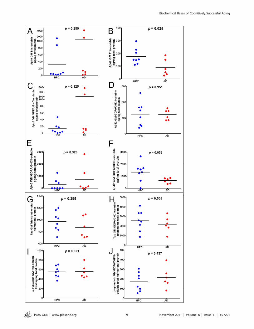
II. ELISA quantificationsImmunoassays for Ab40 and Ab42, tau and a-synuclein were
performed for all ND-HPC and AD cases, as illustrated in the plots
shown in Figure 3. The enormous range of variability that exists
among human subjects is reflected in the scatter plots of Figure 3.
In conformity with the neuropathological observations, quantita-
tion of gray matter soluble and insoluble Ab peptides demon-
strated no statistical differences with the exception of Ab42. In the
Tris-soluble gray matter samples (Figure 3B), the ND-HPC
Figure 2. Neurofibrillary tangles stained by the Gallyas technique. The histological fields are representative of the neurofibrillary tangleabundance shown in 40 mm coronal sections of the frontal lobe and hippocampus. The numbers on the top right corners correspond to the caseidentification numbers given in Table 1 and the letters F and H represent frontal cortex and hippocampus, respectively. Scale bars = 100 mm.doi:10.1371/journal.pone.0027291.g002
Biochemical Bases of Cognitively Successful Aging
PLoS ONE | www.plosone.org 7 November 2011 | Volume 6 | Issue 11 | e27291

demonstrated a greater mean value of Ab42 (ND-HPC = 177 pg/
mg total protein; AD = 89 pg/mg total protein; p = 0.025). The
white matter Ab42, solubilized with GDFA/GHCl, demonstrated
significant differences (p = 0.052) between the ND-HPC and the
AD groups (Figure 3F). Intriguingly, the Ab42 levels were
significantly higher in the ND-HPC gray matter and white matter
versus the AD cases (Figure 3B and 3F), even though they were
not demented. A comparison between ND-HPC and AD white
matter Ab40 was non-significant (p = 0.326; Figure 3E), in spite
of the fact that the 2 cases that had elevated Ab40 in the gray
matter (# 10 and # 14, Table 1) also contained exaggerated
quantities of this peptide in the white matter. Immunoassay
quantification of total tau and a-synuclein (Figure 3G, 3H, 3Iand 3J) did not show any significant differences between the two
groups. Setting aside the increased load of CAA, the presence of
similar or higher Ab in the ND-HPC neuropathological and
biochemical Ab brain parenchymal burdens suggest that the
abundance of amyloid plaques alone is not directly responsible for
the emergence of AD dementia.
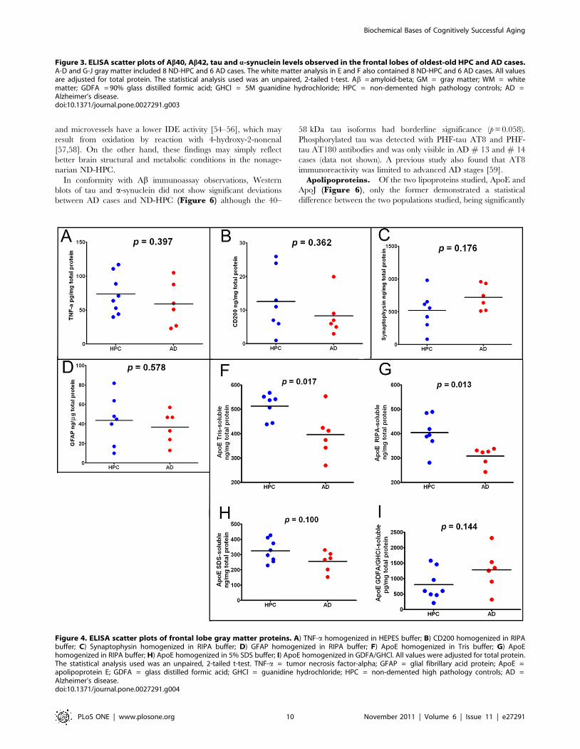
The amounts of gray matter inflammatory TNF-a cytokine
were not significantly different between the ND-HPC and AD
cases (Figure 4A). In addition, as shown in Figure 4B, the
immunosuppressive protein CD200 was decreased in the AD
compared to the ND-HPC, although the levels did not reach
statistical significance (p = 0.362). CD200 is a highly glycosylated
cell surface protein whose only known function is as a ligand for
CD200 receptor. This difference may in part be responsible for
better neuroprotection. CD200 is expressed primarily in neurons
and oligodendrocytes [39], but has also been identified in astroglia
and endothelial cells [40]. In the human brain, CD200 as well as
its microglia receptor are decreased in those regions with
abundant AD pathology. The synaptic vesicle marker synapto-
physin was not significantly different between AD and ND-HPC
when determined by ELISA (Figure 4C, p = 0.176) or Western
blots (see below for discussion). Likewise, GFAP did not show any
significant differences between the two cohorts under investigation
(Figure 4D). This intermediate filament protein is the principal
structural molecule of astrocytes, the most abundant cell type in
the CNS and main homeostatic modulator of neuronal function,
where it regulates motility and shape and is substantially expressed
as a response to trauma, chemical injury and neuroinflammation
[41,42]. GFAP has been found to be increased as the result of
astrogliosis in dementia [43]. Astrogliosis is probably present in
both oldest-old groups examined in the current study due to the
profuse amyloid and NFT insults. GFAP has the distinction of
being one of the most abundant proteins in the brain and can
accumulate an enormous quantity of post-translational modifica-
tions such as phosphorylation and N-and O-glycosylations
resulting in a complex array of isoforms. This intricacy, in the
realm of AD and ND conditions, has been elegantly explored
through proteomic analysis by Korolainen et al. [44].
Quantitative ELISA and Western blot (see below) analyses
demonstrated reduced ApoE levels in AD compared to the ND-
HPC. We analyzed Tris, RIPA, 5% SDS and GDFA/GHCl
soluble ApoE by ELISA (Figure 4F, 4G, 4H, 4I, respectively).
In agreement with Western blot data, ApoE was significantly
reduced in Tris- and RIPA-buffer soluble fractions by ,20% in
AD compared to ND-HPC (Figure 4F and 4G). The 5% SDS-
soluble ApoE was reduced as well, but did not reach significance.
Interestingly, ApoE tended to be higher in the GDFA/GHCl-
soluble fractions in AD samples compared to ND-HPC, but also
did not reach significance (see Western blot section for additional
discussion of ApoE).
III. Western Blots appraisalsA battery of antibodies (Table 2) was utilized to assess proteins
that have been found to be altered in AD. For a final quality
control of total protein loading, actin was used as an internal
standard as shown at the bottom of each of the Western blots
(Figures 5, 6, and 7). Interestingly, the total protein values
present in ,100 mg of wet weight per ml in each of the individual
specimens utilized was 14% lower in the AD group. Although this
difference was not statistically significant (p = 0.09), it nevertheless
suggests a trend in which the ND-HPC have slightly more protein
per unit of brain weight than the AD specimens. This finding may
also be an indication of the general loss of protein and
concomitant increase in water content in the gray matter of AD
subjects resulting from a chronic and emaciating neurodegener-
ative disorder.
APP/Ab, protease-related proteins, tau and a-
synuclein. Figure 5 illustrates the results obtained by
probing with antibodies related to APP/Ab processing and
metabolism which included: 22C11, CT9APP, 4G8, 6E10, A11,
b-site APP cleaving enzyme (BACE-1), insulin degrading enzyme
(IDE), neprilysin and Notch-1. Significant differences between the
ND-HPC and AD groups were only observed with the CT9APP
antibody for the full length APP 110 kDa protein and 35 kDa
peptides (p = 0.010 and 0.053, respectively). Likewise, the 6E10
antibody detected differences between the two cohorts in the full
length APP 110 kDa protein (p = 0.001) while the 4G8 antibody
showed no significant deviations. The results suggest that relative
to the AD values, the ND-HPC APP holoprotein was elevated, as
detected by the CT9APP and 6E10 antibodies, suggesting greater
abundance of important APP-derived peptides such as sAPPa,
sAPPb, APP-carboxy-terminal fragment and the APP intracellular
domain which have multiple neurotrophic roles, intracellular
adaptor protein interactions and transcriptional functions [45–47].
However, differences in APP were not demonstrated with the 4G8
and 22C11 which may be explained by different antibody affinities
or the fact that the 22C11 also detects APLP2 [48]. Another Abrelated molecule which also showed differences with specific
antibodies between the two groups was the Ab*56 oligomer
detected by the A11 antibody that targets a variety of amyloids
rich in b-sheets (p = 0.005). To explain the pathogenesis of AD,
emphasis has been given to the presence of the Ab*56 [49], an
apparently soluble dodecameric Ab aggregate with a ,56 kDa Mr
that can be detected by the A11 antibody. Although this oligomer
may bolster the contention that aggregated Ab acts as a specific
neurotoxic molecule in AD, this putative soluble oligomeric Abhas not been rigorously characterized in the human brain. In
addition, the A11 antibody is not specific for Ab since it also
detects other oligomeric b-sheet conformations in a diverse
number of amyloid proteins [50].
Additionally, the amounts of the b-secretase (BACE-1), a
molecule extensively post-translationally modified in its mature
form [51], are increased in the ND-HPC (70 kDa peptide;
p = 0.052). Others have reported BACE-1 to be increased in AD
brains (reviewed in [52]). Of the Ab related proteolytic enzymes
only the low molecular mass form of IDE 110 kDa was
significantly lower (p = 0.015) in ND-HPC, while neprilysin and
the c-secretase target multifunctional molecule Notch-1 demon-
strated no differences between the 2 cohorts of nonagenarians.
The significant rise in IDE in AD versus ND-HPC could result in
an elevated degradation of insulin thereby making glucose less
available for energy metabolism. Alternatively, in AD, the relative
enhancement of IDE may be a reflection of an inactivated enzyme
by monomeric/dimeric soluble Ab peptides [53]. Interestingly, in
comparison with age-matched controls, AD brain homogenates
Biochemical Bases of Cognitively Successful Aging
PLoS ONE | www.plosone.org 8 November 2011 | Volume 6 | Issue 11 | e27291
Biochemical Bases of Cognitively Successful Aging
PLoS ONE | www.plosone.org 9 November 2011 | Volume 6 | Issue 11 | e27291
and microvessels have a lower IDE activity [54–56], which may
result from oxidation by reaction with 4-hydroxy-2-nonenal
[57,58]. On the other hand, these findings may simply reflect
better brain structural and metabolic conditions in the nonage-
narian ND-HPC.
In conformity with Ab immunoassay observations, Western
blots of tau and a-synuclein did not show significant deviations
between AD cases and ND-HPC (Figure 6) although the 40–
58 kDa tau isoforms had borderline significance (p = 0.058).
Phosphorylated tau was detected with PHF-tau AT8 and PHF-
tau AT180 antibodies and was only visible in AD # 13 and # 14
cases (data not shown). A previous study also found that AT8
immunoreactivity was limited to advanced AD stages [59].
Apolipoproteins. Of the two lipoproteins studied, ApoE and
ApoJ (Figure 6), only the former demonstrated a statistical
difference between the two populations studied, being significantly
Figure 4. ELISA scatter plots of frontal lobe gray matter proteins. A) TNF-a homogenized in HEPES buffer; B) CD200 homogenized in RIPAbuffer; C) Synaptophysin homogenized in RIPA buffer; D) GFAP homogenized in RIPA buffer; F) ApoE homogenized in Tris buffer; G) ApoEhomogenized in RIPA buffer; H) ApoE homogenized in 5% SDS buffer; I) ApoE homogenized in GDFA/GHCl. All values were adjusted for total protein.The statistical analysis used was an unpaired, 2-tailed t-test. TNF-a = tumor necrosis factor-alpha; GFAP = glial fibrillary acid protein; ApoE =apolipoprotein E; GDFA = glass distilled formic acid; GHCl = guanidine hydrochloride; HPC = non-demented high pathology controls; AD =Alzheimer’s disease.doi:10.1371/journal.pone.0027291.g004
Figure 3. ELISA scatter plots of Ab40, Ab42, tau and a-synuclein levels observed in the frontal lobes of oldest-old HPC and AD cases.A-D and G-J gray matter included 8 ND-HPC and 6 AD cases. The white matter analysis in E and F also contained 8 ND-HPC and 6 AD cases. All valuesare adjusted for total protein. The statistical analysis used was an unpaired, 2-tailed t-test. Ab = amyloid-beta; GM = gray matter; WM = whitematter; GDFA = 90% glass distilled formic acid; GHCl = 5M guanidine hydrochloride; HPC = non-demented high pathology controls; AD =Alzheimer’s disease.doi:10.1371/journal.pone.0027291.g003
Biochemical Bases of Cognitively Successful Aging
PLoS ONE | www.plosone.org 10 November 2011 | Volume 6 | Issue 11 | e27291

Biochemical Bases of Cognitively Successful Aging
PLoS ONE | www.plosone.org 11 November 2011 | Volume 6 | Issue 11 | e27291
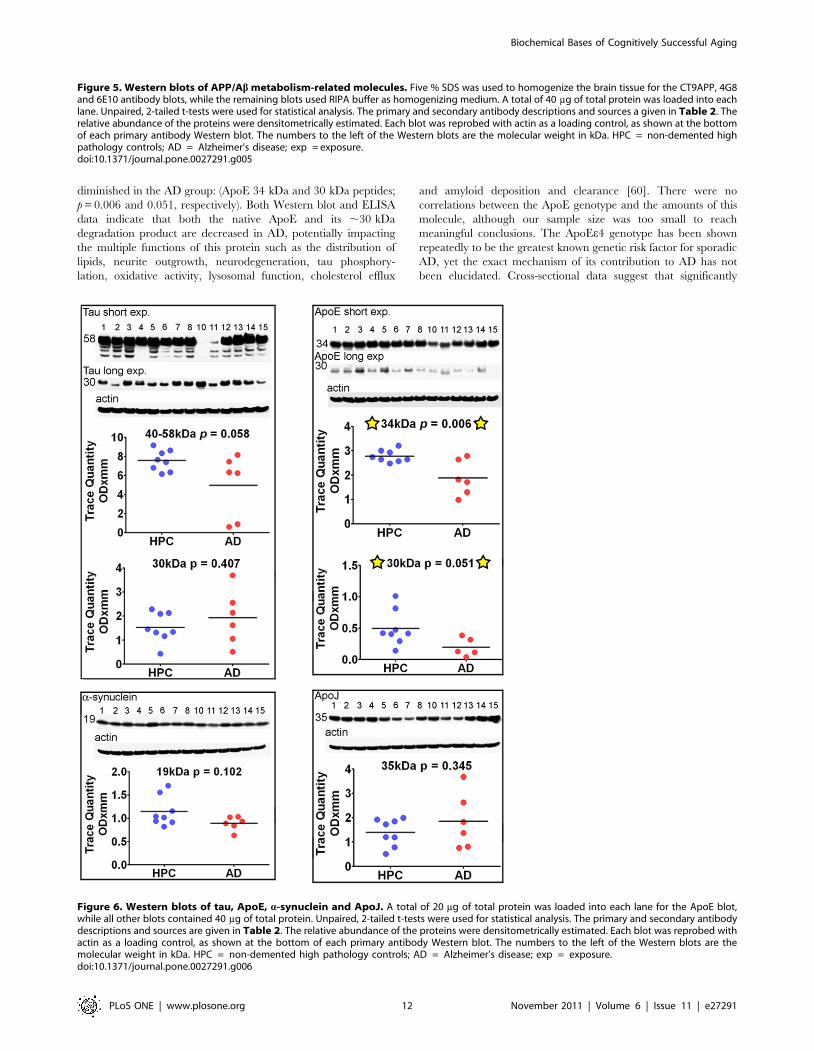
diminished in the AD group: (ApoE 34 kDa and 30 kDa peptides;
p = 0.006 and 0.051, respectively). Both Western blot and ELISA
data indicate that both the native ApoE and its ,30 kDa
degradation product are decreased in AD, potentially impacting
the multiple functions of this protein such as the distribution of
lipids, neurite outgrowth, neurodegeneration, tau phosphory-
lation, oxidative activity, lysosomal function, cholesterol efflux
and amyloid deposition and clearance [60]. There were no
correlations between the ApoE genotype and the amounts of this
molecule, although our sample size was too small to reach
meaningful conclusions. The ApoEe4 genotype has been shown
repeatedly to be the greatest known genetic risk factor for sporadic
AD, yet the exact mechanism of its contribution to AD has not
been elucidated. Cross-sectional data suggest that significantly
Figure 6. Western blots of tau, ApoE, a-synuclein and ApoJ. A total of 20 mg of total protein was loaded into each lane for the ApoE blot,while all other blots contained 40 mg of total protein. Unpaired, 2-tailed t-tests were used for statistical analysis. The primary and secondary antibodydescriptions and sources are given in Table 2. The relative abundance of the proteins were densitometrically estimated. Each blot was reprobed withactin as a loading control, as shown at the bottom of each primary antibody Western blot. The numbers to the left of the Western blots are themolecular weight in kDa. HPC = non-demented high pathology controls; AD = Alzheimer’s disease; exp = exposure.doi:10.1371/journal.pone.0027291.g006
Figure 5. Western blots of APP/Ab metabolism-related molecules. Five % SDS was used to homogenize the brain tissue for the CT9APP, 4G8and 6E10 antibody blots, while the remaining blots used RIPA buffer as homogenizing medium. A total of 40 mg of total protein was loaded into eachlane. Unpaired, 2-tailed t-tests were used for statistical analysis. The primary and secondary antibody descriptions and sources a given in Table 2. Therelative abundance of the proteins were densitometrically estimated. Each blot was reprobed with actin as a loading control, as shown at the bottomof each primary antibody Western blot. The numbers to the left of the Western blots are the molecular weight in kDa. HPC = non-demented highpathology controls; AD = Alzheimer’s disease; exp = exposure.doi:10.1371/journal.pone.0027291.g005
Biochemical Bases of Cognitively Successful Aging
PLoS ONE | www.plosone.org 12 November 2011 | Volume 6 | Issue 11 | e27291

decreased plasma ApoEe4 levels correlate with AD pathology
levels assessed by PiB-PET [61]. ApoE levels have been shown to
be reduced in ApoE mouse models of amyloidosis as well as in AD
human subjects [25]. It has also been suggested that ApoEe4 is
degraded at a higher rate than other variants [62]. We
demonstrate small, but significant decreases in Tris, RIPA- and
5% SDS-soluble ApoE in AD cases, in both Western blot and
immunoassay, when compared to ND-HPC. Interestingly, we
found a trend towards higher ApoE in the GDFA/GHCl-soluble
fractions by ELISA in AD samples compared to ND-HPC
(Figure 4I). Statistical significance was not reached due to the
amount of variability in these fractions. Since ApoE co-localizes
with Ab in CAA [63], it is possible that decreased soluble ApoE in
AD fractions is the result of its selective sequestration by vascular
amyloid deposits. Similarly, the increased levels of insoluble ApoE
in AD versus ND-HPC may result from the 2-fold more abundant
vascular amyloid observed in our AD oldest-old cases. Moreover,
recent investigations suggest that the ApoEe4 isoform is less able to
clear Ab from the brain, thus contributing to dementia [64].
Neurotrophic and vascular-related factors. Biochemical
alterations in the AD brain’s circulatory system promote changes
in vascular structure, blood-brain barrier disturbances and
ultimate microvessel demise [65–67]. These alterations
prompted our investigation of vascular endothelial growth factor
(VEGF) and pigment epithelium-derived factor (PEDF). As
depicted in Figure 7, the Western blots/scanning densitometry
results obtained by specific antibodies to VEGF and PEDF
demonstrated significant differences between the ND-HPC and
AD groups for the 32 kDa peptide (p = 0.017) and 50 kDa peptide
(p = 0.003), respectively. The specific immunohistochemical
reactivity of VEGF has been found to be elevated in AD where
it has been localized to astrocytes, microvessels and amyloid
Figure 7. Western blots of VEGF, PEDF, BDNF, TDP-43, synaptophysin and S100B. Forty mg of total protein was loaded onto VEFG, BDNFand TDP-43 acrylamide gels. The PEDF and synpaptophysin blots contained 20 mg and 10 mg of total protein, respectively. Unpaired, 2-tailed t-testswere used for statistical analysis. The primary and secondary antibody descriptions and sources are given in Table 2. The relative abundance of theproteins were densitometrically estimated. Each blot was reprobed with actin as a loading control, as shown at the bottom of each primary antibodyWestern blot. The numbers to the left of the Western blots are the molecular weight in kDa. HPC = non-demented high pathology controls; AD =Alzheimer’s disease; exp = exposure.doi:10.1371/journal.pone.0027291.g007
Biochemical Bases of Cognitively Successful Aging
PLoS ONE | www.plosone.org 13 November 2011 | Volume 6 | Issue 11 | e27291

plaques [68–70]. In the present investigation the pro-angiogenic
VEGF was significantly decreased in AD cases relative to ND-
HPC. The relative increase of this factor in the ND-HPC may
reflect its neuroprotective effects when confronted with brain
hypoperfusion, glucose deprivation [71–73] and the anti-
angiogenic activity of Ab [74]. The decreased levels of VEGF
are in contrast with the concomitant increase of the anti-
angiogenic PEDF in our AD specimens, a factor that naturally
decreases in normal aging [75,76]. In the AD brain, PEDF has a
strong immunoreactivity in cortical neurons and astrocytes. This
elevation may be explained as a defense response in AD, since
PEDF has potent anti-inflammatory, anti-oxidant, anti-
thrombotic, and neuroprotective properties [77–81]. However,
an increase in PEDF may also have a negative function by
preventing blood vessel formation and inducing apoptosis in
proliferating endothelial cells [82–85], eventually leading to
ischemia and neurodegeneration. VEGF and PEDF apparently
have paradoxical functions on the microcirculation, since capillary
permeability is increased by VEGF and inhibited by PEDF [86].
The imbalance between VEGF and PEDF has also been observed
in other human diseases [87–91]. Interestingly, in two previous
proteomic studies of CSF biomarkers, performed in our
laboratory, in which neuropathologically confirmed AD and ND
cases were examined by 2-D electrophoresis proteomic
methodologies, PEDF was significantly increased in the CSF of
an AD pool [92,93].
Brain-derived neurotrophic factor (BDNF) is a powerful growth
factor that stimulates neuronal function, prevents cell death in
adulthood and is thought to be deficient in AD. Administration of
BDNF into mouse models of AD, aged rats and lesion-induced
primate models apparently restored learning and memory and
prevented or delayed neuronal death (reviewed in [94]), suggesting
BDNF as a good candidate for neurodegenerative disease clinical
trials. However, investigations of BDNF levels in patients with AD
and deletion studies of BDNF in mice have been contradictory
(reviewed in [95]). In our Western blots, the 32 kDa N-
glycosylated and glucosulfated forms of the pro-BDNF yielded
no significant differences between AD cases and ND-HPC. This
molecule is normally cleaved to yield the mature 14 kDa protein
that was faintly visible in our oldest-old population. Of the 4 bands
detected by the BDNF antibody only the 28 kDa yielded
significant differences between AD and ND-HPC being increased
in the former group (Figure 7; p = 0.009). This molecule may
represent a truncated form of pro-BDNF that is aberrantly
processed [96].
Multifunctional molecules. TDP-43, an important molecule
with multifunctional RNA binding functions, apparently plays an
important role in several neurodegenerative disorders, including
AD, by generating intracellular inclusions [97,98]. It is normally
found in the nucleus, but under pathological conditions, moves to
the cytoplasm where it is ubiquitinated, phosphorylated and
cleaved to generate C-terminal fragments (reviewed in [99]).
Interestingly, this molecule is elevated in traumatic brain injury
[100]. No statistical differences between the two groups were
detected for the transcriptional factor TDP-43 although half of the
AD cases did not have detectable TDP-43, while this was true in
only two of the 8 ND-HPC (Figure 7). All groups had TDP-43
fragments [101,102], including the C-terminal 35 kDa which was
marginally elevated in AD (Figure 7, p = 0.073).
Similarly the presynaptic vesicular marker synaptophysin did
not show statistical differences by ELISA (Figure 4C) which was
confirmed by Western blot (Figure 7). This was an unexpected
observation since by immunocytochemistry there is a remarkable
loss of synapses (,45%) in AD cases when compared to ND
controls [103], although our ND-HPC are uniquely different from
normal ND controls in that it has an amyloid plaque burden
similar to AD. Our own Western blot experiments confirmed a
significant reduction of synaptophysin in a younger population of
AD subjects (n = 31) when compared to ND age-matched controls
(n = 22) (p = 0.018; A.E. Roher, unpublished observations). We did
not quantify the number of synapses in our specimens, but the
observation of a lack of significant difference between synapto-
physin levels of ND-HPC and AD groups presents a logical
conundrum. However, synaptophysin is a marker for synaptic
vesicles that is extrapolated as a proxy to reveal the conditions of
the synapses. This assumes physiological equivalence between the
two groups, an assumption that may be confounded given the
capacity for compensation under slowly emerging stressful
conditions. Interestingly, synaptophysin is not essential for
neurotransmitter release [104], although mice lacking synapto-
physin develop behavioral and learning dysfunctions [105].
S100B is a 92 amino acid long calcium binding protein that is
functionally expressed as a 21 kDa homodimer and is produced by
astrocytes around blood vessels. This molecule was significantly
increased in ND-HPC relative to AD (p = 0.05) (Figure 7). S100B
is an important cellular mediator of protein phosphorylation,
protein degradation, cell locomotion, regulation of transcription
factors, cell proliferation and differentiation, cytoskeleton assem-
bly, regulation of enzyme activities and receptor function [106–
108]. S100B has also been linked to survival of neurons and when
generated at micromolar concentrations, enhance the production
of inflammatory cytokines [107]. In addition, S100B is considered
a marker of brain damage and neurodegeneration since it is
elevated in global hypoxia, ischemia and hemorrhagic stroke
[106,107,109]. It has also been associated with the density of
amyloid plaques [110,111] and it is increased in AD CSF
[112,113]. Our observation of a modest elevation of S100B in
ND-HPC could be interpreted as a positive effect since this
molecule has, as mentioned above, a large number of beneficial
functions that may have a role in the prevention of dementia
[114]. Furthermore, S100B is a potent neuroprotective factor for
cholinergic neurons during oxygen/glucose deprivation [115].
Summary and conclusionsOur goal was to evaluate the biochemical differences that
distinguish the oldest-old AD population from ND-HPC. Within
the framework of the multifaceted pathogenesis of AD our data
suggest a compromised brain perfusion as one of the underlying
causes of dementia. The brain samples showed that the severity of
WMR in the AD group was over 2 times higher than the
corresponding value in the ND-HPC cases. A general decline in
cerebral blood flow observed in AD [116,117] is apparently
associated with loss of white matter axons and demyelination
[38,118]. When compared to a ND population, AD patients
revealed a significant decrease in diastolic flow velocities (cm/sec)
in the common and internal carotid arteries [35] and a decreased
total and regional cerebral blood flow volumes [119–123]. The
AD brain also exhibits increased vascular resistance [35] a
manifestation of diffuse microvascular disease. A dysfunctional
microcirculation in AD is supported by our observations related to
decreased pro-angiogenic VEGF and increased anti-angiogenic
PEDF, relative to the ND-HPC, which would restrict the ability of
de novo vessel formation that could alleviate brain hypoperfusion. In
addition, our oldest-old AD cases also had a significant decrease of
S100B, an important multifunctional regulatory molecule [106–
108]. In line with a global circulatory compromise in AD, there is
an increased dilation of the white matter perivascular spaces (etat
crible) suggesting interstitial fluid stagnation and compromised
Biochemical Bases of Cognitively Successful Aging
PLoS ONE | www.plosone.org 14 November 2011 | Volume 6 | Issue 11 | e27291

cerebral venous outflow [37]. These data are supported by an
increase in the load of vascular amyloid (total CAA score) that was
on the average 2 times more abundant in AD cases than in the
ND-HPC, further endorsing the contention of a compromised
cerebral microvasculature, disturbed BBB and dysfunctional brain
perfusion. Other studies of the oldest-old populations have also
found unusual blood vessel architecture and function in AD cases
compared to ND-HPC (reviewed in [5]).
Four out of the six AD cases studied showed NFT in the frontal
cortex while none of the ND-HPC exhibited these lesions,
reminiscent of a better preservation of the associative, executive
and short-term/working memory functions in the latter group.
Overall total NFT scores were about 25% higher in the AD
subjects than in the ND-HPC indicative of a conserved neuronal
morphology and function in the latter group. The lack of
quantitative significance in the total amount of tau between the
2 groups under study emanates from the wide range of variability
that characterizes tau and NFT pathology in the elderly
population. Likewise, only Ab42 revealed significant differences,
but intriguingly was elevated in the ND-HPC. These data support
the contention that the Ab burden per se, whether soluble or
insoluble, is not the decisive factor in determining the dementia
status in the oldest-old subjects.
These observations are very revealing and instructive about
parameters, other than or in addition to Ab and amyloid plaque
deposition, that may contribute to the conserved cognitive
integrity of the ND-HPC. The lack of any clear pathological
and biochemical demarcation between demented and ND groups
suggests that the near exclusive focus on amyloid plaques and their
components, long presumed to play dominant roles in cognitive
failure, may be misguided. Along the process of aging, multi-
system decay and failure to adapt and repair play a decisive role in
the development of dementia. Complex environmental and
molecular pleiotropic interactions are likely to govern parameters
such as time of onset of disease and severity among affected
individuals. These issues are well illustrated by the disease
modifying effects of the ApoEe4 genotype on numerous essential
functions. Characterization of pathologically deviant molecules as
well as of those that promote healthy mental aging will enormously
help in the identification of new targets for therapeutic
interventions that will prevent, delay the onset or mitigate the
clinical progression of this devastating dementia.
Acknowledgments
We express our gratitude to Dr. Walter M. Kalback and Dr. Dean C.
Luehrs for critical review of the manuscript.
Author Contributions
Conceived and designed the experiments: CLM TAK DGW JMH TGB
AER. Performed the experiments: CLM IDD DGW JMH JCK. Analyzed
the data: CLM TAK DGW JMH TGB AER. Contributed reagents/
materials/analysis tools: TGB AER. Wrote the paper: CLM TAK DGW
JMH RW JK EMC MNS TGB AER.
References
1. Castellani RJ, Rolston RK, Smith MA (2010) Alzheimer disease. Dis Mon 56:
484–546.
2. Corrada MM, Brookmeyer R, Paganini-Hill A, Berlau D, Kawas CH (2010)
Dementia incidence continues to increase with age in the oldest old: the 90+study. Ann Neurol 67: 114–121.
3. Kobayashi M, Sato T, Sato A, Imamura T (2009) [Oldest-old dementia in a
Japanese memory clinic]. Brain Nerve 61: 972–978.
4. Kawas CH, Corrada MM (2006) Alzheimer’s and dementia in the oldest-old: a
century of challenges. Curr Alzheimer Res 3: 411–419.
5. von Gunten A, Ebbing K, Imhof A, Giannakopoulos P, Kovari E (2010) Brain
aging in the oldest-old. Curr Gerontol Geriatr Res 2010: 358531.
6. Kinsella K, He W (2009) An Aging World: 2008 International Population
Reports, U.S. Government Printing Office. Available: http://www.census.
gov/prod/2009pubs/p95-09-1.pdf.
7. Selkoe DJ (1989) Amyloid beta protein precursor and the pathogenesis of
Alzheimer’s disease. Cell 58: 611–612.
8. Korczyn AD (2008) The amyloid cascade hypothesis. Alzheimers Dement 4:
176–178.
9. Plassman BL, Khachaturian AS, Townsend JJ, Ball MJ, Steffens DC, et al.
(2006) Comparison of clinical and neuropathologic diagnoses of Alzheimer’s
disease in 3 epidemiologic samples. Alzheimers Dement 2: 2–11.
10. Jellinger KA (1995) Alzheimer’s changes in non-demented and demented
patients. Acta Neuropathol 89: 112–113.
11. Bertram L, Lill CM, Tanzi RE (2010) The genetics of Alzheimer disease: back
to the future. Neuron 68: 270–281.
12. Terry RD, Masliah E, Salmon DP, Butters N, DeTeresa R, et al. (1991)
Physical basis of cognitive alterations in Alzheimer’s disease: synapse loss is the
major correlate of cognitive impairment. Ann Neurol 30: 572–580.
13. Fagan AM, Shaw LM, Xiong C, Vanderstichele H, Mintun MA, et al. (2011)
Comparison of Analytical Platforms for Cerebrospinal Fluid Measures of
{beta}-Amyloid 1-42, Total tau, and P-tau181 for Identifying Alzheimer
Disease Amyloid Plaque Pathology. Arch Neurol.
14. Hampel H, Shen Y, Walsh DM, Aisen P, Shaw LM, et al. (2010) Biological
markers of amyloid beta-related mechanisms in Alzheimer’s disease. Exp
Neurol 223: 334–346.
15. Trojanowski JQ, Vandeerstichele H, Korecka M, Clark CM, Aisen PS, et al.
(2010) Update on the biomarker core of the Alzheimer’s Disease Neuroimaging
Initiative subjects. Alzheimers Dement 6: 230–238.
16. Quigley H, Colloby SJ, O’Brien JT (2010) PET imaging of brain amyloid in
dementia: a review. Int J Geriatr Psychiatry 26(10): 991–999.
17. Jack CR, Jr., Lowe VJ, Weigand SD, Wiste HJ, Senjem ML, et al. (2009) Serial
PIB and MRI in normal, mild cognitive impairment and Alzheimer’s disease:
implications for sequence of pathological events in Alzheimer’s disease. Brain
132: 1355–1365.
18. Beach TG, Sue LI, Walker DG, Roher AE, Lue L, et al. (2008) The Sun
Health Research Institute Brain Donation Program: description and experi-ence, 1987-2007. Cell Tissue Bank 9: 229–245.
19. Hyman BT, Trojanowski JQ (1997) Consensus recommendations for thepostmortem diagnosis of Alzheimer disease from the National Institute on
Aging and the Reagan Institute Working Group on diagnostic criteria for the
neuropathological assessment of Alzheimer disease. J Neuropathol Exp Neurol56: 1095–1097.
20. Mirra SS (1997) The CERAD neuropathology protocol and consensusrecommendations for the postmortem diagnosis of Alzheimer’s disease: a
commentary. Neurobiol Aging 18: S91–S94.
21. Mirra SS, Heyman A, McKeel D, Sumi SM, Crain BJ, et al. (1991) TheConsortium to Establish a Registry for Alzheimer’s Disease (CERAD). Part II.
Standardization of the neuropathologic assessment of Alzheimer’s disease.Neurology 41: 479–486.
22. Braak H, Braak E (1991) Neuropathological stageing of Alzheimer-related
changes. Acta Neuropathol 82: 239–259.
23. Hixson JE, Vernier DT (1990) Restriction isotyping of human apolipoprotein E
by gene amplification and cleavage with HhaI. J Lipid Res 31: 545–548.
24. Mulugeta E, Molina-Holgado F, Elliott MS, Hortobagyi T, Perry R, et al.
(2008) Inflammatory mediators in the frontal lobe of patients with mixed and
vascular dementia. Dement Geriatr Cogn Disord 25: 278–286.
25. Sullivan PM, Han B, Liu F, Mace BE, Ervin JF, et al. (2011) Reduced levels of
human apoE4 protein in an animal model of cognitive impairment. NeurobiolAging 32: 791–801.
26. Silbert LC, Quinn JF, Moore MM, Corbridge E, Ball MJ, et al. (2003) Changes
in premorbid brain volume predict Alzheimer’s disease pathology. Neurology61: 487–492.
27. Josephs KA, Whitwell JL, Ahmed Z, Shiung MM, Weigand SD, et al. (2008)Beta-amyloid burden is not associated with rates of brain atrophy. Ann Neurol
63: 204–212.
28. de Calignon A, Fox LM, Pitstick R, Carlson GA, Bacskai BJ, et al. (2010)Caspase activation precedes and leads to tangles. Nature 464: 1201–1204.
29. Gray EG, Paula-Barbosa M, Roher A (1987) Alzheimer’s disease: paired helicalfilaments and cytomembranes. Neuropathol Appl Neurobiol 13: 91–110.
30. Goux WJ, Rodriguez S, Sparkman DR (1995) Analysis of the core components
of Alzheimer paired helical filaments. A gas chromatography/mass spectrom-etry characterization of fatty acids, carbohydrates and long-chain bases. FEBS
Lett 366: 81–85.
31. Goux WJ, Rodriguez S, Sparkman DR (1996) Characterization of the
glycolipid associated with Alzheimer paired helical filaments. J Neurochem 67:
723–733.
32. Jefferson AL, Tate DF, Poppas A, Brickman AM, Paul RH, et al. (2007) Lower
cardiac output is associated with greater white matter hyperintensities in olderadults with cardiovascular disease. J Am Geriatr Soc 55: 1044–1048.
Biochemical Bases of Cognitively Successful Aging
PLoS ONE | www.plosone.org 15 November 2011 | Volume 6 | Issue 11 | e27291
33. Jefferson AL, Himali JJ, Beiser AS, Au R, Massaro JM, et al. (2010) Cardiac
index is associated with brain aging: the Framingham Heart Study. Circulation
122: 690–697.
34. Belohlavek M, Jiamsripong P, Calleja AM, McMahon EM, Maarouf CL, et al.
(2009) Patients with Alzheimer disease have altered transmitral flow:
echocardiographic analysis of the vortex formation time. J Ultrasound Med
28: 1493–1500.
35. Roher AE, Garami Z, Tyas SL, Maarouf CL, Kokjohn TA, et al. (2011)
Transcranial Doppler ultrasound blood flow velocity and pulsatility index as
systemic indicators for Alzheimer’s disease. Alzheimers Dement 7: 445–455.
36. Roher AE, Tyas SL, Maarouf CL, Daugs ID, Kokjohn TA, et al. (2011)
Intracranial atherosclerosis as a contributing factor to Alzheimer’s disease
dementia. Alzheimers Dement 7: 436–444.
37. Roher AE, Kuo YM, Esh C, Knebel C, Weiss N, et al. (2003) Cortical and
leptomeningeal cerebrovascular amyloid and white matter pathology in
Alzheimer’s disease. Mol Med 9: 112–122.
38. Kalback W, Esh C, Castano EM, Rahman A, Kokjohn T, et al. (2004)
Atherosclerosis, vascular amyloidosis and brain hypoperfusion in the
pathogenesis of sporadic Alzheimer’s disease. Neurol Res 26: 525–539.
39. Walker DG, Dalsing-Hernandez JE, Campbell NA, Lue LF (2009) Decreased
expression of CD200 and CD200 receptor in Alzheimer’s disease: a potential
mechanism leading to chronic inflammation. Exp Neurol 215: 5–19.
40. Lue LF, Kuo YM, Beach T, Walker DG (2010) Microglia activation and anti-
inflammatory regulation in Alzheimer’s disease. Mol Neurobiol 41: 115–128.
41. Eng LF, Ghirnikar RS, Lee YL (2000) Glial fibrillary acidic protein: GFAP-
thirty-one years (1969-2000). Neurochem Res 25: 1439–1451.
42. Norenberg MD (1994) Astrocyte responses to CNS injury. J Neuropathol Exp
Neurol 53: 213–220.
43. Kashon ML, Ross GW, O’Callaghan JP, Miller DB, Petrovitch H, et al. (2004)
Associations of cortical astrogliosis with cognitive performance and dementia
status. J Alzheimers Dis 6: 595–604.
44. Korolainen MA, Auriola S, Nyman TA, Alafuzoff I, Pirttila T (2005) Proteomic
analysis of glial fibrillary acidic protein in Alzheimer’s disease and aging brain.
Neurobiol Dis 20: 858–870.
45. Mattson MP (1997) Cellular actions of beta-amyloid precursor protein and its
soluble and fibrillogenic derivatives. Physiol Rev 77: 1081–1132.
46. Schettini G, Govoni S, Racchi M, Rodriguez G (2010) Phosphorylation of
APP-CTF-AICD domains and interaction with adaptor proteins: signal
transduction and/or transcriptional role–relevance for Alzheimer pathology.
J Neurochem 115: 1299–1308.
47. Turner PR, O’Connor K, Tate WP, Abraham WC (2003) Roles of amyloid
precursor protein and its fragments in regulating neural activity, plasticity and
memory. Prog Neurobiol 70: 1–32.
48. Slunt HH, Thinakaran G, Von Koch C, Lo AC, Tanzi RE, et al. (1994)
Expression of a ubiquitous, cross-reactive homologue of the mouse beta-
amyloid precursor protein (APP). J Biol Chem 269: 2637–2644.
49. Lesne S, Koh MT, Kotilinek L, Kayed R, Glabe CG, et al. (2006) A specific
amyloid-beta protein assembly in the brain impairs memory. Nature 440:
352–357.
50. Kayed R, Head E, Thompson JL, McIntire TM, Milton SC, et al. (2003)
Common structure of soluble amyloid oligomers implies common mechanism
of pathogenesis. Science 300: 486–489.
51. Haniu M, Denis P, Young Y, Mendiaz EA, Fuller J, et al. (2000)
Characterization of Alzheimer’s beta -secretase protein BACE. A pepsin
family member with unusual properties. J Biol Chem 275: 21099–21106.
52. Vassar R, Kovacs DM, Yan R, Wong PC (2009) The beta-secretase enzyme
BACE in health and Alzheimer’s disease: regulation, cell biology, function, and
therapeutic potential. J Neurosci 29: 12787–12794.
53. Llovera RE, de Tullio M, Alonso LG, Leissring MA, Kaufman SB, et al. (2008)
The catalytic domain of insulin-degrading enzyme forms a denaturant-resistant
complex with amyloid beta peptide: implications for Alzheimer disease
pathogenesis. J Biol Chem 283: 17039–17048.
54. Perez A, Morelli L, Cresto JC, Castano EM (2000) Degradation of soluble
amyloid beta-peptides 1-40, 1-42, and the Dutch variant 1-40Q by insulin
degrading enzyme from Alzheimer disease and control brains. Neurochem Res
25: 247–255.
55. Morelli L, Llovera RE, Mathov I, Lue LF, Frangione B, et al. (2004) Insulin-
degrading enzyme in brain microvessels: proteolysis of amyloid {beta}
vasculotropic variants and reduced activity in cerebral amyloid angiopathy.
J Biol Chem 279: 56004–56013.
56. de Tullio MB, Morelli L, Castano EM (2008) The irreversible binding of
amyloid peptide substrates to insulin-degrading enzyme: a biological perspec-
tive. Prion 2: 51–56.
57. Shinall H, Song ES, Hersh LB (2005) Susceptibility of amyloid beta peptide
degrading enzymes to oxidative damage: a potential Alzheimer’s disease spiral.
Biochemistry 44: 15345–15350.
58. Caccamo A, Oddo S, Sugarman MC, Akbari Y, LaFerla FM (2005) Age- and
region-dependent alterations in Abeta-degrading enzymes: implications for
Abeta-induced disorders. Neurobiol Aging 26: 645–654.
59. Muntane G, Dalfo E, Martinez A, Ferrer I (2008) Phosphorylation of tau and
alpha-synuclein in synaptic-enriched fractions of the frontal cortex in
Alzheimer’s disease, and in Parkinson’s disease and related alpha-synucleino-
pathies. Neuroscience 152: 913–923.
60. Mahley RW, Weisgraber KH, Huang Y (2006) Apolipoprotein E4: a causativefactor and therapeutic target in neuropathology, including Alzheimer’s disease.
Proc Natl Acad Sci U S A 103: 5644–5651.
61. Gupta VB, Laws SM, Villemagne VL, Ames D, Bush AI, et al. (2011) Plasmaapolipoprotein E and Alzheimer disease risk: the AIBL study of aging.
Neurology 76: 1091–1098.
62. Mahley RW, Huang Y, Weisgraber KH (2007) Detrimental effects of
apolipoprotein E4: potential therapeutic targets in Alzheimer’s disease. Curr
Alzheimer Res 4: 537–540.
63. Tanskanen M, Lindsberg PJ, Tienari PJ, Polvikoski T, Sulkava R, et al. (2005)
Cerebral amyloid angiopathy in a 95+ cohort: complement activation andapolipoprotein E (ApoE) genotype. Neuropathol Appl Neurobiol 31: 589–599.
64. Castellano JM, Kim J, Stewart FR, Jiang H, DeMattos RB, et al. (2011)
Human apoE Isoforms Differentially Regulate Brain Amyloid-b PeptideClearance. Science Translational Medicine 3: 89ra57.
65. Kalaria RN (2002) Small vessel disease and Alzheimer’s dementia: pathologicalconsiderations. Cerebrovasc Dis 13 Suppl 2: 48–52.
66. Grammas P, Yamada M, Zlokovic B (2002) The cerebromicrovasculature: a
key player in the pathogenesis of Alzheimer’s disease. J Alzheimers Dis 4:217–223.
67. Kalaria RN (2010) Vascular basis for brain degeneration: faltering controls and
risk factors for dementia. Nutr Rev 68 Suppl 2: S74–S87.
68. Kalaria RN, Cohen DL, Premkumar DR, Nag S, LaManna JC, et al. (1998)
Vascular endothelial growth factor in Alzheimer’s disease and experimentalcerebral ischemia. Brain Res Mol Brain Res 62: 101–105.
69. Thirumangalakudi L, Samany PG, Owoso A, Wiskar B, Grammas P (2006)
Angiogenic proteins are expressed by brain blood vessels in Alzheimer’s disease.J Alzheimers Dis 10: 111–118.
70. Yang SP, Bae DG, Kang HJ, Gwag BJ, Gho YS, et al. (2004) Co-accumulationof vascular endothelial growth factor with beta-amyloid in the brain of patients
with Alzheimer’s disease. Neurobiol Aging 25: 283–290.
71. Storkebaum E, Lambrechts D, Carmeliet P (2004) VEGF: once regarded as aspecific angiogenic factor, now implicated in neuroprotection. Bioessays 26:
943–954.
72. Hai J, Li ST, Lin Q, Pan QG, Gao F, et al. (2003) Vascular endothelial growthfactor expression and angiogenesis induced by chronic cerebral hypoperfusion
in rat brain. Neurosurgery 53: 963–970.
73. Gora-Kupilas K, Josko J (2005) The neuroprotective function of vascular
endothelial growth factor (VEGF). Folia Neuropathol 43: 31–39.
74. Paris D, Townsend K, Quadros A, Humphrey J, Sun J, et al. (2004) Inhibitionof angiogenesis by Abeta peptides. Angiogenesis 7: 75–85.
75. Tombran-Tink J, Shivaram SM, Chader GJ, Johnson LV, Bok D (1995)Expression, secretion, and age-related downregulation of pigment epithelium-
derived factor, a serpin with neurotrophic activity. J Neurosci 15: 4992–5003.
76. Chader GJ (2001) PEDF: Raising both hopes and questions in controllingangiogenesis. Proc Natl Acad Sci U S A 98: 2122–2124.
77. Yamagishi S, Inagaki Y, Takeuchi M, Sasaki N (2004) Is pigment epithelium-derived factor level in cerebrospinal fluid a promising biomarker for early
diagnosis of Alzheimer’s disease? Med Hypotheses 63: 115–117.
78. Rychli K, Huber K, Wojta J (2009) Pigment epithelium-derived factor (PEDF)as a therapeutic target in cardiovascular disease. Expert Opin Ther Targets 13:
1295–1302.
79. Yabe T, Sanagi T, Yamada H (2010) The neuroprotective role of PEDF:implication for the therapy of neurological disorders. Curr Mol Med 10:
259–266.
80. Bilak MM, Corse AM, Bilak SR, Lehar M, Tombran-Tink J, et al. (1999)
Pigment epithelium-derived factor (PEDF) protects motor neurons from
chronic glutamate-mediated neurodegeneration. J Neuropathol Exp Neurol58: 719–728.
81. Bilak MM, Becerra SP, Vincent AM, Moss BH, Aymerich MS, et al. (2002)Identification of the neuroprotective molecular region of pigment epithelium-
derived factor and its binding sites on motor neurons. J Neurosci 22:
9378–9386.
82. Abramson LP, Stellmach V, Doll JA, Cornwell M, Arensman RM, et al. (2003)
Wilms’ tumor growth is suppressed by antiangiogenic pigment epithelium-derived factor in a xenograft model. J Pediatr Surg 38: 336–342.
83. Tombran-Tink J, Barnstable CJ (2003) Therapeutic prospects for PEDF: more
than a promising angiogenesis inhibitor. Trends Mol Med 9: 244–250.
84. Doll JA, Stellmach VM, Bouck NP, Bergh AR, Lee C, et al. (2003) Pigment
epithelium-derived factor regulates the vasculature and mass of the prostateand pancreas. Nat Med 9: 774–780.
85. Abe R, Shimizu T, Yamagishi S, Shibaki A, Amano S, et al. (2004)
Overexpression of pigment epithelium-derived factor decreases angiogenesisand inhibits the growth of human malignant melanoma cells in vivo.
Am J Pathol 164: 1225–1232.
86. Liu H, Ren JG, Cooper WL, Hawkins CE, Cowan MR, et al. (2004)
Identification of the antivasopermeability effect of pigment epithelium-derived
factor and its active site. Proc Natl Acad Sci U S A 101: 6605–6610.
87. Sawant S, Aparicio S, Tink AR, Lara N, Barnstable CJ, et al. (2004) Regulation
of factors controlling angiogenesis in liver development: a role for PEDF in theformation and maintenance of normal vasculature. Biochem Biophys Res
Commun 325: 408–413.
88. Ohno-Matsui K, Morita I, Tombran-Tink J, Mrazek D, Onodera M, et al.(2001) Novel mechanism for age-related macular degeneration: an equilibrium
Biochemical Bases of Cognitively Successful Aging
PLoS ONE | www.plosone.org 16 November 2011 | Volume 6 | Issue 11 | e27291

shift between the angiogenesis factors VEGF and PEDF. J Cell Physiol 189:
323–333.89. Duh EJ, Yang HS, Haller JA, De JE, Humayun MS, et al. (2004) Vitreous
levels of pigment epithelium-derived factor and vascular endothelial growth
factor: implications for ocular angiogenesis. Am J Ophthalmol 137: 668–674.90. Jin J, Guan M, Sima J, Gao G, Zhang M, et al. (2003) Decreased pigment
epithelium-derived factor and increased vascular endothelial growth factorlevels in pterygia. Cornea 22: 473–477.
91. Cosgrove GP, Brown KK, Schiemann WP, Serls AE, Parr JE, et al. (2004)
Pigment epithelium-derived factor in idiopathic pulmonary fibrosis: a role inaberrant angiogenesis. Am J Respir Crit Care Med 170: 242–251.
92. Castano EM, Roher AE, Esh CL, Kokjohn TA, Beach T (2006) Comparativeproteomics of cerebrospinal fluid in neuropathologically-confirmed Alzheimer’s
disease and non-demented elderly subjects. Neurol Res 28: 155–163.93. Maarouf CL, Andacht TM, Kokjohn TA, Castano EM, Sue LI, et al. (2009)
Proteomic analysis of Alzheimer’s disease cerebrospinal fluid from neuro-
pathologically diagnosed subjects. Curr Alzheimer Res 6: 399–406.94. Nagahara AH, Tuszynski MH (2011) Potential therapeutic uses of BDNF in
neurological and psychiatric disorders. Nat Rev Drug Discov 10: 209–219.95. Zuccato C, Cattaneo E (2009) Brain-derived neurotrophic factor in
neurodegenerative diseases. Nat Rev Neurol 5: 311–322.
96. Mowla SJ, Farhadi HF, Pareek S, Atwal JK, Morris SJ, et al. (2001)Biosynthesis and post-translational processing of the precursor to brain-derived
neurotrophic factor. J Biol Chem 276: 12660–12666.97. Pinto S, Vlahovicek K, Buratti E (2011) PRO-MINE: A bioinformatics
repository and analytical tool for TARDBP mutations. Hum Mutat 32:E1948–E1958.
98. Wilson AC, Dugger BN, Dickson DW, Wang DS (2011) TDP-43 in aging and
Alzheimer’s disease - a review. Int J Clin Exp Pathol 4: 147–155.99. Chen-Plotkin AS, Lee VM, Trojanowski JQ (2010) TAR DNA-binding protein
43 in neurodegenerative disease. Nat Rev Neurol 6: 211–220.100. McKee AC, Gavett BE, Stern RA, Nowinski CJ, Cantu RC, et al. (2010) TDP-
43 proteinopathy and motor neuron disease in chronic traumatic encephalop-
athy. J Neuropathol Exp Neurol 69: 918–929.101. Caccamo A, Magri A, Oddo S (2010) Age-dependent changes in TDP-43 levels
in a mouse model of Alzheimer disease are linked to Abeta oligomersaccumulation. Mol Neurodegener 5: 51.
102. Amador-Ortiz C, Lin WL, Ahmed Z, Personett D, Davies P, et al. (2007) TDP-43 immunoreactivity in hippocampal sclerosis and Alzheimer’s disease. Ann
Neurol 61: 435–445.
103. Masliah E, Terry RD, Alford M, DeTeresa R, Hansen LA (1991) Cortical andsubcortical patterns of synaptophysinlike immunoreactivity in Alzheimer’s
disease. Am J Pathol 138: 235–246.104. McMahon HT, Bolshakov VY, Janz R, Hammer RE, Siegelbaum SA, et al.
(1996) Synaptophysin, a major synaptic vesicle protein, is not essential for
neurotransmitter release. Proc Natl Acad Sci U S A 93: 4760–4764.105. Schmitt U, Tanimoto N, Seeliger M, Schaeffel F, Leube RE (2009) Detection
of behavioral alterations and learning deficits in mice lacking synaptophysin.Neuroscience 162: 234–243.
106. Donato R, Sorci G, Riuzzi F, Arcuri C, Bianchi R, et al. (2009) S100B’s doublelife: intracellular regulator and extracellular signal. Biochim Biophys Acta
1793: 1008–1022.
107. Rothermundt M, Peters M, Prehn JH, Arolt V (2003) S100B in brain damage
and neurodegeneration. Microsc Res Tech 60: 614–632.108. Esposito G, Scuderi C, Lu J, Savani C, De Filippis D, et al. (2008) S100B
induces tau protein hyperphosphorylation via Dickopff-1 up-regulation and
disrupts the Wnt pathway in human neural stem cells. J Cell Mol Med 12:914–927.
109. Stroick M, Fatar M, Ragoschke-Schumm A, Fassbender K, Bertsch T, et al.(2006) Protein S-100B–a prognostic marker for cerebral damage. Curr Med
Chem 13: 3053–3060.
110. Mrak RE, Griffin WS (2001) The role of activated astrocytes and of theneurotrophic cytokine S100B in the pathogenesis of Alzheimer’s disease.
Neurobiol Aging 22: 915–922.111. Van Eldik LJ, Griffin WS (1994) S100 beta expression in Alzheimer’s disease:
relation to neuropathology in brain regions. Biochim Biophys Acta 1223:398–403.
112. Peskind ER, Griffin WS, Akama KT, Raskind MA, Van Eldik LJ (2001)
Cerebrospinal fluid S100B is elevated in the earlier stages of Alzheimer’sdisease. Neurochem Int 39: 409–413.
113. Petzold A, Jenkins R, Watt HC, Green AJ, Thompson EJ, et al. (2003)Cerebrospinal fluid S100B correlates with brain atrophy in Alzheimer’s disease.
Neurosci Lett 336: 167–170.
114. Mori T, Asano T, Town T (2010) Targeting S100B in Cerebral Ischemia andin Alzheimer’s Disease. Cardiovasc Psychiatry Neurol 2010.
115. Serbinek D, Ullrich C, Pirchl M, Hochstrasser T, Schmidt-Kastner R, et al.(2010) S100b counteracts neurodegeneration of rat cholinergic neurons in
brain slices after oxygen-glucose deprivation. Cardiovasc Psychiatry Neurol2010: 106123.
116. Spilt A, Weverling-Rijnsburger AW, Middelkoop HA, van Der Flier WM,
Gussekloo J, et al. (2005) Late-onset dementia: structural brain damage andtotal cerebral blood flow. Radiology 236: 990–995.
117. Maalikjy Akkawi N, Borroni B, Agosti C, Pezzini A, Magoni M, et al. (2003)Volume reduction in cerebral blood flow in patients with Alzheimer’s disease: a
sonographic study. Dement Geriatr Cogn Disord 16: 163–169.
118. Roher AE, Weiss N, Kokjohn TA, Kuo YM, Kalback W, et al. (2002)Increased A beta peptides and reduced cholesterol and myelin proteins
characterize white matter degeneration in Alzheimer’s disease. Biochemistry41: 11080–11090.
119. Postiglione A, Lassen NA, Holman BL (1993) Cerebral blood flow in patientswith dementia of Alzheimer’s type. Aging (Milano) 5: 19–26.
120. Streitparth F, Wieners G, Kamena A, Schroder RJ, Stiepani H, et al. (2008)
[Diagnostic value of multislice perfusion CT in dementia patients]. Radiologe48: 175–183.
121. Kobayashi S, Tateno M, Utsumi K, Takahashi A, Saitoh M, et al. (2008)Quantitative analysis of brain perfusion SPECT in Alzheimer’s disease using a
fully automated regional cerebral blood flow quantification software, 3DSRT.
J Neurol Sci 264: 27–33.122. Nishimura T, Hashikawa K, Fukuyama H, Kubota T, Kitamura S, et al. (2007)
Decreased cerebral blood flow and prognosis of Alzheimer’s disease: amulticenter HMPAO-SPECT study. Ann Nucl Med 21: 15–23.
123. Matsuda H, Kanetaka H, Ohnishi T, Asada T, Imabayashi E, et al. (2002)Brain SPET abnormalities in Alzheimer’s disease before and after atrophy
correction. Eur J Nucl Med Mol Imaging 29: 1502–1505.
Biochemical Bases of Cognitively Successful Aging
PLoS ONE | www.plosone.org 17 November 2011 | Volume 6 | Issue 11 | e27291