altered immune response differentially enhances ... · altered immune response differentially...
TRANSCRIPT
Altered Immune Response Differentially Enhances Susceptibility toCryptococcus neoformans and Cryptococcus gattii Infection in MiceExpressing the HIV-1 Transgene
Kassandre Leongson,a Vincent Cousineau-Côté,a Mathieu Goupil,a Francine Aumont,a Serge Sénéchal,a Louis Gaboury,b
Paul Jolicoeur,a,c,d James W. Kronstad,e Louis de Repentignya
Departments of Microbiology and Immunologya and Pathology and Cell Biology,b Faculty of Medicine, University of Montreal, Laboratory of Molecular Biology, ClinicalResearch Institute of Montreal,c and Division of Experimental Medicine, McGill University,d Montreal, Quebec, Canada; The Michael Smith Laboratories, Department ofMicrobiology and Immunology, University of British Columbia, Vancouver, British Columbia, Canadae
Cryptococcus neoformans var. grubii is the most frequent cause of AIDS-associated cryptococcosis worldwide, while Cryptococcus gat-tii usually infects immunocompetent people. To understand the mechanisms which cause differential susceptibility to these cryptococ-cal species in HIV infection, we established and characterized a model of cryptococcosis in CD4C/HIVMutA transgenic (Tg) mice ex-pressing gene products of HIV-1 and developing an AIDS-like disease. Tg mice infected intranasally with C. neoformans var. grubiistrain H99 or C23 consistently displayed reduced survival compared to non-Tg mice at three graded inocula, while shortened survivalof Tg mice infected with C. gattii strain R265 or R272 was restricted to a single high inoculum. HIV-1 transgene expression selectivelyaugmented systemic dissemination to the liver and spleen for strains H99 and C23 but not strains R265 and R272. Histopathologic ex-amination of lungs of Tg mice revealed large numbers of widely scattered H99 cells, with a minimal inflammatory cell response, whilein the non-Tg mice H99 was almost completely embedded within extensive mixed inflammatory cell infiltrates. In contrast to H99,R265 was dispersed throughout the lung parenchyma and failed to induce a strong inflammatory response in both Tg and non-Tgmice. HIV-1 transgene expression reduced pulmonary production of CCL2 and CCL5 after infection with H99 or R265, and produc-tion of these two chemokines was lower after infection with R265. These results indicate that an altered immune response in these Tgmice markedly enhances C. neoformans but not C. gattii infection. This model therefore provides a powerful new tool to further inves-tigate the immunopathogenesis of cryptococcosis.
Cryptococcal meningitis is one of the most important HIV-related opportunistic infections worldwide, especially in sub-
Saharan Africa (1). Globally, approximately 957,900 cases occureach year, resulting in 624,700 deaths among persons living withHIV/AIDS (1). Although cryptococcosis can occur in apparentlyhealthy hosts, most infections are observed in HIV-infected pa-tients, who are particularly susceptible to this life-threatening fun-gal infection (1). Inhalation of basidiospores or yeast cells of Cryp-tococcus from the environment results in pulmonary infection andpreferential dissemination to the central nervous system, causingmeningoencephalitis. Cryptococcus neoformans var. grubii (sero-type A) is by far the most frequent cause of AIDS-associated cryp-tococcosis worldwide, with fewer cases caused by Cryptococcusneoformans var. neoformans (serotype D), Cryptococcus gattii (se-rotypes B and C) (2–7), or, exceptionally, a C. neoformans var.grubii serotype A � C. gattii serotype B hybrid (8, 9). In contrast toC. neoformans var. grubii, C. gattii usually infects immunocompe-tent people (10) and is only occasionally found in patients withHIV/AIDS (2–6). In a survey from South Africa, however, al-though only 2.4% of all Cryptococcus isolates were confirmed to beC. gattii, 24 of these cases occurred in HIV-infected patients, andonly a single case involved an HIV-uninfected person (6). Accord-ingly, although HIV/AIDS may potentially augment susceptibilityto C. gattii infection in specific circumstances combining bothenvironmental exposure in an area of endemicity and limited ac-cess to antiretroviral therapy, most of the enhanced burden ofcryptococcal infection in HIV/AIDS is caused by the ubiquitous C.neoformans var. grubii (6).
A major endemic outbreak of C. gattii infection that began on
Vancouver Island in 1999 led to 239 reported cases and at least 19deaths by the end of 2008 (10–12; www.BCCDC.ca), and it hasnow spread to mainland British Columbia and the Pacific North-west in the United States (10, 13–15). Consistent with the epide-miology of C. gattii infections in Australia and New Zealand (7,16), these infections in the British Columbia outbreak occurredmainly in immunocompetent people, and only 6.2% of confirmedC. gattii-infected patients were infected with HIV (12).
The mechanisms underlying the differential ability of C. gattiiand C. neoformans var. grubii to cause disease in healthy persons orpatients with HIV/AIDS are largely unknown. As a first step to-ward understanding the ability of C. gattii to cause disease in im-munocompetent hosts, a previous study revealed reduced levels ofneutrophil infiltration and reduced inflammatory cytokine pro-duction in the lungs of C57BL/6 mice infected with C. gattii com-
Received 26 November 2012 Returned for modification 15 December 2012Accepted 12 January 2013
Published ahead of print 22 January 2013
Editor: G. S. Deepe, Jr.
Address correspondence to Louis de Repentigny,[email protected].
K.L. and V.C.-C. contributed equally to this article.
Supplemental material for this article may be found at http://dx.doi.org/10.1128/IAI.01339-12.
Copyright © 2013, American Society for Microbiology. All Rights Reserved.
doi:10.1128/IAI.01339-12
1100 iai.asm.org Infection and Immunity p. 1100–1113 April 2013 Volume 81 Number 4
on May 1, 2020 by guest
http://iai.asm.org/
Dow
nloaded from
pared to those of mice infected with C. neoformans var. grubii (17).However, a comprehensive analysis of virulence and host immunecell responses to these Cryptococcus species would be facilitatedgreatly by the availability of a relevant animal model of cryptococ-cosis in HIV infection. We previously devised a novel model ofmucosal candidiasis in CD4C/HIV transgenic (Tg) mice express-ing gene products of HIV-1 in immune cells and developing anAIDS-like disease (18). These CD4C/HIV Tg mice are immuno-deficient and exhibit severe atrophy and fibrosis of lymphoid or-gans and a preferential depletion of CD4� T cells, with alteredCD4� T-cell proliferation in vitro, loss of CD4� T-cell help, CD4�
T-cell and B-cell activation, and impaired dendritic cell (DC)function (19–23). In addition, diseases of the lung (lymphocyticinterstitial pneumonitis), heart (myocytolysis and myocarditis),and kidney (tubulointerstitial nephritis, segmental glomerulo-sclerosis, and microcystic dilatation) develop in these Tg mice (19,24). Mucosal Candida infection in these Tg mice closely mimicsthe clinical and pathological features of candidal infection in hu-man HIV infection (18, 25) and has allowed us to perform con-trolled studies on the immunopathogenesis of mucosal candidia-sis in HIV infection (26–28).
With the recognition that a cause-and-effect analysis of theimmunopathogenesis of cryptococcosis and the virulence ofCryptococcus species could potentially be achieved with these Tgmice, the present study was undertaken to establish and charac-terize a novel model of cryptococcosis in these animals and toexamine the infections caused by C. neoformans var. grubii and C.gattii, using survival assays, organ fungal burdens, histopathology,and assessments of the host immune response during a timecourse of infection.
MATERIALS AND METHODSStrains. C. neoformans var. grubii strains H99 and C23 and C. gattii strainsR265 and R272 were used in this study. Clinical strains H99 and C23, bothof molecular type VNI (29), were obtained from Joseph Heitman andThomas Mitchell (Duke University Medical Center). R265 and R272 wereboth isolated in 2001 from the bronchial washings of immunocompetentpatients infected during the outbreak on Vancouver Island and belong tothe major VGIIa and less frequent VGIIb molecular types of C. gattiicausing this outbreak, respectively (11).
Infection of Tg mice expressing HIV-1. CD4C/HIVMutA Tg micehave been described elsewhere (19). CD4C/HIVMutA mutant DNA har-bors mouse CD4 enhancer and human CD4 promoter elements to driveexpression of the nef, env, and rev genes of HIV-1 in CD4� CD8� andCD4� thymocytes, peripheral CD4� T cells, macrophages, and DCs. Thefounder mouse F21388 was bred on the C3H background. Animals fromthis line express moderate levels of the transgene, with 50% survival at 3months (19). Several HIV-1 genes (gag, pol, vif, vpr, tat, and vpu) aremutated in the CD4C/HIVMutA DNA, whereas nef, env, and rev are intact.Specific-pathogen-free male and female Tg mice and non-Tg littermateswere housed in sterilized individual cages equipped with filter hoods,supplied with sterile water, and fed with sterile mouse chow. All animalexperiments were approved by the animal care committee of the Univer-sity of Montreal.
Cryptococcus strains were grown in yeast extract-peptone-dextrose(YPD) medium for 24 h at 30°C, washed twice with phosphate-bufferedsaline (PBS), counted in a hemacytometer, and resuspended in PBS at adensity of 2.5 � 106 or 2.5 � 105 yeast cells/ml. Intranasal inoculation ofthe mice was performed as described previously (17). For the survivalassay, animals reaching predetermined morbidity endpoints (�20%weight loss, immobile, no response when stimulated, or irregular/laboredabdominal respiration) were designated premortem and euthanized witha lethal dose of ketamine and xylazine (18). For all other assays, mice were
euthanized on the indicated days. Quantification of Cryptococcus in inter-nal organs, histopathology, and determination of Cryptococcus cell bodydiameters and capsule thicknesses in mucicarmine-stained tissue sectionswere done using methods described elsewhere (17, 18, 30).
Flow cytometry analysis of lung immune cell populations. Groups offive CD4C/HIVMutA Tg and non-Tg littermates (42 to 69 days old) wereinfected intranasally with 1.25 � 104 CFU of C. neoformans H99 or 1.25 �105 CFU of C. gattii R265 and assessed at 7 and 14 days postinfection.Uninfected control mice received intranasal PBS alone. Independent ex-periments were conducted by pooling cells from all mice within eachgroup. Mice were anesthetized with a mixture of ketamine and xylazineand then exsanguinated with 0.9% NaCl. Single-cell suspensions of lungtissue were prepared by mechanical disruption in a mortar containing 3ml of PBS and incubation at 37°C for 1 h with 1% collagenase type IV(Sigma) in RPMI 1640 medium (Wisent Inc., St. Bruno, Canada) supple-mented with 5% heat-inactivated fetal bovine serum (FBS; Wisent), 100U/ml penicillin-streptomycin, and 50 �g/ml gentamicin. Cells were fil-tered through a sterile nylon mesh (pore size, 80 �m) to obtain a homo-geneous suspension. Cells were surface stained with anti-mouse anti-CD45, anti-CD11b, anti-CD11c, and anti-F4/80 fluorescence-labeledmonoclonal antibodies and their respective isotype controls (all from Bio-Legend, San Diego, CA) for quantitation of interstitial (CD45� CD11b�
CD11c� F4/80�) and alveolar (CD45� CD11b� CD11c� F4/80�) mac-rophages and dendritic cells (CD45� CD11b� CD11c� F4/80�); withanti-CD45, anti-CD3, and anti-Gr-1 to quantitate Gr-1� cells (CD45�
CD3� Gr-1�); and with anti-CD45, anti-CD4, and anti-CD8 to quanti-tate CD4� (CD45� CD4� CD8�) and CD8� (CD45� CD4� CD8�)T-cell populations. Red blood cells were removed with FACS lysing solu-tion (BD Biosciences), and the remaining total extracted cells werecounted using a hemacytometer. Cell surface marker analysis was con-ducted on a FACSCalibur flow cytometer (BD Biosciences) equipped withCellQuest software. Data were acquired for 30,000 events by gating onCD45� cells. Results for each immune cell population were calculated asboth the percentage of CD45� cells and the absolute number of cellsextracted from the lungs of a single mouse.
Production of cytokines. To assay the production of cytokines, lungswere harvested from CD4C/HIVMutA Tg mice and non-Tg littermates 7 or14 days after intranasal infection with 1.25 � 104 CFU of C. neoformansH99 or 1.25 � 105 CFU of C. gattii R265. Uninfected control mice re-ceived intranasal PBS. Lungs were mechanically disrupted in a mortarcontaining 2 ml of PBS. Lung homogenates were centrifuged, and super-natants were stored at �80°C. Cytokines in supernatants were assayedusing a BD Flex cytometric bead array set (BD Biosciences) according tothe manufacturer’s protocol on a FACSCalibur flow cytometer equippedwith BD CellQuest software. Data analysis was performed using BD FCAParray software 3.0.
Statistical analysis. Kaplan-Meier modeling and a log rank (Mantel-Cox) test were used to compare survival of C. neoformans var. grubii- andC. gattii-infected Tg and non-Tg mice. Organ burdens of Cryptococcuswere compared using the Kruskal-Wallis test, and significant interactionswere further analyzed by use of the Mann-Whitney test. Cryptococcus cellbody diameters and capsule thicknesses, lung immune cell populations,and cytokine production were analyzed with SPSS, version 19, software(SPSS, Chicago, IL), using analysis of variance. Differences were consid-ered significant if the P value was �0.05.
RESULTSEnhanced susceptibility to cryptococcosis in Tg mice. Tg andnon-Tg mice were infected intranasally with three graded inoculaof C. neoformans (strain H99 or C23) or C. gattii (strain R265 orR272) and then assessed for survival and organ burdens. Survivalof both Tg and non-Tg mice was inversely correlated with theinoculum size of C. neoformans and C. gattii, with the single ex-ception of Tg mice infected with strain R265 (Fig. 1A). AlthoughC. neoformans strain C23 was less virulent than C. neoformans
Cryptococcus Infection in HIV Gene-Expressing Tg Mice
April 2013 Volume 81 Number 4 iai.asm.org 1101
on May 1, 2020 by guest
http://iai.asm.org/
Dow
nloaded from
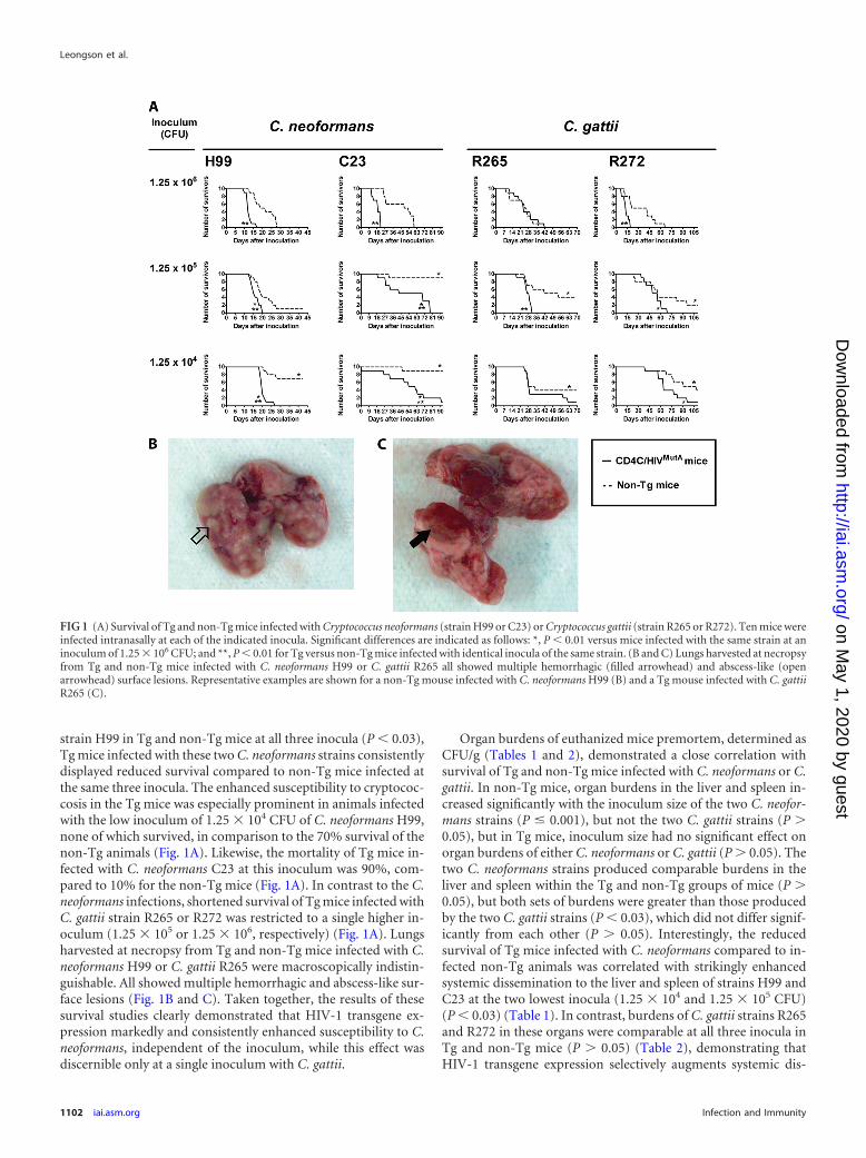
strain H99 in Tg and non-Tg mice at all three inocula (P � 0.03),Tg mice infected with these two C. neoformans strains consistentlydisplayed reduced survival compared to non-Tg mice infected atthe same three inocula. The enhanced susceptibility to cryptococ-cosis in the Tg mice was especially prominent in animals infectedwith the low inoculum of 1.25 � 104 CFU of C. neoformans H99,none of which survived, in comparison to the 70% survival of thenon-Tg animals (Fig. 1A). Likewise, the mortality of Tg mice in-fected with C. neoformans C23 at this inoculum was 90%, com-pared to 10% for the non-Tg mice (Fig. 1A). In contrast to the C.neoformans infections, shortened survival of Tg mice infected withC. gattii strain R265 or R272 was restricted to a single higher in-oculum (1.25 � 105 or 1.25 � 106, respectively) (Fig. 1A). Lungsharvested at necropsy from Tg and non-Tg mice infected with C.neoformans H99 or C. gattii R265 were macroscopically indistin-guishable. All showed multiple hemorrhagic and abscess-like sur-face lesions (Fig. 1B and C). Taken together, the results of thesesurvival studies clearly demonstrated that HIV-1 transgene ex-pression markedly and consistently enhanced susceptibility to C.neoformans, independent of the inoculum, while this effect wasdiscernible only at a single inoculum with C. gattii.
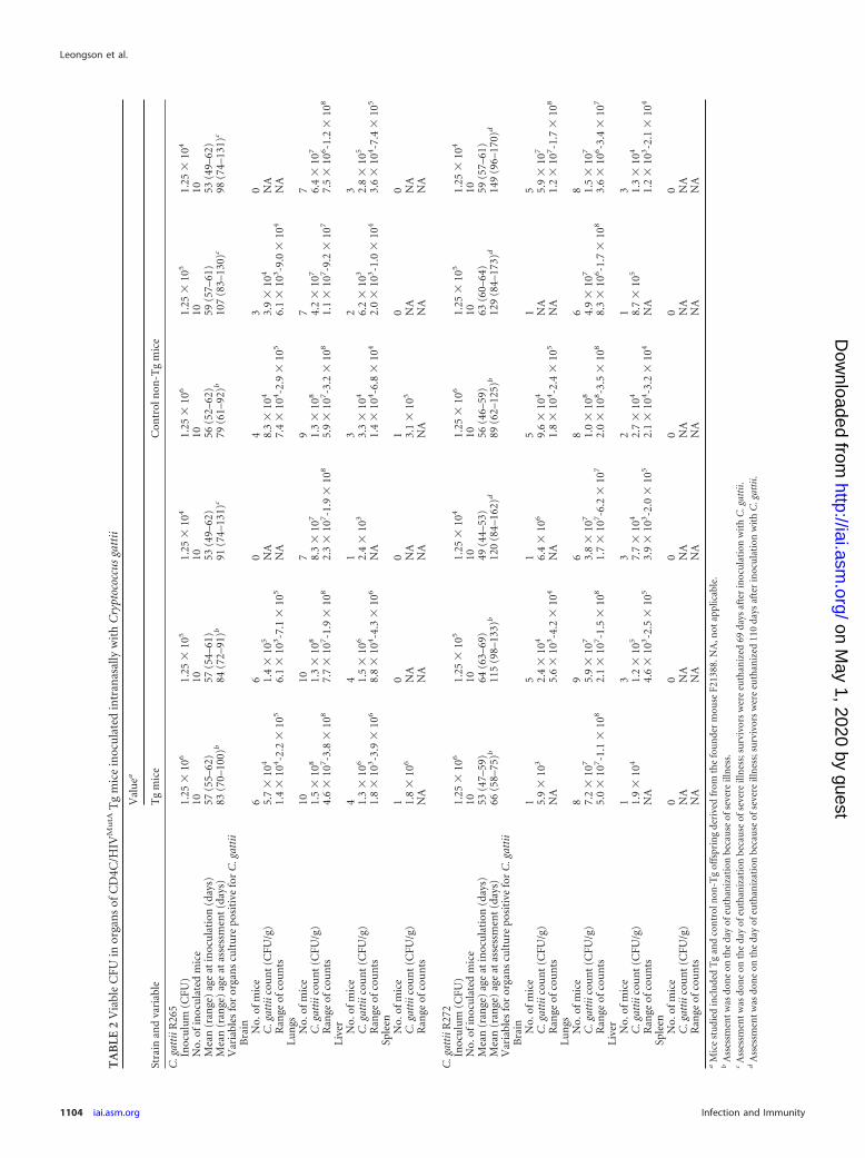
Organ burdens of euthanized mice premortem, determined asCFU/g (Tables 1 and 2), demonstrated a close correlation withsurvival of Tg and non-Tg mice infected with C. neoformans or C.gattii. In non-Tg mice, organ burdens in the liver and spleen in-creased significantly with the inoculum size of the two C. neofor-mans strains (P � 0.001), but not the two C. gattii strains (P �0.05), but in Tg mice, inoculum size had no significant effect onorgan burdens of either C. neoformans or C. gattii (P � 0.05). Thetwo C. neoformans strains produced comparable burdens in theliver and spleen within the Tg and non-Tg groups of mice (P �0.05), but both sets of burdens were greater than those producedby the two C. gattii strains (P � 0.03), which did not differ signif-icantly from each other (P � 0.05). Interestingly, the reducedsurvival of Tg mice infected with C. neoformans compared to in-fected non-Tg animals was correlated with strikingly enhancedsystemic dissemination to the liver and spleen of strains H99 andC23 at the two lowest inocula (1.25 � 104 and 1.25 � 105 CFU)(P � 0.03) (Table 1). In contrast, burdens of C. gattii strains R265and R272 in these organs were comparable at all three inocula inTg and non-Tg mice (P � 0.05) (Table 2), demonstrating thatHIV-1 transgene expression selectively augments systemic dis-
FIG 1 (A) Survival of Tg and non-Tg mice infected with Cryptococcus neoformans (strain H99 or C23) or Cryptococcus gattii (strain R265 or R272). Ten mice wereinfected intranasally at each of the indicated inocula. Significant differences are indicated as follows: *, P � 0.01 versus mice infected with the same strain at aninoculum of 1.25 � 106 CFU; and **, P � 0.01 for Tg versus non-Tg mice infected with identical inocula of the same strain. (B and C) Lungs harvested at necropsyfrom Tg and non-Tg mice infected with C. neoformans H99 or C. gattii R265 all showed multiple hemorrhagic (filled arrowhead) and abscess-like (openarrowhead) surface lesions. Representative examples are shown for a non-Tg mouse infected with C. neoformans H99 (B) and a Tg mouse infected with C. gattiiR265 (C).
Leongson et al.
1102 iai.asm.org Infection and Immunity
on May 1, 2020 by guest
http://iai.asm.org/
Dow
nloaded from
TA
BLE
1V
iableC
FUin
organs
ofC
D4C
/HIV
Mu
tAT
gm
icein
oculated
intran
asallyw
ithC
ryptococcusneoform
ans
Strainan
dvariable
Valu
ea
Tg
mice
Con
trolnon
-Tg
mice
C.neoform
ansH
99In
oculu
m(C
FU)
1.25�
106
1.25�
105
1.25�
104
1.25�
106
1.25�
105
1.25�
104
No.ofm
icein
oculated
1010
1010
1010
Mean
(range)
ageat
inocu
lation(days)
59(49–63)
54(50–57)
60(50–64)
49(42–63)
53(45–67)
61(50–64)
Mean
(range)
ageat
assessmen
t(days)
72(61–80)
b70
(64–75)b
80(70–84)
b70
(58–80)b
76(65–93)
c96
(73–106)c
Variables
fororgan
scu
lture
positivefor
C.neoform
ansB
rainN
o.ofmice
79
910
85
C.neoform
anscou
nt
(CFU
/g)8.8
�10
72.5
�10
77.1
�10
76.0
�10
75.3
�10
76.2
�10
7
Ran
geofcou
nts
7.4�
103-3.8
�10
81.4
�10
4-1.0�
108
2.7�
104-2.8
�10
83.3
�10
5-1.6�
108
2.7�
104-1.3
�10
87.9
�10
4-2.0�
108
Lun
gsN
o.ofmice
910
910
96
C.neoform
anscou
nt
(CFU
/g)2.0
�10
91.3
�10
92.5
�10
91.2
�10
91.0
�10
93.8
�10
8
Ran
geofcou
nts
2.7�
108-8.7
�10
91.8
�10
7-5.0�
109
9.6�
107-4.7
�10
98.6
�10
7-5.4�
109
6.2�
106-6.3
�10
96.9
�10
5-6.9�
108
LiverN
o.ofmice
910
910
94
C.neoform
anscou
nt
(CFU
/g)4.6
�10
51.3
�10
63.5
�10
52.9
�10
58.8
�10
47.3
�10
4
Ran
geofcou
nts
2.5�
104-2.5
�10
64.2
�10
4-7.6�
106
1.3�
105-6.5
�10
52.9
�10
4-8.4�
105
1.1�
104-3.2
�10
51.9
�10
4-2.1�
105
SpleenN
o.ofmice
89
910
83
C.neoform
anscou
nt
(CFU
/g)3.6
�10
61.5
�10
61.5
�10
64.0
�10
52.2
�10
56.0
�10
4
Ran
geofcou
nts
1.1�
105-2.2
�10
72.3
�10
5-2.8�
106
3.6�
105-4.0
�10
65.4
�10
4-1.1�
106
1.8�
104-6.5
�10
59.1
�10
3-1.2�
105
C.neoform
ansC
23In
oculu
m(C
FU)
1.25�
106
1.25�
105
1.25�
104
1.25�
106
1.25�
105
1.25�
104
No.ofin
oculated
mice
1010
1010
1010
Mean
(range)
ageat
inocu
lation(days)
53(50–56)
53(46–60)
46(43–52)
45(43–57)
49(49–50)
52(50–54)
Mean
(range)
ageat
assessmen
t(days)
71(62–77)
b105
(65–134)b
100(53–135)
d90
(69–113)b
135(81–142)
d140
(99–146)d
Variables
fororgan
scu
lture
positivefor
C.neoform
ansB
rainN
o.ofmice
87
66
00
C.neoform
anscou
nt
(CFU
/g)2.2
�10
74.4
�10
68.1
�10
61.2
�10
7N
AN
AR
ange
ofcoun
ts3.6
�10
4-7.1�
107
5.9�
103-2.0
�10
79.5
�10
3-1.4�
107
8.2�
105-4.3
�10
7N
AN
ALu
ngs
No.ofm
ice10
74
81
0C
.neoformans
coun
t(C
FU/g)
4.2�
108
1.3�
108
2.2�
108
5.7�
108
4.4�
106
NA
Ran
geofcou
nts
1.2�
108-9.4
�10
87.7
�10
5-3.2�
108
2.1�
106-3.9
�10
81.7
�10
7-1.5�
109
NA
NA
LiverN
o.ofmice
107
47
10
C.neoform
anscou
nt
(CFU
/g)6.1
�10
56.0
�10
52.2
�10
63.4
�10
64.4
�10
3N
AR
ange
ofcoun
ts3.9
�10
4-2.5�
106
1.5�
103-2.1
�10
61.4
�10
4-4.3�
106
9.6�
103-8.6
�10
6N
AN
ASpleen
No.ofm
ice10
54
50
0C
.neoformans
coun
t(C
FU/g)
2.8�
106
2.8�
106
3.1�
107
4.1�
106
NA
NA
Ran
geofcou
nts
1.8�
105-1.7
�10
72.0
�10
5-7.7�
106
1.0�
105-7.4
�10
71.6
�10
5-8.5�
106
NA
NA
aM
icestu
diedin
cluded
Tg
and
controln
on-T
goffsprin
gderived
fromth
efou
nder
mou
seF21388.N
A,n
otapplicable.
bA
ssessmen
tw
asdon
eon
the
dayofeu
than
izationbecau
seofsevere
illness.
cAssessm
ent
was
done
onth
eday
ofeuth
anization
because
ofsevereilln
ess;survivors
were
euth
anized
42days
afterin
oculation
with
C.neoform
ans.d
Assessm
ent
was
done
onth
eday
ofeuth
anization
because
ofsevereilln
ess;survivors
were
euth
anized
92days
afterin
oculation
with
C.neoform
ans.
Cryptococcus Infection in HIV Gene-Expressing Tg Mice
April 2013 Volume 81 Number 4 iai.asm.org 1103
on May 1, 2020 by guest
http://iai.asm.org/
Dow
nloaded from
TA
BLE
2V
iabl
eC
FUin
orga
ns
ofC
D4C
/HIV
Mu
tAT
gm
ice
inoc
ula
ted
intr
anas
ally
wit
hC
rypt
ococ
cus
gatt
ii
Stra
inan
dva
riab
le
Val
uea
Tg
mic
eC
ontr
oln
on-T
gm
ice
C.g
atti
iR26
5In
ocu
lum
(CFU
)1.
25�
106
1.25
�10
51.
25�
104
1.25
�10
61.
25�
105
1.25
�10
4
No.
ofin
ocu
late
dm
ice
1010
1010
1010
Mea
n(r
ange
)ag
eat
inoc
ula
tion
(day
s)57
(55–
62)
57(5
4–61
)53
(49–
62)
56(5
2–62
)59
(57–
61)
53(4
9–62
)M
ean
(ran
ge)
age
atas
sess
men
t(d
ays)
83(7
0–10
0)b
84(7
2–91
)b91
(74–
131)
c79
(61–
92)b
107
(83–
130)
c98
(74–
131)
c
Var
iabl
esfo
ror
gan
scu
ltu
repo
siti
vefo
rC
.gat
tii
Bra
in No.
ofm
ice
66
04
30
C.g
atti
icou
nt
(CFU
/g)
5.7
�10
41.
4�
105
NA
8.3
�10
43.
9�
104
NA
Ran
geof
cou
nts
1.4
�10
4-2
.2�
105
6.1
�10
3-7
.1�
105
NA
7.4
�10
4-2
.9�
105
6.1
�10
3-9
.0�
104
NA
Lu
ngs
No.
ofm
ice
1010
79
77
C.g
atti
icou
nt
(CFU
/g)
1.5
�10
81.
3�
108
8.3
�10
71.
3�
108
4.2
�10
76.
4�
107
Ran
geof
cou
nts
4.6
�10
7-3
.8�
108
7.7
�10
7-1
.9�
108
2.3
�10
7-1
.9�
108
5.9
�10
7-3
.2�
108
1.1
�10
7-9
.2�
107
7.5
�10
6-1
.2�
108
Live
rN
o.of
mic
e4
41
32
3C
.gat
tiic
oun
t(C
FU/g
)1.
3�
106
1.5
�10
62.
4�
103
3.3
�10
46.
2�
103
2.8
�10
5
Ran
geof
cou
nts
1.8
�10
3-3
.9�
106
8.8
�10
4-4
.3�
106
NA
1.4
�10
4-6
.8�
104
2.0
�10
3-1
.0�
104
3.6
�10
4-7
.4�
105
Sple
enN
o.of
mic
e1
00
10
0C
.gat
tiic
oun
t(C
FU/g
)1.
8�
106
NA
NA
3.1
�10
5N
AN
AR
ange
ofco
un
tsN
AN
AN
AN
AN
AN
A
C.g
atti
iR27
2In
ocu
lum
(CFU
)1.
25�
106
1.25
�10
51.
25�
104
1.25
�10
61.
25�
105
1.25
�10
4
No.
ofin
ocu
late
dm
ice
1010
1010
1010
Mea
n(r
ange
)ag
eat
inoc
ula
tion
(day
s)53
(47–
59)
64(6
3–69
)49
(44–
53)
56(4
6–59
)63
(60–
64)
59(5
7–61
)M
ean
(ran
ge)
age
atas
sess
men
t(d
ays)
66(5
8–75
)b11
5(9
8–13
3)b
120
(84–
162)
d89
(62–
125)
b12
9(8
4–17
3)d
149
(96–
170)
d
Var
iabl
esfo
ror
gan
scu
ltu
repo
siti
vefo
rC
.gat
tii
Bra
in No.
ofm
ice
15
15
15
C.g
atti
icou
nt
(CFU
/g)
5.9
�10
32.
4�
104
6.4
�10
69.
6�
104
NA
5.9
�10
7
Ran
geof
cou
nts
NA
5.6
�10
3-4
.2�
104
NA
1.8
�10
4-2
.4�
105
NA
1.2
�10
7-1
.7�
108
Lun
gsN
o.of
mic
e8
96
86
8C
.gat
tiic
oun
t(C
FU/g
)7.
2�
107
5.9
�10
73.
8�
107
1.0
�10
84.
9�
107
1.5
�10
7
Ran
geof
cou
nts
5.0
�10
7-1
.1�
108
2.1
�10
7-1
.5�
108
1.7
�10
7-6
.2�
107
2.0
�10
8-3
.5�
108
8.3
�10
6-1
.7�
108
3.6
�10
6-3
.4�
107
Live
rN
o.of
mic
e1
33
21
3C
.gat
tiic
oun
t(C
FU/g
)1.
9�
104
1.2
�10
57.
7�
104
2.7
�10
48.
7�
105
1.3
�10
4
Ran
geof
cou
nts
NA
4.6
�10
3-2
.5�
105
3.9
�10
3-2
.0�
105
2.1
�10
4-3
.2�
104
NA
1.2
�10
3-2
.1�
104
Sple
enN
o.of
mic
e0
00
00
0C
.gat
tiic
oun
t(C
FU/g
)N
AN
AN
AN
AN
AN
AR
ange
ofco
un
tsN
AN
AN
AN
AN
AN
Aa
Mic
est
udi
edin
clu
ded
Tg
and
con
trol
non
-Tg
offs
prin
gde
rive
dfr
omth
efo
un
der
mou
seF2
1388
.NA
,not
appl
icab
le.
bA
sses
smen
tw
asdo
ne
onth
eda
yof
euth
aniz
atio
nbe
cau
seof
seve
reill
nes
s.c
Ass
essm
ent
was
don
eon
the
day
ofeu
than
izat
ion
beca
use
ofse
vere
illn
ess;
surv
ivor
sw
ere
euth
aniz
ed69
days
afte
rin
ocu
lati
onw
ith
C.g
atti
i.d
Ass
essm
ent
was
don
eon
the
day
ofeu
than
izat
ion
beca
use
ofse
vere
illn
ess;
surv
ivor
sw
ere
euth
aniz
ed11
0da
ysaf
ter
inoc
ula
tion
wit
hC
.gat
tii.
Leongson et al.
1104 iai.asm.org Infection and Immunity
on May 1, 2020 by guest
http://iai.asm.org/
Dow
nloaded from
semination to the liver and spleen for C. neoformans but not C.gattii. However, enhanced burdens in brains of Tg compared tonon-Tg mice were observed at the two lowest inocula with C.neoformans strain C23 only (P � 0.002), not strain H99 (P � 0.05)or the two C. gattii strains (P � 0.05) (Tables 1 and 2), showingthat HIV-1 transgene-mediated augmentation of C. neoformansdissemination to the brain may be strain dependent.
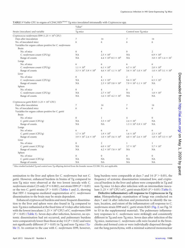
Enhanced cryptococcal burdens and more frequent dissemina-tion to the liver and spleen were also found in Tg compared tonon-Tg mice euthanized at the fixed time of 14 days after infectionwith the lowest inoculum (1.25 � 104 CFU) of C. neoformans H99(P � 0.05) (Table 3). Seven days after infection, however, no sys-temic dissemination had yet occurred, and pulmonary burdenswere comparatively lower than those at day 14 (P � 0.02) and werenot significantly different (P � 0.05) in Tg and non-Tg mice (Ta-ble 3). In contrast to the case with C. neoformans H99, however,
lung burdens were comparable at days 7 and 14 (P � 0.05), thefrequency of systemic dissemination remained low, and crypto-coccal burdens in the liver and spleen were comparable in Tg andnon-Tg mice 14 days after infection with an intermediate inocu-lum (1.25 � 105 CFU) of C. gattii strain R265 (P � 0.05) (Table 3).
Defective inflammatory cell response to Cryptococcus in Tgmice. Histopathologic examination of lungs was conducted ondays 7 and 14 after infection and premortem to identify the na-ture, location, and extent of the inflammatory cell response to C.neoformans strain H99 and C. gattii strain R265 (Fig. 2; see Fig. S1to S3 in the supplemental material). The pulmonary inflamma-tory responses to C. neoformans were strikingly and consistentlydifferent in Tg and non-Tg mice. Seven days after infection of theTg mice, numerous C. neoformans cells were located in the bron-chioles and formed cysts or were individually dispersed through-out the lung parenchyma, with a minimal scattered mononuclear
TABLE 3 Viable CFU in organs of CD4C/HIVMutA Tg mice inoculated intranasally with Cryptococcus spp.
Strain (inoculum) and variable
Valuea
Tg mice Control non-Tg mice
Cryptococcus neoformans H99 (1.25 � 104 CFU)Days after inoculation 7 14 7 14No. of inoculated mice 6 6 6 6Variables for organs culture positive for C. neoformans
BrainNo. of mice 0 4 0 2C. neoformans count (CFU/g) NA 2.5 � 106 NA 6.9 � 106
Range of counts NA 4.4 � 104-8.5 � 106 NA 3.8 � 105-1.4 � 107
LungsNo. of mice 6 6 6 6C. neoformans count (CFU/g) 1.1 � 108 6.1 � 108 6.7 � 107 2.0 � 108
Range of counts 3.3 � 107-3.9 � 108 4.6 � 107-1.1 � 109 3.6 � 107-1.0 � 108 1.0 � 108-3.1 � 108
LiverNo. of mice 0 4 2 1C. neoformans count (CFU/g) NA 4.1 � 104 9.6 � 103 6.5 � 103
Range of counts NA 2.3 � 104-5.8 � 104 7.9 � 103-1.1 � 104 NASpleen
No. of mice 0 4 0 1C. neoformans count (CFU/g) NA 2.7 � 105 NA 1.8 � 104
Range of counts NA 1.6 � 105-4.2 � 105 NA NA
Cryptococcus gattii R265 (1.25 � 105 CFU)Days after inoculation 7 14 7 14No. of inoculated mice 6 6 6 6Variables for organs culture positive for C. gattii
BrainNo. of mice 0 1 4 0C. gattii count (CFU/g) NA 3.5 � 105 2.4 � 104 NARange of counts NA NA 1.3 � 104-5.9 � 104 NA
LungsNo. of mice 6 6 6 6C. gattii count (CFU/g) 1.7 � 108 1.9 � 108 1.6 � 108 2.5 � 108
Range of counts 3.9 � 107-2.4 � 108 1.0 � 108-3.6 � 108 4.0 � 107-2.6 � 108 1.8 � 108-3.3 � 108
LiverNo. of mice 0 2 1 1C. gattii count (CFU/g) NA 6.0 � 103 3.7 � 103 5.7 � 104
Range of counts NA 4.2 � 103-7.8 � 103 NA NASpleen
No. of mice 0 0 0 0C. gattii count (CFU/g) NA NA NA NARange of counts NA NA NA NA
a Mice studied included Tg and control non-Tg offspring derived from the founder mouse F21388. NA, not applicable.
Cryptococcus Infection in HIV Gene-Expressing Tg Mice
April 2013 Volume 81 Number 4 iai.asm.org 1105
on May 1, 2020 by guest
http://iai.asm.org/
Dow
nloaded from
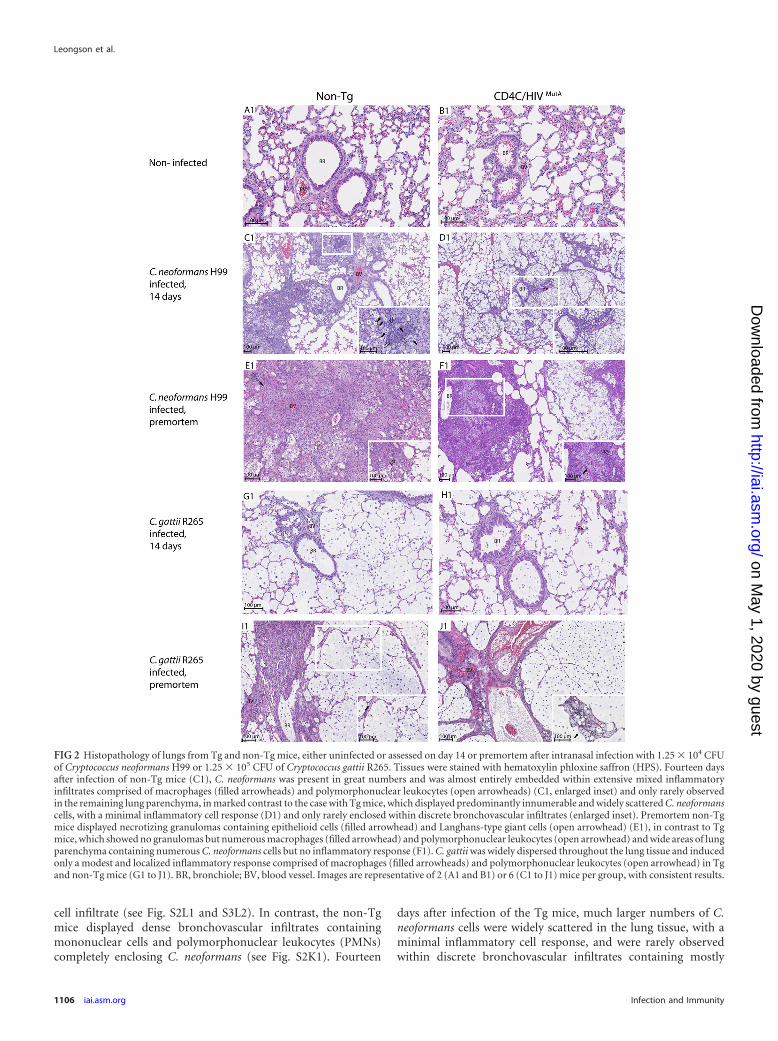
cell infiltrate (see Fig. S2L1 and S3L2). In contrast, the non-Tgmice displayed dense bronchovascular infiltrates containingmononuclear cells and polymorphonuclear leukocytes (PMNs)completely enclosing C. neoformans (see Fig. S2K1). Fourteen
days after infection of the Tg mice, much larger numbers of C.neoformans cells were widely scattered in the lung tissue, with aminimal inflammatory cell response, and were rarely observedwithin discrete bronchovascular infiltrates containing mostly
FIG 2 Histopathology of lungs from Tg and non-Tg mice, either uninfected or assessed on day 14 or premortem after intranasal infection with 1.25 � 104 CFUof Cryptococcus neoformans H99 or 1.25 � 105 CFU of Cryptococcus gattii R265. Tissues were stained with hematoxylin phloxine saffron (HPS). Fourteen daysafter infection of non-Tg mice (C1), C. neoformans was present in great numbers and was almost entirely embedded within extensive mixed inflammatoryinfiltrates comprised of macrophages (filled arrowheads) and polymorphonuclear leukocytes (open arrowheads) (C1, enlarged inset) and only rarely observedin the remaining lung parenchyma, in marked contrast to the case with Tg mice, which displayed predominantly innumerable and widely scattered C. neoformanscells, with a minimal inflammatory cell response (D1) and only rarely enclosed within discrete bronchovascular infiltrates (enlarged inset). Premortem non-Tgmice displayed necrotizing granulomas containing epithelioid cells (filled arrowhead) and Langhans-type giant cells (open arrowhead) (E1), in contrast to Tgmice, which showed no granulomas but numerous macrophages (filled arrowhead) and polymorphonuclear leukocytes (open arrowhead) and wide areas of lungparenchyma containing numerous C. neoformans cells but no inflammatory response (F1). C. gattii was widely dispersed throughout the lung tissue and inducedonly a modest and localized inflammatory response comprised of macrophages (filled arrowheads) and polymorphonuclear leukocytes (open arrowhead) in Tgand non-Tg mice (G1 to J1). BR, bronchiole; BV, blood vessel. Images are representative of 2 (A1 and B1) or 6 (C1 to J1) mice per group, with consistent results.
Leongson et al.
1106 iai.asm.org Infection and Immunity
on May 1, 2020 by guest
http://iai.asm.org/
Dow
nloaded from
PMNs and a few mononuclear cells (Fig. 2D1; see Fig. S1D2). Instriking contrast, in non-Tg mice, C. neoformans cells were almostentirely embedded within far more extensive mixed inflammatoryinfiltrates comprised of PMNs and macrophages and were seldomobserved in the remaining lung parenchyma, which was devoid ofinflammatory cells (Fig. 2C1). Finally, premortem non-Tg miceagain displayed a widespread inflammatory response, with theadded appearance at this late time point of necrotizing granulo-mas containing epithelioid cells and Langhans-type giant cells(Fig. 2E1). This was in contrast to the Tg mice, which displayedmore limited inflammatory foci containing abundant macro-phages and PMNs but no granulomas, as well as broad areas oflung parenchyma containing numerous C. neoformans cells butno inflammatory response (Fig. 2F1).
In sharp contrast to the case for infection with C. neoformans,numerous C. gattii cells were widely dispersed throughout thelung tissue and induced only a sparse inflammatory response ondays 7 and 14 after infection in both Tg and non-Tg mice (Fig. 2G1and H1; see Fig. S1 to S3 in the supplemental material). A modestand circumscribed inflammatory response comprised of macro-phages and PMNs appeared only in premortem animals and wasindependent of HIV-1 transgene expression (Fig. 2I1 and J1).
Interestingly, macrophages in lung tissue sections from Tg andnon-Tg mice infected with C. neoformans or C. gattii often dis-played the distinctive appearance of “hueco” cells filled with ves-icles containing capsular polysaccharide (31, 32). These cells wereobserved beginning on day 14 after infection and became moreabundant in mice assessed premortem.
Histopathologic examination of the brains of Tg and non-Tgmice on day 7 after infection with C. neoformans showed that thebrains were entirely normal, in accordance with the absence ofsystemic dissemination to this organ at this early time point (Table3). On day 14 after infection, however, histopathology revealed C.neoformans in the brain parenchyma of a single non-Tg mousewhich displayed culture evidence of dissemination to this organ,but not in the other animals, which were either culture positive ornegative (Table 3). Taken together with the organ burdens, theseresults indicated that the onset of dissemination to the brain for C.neoformans was detectable more than 7 days after infection in bothTg and non-Tg mice and did not occur earlier in the Tg mice,despite their enhanced frequency of systemic dissemination (Ta-bles 1 and 2). Examination of the brains of Tg and non-Tg mice 7and 14 days after infection with C. gattii did not show histopatho-logic evidence of the fungus, in accordance with lower burdens ofC. gattii than of C. neoformans in this organ (Table 3).
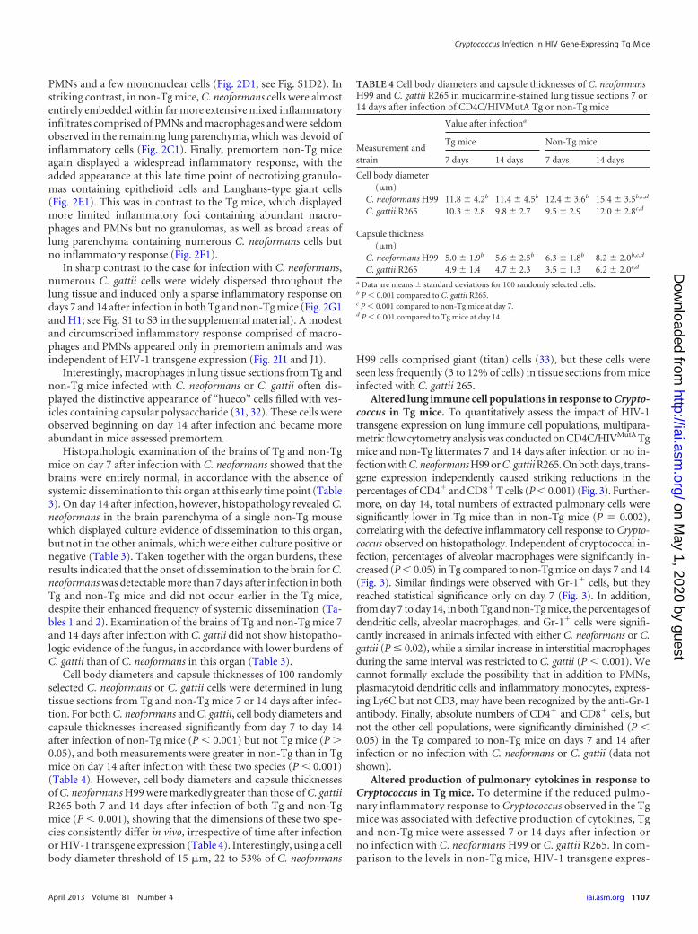
Cell body diameters and capsule thicknesses of 100 randomlyselected C. neoformans or C. gattii cells were determined in lungtissue sections from Tg and non-Tg mice 7 or 14 days after infec-tion. For both C. neoformans and C. gattii, cell body diameters andcapsule thicknesses increased significantly from day 7 to day 14after infection of non-Tg mice (P � 0.001) but not Tg mice (P �0.05), and both measurements were greater in non-Tg than in Tgmice on day 14 after infection with these two species (P � 0.001)(Table 4). However, cell body diameters and capsule thicknessesof C. neoformans H99 were markedly greater than those of C. gattiiR265 both 7 and 14 days after infection of both Tg and non-Tgmice (P � 0.001), showing that the dimensions of these two spe-cies consistently differ in vivo, irrespective of time after infectionor HIV-1 transgene expression (Table 4). Interestingly, using a cellbody diameter threshold of 15 �m, 22 to 53% of C. neoformans
H99 cells comprised giant (titan) cells (33), but these cells wereseen less frequently (3 to 12% of cells) in tissue sections from miceinfected with C. gattii 265.
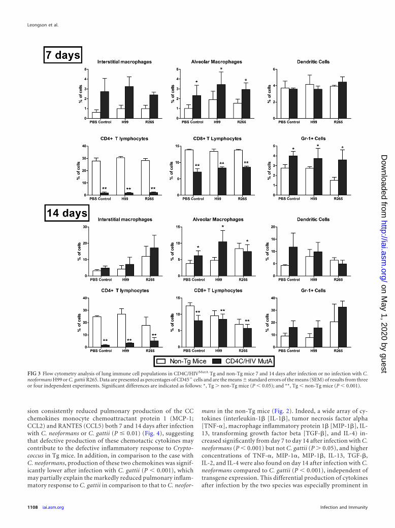
Altered lung immune cell populations in response to Crypto-coccus in Tg mice. To quantitatively assess the impact of HIV-1transgene expression on lung immune cell populations, multipara-metric flow cytometry analysis was conducted on CD4C/HIVMutA Tgmice and non-Tg littermates 7 and 14 days after infection or no in-fection with C. neoformans H99 or C. gattii R265. On both days, trans-gene expression independently caused striking reductions in thepercentages of CD4� and CD8� T cells (P � 0.001) (Fig. 3). Further-more, on day 14, total numbers of extracted pulmonary cells weresignificantly lower in Tg mice than in non-Tg mice (P � 0.002),correlating with the defective inflammatory cell response to Crypto-coccus observed on histopathology. Independent of cryptococcal in-fection, percentages of alveolar macrophages were significantly in-creased (P � 0.05) in Tg compared to non-Tg mice on days 7 and 14(Fig. 3). Similar findings were observed with Gr-1� cells, but theyreached statistical significance only on day 7 (Fig. 3). In addition,from day 7 to day 14, in both Tg and non-Tg mice, the percentages ofdendritic cells, alveolar macrophages, and Gr-1� cells were signifi-cantly increased in animals infected with either C. neoformans or C.gattii (P � 0.02), while a similar increase in interstitial macrophagesduring the same interval was restricted to C. gattii (P � 0.001). Wecannot formally exclude the possibility that in addition to PMNs,plasmacytoid dendritic cells and inflammatory monocytes, express-ing Ly6C but not CD3, may have been recognized by the anti-Gr-1antibody. Finally, absolute numbers of CD4� and CD8� cells, butnot the other cell populations, were significantly diminished (P �0.05) in the Tg compared to non-Tg mice on days 7 and 14 afterinfection or no infection with C. neoformans or C. gattii (data notshown).
Altered production of pulmonary cytokines in response toCryptococcus in Tg mice. To determine if the reduced pulmo-nary inflammatory response to Cryptococcus observed in the Tgmice was associated with defective production of cytokines, Tgand non-Tg mice were assessed 7 or 14 days after infection orno infection with C. neoformans H99 or C. gattii R265. In com-parison to the levels in non-Tg mice, HIV-1 transgene expres-
TABLE 4 Cell body diameters and capsule thicknesses of C. neoformansH99 and C. gattii R265 in mucicarmine-stained lung tissue sections 7 or14 days after infection of CD4C/HIVMutA Tg or non-Tg mice
Measurement andstrain
Value after infectiona
Tg mice Non-Tg mice
7 days 14 days 7 days 14 days
Cell body diameter(�m)
C. neoformans H99 11.8 � 4.2b 11.4 � 4.5b 12.4 � 3.6b 15.4 � 3.5b,c,d
C. gattii R265 10.3 � 2.8 9.8 � 2.7 9.5 � 2.9 12.0 � 2.8c,d
Capsule thickness(�m)
C. neoformans H99 5.0 � 1.9b 5.6 � 2.5b 6.3 � 1.8b 8.2 � 2.0b,c,d
C. gattii R265 4.9 � 1.4 4.7 � 2.3 3.5 � 1.3 6.2 � 2.0c,d
a Data are means � standard deviations for 100 randomly selected cells.b P � 0.001 compared to C. gattii R265.c P � 0.001 compared to non-Tg mice at day 7.d P � 0.001 compared to Tg mice at day 14.
Cryptococcus Infection in HIV Gene-Expressing Tg Mice
April 2013 Volume 81 Number 4 iai.asm.org 1107
on May 1, 2020 by guest
http://iai.asm.org/
Dow
nloaded from
sion consistently reduced pulmonary production of the CCchemokines monocyte chemoattractant protein 1 (MCP-1;CCL2) and RANTES (CCL5) both 7 and 14 days after infectionwith C. neoformans or C. gattii (P � 0.01) (Fig. 4), suggestingthat defective production of these chemotactic cytokines maycontribute to the defective inflammatory response to Crypto-coccus in Tg mice. In addition, in comparison to the case withC. neoformans, production of these two chemokines was signif-icantly lower after infection with C. gattii (P � 0.001), whichmay partially explain the markedly reduced pulmonary inflam-matory response to C. gattii in comparison to that to C. neofor-
mans in the non-Tg mice (Fig. 2). Indeed, a wide array of cy-tokines (interleukin-1 [IL-1], tumor necrosis factor alpha[TNF-], macrophage inflammatory protein 1 [MIP-1], IL-13, transforming growth factor beta [TGF-], and IL-4) in-creased significantly from day 7 to day 14 after infection with C.neoformans (P � 0.001) but not C. gattii (P � 0.05), and higherconcentrations of TNF-, MIP-1, MIP-1, IL-13, TGF-,IL-2, and IL-4 were also found on day 14 after infection with C.neoformans compared to C. gattii (P � 0.001), independent oftransgene expression. This differential production of cytokinesafter infection by the two species was especially prominent in
FIG 3 Flow cytometry analysis of lung immune cell populations in CD4C/HIVMutA Tg and non-Tg mice 7 and 14 days after infection or no infection with C.neoformans H99 or C. gattii R265. Data are presented as percentages of CD45� cells and are the means � standard errors of the means (SEM) of results from threeor four independent experiments. Significant differences are indicated as follows: *, Tg � non-Tg mice (P � 0.05); and **, Tg � non-Tg mice (P � 0.001).
Leongson et al.
1108 iai.asm.org Infection and Immunity
on May 1, 2020 by guest
http://iai.asm.org/
Dow
nloaded from
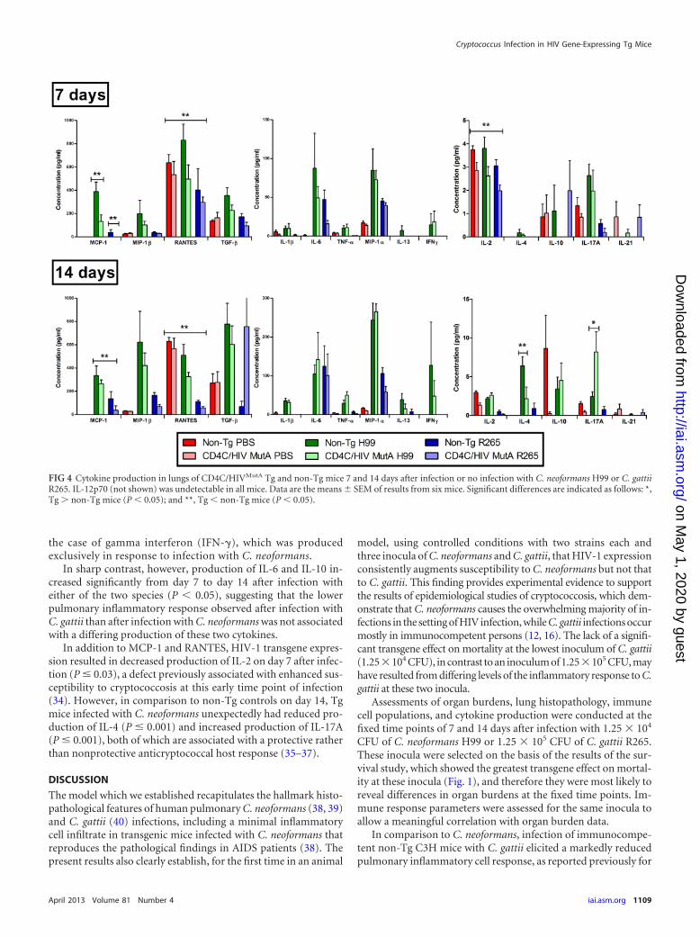
the case of gamma interferon (IFN-�), which was producedexclusively in response to infection with C. neoformans.
In sharp contrast, however, production of IL-6 and IL-10 in-creased significantly from day 7 to day 14 after infection witheither of the two species (P � 0.05), suggesting that the lowerpulmonary inflammatory response observed after infection withC. gattii than after infection with C. neoformans was not associatedwith a differing production of these two cytokines.
In addition to MCP-1 and RANTES, HIV-1 transgene expres-sion resulted in decreased production of IL-2 on day 7 after infec-tion (P � 0.03), a defect previously associated with enhanced sus-ceptibility to cryptococcosis at this early time point of infection(34). However, in comparison to non-Tg controls on day 14, Tgmice infected with C. neoformans unexpectedly had reduced pro-duction of IL-4 (P � 0.001) and increased production of IL-17A(P � 0.001), both of which are associated with a protective ratherthan nonprotective anticryptococcal host response (35–37).
DISCUSSION
The model which we established recapitulates the hallmark histo-pathological features of human pulmonary C. neoformans (38, 39)and C. gattii (40) infections, including a minimal inflammatorycell infiltrate in transgenic mice infected with C. neoformans thatreproduces the pathological findings in AIDS patients (38). Thepresent results also clearly establish, for the first time in an animal
model, using controlled conditions with two strains each andthree inocula of C. neoformans and C. gattii, that HIV-1 expressionconsistently augments susceptibility to C. neoformans but not thatto C. gattii. This finding provides experimental evidence to supportthe results of epidemiological studies of cryptococcosis, which dem-onstrate that C. neoformans causes the overwhelming majority of in-fections in the setting of HIV infection, while C. gattii infections occurmostly in immunocompetent persons (12, 16). The lack of a signifi-cant transgene effect on mortality at the lowest inoculum of C. gattii(1.25�104 CFU), in contrast to an inoculum of 1.25�105 CFU, mayhave resulted from differing levels of the inflammatory response to C.gattii at these two inocula.
Assessments of organ burdens, lung histopathology, immunecell populations, and cytokine production were conducted at thefixed time points of 7 and 14 days after infection with 1.25 � 104
CFU of C. neoformans H99 or 1.25 � 105 CFU of C. gattii R265.These inocula were selected on the basis of the results of the sur-vival study, which showed the greatest transgene effect on mortal-ity at these inocula (Fig. 1), and therefore they were most likely toreveal differences in organ burdens at the fixed time points. Im-mune response parameters were assessed for the same inocula toallow a meaningful correlation with organ burden data.
In comparison to C. neoformans, infection of immunocompe-tent non-Tg C3H mice with C. gattii elicited a markedly reducedpulmonary inflammatory cell response, as reported previously for
FIG 4 Cytokine production in lungs of CD4C/HIVMutA Tg and non-Tg mice 7 and 14 days after infection or no infection with C. neoformans H99 or C. gattiiR265. IL-12p70 (not shown) was undetectable in all mice. Data are the means � SEM of results from six mice. Significant differences are indicated as follows: *,Tg � non-Tg mice (P � 0.05); and **, Tg � non-Tg mice (P � 0.05).
Cryptococcus Infection in HIV Gene-Expressing Tg Mice
April 2013 Volume 81 Number 4 iai.asm.org 1109
on May 1, 2020 by guest
http://iai.asm.org/
Dow
nloaded from
C57BL/6 and A/JCr mice infected with identical inocula of the twospecies (17, 41). It is therefore unlikely that the less robust pulmo-nary inflammatory cell response to C. gattii than that to C. neofor-mans which we found in the non-Tg mice was caused by the higherinoculum.
The lower pulmonary inflammatory cell response to C. gattiiwas closely correlated with diminished production of several cy-tokines and chemokines, including MCP-1, RANTES, MIP-1,MIP-1, IL-1, IL-2, IL-4, IL-13, TNF-, IFN-�, and TGF-.Among these, MCP-1, MIP-1, TNF-, and IFN-� all play a rolein leukocyte recruitment to the lungs in response to C. neoformansinfection (42–52). Accordingly, reduced production of these fourcytokines may explain, at least in part, the strikingly sparse inflam-matory cell response to C. gattii compared to that to C. neoformansin the non-Tg C3H mice. Interestingly, we found greater capsulethicknesses of C. neoformans than C. gattii, and it has been re-ported that increasing capsule thicknesses of C. neoformans aug-ment the magnitudes of IL-1 and TNF- release by humanPMNs (53). It would be relevant in future work to examine infec-tion by C. neoformans 145A, which like C. gattii R265 induces alimited pulmonary inflammatory response (54), to determine if itbehaves similarly to C. gattii in HIV-1-expressing Tg mice.
Despite these strikingly dissimilar host immune responses toC. neoformans and C. gattii, comparable lung burdens of bothcryptococcal species were found on days 7 and 14 after infectionand premortem. This seemingly paradoxical finding could possi-bly be explained by the antiphagocytic properties of the crypto-coccal capsule (55) and the reduced phagocytosis of cryptococcalgiant (titan) cells (33, 56–58), which would allow C. neoformans toproliferate at a rate comparable to that of C. gattii despite theenhanced inflammatory cell response. However, in a recent report(41), C. gattii R265 produced higher lung burdens than those of C.neoformans H99 after infection of C57BL/6 and BALB/c mice, sug-gesting that the protective pulmonary immune responses to Cryp-tococcus of these two mouse strains may differ qualitatively orquantitatively from those of non-Tg C3H mice. Nevertheless, inthe non-Tg C3H mice, dissemination of C. neoformans to the liverand spleen at the time of euthanasia largely exceeded that of C.gattii, demonstrating a greater capacity of C. neoformans for sys-temic dissemination in the immunocompetent host (41). Thegreater capsule thickness of C. neoformans than that of C. gattii,providing protection against reactive oxygen and nitrogen specieswithin phagocytes (55), may have facilitated dissemination by a“Trojan horse” mechanism (59). Despite this enhanced dissemi-nation, however, the survival of non-Tg C3H mice infected withthe C. neoformans and C. gattii strains did not differ significantly,suggesting that the variable virulence of strains within each speciesoutweighs any potentially consistent difference in virulence be-tween these two cryptococcal species. In fact, previous studiescomparing the virulence of C. neoformans H99 and C. gattii R265in C57BL/6 and BALB/c mice produced inconsistent results (17,41), indicating that the virulence of C. neoformans and C. gattii islikely comparable in many, if not most, strains of immunocom-petent mice. This interpretation is supported by the balanced up-regulation in production of protective (IFN-�) and nonprotective(IL-4 and IL-13) cytokines (36, 51, 52, 60, 61) in non-Tg C3Hmice infected with C. neoformans compared to those infected withC. gattii. Taken together, the results of our survival studies dem-onstrate that HIV-1 transgene expression alters the course of cryp-tococcal infection to a far larger degree than any intrinsic differ-
ences in virulence, systemic dissemination, or host immuneresponses between C. neoformans and C. gattii.
Enhanced susceptibility to C. neoformans infection in the Tgmice was associated with a sharply reduced pulmonary inflamma-tory cell response and decreased production of the CC chemo-kines MCP-1 (CCL2) and RANTES (CCL5). The striking deple-tion of pulmonary CD4� and CD8� T cells in infected oruninfected Tg mice is congruent with the quantitative reductionsof these cell populations in the oral mucosa, secondary lymphoidorgans, and peripheral blood of these Tg mice (18, 23). The pres-ent results therefore suggest that the defective pulmonary CD4�
and CD8� T-cell response to C. neoformans infection in Tg miceresulted from the primary depletion of these cell populations as aconsequence of HIV-1 transgene expression, combined with afailure of their recruitment as a result of reduced production of thechemokines MCP-1 and RANTES, which attract activated T cells,monocytes, and dendritic cells. During pulmonary C. neoformansinfection, upregulation of MCP-1 and MCP-3 (CCL7) productionis required for CCR2-mediated recruitment of T cells, dendriticcells, and macrophages, formation of bronchovascular cell infil-trates, and development of protective Th1 immunity (42–48).Furthermore, SJL/J mice, which are resistant to C. neoformansinfection, show enhanced MCP-1 mRNA expression compared tosusceptible C57BL/6 mice (62). Potential cellular sources ofMCP-1 in the lungs include epithelial cells, endothelial cells, fibro-blasts, and macrophages (42). Of these specific cell populations,only macrophages express the HIV-1 transgene (19) and wouldthus be susceptible primarily to altered cytokine expression. Inthis regard, we have previously shown that F4/80� macrophagesrecruited to the gastric submucosa and oral mucosa of HIV-1-expressing Tg mice in response to Candida albicans infection ex-press the mannose receptor (CD206) almost uniformly, butMCP-1 only very infrequently (26), consistent with an alterna-tively activated (M2) phenotype known to be associated with sus-ceptibility to cryptococcosis (36, 52). Furthermore, because it hasbeen shown that experimental depletion of CD4� and CD8� Tcells independently abrogates the appearance of a protective in-flammatory response to pulmonary C. neoformans infection andaugments systemic dissemination (63–65), it is likely that the de-pletion of these T-cell populations in the Tg mice contributed tothe reduced pulmonary inflammatory cell response to C. neofor-mans and the augmented systemic dissemination to the liver andspleen. Despite the defective pulmonary inflammatory cell re-sponse to C. neoformans in the Tg mice, pulmonary fungal bur-dens were remarkably comparable to those in non-Tg mice, sug-gesting that reduced survival of the Tg mice was caused primarilyby enhanced systemic dissemination rather than increased prolif-eration of C. neoformans in the lungs (66). Surprisingly, aug-mented susceptibility of the Tg mice to C. neoformans infectionwas associated with diminished pulmonary production of IL-4and increased production of IL-17A, which result in an alterationof the Th1-Th2-Th17 balance associated with a protective ratherthan a nonprotective host response to C. neoformans (35–37, 55).The augmented dissemination of C. neoformans to the liver andspleen in Tg mice, also previously observed in IL-23p19�/� micewith impaired production of IL-17 (35), was therefore likelycaused by perturbations other than a defective Th17 response.
Capsule thicknesses of C. neoformans and C. gattii in the lungsincreased significantly during the course of infection of non-Tgmice (30) but not Tg mice. The mechanisms responsible for dif-
Leongson et al.
1110 iai.asm.org Infection and Immunity
on May 1, 2020 by guest
http://iai.asm.org/
Dow
nloaded from
ferences in capsule thickness in vivo are unknown (30) but couldpotentially include variations in iron, CO2, and nutrient concen-trations in host tissues (30, 67). Interestingly, CD4C/HIVNef
transgenic mice display increased circulating ferritin levels due toNef-dependent release of ferritin from macrophages, and plasmaferritin levels are correlated with viral RNA in HIV-1-infectedpatients (68). C. neoformans can acquire iron bound to the majorcarrier transferrin by a reductive iron uptake pathway (69). Be-cause growth of C. neoformans at high iron concentrations resultsin cells with thinner capsules (30) and lower expression of theCAP60 gene that is required for capsule production (70), in-creased availability of iron from the ferritin carrier may have con-tributed to the lack of capsule thickening during the course ofcryptococcal infection in the Tg mice. However, despite the ab-sence of capsule thickening during infection by both species, thecapsule thickness of C. neoformans remained greater than that ofC. gattii in the Tg mice and may have contributed to its enhancedsystemic dissemination to the liver and spleen, which was alsoobserved in the non-Tg mice.
The percentages of pulmonary dendritic cells, alveolar macro-phages, and Gr-1� cells increased from day 7 to day 14 after infec-tion of Tg and non-Tg mice with C. neoformans, and absolutenumbers of these cell populations extracted from the lungs werenot significantly diminished in the Tg mice. Dendritic cells inCD4C/HIVMutA Tg mice have an immature phenotype, with lowexpression of major histocompatibility complex (MHC) class IIand costimulatory molecules and a decreased capacity to presentantigen in vitro (20, 27). In view of the defective production ofMCP-1 in the Tg mice, dendritic cells could potentially have failedto accumulate in the lungs in response to C. neoformans infectionbecause of defective CCR2-mediated recruitment and differenti-ation of monocytes (46). Preserved production of other CCR2agonists, such as MCP-2 and MCP-3, may have compensated forthe defective production of MCP-1. Because dendritic cells andalveolar macrophages play a critical role in the early innate pro-tective host response against C. neoformans (71) and are associatedwith natural resistance to progressive infection (62), it is likely thatfunctional defects of these cell populations also contributed to theincreased susceptibility of the Tg mice to C. neoformans infection.Blood monocytes and alveolar macrophages from HIV-infectedpatients have impaired fungistatic activity against C. neoformans(72–76).
In summary, the present findings clearly demonstrate thatHIV-1 transgene expression consistently augments susceptibilityto C. neoformans but not C. gattii infection, and it reduces thepulmonary inflammatory cell response by both depletion of im-mune cells and diminished production of chemokines. In the ab-sence of this protective host response in Tg mice, the greater cap-sule thickness of C. neoformans than that of C. gattii in vivo maybecome a primary determinant of the host-pathogen interactionand result in selectively enhanced virulence of C. neoformans, con-sidering that both species qualitatively share all of the known ma-jor C. neoformans virulence traits (7, 77).
ACKNOWLEDGMENTS
This work was supported by the Canadian Institutes of Health Research(grant MOP-93597). Kassandre Leongson and Mathieu Goupil are recip-ients of a studentship award from the University of Montreal.
We thank Marie-Andrée Laniel for support in maintaining the Tg
mouse colony, Christian Charbonneau for assistance with photomicrog-raphy, and Miguel Chagnon for statistical analysis.
REFERENCES1. Park BJ, Wannemuehler KA, Marston BJ, Govender N, Pappas PG,
Chiller TM. 2009. Estimation of the current global burden of cryptococcalmeningitis among persons living with HIV/AIDS. AIDS 23:525–530.
2. Bodasing N, Seaton RA, Shankland GS, Kennedy D. 2004. Cryptococcusneoformans var. gattii meningitis in an HIV-positive patient: first obser-vation in the United Kingdom. J. Infect. 49:253–255.
3. Chaturvedi S, Dyavaiah M, Larsen RA, Chaturvedi V. 2005. Cryptococ-cus gattii in AIDS patients, southern California. Emerg. Infect. Dis. 11:1686 –1692.
4. Hoang LM, Maguire JA, Doyle P, Fyfe M, Roscoe DL. 2004. Crypto-coccus neoformans infections at Vancouver Hospital and Health SciencesCentre (1997–2002): epidemiology, microbiology and histopathology. J.Med. Microbiol. 53:935–940.
5. Karstaedt AS, Crewe-Brown HH, Dromer F. 2002. Cryptococcal men-ingitis caused by Cryptococcus neoformans var. gattii, serotype C, in AIDSpatients in Soweto, South Africa. Med. Mycol. 40:7–11.
6. Morgan J, McCarthy KM, Gould S, Fan K, Arthington-Skaggs B, IqbalN, Stamey K, Hajjeh RA, Brandt ME, Gauteng Cryptococcal Surveil-lance Initiative Group. 2006. Cryptococcus gattii infection: characteris-tics and epidemiology of cases identified in a South African province withhigh HIV seroprevalence, 2002–2004. Clin. Infect. Dis. 43:1077–1080.
7. Sorrell TC. 2001. Cryptococcus neoformans variety gattii. Med. Mycol.39:155–168.
8. Bovers M, Hagen F, Kuramae EE, Hoogveld HL, Dromer F, St-GermainG, Boekhout T. 2008. AIDS patient death caused by novel Cryptococcusneoformans � C. gattii hybrid. Emerg. Infect. Dis. 14:1105–1108.
9. St-Germain G, Noel G, Chung KJ. 1988. Disseminated cryptococcosisdue to Cryptococcus neoformans variety gattii in a Canadian patient withAIDS. Eur. J. Clin. Microbiol. Infect. Dis. 7:587–588.
10. Galanis E, Hoang L, Kibsey P, Morshed M, Phillips P. 2009. Clinicalpresentation, diagnosis and management of Cryptococcus gattii cases: les-sons learned from British Columbia. Can. J. Infect. Dis. Med. Microbiol.20:23–28.
11. Kidd SE, Hagen F, Tscharke RL, Huynh M, Bartlett KH, Fyfe M,Macdougall L, Boekhout T, Kwon-Chung KJ, Meyer W. 2004. A raregenotype of Cryptococcus gattii caused the cryptococcosis outbreak onVancouver Island (British Columbia, Canada). Proc. Natl. Acad. Sci.U. S. A. 101:17258 –17263.
12. Galanis E, Macdougall L. 2010. Epidemiology of Cryptococcus gattii,British Columbia, Canada, 1999 –2007. Emerg. Infect. Dis. 16:251–257.
13. Byrnes EJ, 3rd, Bildfell RJ, Frank SA, Mitchell TG, Marr KA, HeitmanJ. 2009. Molecular evidence that the range of the Vancouver Island out-break of Cryptococcus gattii infection has expanded into the PacificNorthwest in the United States. J. Infect. Dis. 199:1081–1086.
14. Datta K, Bartlett KH, Baer R, Byrnes E, Galanis E, Heitman J, HoangL, Leslie MJ, MacDougall L, Magill SS, Morshed MG, Marr KA, Cryp-tococcus gattii Working Group of the Pacific Northwest. 2009. Spreadof Cryptococcus gattii into Pacific Northwest region of the United States.Emerg. Infect. Dis. 15:1185–1191.
15. Fyfe M, MacDougall L, Romney M, Starr M, Pearce M, Mak S, MithaniS, Kibsey P. 2008. Cryptococcus gattii infections on Vancouver Island,British Columbia, Canada: emergence of a tropical fungus in a temperateenvironment. Can. Commun. Dis. Rep. 34:1–12.
16. Chen S, Sorrell T, Nimmo G, Speed B, Currie B, Ellis D, Marriott D,Pfeiffer T, Parr D, Byth K. 2000. Epidemiology and host- and variety-dependent characteristics of infection due to Cryptococcus neoformans inAustralia and New Zealand. Clin. Infect. Dis. 31:499 –508.
17. Cheng PY, Sham A, Kronstad JW. 2009. Cryptococcus gattii isolatesfrom the British Columbia cryptococcosis outbreak induce less protectiveinflammation in a murine model of infection than Cryptococcus neofor-mans. Infect. Immun. 77:4284 – 4294.
18. de Repentigny L, Aumont F, Ripeau JS, Fiorillo M, Kay DG, Hanna Z,Jolicoeur P. 2002. Mucosal candidiasis in transgenic mice expressing hu-man immunodeficiency virus type 1. J. Infect. Dis. 185:1103–1114.
19. Hanna Z, Kay DG, Rebai N, Guimond A, Jothy S, Jolicoeur P. 1998. Nefharbors a major determinant of pathogenicity for an AIDS-like diseaseinduced by HIV-1 in transgenic mice. Cell 95:163–175.
20. Poudrier J, Weng X, Kay DG, Hanna Z, Jolicoeur P. 2003. The AIDS-
Cryptococcus Infection in HIV Gene-Expressing Tg Mice
April 2013 Volume 81 Number 4 iai.asm.org 1111
on May 1, 2020 by guest
http://iai.asm.org/
Dow
nloaded from
like disease of CD4C/human immunodeficiency virus transgenic mice isassociated with accumulation of immature CD11bHi dendritic cells. J.Virol. 77:11733–11744.
21. Poudrier J, Weng X, Kay DG, Pare G, Calvo EL, Hanna Z, Kosco-Vilbois MH, Jolicoeur P. 2001. The AIDS disease of CD4C/HIV trans-genic mice shows impaired germinal centers and autoantibodies and de-velops in the absence of IFN-gamma and IL-6. Immunity 15:173–185.
22. Priceputu E, Rodrigue I, Chrobak P, Poudrier J, Mak TW, Hanna Z, HuC, Kay DG, Jolicoeur P. 2005. The Nef-mediated AIDS-like disease ofCD4C/human immunodeficiency virus transgenic mice is associated withincreased Fas/FasL expression on T cells and T-cell death but is not pre-vented in Fas-, FasL-, tumor necrosis factor receptor 1-, or interleukin-1beta-converting enzyme-deficient or Bcl2-expressing transgenic mice. J.Virol. 79:6377– 6391.
23. Weng X, Priceputu E, Chrobak P, Poudrier J, Kay DG, Hanna Z, MakTW, Jolicoeur P. 2004. CD4� T cells from CD4C/HIVNef transgenicmice show enhanced activation in vivo with impaired proliferation invitro but are dispensable for the development of a severe AIDS-like organdisease. J. Virol. 78:5244 –5257.
24. Kay DG, Yue P, Hanna Z, Jothy S, Tremblay E, Jolicoeur P. 2002.Cardiac disease in transgenic mice expressing human immunodeficiencyvirus-1 nef in cells of the immune system. Am. J. Pathol. 161:321–335.
25. de Repentigny L, Lewandowski D, Jolicoeur P. 2004. Immunopatho-genesis of oropharyngeal candidiasis in human immunodeficiency virusinfection. Clin. Microbiol. Rev. 17:729 –759.
26. Goupil M, Trudelle EB, Dugas V, Racicot-Bergeron C, Aumont F,Senechal S, Hanna Z, Jolicoeur P, de Repentigny L. 2009. Macrophage-mediated responses to Candida albicans in mice expressing the humanimmunodeficiency virus type 1 transgene. Infect. Immun. 77:4136 – 4149.
27. Lewandowski D, Marquis M, Aumont F, Lussier-Morin AC, RaymondM, Senechal S, Hanna Z, Jolicoeur P, de Repentigny L. 2006. AlteredCD4� T cell phenotype and function determine the susceptibility to mu-cosal candidiasis in transgenic mice expressing HIV-1. J. Immunol. 177:479 – 491.
28. Marquis M, Lewandowski D, Dugas V, Aumont F, Senechal S, JolicoeurP, Hanna Z, de Repentigny L. 2006. CD8� T cells but not polymorpho-nuclear leukocytes are required to limit chronic oral carriage of Candidaalbicans in transgenic mice expressing human immunodeficiency virustype 1. Infect. Immun. 74:2382–2391.
29. Litvintseva AP, Mitchell TG. 2009. Most environmental isolates of Cryp-tococcus neoformans var. grubii (serotype A) are not lethal for mice. In-fect. Immun. 77:3188 –3195.
30. Rivera J, Feldmesser M, Cammer M, Casadevall A. 1998. Organ-dependent variation of capsule thickness in Cryptococcus neoformansduring experimental murine infection. Infect. Immun. 66:5027–5030.
31. Feldmesser M, Kress Y, Novikoff P, Casadevall A. 2000. Cryptococcusneoformans is a facultative intracellular pathogen in murine pulmonaryinfection. Infect. Immun. 68:4225– 4237.
32. Feldmesser M, Tucker S, Casadevall A. 2001. Intracellular parasitism ofmacrophages by Cryptococcus neoformans. Trends Microbiol. 9:273–278.
33. Feldmesser M, Kress Y, Casadevall A. 2001. Dynamic changes in themorphology of Cryptococcus neoformans during murine pulmonary in-fection. Microbiology 147:2355–2365.
34. Hoag KA, Street NE, Huffnagle GB, Lipscomb MF. 1995. Early cytokineproduction in pulmonary Cryptococcus neoformans infections distin-guishes susceptible and resistant mice. Am. J. Respir. Cell Mol. Biol. 13:487– 495.
35. Kleinschek MA, Muller U, Brodie SJ, Stenzel W, Kohler G, Blumens-chein WM, Straubinger RK, McClanahan T, Kastelein RA, Alber G.2006. IL-23 enhances the inflammatory cell response in Cryptococcusneoformans infection and induces a cytokine pattern distinct from IL-12.J. Immunol. 176:1098 –1106.
36. Muller U, Stenzel W, Kohler G, Werner C, Polte T, Hansen G, SchutzeN, Straubinger RK, Blessing M, McKenzie AN, Brombacher F, Alber G.2007. IL-13 induces disease-promoting type 2 cytokines, alternatively ac-tivated macrophages and allergic inflammation during pulmonary infec-tion of mice with Cryptococcus neoformans. J. Immunol. 179:5367–5377.
37. Voelz K, Lammas DA, May RC. 2009. Cytokine signaling regulates theoutcome of intracellular macrophage parasitism by Cryptococcus neofor-mans. Infect. Immun. 77:3450 –3457.
38. Gal AA, Koss MN, Hawkins J, Evans S, Einstein H. 1986. The pathology
of pulmonary cryptococcal infections in the acquired immunodeficiencysyndrome. Arch. Pathol. Lab. Med. 110:502–507.
39. McDonnell JM, Hutchins GM. 1985. Pulmonary cryptococcosis. Hum.Pathol. 16:121–128.
40. Torda A, Kumar RK, Jones PD. 2001. The pathology of human andmurine pulmonary infection with Cryptococcus neoformans var. gattii.Pathology 33:475– 478.
41. Ngamskulrungroj P, Chang Y, Sionov E, Kwon-Chung KJ. 2012. Theprimary target organ of Cryptococcus gattii is different from that of Cryp-tococcus neoformans in a murine model. mBio 3:e00103–12. doi:10.1128/mBio.00103-12.
42. Huffnagle GB, Strieter RM, Standiford TJ, McDonald RA, Burdick MD,Kunkel SL, Toews GB. 1995. The role of monocyte chemotactic protein-1(MCP-1) in the recruitment of monocytes and CD4� T cells during apulmonary Cryptococcus neoformans infection. J. Immunol. 155:4790 –4797.
43. Traynor TR, Herring AC, Dorf ME, Kuziel WA, Toews GB, HuffnagleGB. 2002. Differential roles of CC chemokine ligand 2/monocyte chemot-actic protein-1 and CCR2 in the development of T1 immunity. J. Immu-nol. 168:4659 – 4666.
44. Osterholzer JJ, Chen GH, Olszewski MA, Zhang YM, Curtis JL,Huffnagle GB, Toews GB. 2011. Chemokine receptor 2-mediated accu-mulation of fungicidal exudate macrophages in mice that clear cryptococ-cal lung infection. Am. J. Pathol. 178:198 –211.
45. Osterholzer JJ, Curtis JL, Polak T, Ames T, Chen GH, McDonald R,Huffnagle GB, Toews GB. 2008. CCR2 mediates conventional dendriticcell recruitment and the formation of bronchovascular mononuclear cellinfiltrates in the lungs of mice infected with Cryptococcus neoformans. J.Immunol. 181:610 – 620.
46. Osterholzer JJ, Chen GH, Olszewski MA, Curtis JL, Huffnagle GB,Toews GB. 2009. Accumulation of CD11b� lung dendritic cells in re-sponse to fungal infection results from the CCR2-mediated recruitmentand differentiation of Ly-6Chigh monocytes. J. Immunol. 183:8044 –8053.
47. Qiu Y, Zeltzer S, Zhang Y, Wang F, Chen GH, Dayrit J, Murdock BJ,Bhan U, Toews GB, Osterholzer JJ, Standiford TJ, Olszewski MA. 2012.Early induction of CCL7 downstream of TLR9 signaling promotes thedevelopment of robust immunity to cryptococcal infection. J. Immunol.188:3940 –3948.
48. Huffnagle GB, Traynor TR, McDonald RA, Olszewski MA, Lindell DM,Herring AC, Toews GB. 2000. Leukocyte recruitment during pulmonaryCryptococcus neoformans infection. Immunopharmacology 48:231–236.
49. Huffnagle GB, Strieter RM, McNeil LK, McDonald RA, Burdick MD,Kunkel SL, Toews GB. 1997. Macrophage inflammatory protein-1alpha(MIP-1alpha) is required for the efferent phase of pulmonary cell-mediated immunity to a Cryptococcus neoformans infection. J. Immunol.159:318 –327.
50. Huffnagle GB, Toews GB, Burdick MD, Boyd MB, McAllister KS,McDonald RA, Kunkel SL, Strieter RM. 1996. Afferent phase productionof TNF-alpha is required for the development of protective T cell immu-nity to Cryptococcus neoformans. J. Immunol. 157:4529 – 4536.
51. Kawakami K, Tohyama M, Teruya K, Kudeken N, Xie Q, Saito A. 1996.Contribution of interferon-gamma in protecting mice during pulmonaryand disseminated infection with Cryptococcus neoformans. FEMS Im-munol. Med. Microbiol. 13:123–130.
52. Arora S, Hernandez Y, Erb-Downward JR, McDonald RA, Toews GB,Huffnagle GB. 2005. Role of IFN-gamma in regulating T2 immunity andthe development of alternatively activated macrophages during allergicbronchopulmonary mycosis. J. Immunol. 174:6346 – 6356.
53. Retini C, Vecchiarelli A, Monari C, Tascini C, Bistoni F, Kozel TR.1996. Capsular polysaccharide of Cryptococcus neoformans induces pro-inflammatory cytokine release by human neutrophils. Infect. Immun. 64:2897–2903.
54. Curtis JL, Huffnagle GB, Chen GH, Warnock ML, Gyetko MR, Mc-Donald RA, Scott PJ, Toews GB. 1994. Experimental murine pulmonarycryptococcosis. Differences in pulmonary inflammation and lymphocyterecruitment induced by two encapsulated strains of Cryptococcus neofor-mans. Lab. Invest. 71:113–126.
55. Voelz K, May RC. 2010. Cryptococcal interactions with the host immunesystem. Eukaryot. Cell 9:835– 846.
56. Okagaki LH, Strain AK, Nielsen JN, Charlier C, Baltes NJ, Chretien F,Heitman J, Dromer F, Nielsen K. 2010. Cryptococcal cell morphology
Leongson et al.
1112 iai.asm.org Infection and Immunity
on May 1, 2020 by guest
http://iai.asm.org/
Dow
nloaded from
affects host cell interactions and pathogenicity. PLoS Pathog. 6:e1000953.doi:10.1371/journal.ppat.1000953.
57. Zaragoza O, Garcia-Rodas R, Nosanchuk JD, Cuenca-Estrella M, Ro-driguez-Tudela JL, Casadevall A. 2010. Fungal cell gigantism duringmammalian infection. PLoS Pathog. 6:e1000945. doi:10.1371/journal.ppat.1000945.
58. Okagaki LH, Nielsen K. 2012. Titan cells confer protection from phago-cytosis in Cryptococcus neoformans infections. Eukaryot. Cell 11:820 –826.
59. Casadevall A. 2010. Cryptococci at the brain gate: break and enter or usea Trojan horse? J. Clin. Invest. 120:1389 –1392.
60. Hernandez Y, Arora S, Erb-Downward JR, McDonald RA, Toews GB,Huffnagle GB. 2005. Distinct roles for IL-4 and IL-10 in regulating T2immunity during allergic bronchopulmonary mycosis. J. Immunol. 174:1027–1036.
61. Kawakami K, Hossain Qureshi M, Zhang T, Koguchi Y, Xie Q, Ku-rimoto M, Saito A. 1999. Interleukin-4 weakens host resistance to pul-monary and disseminated cryptococcal infection caused by combinedtreatment with interferon-gamma-inducing cytokines. Cell. Immunol.197:55– 61.
62. Guillot L, Carroll SF, Homer R, Qureshi ST. 2008. Enhanced innateimmune responsiveness to pulmonary Cryptococcus neoformans infec-tion is associated with resistance to progressive infection. Infect. Immun.76:4745– 4756.
63. Huffnagle GB, Lipscomb MF, Lovchik JA, Hoag KA, Street NE. 1994.The role of CD4� and CD8� T cells in the protective inflammatory re-sponse to a pulmonary cryptococcal infection. J. Leukoc. Biol. 55:35– 42.
64. Huffnagle GB, Yates JL, Lipscomb MF. 1991. Immunity to a pulmonaryCryptococcus neoformans infection requires both CD4� and CD8� Tcells. J. Exp. Med. 173:793– 800.
65. Huffnagle GB, Yates JL, Lipscomb MF. 1991. T cell-mediated immunityin the lung: a Cryptococcus neoformans pulmonary infection model usingSCID and athymic nude mice. Infect. Immun. 59:1423–1433.
66. Wang JP, Lee CK, Akalin A, Finberg RW, Levitz SM. 2011. Contribu-tions of the MyD88-dependent receptors IL-18R, IL-1R, and TLR9 to hostdefenses following pulmonary challenge with Cryptococcus neoformans.PLoS One 6:e26232. doi:10.1371/journal.pone.0026232.
67. Mogensen EG, Janbon G, Chaloupka J, Steegborn C, Fu MS, Moyrand
F, Klengel T, Pearson DS, Geeves MA, Buck J, Levin LR, MuhlschlegelFA. 2006. Cryptococcus neoformans senses CO2 through the carbonicanhydrase Can2 and the adenylyl cyclase Cac1. Eukaryot. Cell 5:103–111.
68. Swingler S, Zhou J, Swingler C, Dauphin A, Greenough T, Jolicoeur P,Stevenson M. 2008. Evidence for a pathogenic determinant in HIV-1 Nefinvolved in B cell dysfunction in HIV/AIDS. Cell Host Microbe 4:63–76.
69. Jung WH, Sham A, Lian T, Singh A, Kosman DJ, Kronstad JW. 2008.Iron source preference and regulation of iron uptake in Cryptococcusneoformans. PLoS Pathog. 4:e45. doi:10.1371/journal.ppat.0040045.
70. Lian T, Simmer MI, D’Souza CA, Steen BR, Zuyderduyn SD, Jones SJ,Marra MA, Kronstad JW. 2005. Iron-regulated transcription and capsuleformation in the fungal pathogen Cryptococcus neoformans. Mol. Micro-biol. 55:1452–1472.
71. Osterholzer JJ, Milam JE, Chen GH, Toews GB, Huffnagle GB, Olsze-wski MA. 2009. Role of dendritic cells and alveolar macrophages in reg-ulating early host defense against pulmonary infection with Cryptococcusneoformans. Infect. Immun. 77:3749 –3758.
72. Reardon CC, Kim SJ, Wagner RP, Koziel H, Kornfeld H. 1996. Phago-cytosis and growth inhibition of Cryptococcus neoformans by humanalveolar macrophages: effects of HIV-1 infection. AIDS 10:613– 618.
73. Ieong MH, Reardon CC, Levitz SM, Kornfeld H. 2000. Human immu-nodeficiency virus type 1 infection of alveolar macrophages impairs theirinnate fungicidal activity. Am. J. Respir. Crit. Care Med. 162:966 –970.
74. Cameron ML, Granger DL, Matthews TJ, Weinberg JB. 1994. Humanimmunodeficiency virus (HIV)-infected human blood monocytes andperitoneal macrophages have reduced anticryptococcal activity whereasHIV-infected alveolar macrophages retain normal activity. J. Infect. Dis.170:60 – 67.
75. Harrison TS, Kornfeld H, Levitz SM. 1995. The effect of infection withhuman immunodeficiency virus on the anticryptococcal activity of lym-phocytes and monocytes. J. Infect. Dis. 172:665– 671.
76. Harrison TS, Levitz SM. 1997. Mechanisms of impaired anticryptococcalactivity of monocytes from donors infected with human immunodefi-ciency virus. J. Infect. Dis. 176:537–540.
77. Kronstad JW, Attarian R, Cadieux B, Choi J, D’Souza CA, Griffiths EJ,Geddes JM, Hu G, Jung WH, Kretschmer M, Saikia S, Wang J. 2011.Expanding fungal pathogenesis: Cryptococcus breaks out of the opportu-nistic box. Nat. Rev. Microbiol. 9:193–203.
Cryptococcus Infection in HIV Gene-Expressing Tg Mice
April 2013 Volume 81 Number 4 iai.asm.org 1113
on May 1, 2020 by guest
http://iai.asm.org/
Dow
nloaded from