altered growth, differentiation, and responsiveness to...
TRANSCRIPT
Altered Growth, Differentiation, and Responsivenessto Epidermal Growth Factor of Human Embryonic Mesenchymal Cellsof Palate by Persistent Rubella Virus InfectionToshiyuki Yoneda, Masahiro Urade, Masayoshi Sakuda, and Tadashi MiyazakiFirst and Second Department of Oral and Maxillofacial Surgery, Faculty of Dentistry, Osaka University, Suita, Osaka 565 Japan
Abstract
Wepreviously demonstrated that human embryonic mesenchy-mal cells derived from the palate (HEMPcells) retain alkalinephosphatase (ALP) content and capacity for collagen synthesisafter long-term culture, and their growth is markedly stimulatedby epidermal growth factor (EGF). There was a dramatic de-crease in ALP content and capacity to synthesize collagen inHEMPcells (HEMP-RV cells) persistently infected with rubellavirus (RV). EGFincreased ALP activity and decreased collagensynthesis in HEMPcells, whereas EGF showed no effect onthese activities in HEMP-RVcells. Growth of HEMP-RVcellswas slightly reduced compared with that of HEMPcells. EGFstimulated growth of HEMPcells and to a lesser extent ofHEMP-RVcells. Binding of '"I-EGF to cell-surface receptorsin HEMP-RVcells was, to our surprise, twice as much as thatin HEMPcells. However, internalization of bound '"I-EGF inHEMP-RVcells was profoundly diminished. Thus, persistentRV infection causes not only changes in HEMPcell growth anddifferentiation but a decrease in or loss of HEMPcell respon-siveness to EGF. The effects of persistent RVinfection on palatalcell differentiation as well as growth may be responsible for thepathogenesis of congenital rubella. Furthermore, since HEMPcells appear to be closely related to osteoblasts, these resultssuggest a mechanism for RV-induced osseous abnormalitiesmanifested in congenital rubella patients.
Introduction
Since the first report described by Gregg in 1941 (1), much at-tention has been paid to the teratogenic and pathogenic effectsof rubella virus (RV).' Clinical studies on wide-spread and vir-ulent rubella epidemics that occurred throughout the UnitedStates in 1964 (2, 3) have indicated that RV is one of the mostdangerous viruses that affects developing fetuses to cause con-genital malformations. The virulence of RVwas emphasized by
Requests for reprints should be addressed to Dr. Yoneda, Second De-partment of Oral and Maxillofacial Surgery, Faculty of Dentistry, OsakaUniversity, 1-8 Yamadaoka, Suita, Osaka 565, Japan.
Received for publication 27 November 1984 and in revised form 13December 198S.
1. Abbreviations used in this paper: ALP, alkaline phosphatase; DME,Dulbecco's modified Eagle's medium; EGF, epidermal growth factor;FBS, fetal bovine serum; HEMPcell, human embryonic mesenchymalcells from palate; HEMP-RVcells, HEMPcells persistently infected withrubella virus; pfu, plaque-forming unit; RV, rubella virus.
the experiments of Cotlier et al., who found that maternal in-fection with RV results in an induction of congenital rubelladiseases in rat embryos and newborns similar to those seen inhuman (4). Severe clinical manifestations such as heart disease,eye defects, and deafness may be first noted in infancy (5) andit is likely that these manifestations resulted from the effects ofRVon the developing fetus during a critical period of organo-genesis. However, little is known of the mechanisms by whichRV induces these malformations, partly because a suitable invitro experimental system has not been developed.
Wehave recently isolated human embryonic mesenchymalcells from palate cells (HEMPcells) (6). Wehave found that thecells possess specific receptors for epidermal growth factor (EGF)and their growth is markedly stimulated by EGF (6, 7). Fur-thermore, recently we have found that HEMPcells show in-creasing alkaline phosphatase activity (ALP) after confluencyand synthesize type 1 collagen (8),2 similar to cells with the ma-ture osteoblast phenotype. These results suggest that HEMPcellsprovide a useful model for biochemical studies on the effects ofpersistent RV infection on cellular growth and differentiation.
In the present experiments, we first attempted to establishHEMPcells persistently infected with RV(HEMP-RV cells) inculture. Wethen studied their biochemical properties in com-parison with those of uninfected HEMPcells in an attempt togain insight into the mechanisms responsible for RV-induceddiseases that occur in human embryos and newborns.
Methods
Culture of HEMPand HEMP-RVcells. HEMPand HEMP-RVcellswere grown in Dulbecco's modified Eagle's medium (DME; Nissui Phar-maceutical Co., Tokyo, Japan) supplemented with 5% (vol/vol) heat-inactivated fetal bovine serum (FBS; Gibco, Grand Island, NY) and 1%(vol/vol) penicillin-streptomycin solution (Flow Laboratories, NorthRyde, Australia) in a humidified atmosphere containing 95%air and 5%CO2 at 370C. The culture medium was changed every 2 d. When thecells became confluent (10-14 d after inoculation) they were harvestedwith 0.05% trypsin-0.02% EDTA solution, and subcultured at a splitratio 1:3 and were grown as described above (9). In the following ex-periments, HEMPcells at the 10th to 20th passage were used.
Virus and virus assay. RV-strain M-33 was plaque purified andpropagated on BHK2 1/WI-2 cell cultures in DMEsupplemented with5%calf serum. The culture medium was harvested at -5 d postinfection,centrifuged, and stored at -70'C as the virus stocks until used. Theinfectious viruses were assayed by plaque forming ability on BHK21/WI-2 cell monolayers (10). The cells were plated onto plastic dishes (Fal-con Plastics, Div. of Becton-Dickinson & Co., Los Angeles, CA) at a
density of 106/60-mm dish and incubated in a humidified atmospherecontaining 95% air and 5% CO2 at 370C. Confluent cell monolayersformed after 2 d of incubation were infected with virus samples in serial10-fold dilutions. After adsorption for 2 h at 370C, the cultures were
2. Yoneda, T., N. Nishikawa, M. Ohmae, and M. Sakuda, manuscriptin preparation.
Persistent Rubella Virus Infection in HumanEmbryonic Cells 1613
J. Clin. Invest.©The American Society for Clinical Investigation, Inc.0021-9738/86/05/1613/09 $ 1.00Volume 77, May 1986, 1613-1621
1614 T. Yoneda, M. Urade, M. Sakuda, and T. Miyazaki
washed once with DME, overlaid with the same fresh medium containing0.7% Agar Noble (Difco Laboratories, Detroit, MI) and incubated for 6d at 370C. Plaques formed were stained with 0.3% neutral red solutionin Dulbecco's phosphate-buffered saline (PBS) ( 11) and counted.
Detection of viral antigens by indirect immunofluorescence antibodytechnique. RV-infected and uninfected cells grown on coverslips at 370Cwere washed three times with PBS and fixed in acetone for 15 min at40C. The coverslips were air-dried and then mounted with anti-RV rabbitantiserum (Flow Laboratories, Rockville, MD) diluted 1:10 in PBS. Afterincubation for 60 min at 370C, the coverslips were washed extensivelywith PBSand incubated with fluorescein isothiocyanate-conjugated anti-rabbit IgG swine antiserum (Dakopatts A/S, Denmark) for 60 min at370C. The coverslips were washed three times with PBSand subsequentlymounted in 90% (vol/vol) glycerol-carbonate buffer (pH 9.4) andexamined on fluorescence microscope (type ELS, Nikon, Tokyo,Japan) (12).
Cell division. HEMPor HEMP-RVcells were plated at a density of2 X 104/17-mm dish in DMEsupplemented with 5%FBS. On the nextday, the medium was replaced by serum-free DMEsupplemented withor without EGF(10 ng/ml) and the cells were grown for 4 d. The mediumwas changed every 2 d. The cells were harvested with 0.05% trypsin-0.02% EDTAsolution and stained with 0.05% trypan blue. Counting ofthe cells that excluded the dye was performed on a hemocytometer.
DNAsynthesis. HEMPor HEMP-RVcells were plated at a densityof 2 X 10⁴/17-mm dish and cultured in DMEsupplemented with 5%FBS until they became subconfluent (5-7 d). The cells were then incu-bated in serum-free DMEsupplemented with or without EGF(10 ng/ml) for 48 h. For the last 4-6 h of incubation, the cells were labelledwith I ACi/mI [3H]thymidine (90-110 Ci/mmol, NewEngland Nuclear,Boston, MA) in fresh serum-free DME. At the end of the incubation,the cells were rinsed twice with ice-cold PBS and treated with 5% tri-chloroacetic acid (TCA) for 30 min at 40C, washed twice with ice-coldTCA and once with ice-cold methanol and were solubilized with I MNaOH. Radioactivity was counted in a liquid scintillation spectrometer(LKB Instruments, Inc., Gaithersburg, MD) (9).
Alkaline phosphatase (ALP). After the cells (2 X I0O/17 mm) werecultured and incubated for periods as indicated in DMEsupplementedwith 5%FBS and containing 5 mMglycerophosphate or in serum-freeDMEcontaining 5 mMglycerophosphate in the presence or absence ofEGF(Collaborative Research, Waltham, MA), they were scraped witha rubber policeman in 0.5 ml distilled water and sonicated. ALP activitywas assayed according to the method of Majeska and Rodan (13) whichwas modified from Lowry (14). The assay mixtures consisted of 0.1 Mcarbonate buffer (pH 10.0), 2 mMMgCl2, 2 mMdisodium P-nitro-phenylphosphate (Wako Pure Chemical Industries, Co., Osaka, Japan),and 1-1.5 jig protein (25 MlA) in a total volume of 0.2 ml. The incubationswere carried out for 20 min and terminated by the addition of 0.8 mlof 0.25 MNaOHand absorbance was read at 410 nm.
Mineral deposition. HEMPor HEMP-RVcells (2 X 10⁵/17-mm dish)were cultured in DMEsupplemented with 5%FBS for 10 d. Confluentphosphate for 15-20 d and then were stained by the von Kossa silvernitrate method (15).
Collagen synthesis. HEMPor HEMP-RVcells (2 X 10⁵/17-mm dish)were cultured in DMEsupplemented with 5%FBS for 10 d. ConfluentHEMPor HEMP-RVcells were cultured in serum-free DMEsupple-mented with 50 ug/ml ascorbic acid (Wako Pure Chemical Industries,in the presence or absence of EGF(10 ng/ml) for 48 h and were incubatedfor 2 h in the same fresh medium containing 5 4Ci/ml L[3H]proline (100MCi/mmol, New England Nuclear) and 64 Ag/ml ,-aminopropionitrilefumarate (Tokyo Chemical Industries, Co., Tokyo). The cell layer wasscraped with rubber policeman, sonicated, dialyzed against 0.15 MNaCI,0.05 MTris-HCI (pH 7.4) containing 1:7 mMphenylmethylsulfonylfluoride (Calbiochem-Behring Corp., La Jolla, CA) and I mML-proline
(16) and digested with chromatographically purified bacterial collagenase(type III, Worthington Diagnosis Systems Inc., Freehold, NJ) for 3 h at370C (9). Weconfirmed that the collagenase used had no protease activityaccording to the method described (17). The reaction was stopped byadding 10%TCAand 10% tannic acid (Wako Pure Chemical Industries,Co.) and the reaction mixture was kept at 40C for 30 min, centrifugedat 1,500 g for 15 min and the supernatants were transferred to countingvials. The precipitates were resuspended in 5%TCAand 0.25% tannicacid, recentrifuged, and the supernatants were combined with the firstsupernatants and their radioactivity was measured (collagenous protein).The precipitates were solubilized with 0.5 MNaOHand their radioactivitywas counted (noncollagenous protein). Percent collagen synthesized wascalculated by the formula described (18).
EGFreceptor assay. Specific '2`I-EGF binding to HEMPor HEMP-RV cells was measured as described previously (6). After HEMPorHEMP-RVcells (2 X 10⁵/17-mm dish) were cultured for 10 d in DMEsupplemented with 5% FBS, the culture media were aspirated and thecells were gently washed twice with 1 ml of binding buffer (DME con-taining 1 mg/ml bovine serum albumin, 50 U/ml penicillin, 50 Mg/mlstreptomycin, and 25 mMNN,-bis-2 hydroxyethyl piperazine-2-ami-noethanesulfonic acid at pH 7.4) at 40C and subsequently I ml of bindingbuffer was added to each dish. 0.05-0.5 MCi of '25I-labeled mouse EGF(1 50-200 MCi/g, NewEngland Nuclear) in 10 Ml of PBSwas then addedand the cells were incubated for 1 h at room temperature. At the end ofincubation, the cells were washed five times with 1 ml of binding bufferat 4°C to remove unbound 25I-EGF and solubilized in 1 ml of lysingbuffer (0.1 MTris-HCI, pH 7.4 containing 0.5% sodium dodecyl sulfateand I mMEDTA) and rinsed with 1 ml of lysing buffer. The combinedcontents were transferred to counting vials and radioactivity was counted.Nonspecific binding was determined in the presence of 2 Mgof unlabeledmouse EGFand was always <5% of the total binding. Specific bindingwas obtained by subtracting nonspecific binding from total binding.
'25I-EGF internalization. The extent of EGFinternalization was de-termined by the method of Haigler et al. (19). Cultures were incubatedwith 0.2 Ci of '25I-EGF for short periods of time at 37°C to allow mem-brane-bound '25I-EGF to internalize. After removal of the unbound EGFby rinsing with I ml ice-cold binding buffer, the cultures were extractedat 4°C for 4 min with 0.2 Macetic acid and 0.5 MNaCl (pH 2.5). Eachplate was rinsed once with the same buffer. The cells were then solubilizedby I ml lysing buffer. Radioactivity in the acid and the lysing buffer wascounted separately and represents the amount of externally bound andinternalized 1II-EGF, respectively.
Protein. Protein was measured by the method of Lowry et al. (20)using bovine serum albumin (Sigma Chemical Co., St. Louis, MO) as astandard.
Statistics. Data were analyzed by Student's t test for unpaired samples.
Results
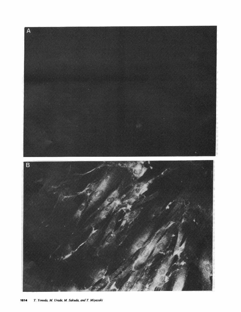
Establishment of HEMP-RV cells. When HEMPcells were in-fected with RV at a multiplicity of infection of 0.1, they sup-ported RV replication and produced infectious viruses at a titerof 104 plaque-forming units (pfu)/ml at 4 d postinfection witha slight cytopathic effect. After 1 mocultivation, infected HEMPcells were maintained in stable culture and grew with a spon-taneous release of infectious virus particles into the culture me-dium. Indirect immunofluorescent antibody staining showed thepresence of RV specific antigens in the cytoplasm of HEMPcells (Fig. 1). These findings show that the HEMPcells werepersistently infected with RV, and so they were designated asHEMP-RVcells. HEMP-RVcells consistently produce infectiousvirus particles at a titer of 103- I04 pfu/ml. HEMP-RVcells
Persistent Rubella Virus Infection in HumanEmbryonic Cells 1615
Figure 1. Indirect immunofluorescent antibody staining of HEMPcells persistently infected with RV. HEMP-RVcells were stained with preim-mune rabbit IgG (A) or anti-RV rabbit antiserum (B) as described in Methods. Note that fluorescence is absent in A and present in cytoplasmin B.
I
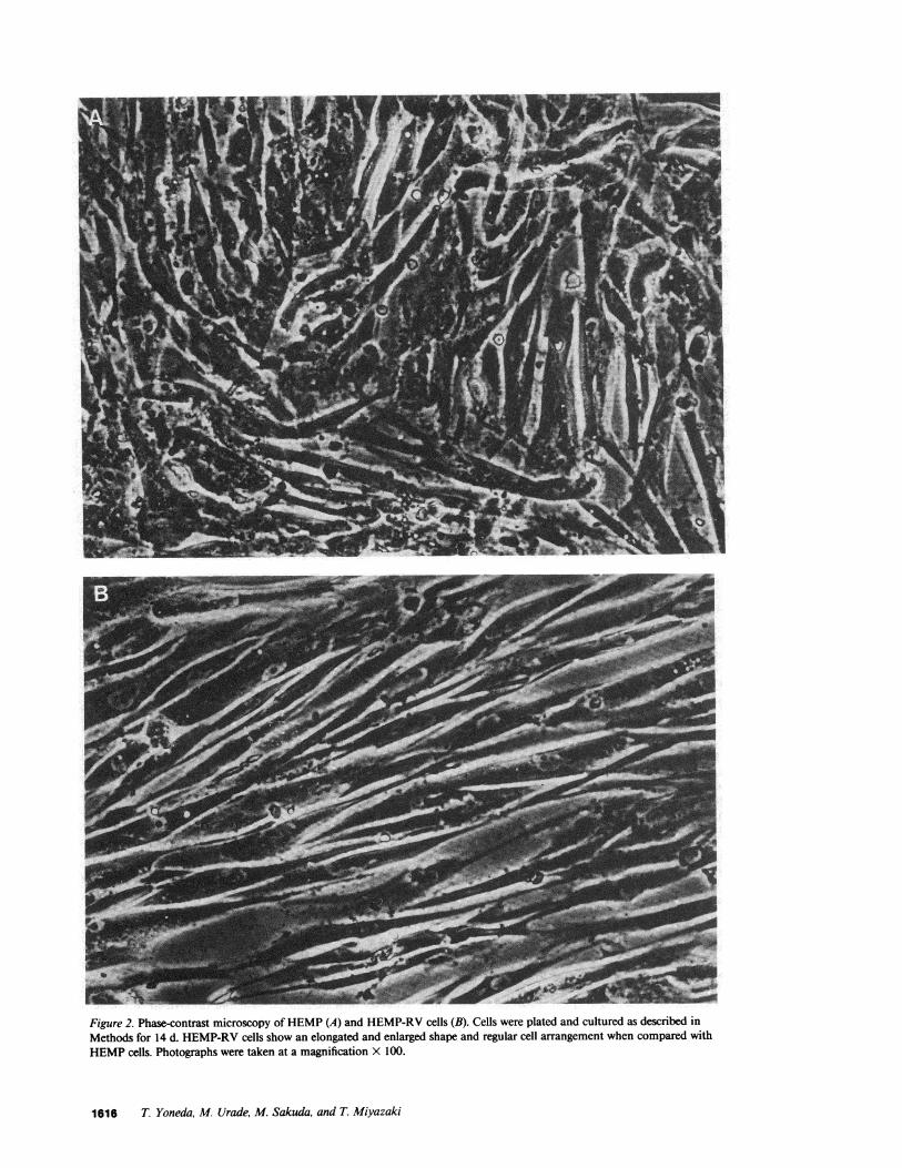
Figure 2. Phase-contrast microscopy of HEMP(A) and HEMP-RVcells (B). Cells were plated and cultured as described inMethods for 14 d. HEMP-RVcells show an elongated and enlarged shape and regular cell arrangement when compared withHEMPcells. Photographs were taken at a magnification x 100.
1616 T Yoneda, M. Urade, M. Sakuda, and T. Miyazaki
demonstrated an elongated and enlarged shape when comparedwith HEMPcells (Fig. 2), which differs from the previousreport (2 1).
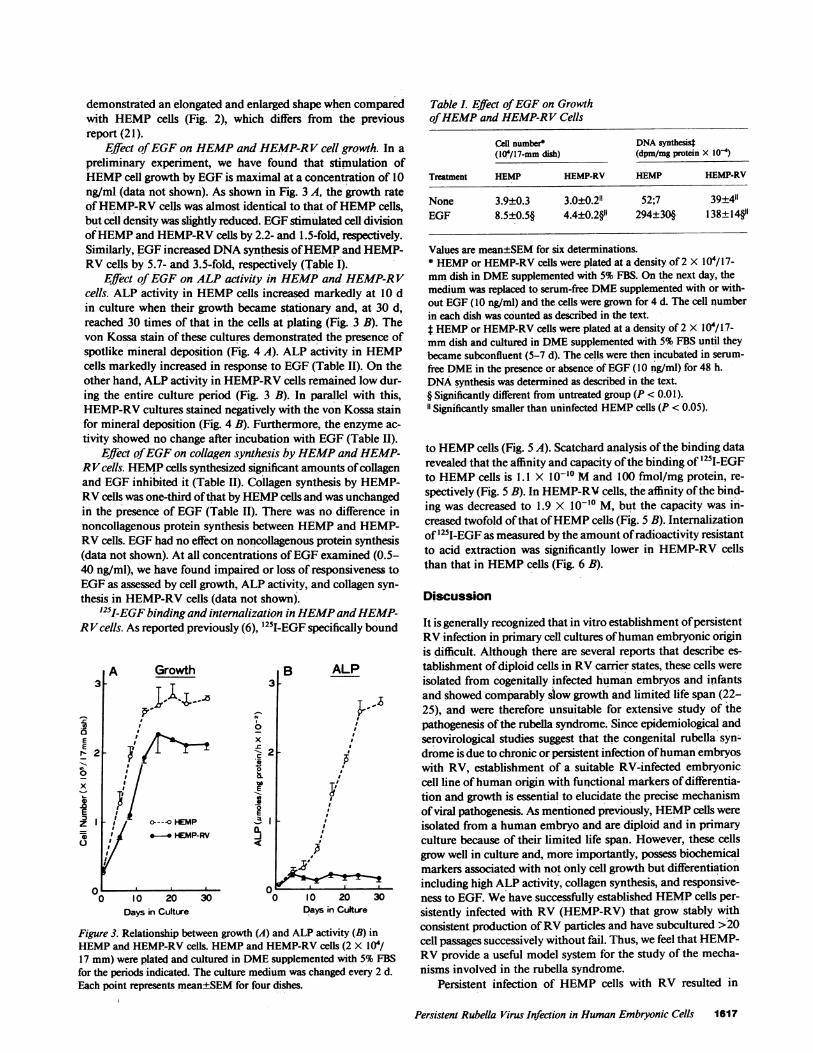
Effect of EGFon HEMPand HEMP-RV cell growth. In apreliminary experiment, we have found that stimulation ofHEMPcell growth by EGFis maximal at a concentration of 10ng/ml (data not shown). As shown in Fig. 3 A, the growth rateof HEMP-RVcells was almost identical to that of HEMPcells,but cell density was slightly reduced. EGFstimulated cell divisionof HEMPand HEMP-RVcells by 2.2- and 1.5-fold, respectively.Similarly, EGFincreased DNAsynthesis of HEMPand HEMP-RVcells by 5.7- and 3.5-fold, respectively (Table I).
Effect of EGFon ALP activity in HEMPand HEMP-RVcells. ALP activity in HEMPcells increased markedly at 10 din culture when their growth became stationary and, at 30 d,reached 30 times of that in the cells at plating (Fig. 3 B). Thevon Kossa stain of these cultures demonstrated the presence ofspotlike mineral deposition (Fig. 4 A). ALP activity in HEMPcells markedly increased in response to EGF(Table II). On theother hand, ALP activity in HEMP-RVcells remained low dur-ing the entire culture period (Fig. 3 B). In parallel with this,HEMP-RVcultures stained negatively with the von Kossa stainfor mineral deposition (Fig. 4 B). Furthermore, the enzyme ac-tivity showed no change after incubation with EGF(Table II).
Effect of EGFon collagen synthesis by HEMPand HEMP-RVcells. HEMPcells synthesized significant amounts of collagenand EGFinhibited it (Table II). Collagen synthesis by HEMP-RVcells was one-third of that by HEMPcells and was unchangedin the presence of EGF(Table II). There was no difference innoncollagenous protein synthesis between HEMPand HEMP-RVcells. EGFhad no effect on noncollagenous protein synthesis(data not shown). At all concentrations of EGFexamined (0.5-40 ng/ml), we have found impaired or loss of responsiveness toEGFas assessed by cell growth, ALP activity, and collagen syn-thesis in HEMP-RVcells (data not shown).
l2s-EGF binding and internalization in HEMPand HEMP-RVcells. As reported previously (6), '25I-EGF specifically bound
0aE
rE2-70x
.SEZ I='1
Growth
CY
x-C
0
-JE
E
M<-1
lo 20Days in Culture
B ALP3
II2 .
C~~~~
02 3
si
.'Yl /
0 I 0 20 30Days in Culture
Figure 3. Relationship between growth (A) and ALP activity (B) inHEMPand HEMP-RVcells. HEMPand HEMP-RVcells (2 X 10⁴/17 mm)were plated and cultured in DMEsupplemented with 5%FBSfor the periods indicated. The culture medium was changed every 2 d.Each point represents mean±SEMfor four dishes.
Table I. Effect of EGFon Growthof HEMPand HEMP-RV Cells
Cell number* DNAsynthesi4(104/17-mm dish) (dpm/mg protein X 104)
Treatment HEMP HEMP-RV HEMP HEMP-RV
None 3.9±0.3 3.0±0.21 52;7 39±4"EGF 8.5±0.5§ 4.4±0.2§1" 294±30§ 138±14§1
Values are mean±SEMfor six determinations.* HEMPor HEMP-RVcells were plated at a density of 2 X l04/17-mmdish in DMEsupplemented with 5% FBS. On the next day, themedium was replaced to serum-free DMEsupplemented with or with-out EGF(10 ng/ml) and the cells were grown for 4 d. The cell numberin each dish was counted as described in the text.t HEMPor HEMP-RVcells were plated at a density of 2 X I0'/17-mmdish and cultured in DMEsupplemented with 5% FBS until theybecame subconfluent (5-7 d). The cells were then incubated in serum-free DMEin the presence or absence of EGF(10 ng/ml) for 48 h.DNAsynthesis was determined as described in the text.§ Significantly different from untreated group (P < 0.01).11 Significantly smaller than uninfected HEMPcells (P < 0.05).
to HEMPcells (Fig. 5 A). Scatchard analysis of the binding datarevealed that the affinity and capacity of the binding of '25I-EGFto HEMPcells is 1.1 X 10⁻¹⁰ Mand 100 fmol/mg protein, re-spectively (Fig. 5 B). In HEMP-RVcells, the affinity of the bind-ing was decreased to 1.9 X 10⁻¹⁰ M, but the capacity was in-creased twofold of that of HEMPcells (Fig. 5 B). Internalizationof '25I-EGF as measured by the amount of radioactivity resistantto acid extraction was significantly lower in HEMP-RVcellsthan that in HEMPcells (Fig. 6 B).
Discussion
It is generally recognized that in vitro establishment of persistentRVinfection in primary cell cultures of human embryonic originis difficult. Although there are several reports that describe es-tablishment of diploid cells in RVcarrier states, these cells wereisolated from cogenitally infected human embryos and infantsand showed comparably slow growth and limited life span (22-25), and were therefore unsuitable for extensive study of thepathogenesis of the rubella syndrome. Since epidemiological andserovirological studies suggest that the congenital rubella syn-drome is due to chronic or persistent infection of human embryoswith RV, establishment of a suitable RV-infected embryoniccell line of human origin with functional markers of differentia-tion and growth is essential to elucidate the precise mechanismof viral pathogenesis. As mentioned previously, HEMPcells wereisolated from a human embryo and are diploid and in primaryculture because of their limited life span. However, these cellsgrow well in culture and, more importantly, possess biochemicalmarkers associated with not only cell growth but differentiationincluding high ALP activity, collagen synthesis, and responsive-ness to EGF. Wehave successfully established HEMPcells per-sistently infected with RV (HEMP-RV) that grow stably withconsistent production of RVparticles and have subcultured >20cell passages successively without fail. Thus, we feel that HEMP-RVprovide a useful model system for the study of the mecha-nisms involved in the rubella syndrome.
Persistent infection of HEMPcells with RV resulted in
Persistent Rubella Virus Infection in HumanEmbryonic Cells 1617
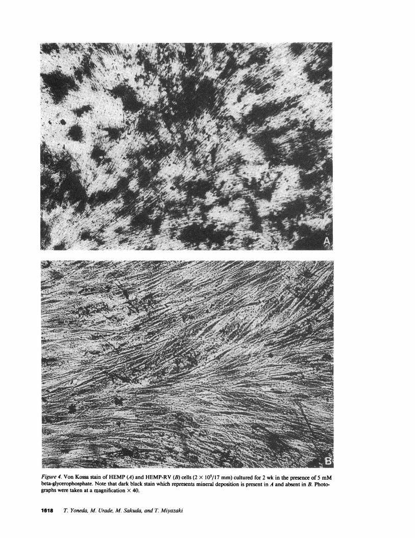
Figure 4. Von Kossa stain of HEMP(A) and HEMP-RV(B) cells (2 X I0O/17 mm)cultured for 2 wk in the presence of 5 mMbeta-glycerophosphate. Note that dark black stain which represents mineral deposition is present in A and absent in B. Photo-graphs were taken at a magnification X 40.
1618 T. Yoneda, M. Urade, M. Sakuda, and T. Miyazaki
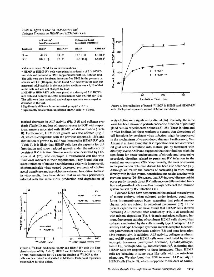
Table II. Effect of EGFon ALPActivity andCollagen Synthesis in HEMPand HEMP-RV Cells
ALP activity* Collagen synthesist(jumol/mg protein/h) (% collagen synthesized)
Treatment HEMP HEMP-RV HEMP HEMP-RV
None 48±5 18±1" 12.3±1.0 4.3±0.3"EGF 102±10§ 17±11 6.3±0.4§ 4.6±0.4"
Values are mean±SEMfor six determinations.* HEMPor HEMP-RVcells were plated at a density of 2 X 10/17-mmdish and cultured in DMEsupplemented with 5%FBS for 10 d.The cells were then incubated in serum-free DMEin the presence orabsence of EGF(10 ng/ml) for 48 h and ALP activity in the cells wasmeasured. ALP activity in the incubation medium was <1/10 of thatin the cells and was not changed by EGF.f HEMPor HEMP-RVcells were plated at a density of 2 X 10/17-mmdish and cultured in DMEsupplemented with 5%FBS for 10 d.The cells were then incubated and collagen synthesis was assayed asdescribed in the text.§ Significantly different from untreated group (P < 0.01).11 Significantly smaller than uninfected HEMPcells (P < 0.01).
marked decreases in ALP activity (Fig. 3 B) and collagen syn-thesis (Table II) and loss of responsiveness to EGFwith respectto parameters associated with HEMPcell differentiation (TableII). Furthermore, HEMPcell growth was also affected (Fig. 3A), which is compatible with the previous reports (21, 25), andstimulation of growth by EGFwas impaired in HEMP-RVcells(Table I). It is likely that HEMPcells lose the capacity for dif-ferentiation and show reduced growth under the influence ofpersistent RV infection. Similar results were described by Old-stone et al. (26), who employed a transformed cell line withfunctional markers in their experiments. They found that per-sistent infection of mouse neuroblastoma cells with lymphocyticchoriomeningitis virus caused decreased synthesis of cholineacetyl transferase and acetylcholine esterase. In addition to thesein vitro results, they have shown that in animals persistentlyinfected with the same virus, production and degradation of
A B Kd10- 1.0-
HEMP 0.1 1 MM
58 0.8 HEMP-RV 0.1 9nM
el 8-Y _HEp 0.2-8
0~~~~~~~
U.4- ~~~~~~0.4-
0-a__ HEMP21 Y *-0 HEMP-RV 002
i 2 3 50 100 150 200
521-EGF (ng/in) 1251-EGF Bould (frroes/mg poemn)
Figure S. '25I-EGF binding to HEMPand HEMP-RVcells (A). Scat-chard analysis of Fig. 5 A (B). HEMPand HEMP-RVcells (2 X 10'/17 mm)were cultured for 10 d and the binding of '25I-EGF to thecells was determined as described in Methods. Each point representsmean±SEMfor four dishes.
X100-
}~~~~
_80- 3
Pi 60-
840~~/ o f ~ON-O HEMPm
09-.-* HEMP-RV
i20 t
10 20 30Incubation Time (min)
Figure 6. Internalization of bound '25I-EGF in HEMPand HEMP-RVcells. Each point represents mean±SEMfor four dishes.
acetylcholine were significantly altered (26). Recently, the samevirus has been shown to perturb endocrine function of pituitarygland cells in experimental animals (27, 28). These in vitro andin vivo findings led these workers to suggest that alterations ofcell functions by persistent virus infection might be implicatedin the mechanisms of virus-induced diseases. Furthermore, VanAlstyne et al. have found that RVreplication was activated whenrat glial cells differentiate into mature glia by treatment withdibutyryl cyclic AMPand suggested that their findings might besignificant for better understanding of chronic and progressiveneurologic disorders related to persistent RV infection in thecentral nervous system (29). Very recently, the roles of reovirusin the production of human disease has been also described (30).Although we realize the hazards of correlating in vitro resultsdirectly with in vivo events, nonetheless our results together withprevious reports (26-30) suggest that RV-induced diseases mightoccur partly through direct RVinfluence on differentiated func-tion and growth of cells as well as through defects of the immunesystem caused by RV infection (31).
Tyler and Koch have demonstrated that palatal mesenchymeof mouse embryo, when cultured under isolated conditions,forms intramembranous bone, suggesting that palatal mesen-chymal cells are related to osteoblast precursors (32). In thepresent experiments, we have found that HEMPcells showedincreasing ALP content after confluency (Fig. 3 B) associatedwith mineral deposition (Fig. 4 A) and synthesized collagen. Im-munofluorescent staining of confluent HEMPcells showed thatcollagen synthesized by the cells is mostly type I collagen.2 ALPactivity and type I collagen synthesis are well-accepted biochem-ical parameters of osteoblastic activity (33) and bone formation(34), respectively. In addition, ALP activity, collagen synthesis,and cAMPcontent of HEMPcells were modulated by the os-teotropic hormones parathyroid hormone, 1,25-dihydroxyvi-tamin D3, prostaglandin E2, and calcitonin (9)2, indicating thatHEMPcells are responsive to these hormones. Thus, HEMPcells are behaving in some ways like cells with the osteoblastphenotype. Wealso found that EGFincreased ALP activity inHEMPcells (Table II), which is opposite to the data of Kume-
Persistent Rubella Virus Infection in HumanEmbryonic Cells 1619
gawa et al. (35) and, similarly to a previous report (36, 37),inhibited collagen synthesis (Table II). EGFhas been shown toinfluence bone cell metabolism (34). It is, therefore, likely thatHEMPcells have some osteoblastic properties. Our findings ofno increase in ALP activity (Fig. 3 B), no mineral deposition(Fig. 4 B), marked decrease in collagen synthesis, and loss ofresponsiveness to EGFin HEMP-RVcells are of particular in-terest in view of the osseous abnormalities seen in congenitalrubella syndrome. RV-induced congenital osteopathy consistingof metaphyseal radiolucent lesion in long bones and poor min-eralization of the calvarium (5, 38-40) has been emphasized inaddition to the clinical manifestations described above. Heggiereported that RV inhibited growth of human embryonic andfetal rat bones in organ culture (4i), raising a possibility thatRVdirectly affects skeletal growth. Concentration of RVin car-tilagenous tissues that presumably results in delayed ossificationand retarded bone growth has been also reported (42). Thus,our findings together with these reports indicate that RV-inducedosteopathy is due to not only indirect nutritional and/or met-abolic disturbances caused by RV infection in other organs butthe direct effect of RVon bone and cartilage cells. We, therefore,would like to propose that alterations of growth and differentia-tion of HEMPcells by persistent RV infection might be impli-cated in the mechanism of the bone lesions seen in congenitalrubella syndrome.
Since the biological actions of EGFare mediated throughspecific and high affinity membrane-bound receptors and wealready demonstrated that HEMPcells possess specific receptorsfor EGFwith high affinity (6), it is likely that impaired respon-siveness of HEMP-RVcells to EGFis due to alteration of EGFreceptor expression by RV infection. To our surprise, however,the number of EGF receptors in HEMP-RVcells was greaterthan that in uninfected counterparts (Fig. 5). Thus, there is anapparent dissociation between EGFbinding and EGFaction inHEMP-RVcells. Recently, a similar observation has been madein streptozotocin-induced diabetic rats (43). In these rats, theeffects of insulin on the liver are diminished despite increasedinsulin binding to its receptors (43). Wehave examined inter-nalization of bound EGF in HEMPand HEMP-RVcells andfound that internalization of bound EGFin HEMP-RVcells isprofoundly reduced compared with that in HEMPcells (Fig. 6).Wespeculate therefore that in HEMP-RVcells a process oc-curring on the plasma membrane subsequent to binding butbefore internalization might be altered by RV infection. Sinceit is known that as a first step of viral infection, virus binds tocell-surface receptors (30) and RV is known to be an envelopedvirus that buds from the plasma membrane in the replicationprocess (44), it is likely that persistent RV infection modifiesplasma membrane structure and function to interfere with bind-ing of EGFand internalization of EGF-receptor complex. Thus,we suggest that decreased responsiveness of HEMP-RVcells toEGF is, at least in part, due to reduced internalization ofbound EGF.
It is unknown whether the results obtained in HEMP-RVcells are specific for RVinfection. However, as mentioned earlier,establishment of HEMPcells persistently infected with a virusis difficult, and it is not unexpected that acute viral infectionresults in alteration of HEMPcell growth and differentiation.In addition, persistent infection of HEMPcells with an oncogenicvirus is unlikely to be related to the purpose of our present study,since congenital RV disease is not a neoplastic disease. Fur-thermore, HEMP-RVcells failed to form colonies in semi-solidagar or to form tumors when transplanted in athymic nude mice
(data not shown), indicating that HEMP-RVcells are not trans-formed. Thus, it should be noted that our data were obtainedin nontransformed HEMPcells chronically infected with RVwith no cytopathic effects, providing an experimental modelmore closely reflecting the in vivo situation of RV infection.Although we did not examine the effect of other viruses onHEMPcell growth and differentiation, we believe our findingsare of potential importance to an understanding of the mecha-nism of virus-induced diseases.
Acknowledgments
Weare grateful to Dr. Gregory R. Mundy for his helpful discussion andMutsuko Tamai and Nancy Garrett for their secretarial assistance.
References
1. Gregg, N. M. 1941. Congenital cataract following German measlesin mother. Trans. Ophthalmol. Soc. Australia. 3:35-46.
2. Rudolph, A. J., M. D. Yow, C. A. Phillips, M. M. Desmond,R. J. Blattner, and J. L. Melnick. 1965. Transplacental rubella infectionin newly born infants. J. Am. Med. Assoc. 191:843-845.
3. Korones, S. B., L. E. Ainger, G. R. G. Monif, J. Roane, J. L.Sever, and F. Fuster. 1965. Congenital rubella syndrome: new clinicalaspects with recovery of virus from affected infants. J. Pediatr. 67:166-181.
4. Cotlier, E., J. Fox, G. Bohigian, C. Beaty, and A. D. Pree. 1968.Pathogenic effects of rubella virus on embryos and newborn rats. Nature(Lond.). 217:38-40.
5. Singleton, E. B., A. J. Rudolph, H. S. Rosenberg, and D. B. Singer.1966. The roentgenographic manifestations of the rubella syndrome innewborn infants. Am. J. Roentgenol. Radium Ther. Nucl. Med. 97:82-91.
6. Yoneda, T., and R. M. Pratt. 1981. Mesenchymal cells from thehuman embryonic palate are highly responsive to epidermal growth fac-tor. Science (Wash. DC). 213:563-565.
7. Yoneda, T., and R. M. Pratt. 1981. Interaction between gluco-corticoids and epidermal growth factor in vitro in the growth of palatalmesenchymal cells from the human embryo. Differentiation. 19:194-198.
8. Yoneda, T., N. Nishikawa, and M. Sakuda. 1984. Responsivenessof mesenchymal cells from human embryonic palate (HEMP cells) tocalcium-regulating hormones. In Endocrine Control of Bone and CalciumMetabolism. D. V. Cohn, T. Fujita; J. T. Potts, and R. V. Talmage,editors. Excerpta Medica, Amsterdam-New York, Oxford. 246-247.
9. Yoneda, T., and R. M. Pratt. 1981. Glucocorticoid receptors inpalatal mesenchymal cells from the human embryo: relevance to humancleft palate formation. J. Cranio. Genet. Dev. Biol. 1:411-423.
10. Sato, M., M. Urade, N. Maeda, T. Miyazaki, Y. Watanabe, T.Shiba, and N. Yamamoto. 1978. Isolation and characterization of a newrubella variant with DNApolymerase activity. Arch. Virol. 56:89-103.
11. Dulbecco, M., and M. Vogt. 1954. Plaque formation and isolationof pure lines with poliomyelotis virus. J. Exp. Med. 99:167-187.
12. Sato, M., N. Maeda, M. Urade, M. Kuribayashi, K. Shirasuna,H. Yoshida, and T. Miyazaki. 1978. Persistent infection of primary hu-man cell cultures with rubella variant carrying DNApolymerase activity.Arch. Virol. 56:181-187.
13. Majeska, R. J., and G. A. Rodan. 1982. Alkaline phosphataseinhibition by parathyroid hormone and isoproterenol in a clonal ratosteosarcoma cell line. Possible mediation by cyclic AMP. Calcif TissueInt. 34:59-66.
14. Lowry, 0. H. 1955. Micromethods for the assay of enzyme. II.Specific procedures. Alkaline phosphatase. Methods Enzymol. 4:371-372.
15. Binderman, I., R. M. Greene, and J. P. Pennypacker. 1979. Cal-cification of differentiation skeletal mesenchyme in vitro. Science (Wash.DC). 206:222-225.
16. Scott, D. M., G. N. Kent, and D. V. Cohn. 1980. Collagen syn-
1620 T. Yoneda, M. Urade, M. Sakuda, and T. Miyazaki
thesis in cultured osteoblast-like cells. Arch. Biochem. Biophys. 201:384-391.
17. Peterkofsky, G., and R. Diegelman. 1971. Use of a mixture ofproteinase-free collagenases for the specific assay of collagen in the pres-ence of other proteins. Biochemistry. 10:988-994.
18. Raisz, L. G., D. M. Maina, S. C. Gworek, J. W. Dietrich, andE. M. Canalis. 1978. Hormonal control of bone collagen synthesis invitro: inhibitory effect of I-hydroxylated vitamin D metabolites. Endo-crinology. 102:731-735.
19. Haigler, H. T., F. R. Maxfield, M. C. Willingham, and I. Pastan.1980. Dansylcadaverine inhibits internalization of '25l epidermal growthfactor in Balb 3T3 cells. J. Biol. Chem. 253:3970-3977.
20. Lowry, 0. H., N. J. Rosebrough, A. L. Farr, and R. J. Randall.1951. Protein measurement with folin phenol reagent. J. Biol. Chem.193:265-275.
21. Plotkin, S. A., and A. Vaheri. 1967. Human fibroblasts infectedwith rubella virus produce a growth inhibitor. Science (Wash. DC). 156:659-661.
22. Kay, H. E. M., M. E. Peppercorn, J. S. Porterfield, K. McCarthy,and C. H. Taylor-Robinson. 1964. Congenital rubella infection of a hu-man embryo. Br. Med. J. 2:166-167.
23. Goffe, A. 1965. A diploid human cell strain with chronic inap-parent rubella infection. Arch. Gesamte Virusforsch. 16:149.
24. Plotkin, S. A., A. Boue, and J. G. Boue. 1965. The in vitro growthof rubella virus in human embryo cells. Am. J. Epidemiol. 81:71-85.
25. Rawls, W. E., and J. L. Melnick. 1966. Rubella virus carriercultures derived from congenitally infected infants. J. Exp. Med. 123:795-816.
26. Oldstone, M. B. A., J. Holmstoen, and R. M. Welsh, Jr. 1977.Alterations of acetylcholine enzymes in neuroblastoma cells persistentlyinfected with lymphocytic choriomeningitis virus. J. Cell. Physiol. 91:459-472.
27. Oldstone, M. B. A., Y. N. Shinha, P. Blout, A. Tishon, M. Rod-riguez, R. von Wedel, and P. W. Lampert. 1982. Virus-induced alterationsin homeostasis: alterations in differentiated functions of infected cells invivo. Science (Wash. DC). 218:1125-1127.
28. Oldstone, M. B. A., M. Rodriguez, W. H. Daughoday, andP. W. Lampert. 1984. Viral perturbation of endocrine function: disor-dered cell function leads to disturbed homeostasis and disease. Nature(Lond.). 307:278-28 1.
29. Alstyne, D. V., and D. W. Paty. 1983. The effect of dibutyryl
cyclic AMPon restricted replication of rubella virus in rat glial cells inculture. Virology. 124:172-180.
30. Sharpe, A. H., and B. N. Fields. 1985. Pathogenesis of viral in-fections. Basic concepts derived from the reovirus model. N. Engl. J.Med. 312:486-497.
31. Rawls, W. E. 1974. Viral persistence in congenital rubella. Prog.Med. Virol. 18:273-288.
32. Tyler, M. S., and W. E. Koch. 1977. In vitro development ofpalatal tissues from embryonic mice. II. Tissue isolation and recombi-nation studies. J. Embryol. Exp. Morphol. 38:19-36.
33. Robinson, R. A., S. B. Doty, and R. R. Cooper. 1973. Electronmicroscopy of mammalian bone. In Biological Mineralization. I. Zipkin,editor. Academic Press, Inc., NewYork. 257-296.
34. Raisz, L. G., and B. E. Kream. 1983. Regulation of bone for-mation. N. Engl. J. Med. 309:29-35, 83-89.
35. Kumegawa, M., M. Hiramatsu, K. Hatakeyama, T. Yajima, H.Kadama, T. Osaki, and K. Kurisu. 1983. Effects of epidermal growthfactor on osteoblastic cells in vitro. Calcif Tissue Int. 35:542-548.
36. Canalis, E. M., and L. G. Raisz. 1979. Effect of epidermal growthfactor on bone formation in vitro. Endocrinology. 104:862-886.
37. Hata, R., H. Hori, Y. Hagai, S. Tanaka, M. Kondo, M. Hiramatsu,N. Utsumi, and M. Kumegawa. 1984. Selective inhibition of type I col-lagen synthesis in osteoblastic cells by epidermal growth factor. Endo-crinology. 115:867-876.
38. Rudolph, A. J., E. B. Singleton, H. S. Rosenberg, D. B. Singer,and C. A. Phillips. 1965. Osseous manifestations of the congenital rubellasyndrome. Am. J. Dis. Child. 110:428-433.
39. Sekeles, E., and A. Ornoy. 1975. Osseous manifestations of ges-tational rubella in young human fetuses. Am. J. Obstet. Gynecol. 122:307-312.
40. Reed, G. B. 1969. Rubella bone lesions. J. Pediatr. 74:208-213.41. Heggie, A. D. 1976. Growth inhibition of human embryonic and
fetal rat bones in organ culture by rubella virus. Teratology. 15:47-56.42. London, W. T., D. A. Fuccillo, B. Anderson, and J. L. Sever.
1970. Concentration of rubella virus antigen in chondrocytes of con-genitally infected rabbits. Nature (Lond.). 226:172-173.
43. Kadowaki, T., M. Kasuga, Y. Akanuma, 0. Ezaki, and F. Takaku.1984. Decreased autophosphorylation of the insulin receptor-kinase instreptozotacin-diabetic rats. J. Biol. Chem. 259:14208-14216.
44. Becker, Y. 1976. Alstiviral drugs: mode of action and chemo-therapy of viral infections of man. Monogr. Virol. I 1: 1-130.
Persistent Rubella Virus Infection in HumanEmbryonic Cells 1621