alterations of paraoxonase and platelet … · 2 alterations of paraoxonase and platelet-activating...
TRANSCRIPT
1
ALTERATIONS OF PARAOXONASE AND PLATELET-ACTIVATING
FACTOR ACETYLHYDROLASE ACTIVITIES IN PATIENTS ON PERITONEAL
DIALYSIS
1Evagelos N Liberopoulos, MD, Senior House Officer
2Eleni Papavasiliou, Postgraduate Student in Biochemistry
1George A Miltiadous, MD, Senior House Officer
3Marios Cariolou, PhD, Molecular Biologist
1Kostas C Siamopoulos, MD, Professor of Nephrology
2Alexandros D Tselepis, MD, PhD, Associate Professor in Biochemistry
1Moses S Elisaf, MD, FACA, FRSH, Professor of Internal Medicine
Running Head: PON1 and PAF-AH in Peritoneal Dialysis
1Department of Internal Medicine, School of Medicine, and 2Department of Biochemistry,
School of Chemistry, University of Ioannina, Ioannina-Greece, 3Molecular Genetics
Department B-DNA Identification Laboratory, the Cyprus Institute of Neurology and
Genetics, Nicosia, Cyprus
Address for correspondence:
Moses S Elisaf, MD, FACA, FRSH
Department of Internal Medicine
University of Ioannina
451 10 Ioannina Greece
Tel +302651-097509
Fax +302651-097016
e-mail: [email protected]
2
ALTERATIONS OF PARAOXONASE AND PLATELET-ACTIVATING
FACTOR ACETYLHYDROLASE ACTIVITIES IN PATIENTS ON PERITONEAL
DIALYSIS
ABSTRACT
Objective: A more atherogenic lipid profile seen in peritoneal dialysis (PD) patients
cannot fully explain the increased incidence of atherosclerosis in this population. Oxidative
modification of low-density lipoproteins (LDL) is considered to play a central role in the
atherogenic process, whereas high-density lipoprotein (HDL) protects LDL from
oxidation. On the other hand, it has been suggested that LDL and HDL from PD patients
are more resistant to oxidation than those from control subjects, while PD-HDL equally
protects LDL from oxidation compared to control-HDL. Two HDL-associated enzymes
have been shown to protect both LDL and HDL from oxidation: paraoxonase (PON1) and
HDL-associated platelet-activating factor acetylhydrolase (HDL-PAF-AH). Furthermore,
low PON1 activity and high total plasma PAF-AH concentration, which mainly represents
the LDL-associated enzyme, have been shown to be independent risk factors for coronary
artery events in the general population. However, there are limited data regarding possible
alterations of these enzymes in PD patients. The aim of our study was to examine the
possible alterations of PON1 and PAF-AH activities in patients undergoing PD.
Design: A cross-sectional study.
Setting: University Medical Center.
Participants: Fifty-six PD patients of Caucasian origin and 86 matched controls were
studied.
Measurements: In all subjects, serum PON1 activity toward paraoxon (paraoxonase)
and phenylacetate (arylesterase), as well as total serum and HDL-associated PAF-AH
3
activities were measured, while PON1 genetic polymorphisms known to influence PON1
activity (Q192R and M55L) were determined.
Results: PD patients exhibited significantly increased serum PON1 (paraoxonase) and
PON1 (arylesterase) activities compared to controls, regardless of the PON1
polymorphisms or the levels of HDL cholesterol. Additionally, PD patients had
significantly elevated activities of total serum PAF-AH and HDL-associated PAF-AH
independently of the levels of LDL or HDL cholesterol, while the ratio of HDL-PAF
AH/total PAF-AH, which has been recently suggested to be a potential marker of
atherogenicity, was decreased in these patients compared to controls. Moreover, no
difference in the prevalence of PON1 polymorphisms between PD patients and controls
was found.
Conclusion: The elevated activities of PON1 and HDL-PAF AH could explain the
increased resistance of PD-HDL to oxidation, while the higher activity of total PAF-AH
and the decreased HDL-PAF AH/total PAF-AH ratio could contribute to the increased
incidence of atherosclerosis in these patients.
Key words: Paraoxonase, Platelet-activating factor acetylhydrolase, Lipoprotein-
associated phospholipase A2, Peritoneal dialysis, CAPD, Lipids, Renal failure,
Atherosclerosis.
4
INTRODUCTION
Patients undergoing peritoneal dialysis (PD) have a high incidence of accelerated
atherosclerosis and coronary artery disease (CAD) [1,2]. Studies have shown a higher
prevalence of dyslipidemic risk factors associated with an increased risk of CAD, such as
elevated levels of total cholesterol, low-density lipoprotein cholesterol (LDL-C),
triglycerides and lipoprotein (a) [Lp(a)], and decreased levels of high-density lipoprotein
cholesterol (HDL-C) in PD patients [3-6]. However, these abnormalities cannot fully
explain the increased incidence of CAD in this population. In this context, recent reports
support a role for non-traditional cardiovascular risk factors, such as the elevated plasma
concentrations of C-reactive protein [7], or the effect of the apolipoprotein E gene
polymorphisms [8].
Oxidative modification of LDL is considered to play a key role in the atherogenic
process [9]. On the other hand, it is well established that HDL plays a protective role
against atherosclerosis [10], while oxidized HDL loses its anti-atherogenic potential [11].
In addition to its role in reverse cholesterol transport, HDL also exhibits anti-inflammatory
and anti-oxidant properties. Two HDL-associated enzymes have been shown to contribute
to these HDL properties, thus protecting both LDL and HDL from oxidation: paraoxonase
(PON1) and HDL-associated platelet-activating factor acetylhydrolase (HDL-PAF AH).
However, despite the higher incidence of atherosclerosis in PD patients, both LDL and
HDL from these patients have been found to be more resistant to in vitro oxidation than
lipoproteins from control subjects, while PD-HDL and control-HDL were equally effective
in preventing cooper-mediated LDL oxidation [12]. Surprisingly, there are limited data
regarding possible alterations of HDL-associated anti-oxidant enzymes in PD patients.
PON1 is an esterase that is exclusively associated with HDL (especially with a HDL
subclass which also contains apolipoprotein AI and clusterin). PON1 can hydrolyze
5
oxidized phospholipids and cholesteryl ester hydroperoxides formed during lipoprotein
peroxidation [13]. The PON1 gene is located on the long arm of chromosome 7. Two main
genetic polymorphisms that determine PON1 activity have been described, one at position
55 (methionine/leukine, M55L) and the other at position 192 (glutamine/arginine, Q192R)
[14]. The substrates for PON1 that have been mostly used in assays in vitro are paraoxon
and phenylacetate. The latter is used to measure the arylesterase activity of PON1, which is
considered to be a more accurate estimate of the PON1 mass. Interestingly, low serum
PON1 activity toward paraoxon has recently been shown to be an independent risk factor
for coronary events in high-risk men [15].
Studies in patients undergoing hemodialysis have revealed a decreased serum PON1
activity in this population, even if it was corrected for HDL-C levels or PON1 genotypes
[16-18]. Furthermore, decreased PON1 activity toward paraoxon, but not phelylacetate,
was found in the only study which examined the serum PON1 activity in PD patients [18].
PAF-AH (also termed lipoprotein-associated phospholipase A2) hydrolyzes short acyl
groups at the sn-2 position of PAF and oxidized phospholipids [19]. In human plasma,
PAF-AH is primarily associated with LDL particles, whereas a small proportion (<20% of
total enzyme activity) is associated with HDL [19]. There has been much controversy
concerning the possible role of the LDL-associated PAF-AH in atherogenesis [19].
However, recent studies suggest that plasma levels of PAF-AH, which are proportional to
its activity and mainly reflect the LDL-associated enzyme, represent an independent risk
factor for coronary artery disease [20,21]. On the contrary, HDL-associated PAF-AH
(HDL-PAF AH) has been shown to protect LDL from oxidation and inhibit the biological
activity of oxidized LDL, thus exhibiting atheroprotective properties [19,22]. In the only
study examining the PAF-AH activity in PD patients, it was shown that total plasma, as
6
well as HDL-associated PAF-AH activities were significantly increased in these patients
compared to the control population [23].
Thus, we undertook the present study to further examine possible alterations of PON1
activity in relation to its genetic polymorphisms, as well as of total and HDL-associated
PAF-AH activities in patients undergoing PD compared with control subjects.
7
MATERIALS AND METHODS
Study population
Our study population consisted of 56 Caucasian patients originating from Northwestern
Greece on chronic ambulatory peritoneal dialysis (PD) for at least six months before their
inclusion in the study. PD patients performed 4 exchanges per day with 2-liter solutions of
1.86 or 3.86% glucose, depending on the individual need for ultrafiltration. All patients
had been free of peritonitis or any other infection at least 3 months preceding blood
sampling. The adequacy of the dialytic treatment was evaluated by the Kt/V ratio, which
ranged from 1.9 to 2.0 (weekly), while in all patients the residual renal function estimated
by the average of the residual creatinine and urea clearances was <5.0 ml/min. The renal
diagnosis was chronic glomerulonephritis in 11 patients, hypertensive nephropathy in 11
patients, chronic pyelonephritis in 12 patients, polycystic kidney disease in 6 patients and
obstructive uropathy in 7 patients, while it remained unknown in the remaining 9 patients.
Patients with a known family history of primary dyslipidemia, excessive alcohol
consumption, diabetes mellitus [fasting serum glucose >6.93 mmol/l (126 mg/dl)], obesity
(body-mass index >30 Kg/m2), liver disease, systemic illness, thyroid disorders, or any
other metabolic or endocrine disorder were excluded from the study. Patients received no
other medications except erythropoietin, multivitamins, calcitriol, iron, phosphate binders
other than sevelamer HCl, angiotensin-converting enzyme inhibitors and calcium channel
blockers. Patients taking lipid-lowering drugs or any other medication known to affect
serum lipids (e.g b-blockers) were also excluded from the study. Furthermore, 86
Caucasian healthy individuals from the same region matched for age, sex and smoking
habits with the PD patients were also studied. These subjects were consecutive healthy
unrelated individuals who underwent a regular check-up in our outpatient internal
medicine clinic. None of these individuals were receiving drugs affecting lipid profile or
8
renal function. Smoking habit was defined as smoking currently. All participants gave
informed consent for genetic analysis, and the study protocol was approved by the ethic
committee of our university hospital. In all participants blood samples were obtained after
a 14-hour overnight fast for gene genotype detection, as well as for the determination of
serum laboratory parameters. All patients were dialyzed during night with a 2-liter solution
of 1.86% glucose. Blood samples were centrifuged for 30 min (3600g) and then the serum
was separated and stored at -80 0C for analysis of laboratory parameters.
Analytical methods
Concentrations of total cholesterol and triglycerides were determined enzymatically on
the Olympus AU600 clinical chemistry analyzer (Olympus Diagnostica, Hamburg,
Germany). High-density lipoprotein cholesterol (HDL-C) was determined in the
supernatant, after precipitation of the apolipoprotein B-containing lipoproteins with
dextran sulphate-Mg++ (Sigma Diagnostics, St. Louis, MO, USA) (HDL-rich serum). Low-
density lipoprotein cholesterol (LDL-C) was calculated using the Friedewald formula if
fasting triglycerides were <4.52mmol/L (400 mg/dl) [24], while the non HDL cholesterol
was calculated by the equation: non HDL-C=Total cholesterol – HDL-C. Apolipoproteins
A1, B, E and the lipoprotein (a) [Lp(a)] were measured with a Behring Nephelometer
BN100 using reagents (antibodies and calibrators) from Date Behring Holding Gmbh
(Liederbach, Germany). The ApoA1 and ApoB assays were calibrated according to the
International Federation of Clinical Chemistry (IFCC) standards. Finally, serum
interleukin-6 (IL-6) levels were measured by ELISA (R&D Systems Europe, Abingdon,
Oxon, UK) in 21 randomly selected PD patients and 32 controls matched for age, sex, BMI
and smoking habits.
9
PON1 assay
Both PON1 (paraoxonase) and PON1 (arylesterase) activities in serum were determined
in the presence of 2 mM Ca+2 in 100 mM Tris-HCl buffer (pH 8.0) for paraoxon as a
substrate, and in 20 mM Tris-HCl buffer (pH 8.0) for phenylacetate as a substrate,
respectively, as previously described [25].
PAF-AH assay
PAF-AH activity was measured by the trichloroacetic acid precipitation procedure [26]
using as a substrate 1-O-hexadecyl-2-[3H-acetyl]-sn-glycero-3-phosphocholine ([3H]-PAF)
(10 Ci/mmol; DuPont-New England Nuclear, Boston, MA) at a final concentration of 100
µΜ [26]. Fifty µl of either serum diluted 1:50, v/v with HEPES buffer, pH 7.4, or the
HDL-containing supernatant after treatment of serum with magnesium chloride-dextran
sulfate (HDL-rich serum) (diluted 1:3, v/v with HEPES) were mixed with HEPES in a
final volume of 90 µl and used as the source of the enzyme. The reaction was performed
for 10 min at 37 oC, and PAF-AH activity was expressed as nmol PAF degraded per min
per ml of serum.
Reproducibility of PON1 (paraoxonase), PON1 (arylesterase) and PAF-AH assays was
determined by intra-assay determination of coefficients of variation (CVs). The CVs for
PON1 (paraoxonase) and PON1 (arylesterase) assays ranged between 6 to 8%, while the
CVs for PAF-AH assays ranged between 4 to 5%.
Determination of PON1 genotypes
Genomic DNA was obtained from leukocytes using standard procedures. The PON1
Q192R and M55L polymorphisms were detected using a previously reported protocol [27].
Briefly, primers for amplification of a 99-bp DNA that contains the coding sequence for
position 192 were 5’TAT TGT TGC TGT GGG ACC TGA G3’ and 5’CAC GCT AAA
CCC AAA TAC ATC TC3’. After an initial denaturation step of 5 min at 95 oC, the PCR
10
was carried out for 40 cycles, with each cycle consisting of 60 sec of denaturation at 94 oC,
45 sec of annealing at 56 oC and 45 sec of extension at 72 oC. PCR product was digested
with 5U of Alw I restriction enzyme, for 3 hours at 37 oC.
For genotyping the M55L polymorphism, the primers for amplification of 144-bp DNA
encoding codon 55 were 5’GAG TGA TGT ATA GCC CCA GTT TC3’ and 5’AGT CCA
TTA GGC AGT ATC TCC G3’. An initial incubation for 5 min at 95 oC was followed by
the step of amplification that was carried out for 40 cycles, with each cycle consisting of 1
min of denaturation at 94 oC, 45 sec of annealing at 61 oC and 45 sec of extension at 72 oC.
PCR product was digested with 5U of Hinf I restriction enzyme for 24 hours at 37 oC.
Statistical analysis
Statistical analysis was performed with STATISTICA 6.0 statistical software. Allele
frequencies were estimated by the gene-counting method. Chi square-test was used to
compare gene frequencies, while Students’ t-test for independent samples and Mann-
Whitney U test were used to test the differences of parametric and non-parametric data,
respectively, between the two study populations. The effect of the PON1 gene
polymorphisms on laboratory parameters was tested using one-way analysis of variance
(one-way ANOVA) followed by the LSD test for pairwise comparisons in case of
significant results, except for serum PON1 (Paraoxonase), PON1 (Paraoxonase)/HDL-C
and PON1 (Paraoxonase)/ApoAI, where the Kruskal-Wallis ANOVA median test was used
followed by the Mann-Whitney U test for pairwise comparisons in case of significant
results. Multiple linear regression analysis was performed to test the overall effect on
PON1 and PAF-AH activities of genetic and other factors shown to influence these
activities in univariate analysis. Log transformation of PON1 (paraoxonase) activity and
serum IL-6 levels was applied because of their skewed distribution. Finally, linkage

11
disequilibrium coefficients for Q192R and M55L genetic variants of PON1 were
calculated using the DnaSP 4.0 software package.
12
RESULTS
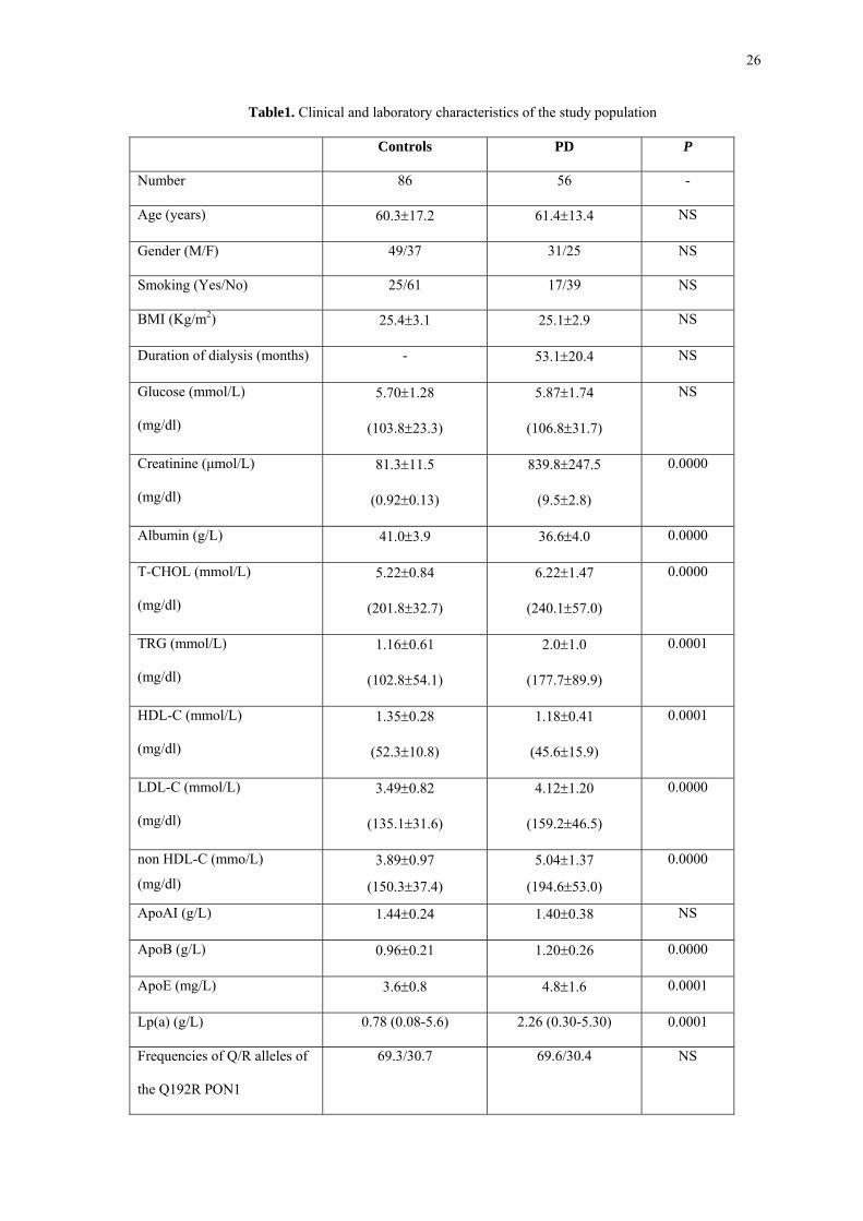
Table 1 shows the clinical and laboratory characteristics of the study population.
Control subjects and PD patients were well-matched with respect to age, gender, smoking
habits, body-mass index (BMI) and serum glucose concentration. The observed genotype
frequencies of Q192R and M55L polymorphisms did not deviate from those predicted by
the Hardy-Weinberg equation, while there was a strong linkage disequilibrium between
them (Lewontin’s D΄ coefficient of 0.75 in the control subjects and 0.71 in PD patients,
p<0.0001 for both groups). Furthermore, frequencies of Q192R and M55L gene alleles
were not different between the two groups. On the other hand, PD patients exhibited
significantly lower serum albumin levels and a more atherogenic lipid profile, consisting of
significantly increased levels of total cholesterol, LDL-C, non HDL-C, triglycerides,
apolipoproteins B and E, as well as elevated concentration of Lp(a), and decreased levels
of HDL-C.
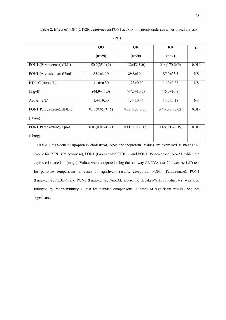
Table 2 demonstrates the effect of Q192R genotypes on PON1 activity in PD patients.
QQ individuals exhibited the lowest activity toward paraoxon, even when corrected for
HDL-C or ApoAI levels. QR patients had intermediate PON1 (paraoxonase) activity, while
RR patients showed the maximum activity. PON1 activity toward phenylacetate was not
influenced by the Q192R polymorphisms. Table 3 presents the effect of M55L genotypes
on PON1 activity in PD patients. LL patients had the highest PON1 (paraoxonase) and
PON1 (arylesterase) activities compared to LM and MM subjects, which had intermediate
and minimum activities toward paraoxon and phenylacetate, respectively. The same trend
remained true even after correction of PON1 activities for HDL-C or ApoAI levels.
Finally, neither Q192R nor M55L polymorphisms had any effect on the levels of serum
lipid parameters (data not shown).
13
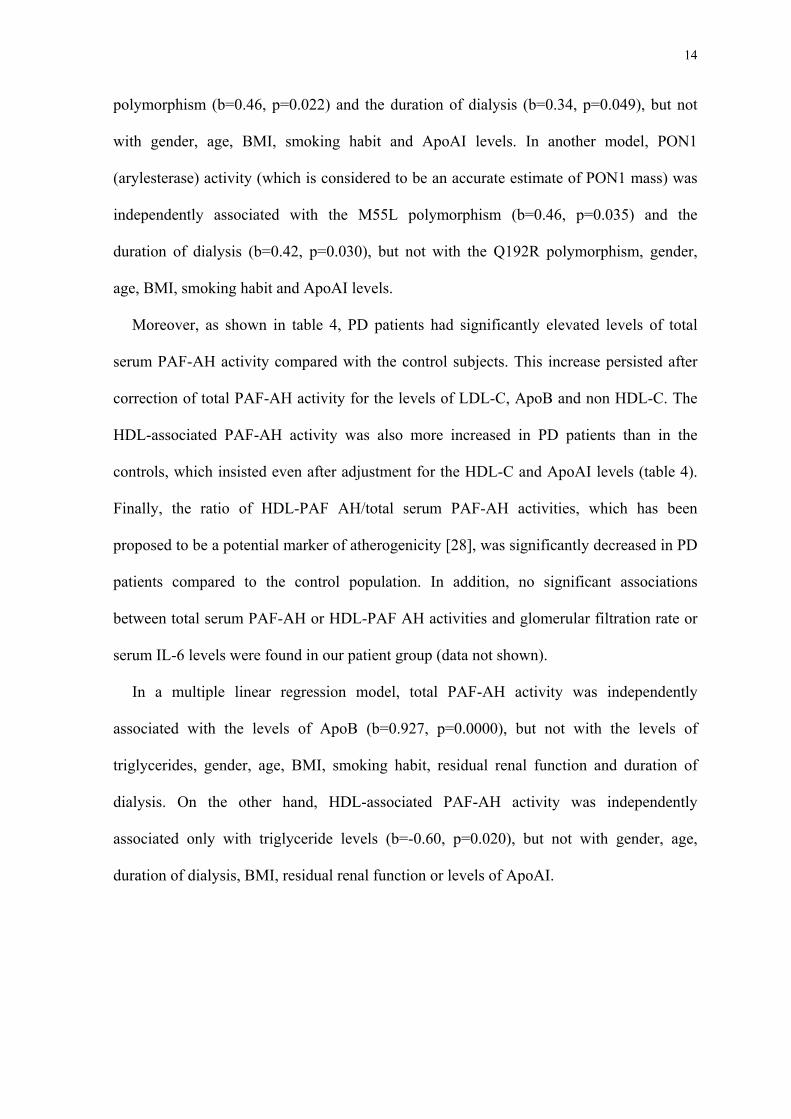
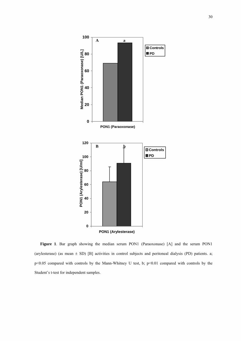
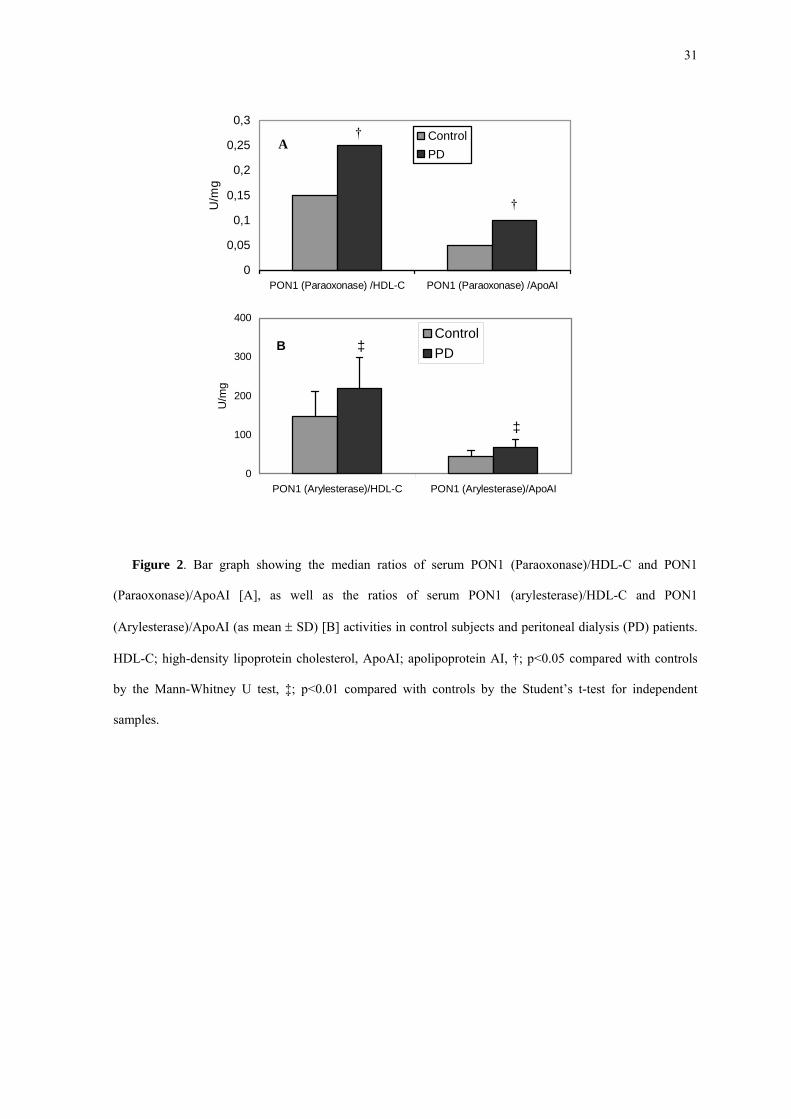
PD patients had significantly increased activities of PON1 (paraoxonase), as well as of
PON1 (arylesterase) compared to control subjects (Fig. 1A and 1B). This difference
persisted even after correction of PON1 (paraoxonase) and PON1 (arylesterase) activities
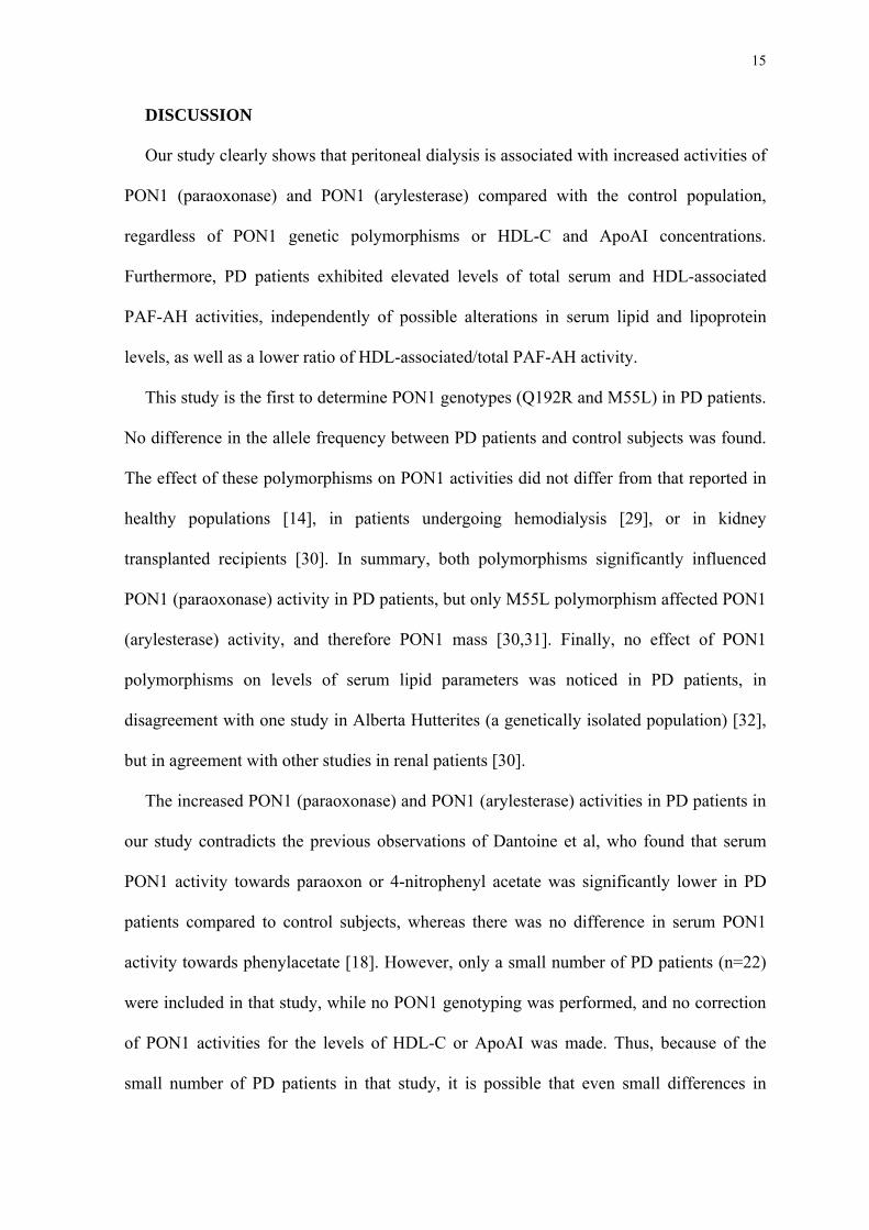
for HDL-C and ApoAI levels (Fig. 2A and 2B), suggesting that the increase in PON1
activities in PD patients is unrelated to the alterations of HDL-C or ApoAI levels seen in
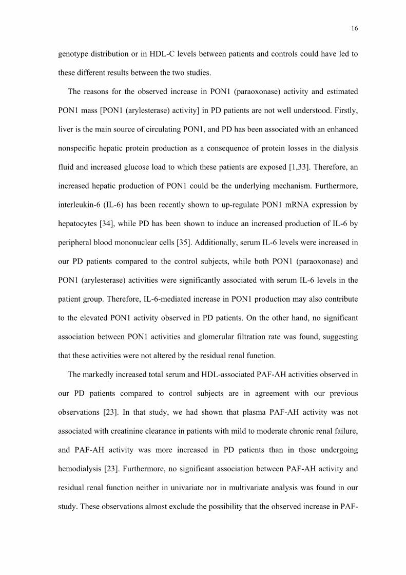
this population. Furthermore, because of the powerful effect of PON1 genotypes on PON1
activities, different genotype combinations could result in different PON1 activities
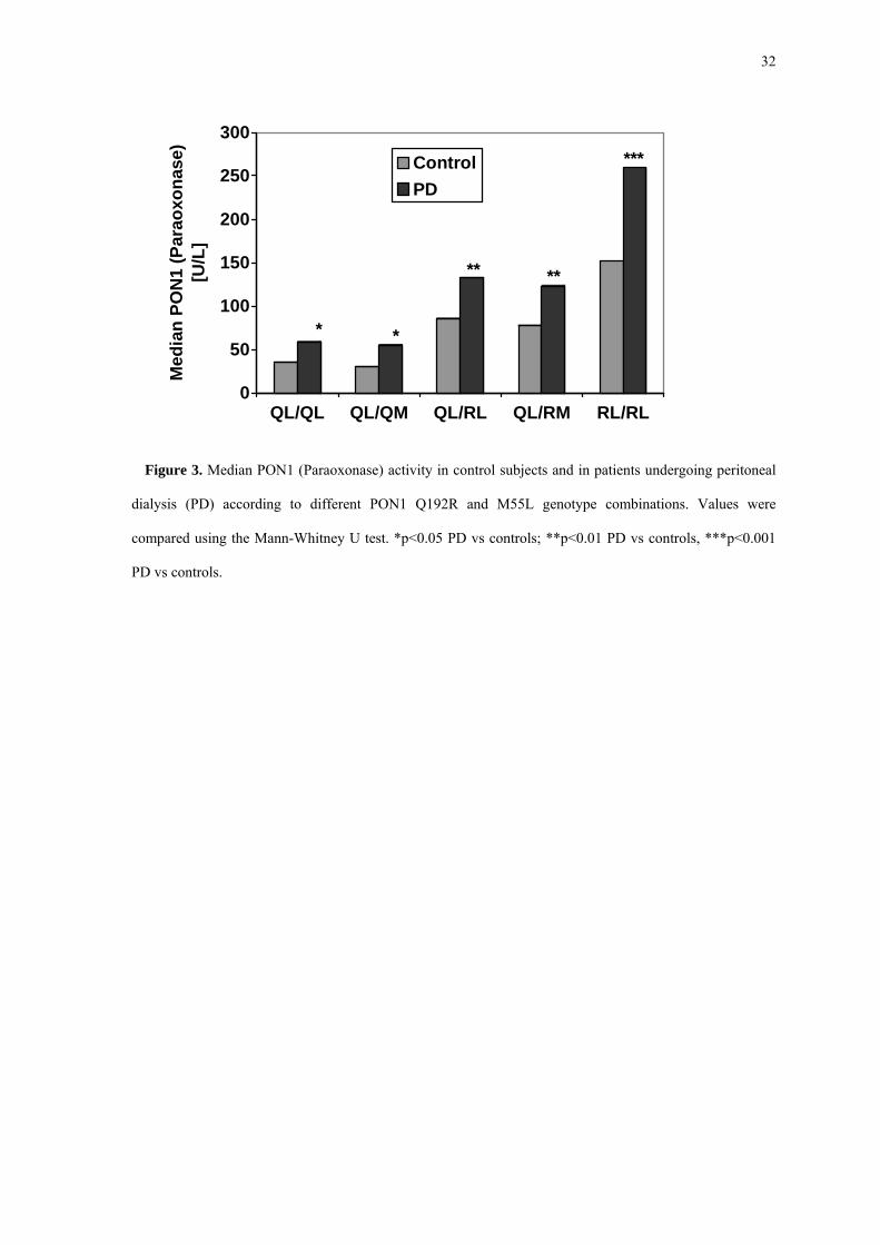
between the two groups. However, as shown in figure 3, median PON1 (paraoxonase)
activity remained significantly higher in PD patients, even if certain PON1 combined
haplotypes between the two groups were compared. Interestingly, the most pronounced
differences were observed between subjects and patients carrying the high-activity
haplotypes (RL/RL). The same observations were also made for the PON1 (arylesterase)
activity, as well as for the corrected PON1 (paraoxonase) and PON1 (arylesterase)
activities (data not shown). Therefore, PON1 activities are increased in PD patients
independently of PON1 genotypes or HDL-C and ApoAI concentrations.
Moreover, PD patients exhibited significantly increased serum IL-6 levels compared
with the controls subjects (5.3±3.9 vs 1.4±0.9 ng/L, respectively, p<0.001 by the Mann-
Whitney U test). Moreover, a significant association between serum Log IL-6 levels and
Log PON1 (paraoxonase) activity (r=0.27, p=0.03), as well as between serum Log IL-6
levels and PON1 (arylesterase) activity (r=0.31, p=0.01) was found in PD patients, but not
in the control subjects (r=0.11 and 0.12, respectively, p=NS). Additionally, no correlation
between Log PON1 (paraoxonase) or PON1 (arylesterase) activities and glomerular
filtration rate was found in our patient group (r=0.03 and 0.09, respectively, p=NS).
In a multiple linear regression model, Log PON1 (paraoxonase) activity in PD patients
was independently associated with the Q192R polymorphism (b=0.51, p=0.007), M55L
14
polymorphism (b=0.46, p=0.022) and the duration of dialysis (b=0.34, p=0.049), but not
with gender, age, BMI, smoking habit and ApoAI levels. In another model, PON1
(arylesterase) activity (which is considered to be an accurate estimate of PON1 mass) was
independently associated with the M55L polymorphism (b=0.46, p=0.035) and the
duration of dialysis (b=0.42, p=0.030), but not with the Q192R polymorphism, gender,
age, BMI, smoking habit and ApoAI levels.
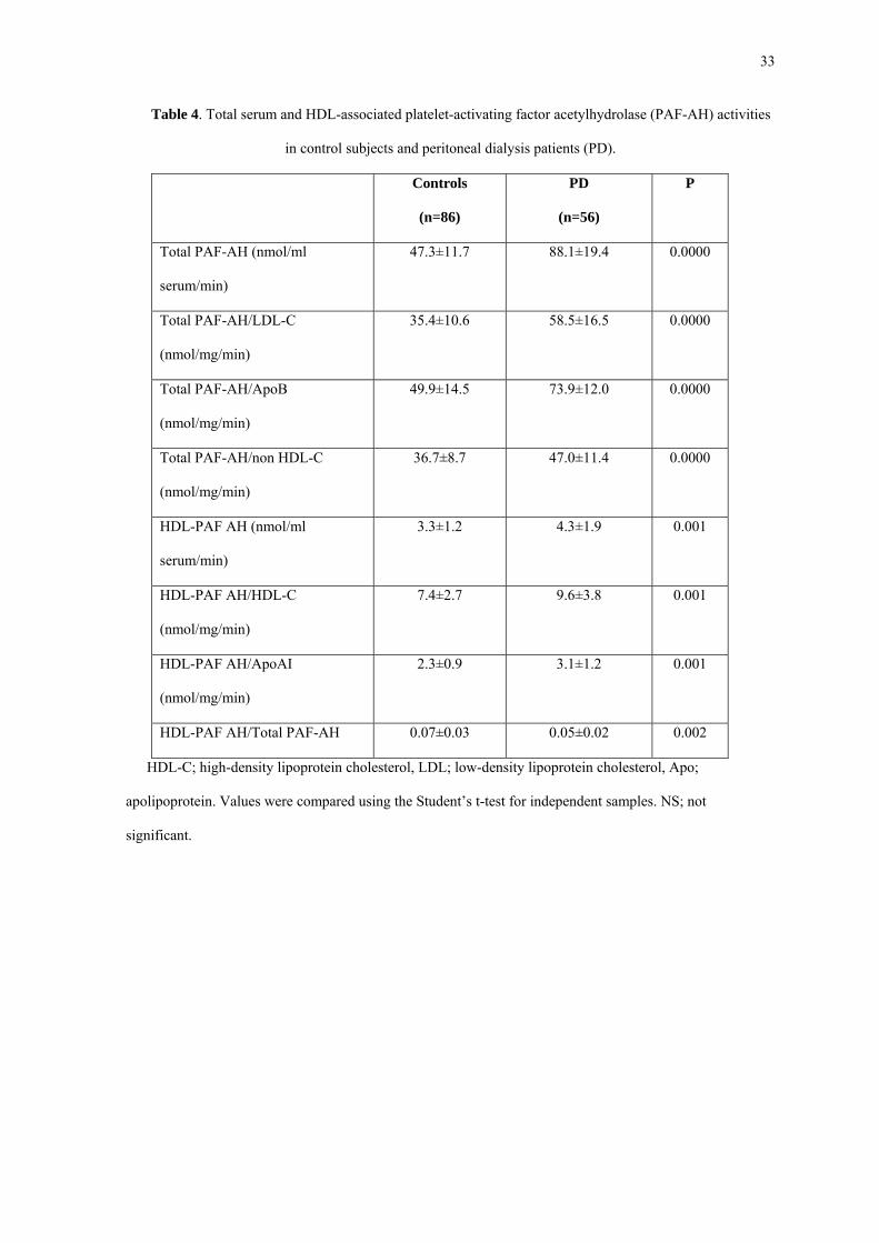
Moreover, as shown in table 4, PD patients had significantly elevated levels of total
serum PAF-AH activity compared with the control subjects. This increase persisted after
correction of total PAF-AH activity for the levels of LDL-C, ApoB and non HDL-C. The
HDL-associated PAF-AH activity was also more increased in PD patients than in the
controls, which insisted even after adjustment for the HDL-C and ApoAI levels (table 4).
Finally, the ratio of HDL-PAF AH/total serum PAF-AH activities, which has been
proposed to be a potential marker of atherogenicity [28], was significantly decreased in PD
patients compared to the control population. In addition, no significant associations
between total serum PAF-AH or HDL-PAF AH activities and glomerular filtration rate or
serum IL-6 levels were found in our patient group (data not shown).
In a multiple linear regression model, total PAF-AH activity was independently
associated with the levels of ApoB (b=0.927, p=0.0000), but not with the levels of
triglycerides, gender, age, BMI, smoking habit, residual renal function and duration of
dialysis. On the other hand, HDL-associated PAF-AH activity was independently
associated only with triglyceride levels (b=-0.60, p=0.020), but not with gender, age,
duration of dialysis, BMI, residual renal function or levels of ApoAI.
15
DISCUSSION
Our study clearly shows that peritoneal dialysis is associated with increased activities of
PON1 (paraoxonase) and PON1 (arylesterase) compared with the control population,
regardless of PON1 genetic polymorphisms or HDL-C and ApoAI concentrations.
Furthermore, PD patients exhibited elevated levels of total serum and HDL-associated
PAF-AH activities, independently of possible alterations in serum lipid and lipoprotein
levels, as well as a lower ratio of HDL-associated/total PAF-AH activity.
This study is the first to determine PON1 genotypes (Q192R and M55L) in PD patients.
No difference in the allele frequency between PD patients and control subjects was found.
The effect of these polymorphisms on PON1 activities did not differ from that reported in
healthy populations [14], in patients undergoing hemodialysis [29], or in kidney
transplanted recipients [30]. In summary, both polymorphisms significantly influenced
PON1 (paraoxonase) activity in PD patients, but only M55L polymorphism affected PON1
(arylesterase) activity, and therefore PON1 mass [30,31]. Finally, no effect of PON1
polymorphisms on levels of serum lipid parameters was noticed in PD patients, in
disagreement with one study in Alberta Hutterites (a genetically isolated population) [32],
but in agreement with other studies in renal patients [30].
The increased PON1 (paraoxonase) and PON1 (arylesterase) activities in PD patients in
our study contradicts the previous observations of Dantoine et al, who found that serum
PON1 activity towards paraoxon or 4-nitrophenyl acetate was significantly lower in PD
patients compared to control subjects, whereas there was no difference in serum PON1
activity towards phenylacetate [18]. However, only a small number of PD patients (n=22)
were included in that study, while no PON1 genotyping was performed, and no correction
of PON1 activities for the levels of HDL-C or ApoAI was made. Thus, because of the
small number of PD patients in that study, it is possible that even small differences in
16
genotype distribution or in HDL-C levels between patients and controls could have led to
these different results between the two studies.
The reasons for the observed increase in PON1 (paraoxonase) activity and estimated
PON1 mass [PON1 (arylesterase) activity] in PD patients are not well understood. Firstly,
liver is the main source of circulating PON1, and PD has been associated with an enhanced
nonspecific hepatic protein production as a consequence of protein losses in the dialysis
fluid and increased glucose load to which these patients are exposed [1,33]. Therefore, an
increased hepatic production of PON1 could be the underlying mechanism. Furthermore,
interleukin-6 (IL-6) has been recently shown to up-regulate PON1 mRNA expression by
hepatocytes [34], while PD has been shown to induce an increased production of IL-6 by
peripheral blood mononuclear cells [35]. Additionally, serum IL-6 levels were increased in
our PD patients compared to the control subjects, while both PON1 (paraoxonase) and
PON1 (arylesterase) activities were significantly associated with serum IL-6 levels in the
patient group. Therefore, IL-6-mediated increase in PON1 production may also contribute
to the elevated PON1 activity observed in PD patients. On the other hand, no significant
association between PON1 activities and glomerular filtration rate was found, suggesting
that these activities were not altered by the residual renal function.
The markedly increased total serum and HDL-associated PAF-AH activities observed in
our PD patients compared to control subjects are in agreement with our previous
observations [23]. In that study, we had shown that plasma PAF-AH activity was not
associated with creatinine clearance in patients with mild to moderate chronic renal failure,
and PAF-AH activity was more increased in PD patients than in those undergoing
hemodialysis [23]. Furthermore, no significant association between PAF-AH activity and
residual renal function neither in univariate nor in multivariate analysis was found in our
study. These observations almost exclude the possibility that the observed increase in PAF-
17
AH activity is due to a decreased catabolism/clearance of the enzyme by the kidneys. On
the other hand, total PAF-AH activity has been shown to correlate with LDL-C and ApoB
levels, being preferentially located in small-dense LDL particles [28]. Therefore, the
increased levels of LDL-C and ApoB in our PD patients could, at least in part, explain the
observed increase in total PAF-AH activity in this population, as it is also shown by the
results of multiple linear regression analysis (see above). However, the above proposed
mechanism does not explain why total PAF-AH activity remains higher in the PD
population than the control subjects even if it was adjusted for the levels of LDL-C and
ApoB. Therefore, an increased production of PAF-AH protein in PD patients from its main
cellular sources (macrophages and liver cells) may be another mechanism for the observed
increase in PAF-AH activities in these patients. Indeed, it has been reported that platelet-
activating factor (PAF) levels are increased in patients with renal failure [36], while PAF
has been shown to directly stimulate the synthesis and secretion of PAF-AH by the liver
[37]. Additionally, the increased hepatic protein production in PD patients may contribute
to the elevated PAF-AH activity. Furthermore, we cannot exclude the possibility that
elevated circulating LDL particle numbers in such patients could enhance PAF-AH
secretion from its cellular sources [19,38]. In addition, levels of oxidized LDL are
increased in PD patients [39], and oxidized LDL contains PAF [40], which can in turn
stimulate liver PAF-AH secretion, as previously discussed [37]. Consequently, high LDL
plasma levels, typical of PD patients, may augment secretion of PAF-AH from both
macrophages and liver cells.
The reasons for the observed enrichment of HDL with PAF-AH activity in PD patients
are poorly understood. An altered enzyme glycosylation in PD patients could have resulted
in this HDL enrichment, since it has recently been shown that the degree of enzyme
glycosylation is a factor affecting the preferential association of PAF-AH with LDL versus
18
HDL particles [41]. Furthermore, the increased PON1 activity could have contributed to
the observed increase in HDL-associated PAF-AH activity, since PON1 has been shown to
additionally express PAF-AH-like catalytic activity [42].
The ratio of HDL-PAF-AH/total PAF-AH, which has been recently proposed to be a
marker of the severity of hypercholesterolemia and atherogenicity [28], was decreased in
our PD patients compared to the control subjects, suggesting that the increase of the total
PAF-AH activity, which mainly represents the LDL-associated enzyme, exceeds that of the
HDL-PAF-AH activity [19].
The elevated activities of PON1 and HDL-associated PAF-AH in our PD patients could
explain why HDL from patients on short or long term PD treatment was more resistant to
in vitro copper-mediated oxidation and auto-oxidation, while its capacity to protect LDL
from oxidation was preserved [12,43]. In vivo, however, reduced levels of HDL in PD
patients could result in decreased overall HDL antioxidant capacity.
The clinical significance of the above described alterations of PON1 and PAF-AH
activities in PD patients in terms of atherogenicity and prevalence of coronary events is
uncertain owing to the lack of appropriate clinical studies in this population. Results from
studies in the general population revealed that low PON1 (paraoxonase) activity is an
independent risk factor for coronary events in high-risk men [15]. These results suggest
that PON1 has a direct protective effect against CAD by preventing the oxidation of LDL
[13,15]. On the other hand, recent studies have shown that high total PAH-AH
concentration is an independent risk factor for CAD in men [20], and in both women and
men with low LDL-C levels [21]. The suggested pro-atherogenic role of the LDL-
associated PAF-AH may be related to its preferential location in the small dense
atherogenic LDL particles [26], and the PAF-AH-mediated release of
lysophosphatidylcholine and oxidized fatty acids, which are known to exhibit

19
proatherogenic properties during LDL oxidation [19]. On the contrary, several lines of
evidence suggest that HDL-associated PAF-AH activity, although much lower than that in
LDL, may substantially contribute to the protection of LDL from oxidation and to the
HDL-mediated inhibition of cell stimulation induced by oxidized LDL [19,22]. In this
context, based on results obtained in patients with primary hypercholesterolemia [28] and
in patients with unstable angina [44], our group has proposed that a decreased ratio of
HDL-PAF AH/total PAF-AH could be a marker of atherogenicity [19].
One limitation of our study is that we do not provide data on other specific factors,
which could have contributed to the described alterations of the enzymes’ activities in PD
patients, such as glucose absorption, glucose homeostasis, calculated protein intake,
nutritional status, inflammation markers, oxidative stress parameters and levels of
antioxidants (e.g beta-carotene and alpha-tocopherol). Thus, other possible causes, as well
as the consequences of these alterations have not been completely analyzed.
In conclusion, we have shown that PD patients exhibit enhanced activities of PON1, as
well as total and HDL-associated PAF-AH, along with a decreased HDL-PAF AH/total
PAF-AH ratio. Further prospective studies on the relationship between the altered activities
of these enzymes and the occurrence of coronary events in this population, as well as on
the effects of lipid-lowering drugs on these enzyme activities [45] are absolutely
necessitated.
20
REFERENCES
1. Siamopoulos KC, Elisaf M. Is CAPD atherogenic? Perit Dial Int 1997; 17:227-31
2. Bloembergen WE, Port FK, Mauger EA, Wolfe RA. A comparison of mortality
between patients treated with hemodialysis and peritoneal dialysis. J Am Soc
Nephrol 1995; 6:177-83
3. Elisaf M, Mikhailidis DP, Siamopoulos KC. Dyslipidaemia in patients with renal
diseases. J Drug Dev Clin Pract 1996; 7:331-48
4. Siamopoulos KC, Elisaf MS, Bairaktari HT, Pappas MB, Sferopoulos GD,
Nikolakakis NG. Lipid parameters including lipoprotein (a) in patients undergoing
CAPD and hemodialysis. Perit Dial Int 1995; 15:342-7
5. Moberly JB, Attman PO, Samuelsson O, Johansson AC, Knight-Gibson C,
Alaupovic P. Alterations in lipoprotein composition in peritoneal dialysis patients.
Perit Dial Int 2002; 22:220-8
6. Milionis HJ, Elisaf MS, Tselepis A, Bairaktari E, Karabina SA, Siamopoulos KC.
Apolipoprotein (a) phenotypes and lipoprotein (a) concentrations in patients with
renal failure. Am J Kidney Dis 1999; 33:1100-6
7. Ducloux D, Bresson-Vautrin C, Kribs M, Abdelfatah A, Chalopin J-M. C-reactive
protein and cardiovascular disease in peritoneal dialysis patients. Kidney Int 2002;
62:1417-22
8. Liberopoulos E, Siamopoulos K, Elisaf M. Apolipoprotein E and renal disease.
Am J Kidney Dis 2004; 43:223-33
9. Witztum JL, Steinberg D. Role of oxidized low density lipoprotein in
atherogenesis. J Clin Invest 1991; 88:1785-92
21
10. Gordon T, Castelli WP, Hjortland MC, Kannel WB, Dawber TR. High density
lipoprotein as a protective factor against coronary heart disease: the Framingham
study. Am J Med 1977; 62:707-14
11. Nagano Y, Arai H, Kita T. High density lipoprotein loses its effect to stimulate
efflux of cholesterol from foam cells after oxidative modification. Proc Natl Acad
Sci USA 1991; 88:6457-61
12. Serdyuk AP, Morton RE. Resistance of lipoproteins from continuous ambulatory
peritoneal dialysis patients to in vitro oxidation. Metabolism 1997; 46:833-9
13. Mackness MI, Arrol S, Abbot C, Durrington PN. Protection of low-density
lipoprotein against oxidative modification by high-density lipoprotein associated
paraoxonase. Atherosclerosis 1993; 104:129-35
14. Kakafika AI, Xenofontos S, Tsimihodimos V, Tambaki AP, Lourida ES,
Kalaitzidis R, et al. The PON1 M55L gene polymorphism is associated with
reduced HDL-associated PAF-AH activity in normolipidemic and dyslipidemic
populations. J Lipid Res 2003; 44:1919-26
15. Mackness B, Durrington P, McElduff P, Yarnell J, Azam N, Watt M, et al. Low
paraoxonase activity predicts coronary events in the Caerphilly Prospective Study.
Circulation 2003; 107:2775-9
16. Schiavon R, De Fanti E, Giavarina D, Biasioli S, Cavalcanti G, Guidi G. Serum
paraoxonase activity is decreased in uremic patients. Clin Chim Acta 1996;
247:71-80
17. Paragh G, Asztalos L, Seres I, Balogh Z, Locsey L, Karpati I, et al. Serum
paraoxonase activity changes in uremic and kidney-transplanted patients. Nephron
1999; 83:126-31
22
18. Dantoine TF, Debord J, Charmes J-P, Merle L, Marquet P, Lachatre G, et al.
Decrease of serum paraoxonase activity in chronic renal failure. J Am Soc
Nephrol 1998; 9:2082-8
19. Tselepis AD, Chapman MJ. Inflammation, bioactive lipids and atherosclerosis:
potential roles of a lipoprotein-associated phospholipase A2, platelet-activating
factor acetylhydrolase. Atherosclerosis Suppl 2002; 3:57-68
20. Packard CJ, O’Reilly DSJ, Caslake MJ, McMahon AD, Ford I, Cooney J, et al.
Lipoprotein-associated phospholipase A2 as an independent predictor of coronary
heart disease. N Engl J Med 2000; 343:1148-55
21. Ballantyne CM, Hoogeveen RC, Bang H, Coresh J, Folsom AR, Heiss G, et al.
Lipoprotein-associated phospholipase A2, high-sensitivity C-reactive protein, and
risk for incident coronary heart disease in middle-aged men and women in the
Atherosclerosis Risk in Communities (ARIC) Study. Circulation 2004; 109:837-
42
22. Navab M, Berliner JA, Subbanagounder G, Hama S, Lusis AJ, Castellani LW, et
al. HDL and the inflammatory response induced by LDL-derived oxidized
phospholipids. Arterioscler Thromb Vasc Biol 2001; 21:481-8
23. Milionis HJ, Elisaf MS, Karabina SAP, Bairaktari E, Tselepis AD, Siamopoulos
KC. Plasma and Lp(a)-associated PAF-acetylhydrolase activity in uremic patients
undergoing different dialysis procedures. Kidney Int 1999; 56:2276-85
24. Bairaktari E, Elisaf M, Tzallas C, Karabina SA, Tselepis AD, Siamopoulos KC, et
al. Evaluation of five methods for determining low-density lipoprotein cholesterol
(LDL-C) in hemodialysis patients. Clin Biochem 2001; 34:593-602
25. Tsimihodimos V, Karabina S-AP, Tambaki AP, Bairaktari E, Goudevenos JA,
Chapman MJ, et al. Atorvastatin preferentially reduces LDL-associated platelet
23
activating factor acetylhydrolase activity in dyslipidemias of type IIA and IIB.
Arterioscler Thromb Vasc Biol 2002; 22:306-11
26. Tselepis AD, Dentan C, Karabina S-AP, Chapman MJ, Ninio E. PAF-degrading
acetylhydrolase is preferentially associated with dense LDL and VHDL-1 in
human plasma. Catalytic characteristics and relation to the monocyte-derived
enzyme. Arterioscler Thromb Vasc Biol 1995; 15:1764-73
27. Zama T, Murata M, Matsubara Y, Kawano K, Aoki N, Yoshimo H, et al. A 191Arg
variant of the human paraoxonase (HUMPONA) gene polymorphism is associated
with an increased risk for coronary artery disease in the Japanese. Arterioscler
Thromb Vasc Biol 1997; 17:3565-9
28. Tsimihodimos V, Karabina S-AP, Tambaki AP, Bairaktari E, Miltiadous G,
Goudevenos JA, et al. Altered distribution of PAF-acetylhydrolase activity
between LDL and HDL as a function of the severity of hypercholesterolemia. J
Lipid Res 2002; 43:256-63
29. Biasioli S, Schiavon R, Petrosino L, De Fanti E, Cavalcanti G, Battaglia P, et al.
Paraoxonase activity and paraoxonase 1 gene polymorphism in patients with
uremia. ASAIO J 2003; 49:295-9
30. Hasselwander O, Savage DA, McMaster D, Loughrey CM, McNamee PT,
Middleton D, et al. Paraoxonase polymorphisms are not associated with
cardiovascular risk in renal transplant recipients. Kidney Int 1999; 56:289-98
31. Leviev I, Negro F, James RW. Two alleles of the human paraoxonase gene
produce different amounts of mRNA. An explanation for differences in serum
concentrations of paraoxonase associated with the (Leu-Met54) polymorphism.
Arterioscler Thromb Vasc Biol 1997; 17:2935-9
24
32. Boright AP, Connelly PW, Brunt JH, Scherer SW, Tsui L-C, Hegele RA. Genetic
variation in paraoxonase-1 and paraoxonase-2 is associated with variation in
plasma lipoproteins in Alberta Hutterites. Atherosclerosis 1998; 139:131-6
33. Wheeler DC. Abnormalities of lipoprotein metabolism in CAPD patients. Kidney
Int 1996; 50(Suppl 56):S41-6
34. Kumon Y, Suehiro T, Ikeda Y, Hashimoto K. Human paraoxonase-1 gene
expression by HepG2 cells is downregulated by interleukin-1β and tumor necrosis
factor-α, but is upregulated by interleukin-6. Life Sci 2003; 73:2807-15
35. Libetta C, De Nicola L, Rampino T, De Simone W, Memoli B. Inflammatory
effects of peritoneal dialysis: evidence of systemic monocyte activation. Kidney
Int 1996; 49:506-11
36. Meade CJ, Birke F, Metcalfe S, Watson C, Jamieson N, Neild G. Serum PAF-
acetylhydrolase in severe renal or hepatic disease in man: Relationship to
circulating levels of PAF and effects of nephrectomy or transplantation. J Lipid
Mediat Cell Signal 1994; 9:205-15
37. Satoh K, Imaizumi T, Kawamura Y, Yoshida H, Hiramoto M, Takamatsu S, et al.
Platelet-activating factor (PAF) stimulates the production of PAF acetylhydrolase
by the human hepatoma cell line, HepG2. J Clin Invest 1991; 87:476-81
38. Stafforini DM, Elstad MR, McIntyre TM, Zimmerman GA, Prescott SM. Human
macrophages secrete platelet-activating factor acetylhydrolase. J Biol Chem 1990;
265:9682-7
39. Futatsuyama M, Oiwa T, Komatsu Y. Correlation between oxidized low-density
lipoprotein and other factors in patients on peritoneal dialysis. Adv Perit Dial
2002; 18:192-4
25
40. Liapikos ThA, Antonopoulou S, Karabina S-AP, Tsoukatos DC, Demopoulos CA,
Tselepis AD. Platelet-activating factor formation during oxidative modification of
low-density lipoprotein when PAF-acetylhydrolase has been inactivated. Biochim
Biophys Acta 1994; 1212:353-60
41. Tselepis AD, Karabina S-AP, Stengel D, Piedagnel R, Chapman MJ, Ninio E. N-
linked glycosylation of macrophage-derived PAF-AH is a major determinant of
enzyme association with plasma HDL. J Lipid Res 2001; 42:1645-54
42. Rodrigo L, Macness B, Durrington PN, Hernandez A, Macness MI. Hydrolysis of
platelet-activating factor by serum paraoxonase. Biochem J 2001; 354:1-7
43. Maggi E, Bellazzi R, Falaschi F, Fratonni A, Perani G, Finardi G, et al. Enhanced
LDL oxidation in uremic patients: An additional mechanism for accelarated
atherosclerosis? Kidney Int 1994; 45:876-83
44. Tselepis AD, Goudevenos JA, Tambaki AP, Michalis L, Stroumbis CS, Tsoukatos
DC, et al. Platelet aggregatory response to platelet activating factor (PAF) ex
vivo, and PAFacetylhydrolase activity in patients with unstable angina: effect of
c7E3 Fab (abciximab) therapy. Cardiovasc Res 1999; 43:183-91
45. Elisaf M, Tselepis AD. Effect of hypolipidemic drugs on lipoprotein-associated
platelet activating factor acetylhydrolase. Implication for atherosclerosis.
Biochem Pharmacol 2003; 66:2069-73
26
Table1. Clinical and laboratory characteristics of the study population
Controls PD P
Number 86 56 -
Age (years) 60.3±17.2 61.4±13.4 NS
Gender (M/F) 49/37 31/25 NS
Smoking (Yes/No) 25/61 17/39 NS
BMI (Kg/m2) 25.4±3.1 25.1±2.9 NS
Duration of dialysis (months) - 53.1±20.4 NS
Glucose (mmol/L)
(mg/dl)
5.70±1.28
(103.8±23.3)
5.87±1.74
(106.8±31.7)
NS
Creatinine (µmol/L)
(mg/dl)
81.3±11.5
(0.92±0.13)
839.8±247.5
(9.5±2.8)
0.0000
Albumin (g/L) 41.0±3.9 36.6±4.0 0.0000
T-CHOL (mmol/L)
(mg/dl)
5.22±0.84
(201.8±32.7)
6.22±1.47
(240.1±57.0)
0.0000
TRG (mmol/L)
(mg/dl)
1.16±0.61
(102.8±54.1)
2.0±1.0
(177.7±89.9)
0.0001
HDL-C (mmol/L)
(mg/dl)
1.35±0.28
(52.3±10.8)
1.18±0.41
(45.6±15.9)
0.0001
LDL-C (mmol/L)
(mg/dl)
3.49±0.82
(135.1±31.6)
4.12±1.20
(159.2±46.5)
0.0000
non HDL-C (mmo/L)
(mg/dl)
3.89±0.97
(150.3±37.4)
5.04±1.37
(194.6±53.0)
0.0000
ApoAI (g/L) 1.44±0.24 1.40±0.38 NS
ApoB (g/L) 0.96±0.21 1.20±0.26 0.0000
ApoE (mg/L) 3.6±0.8 4.8±1.6 0.0001
Lp(a) (g/L) 0.78 (0.08-5.6) 2.26 (0.30-5.30) 0.0001
Frequencies of Q/R alleles of
the Q192R PON1
69.3/30.7 69.6/30.4 NS
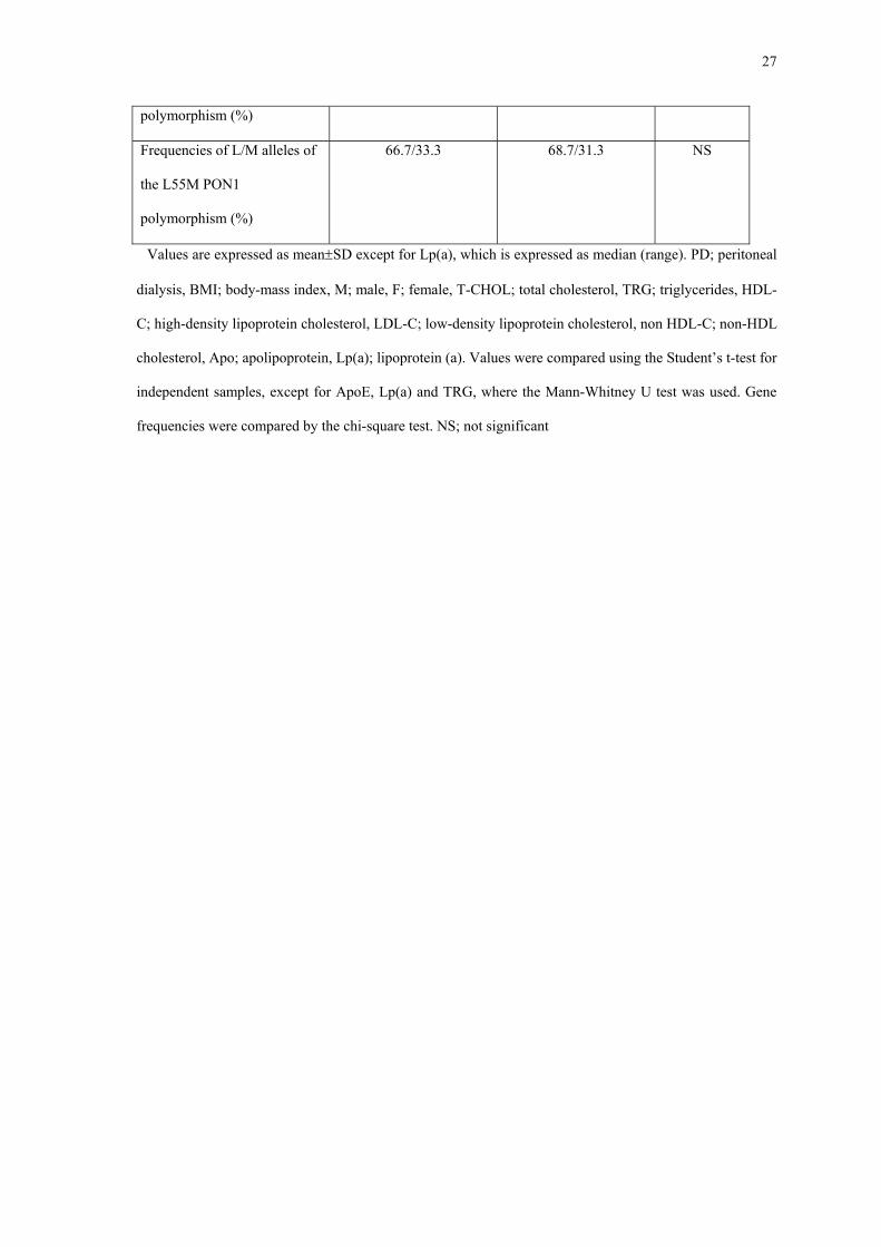
27
polymorphism (%)
Frequencies of L/M alleles of
the L55M PON1
polymorphism (%)
66.7/33.3 68.7/31.3 NS
Values are expressed as mean±SD except for Lp(a), which is expressed as median (range). PD; peritoneal
dialysis, BMI; body-mass index, M; male, F; female, T-CHOL; total cholesterol, TRG; triglycerides, HDL-
C; high-density lipoprotein cholesterol, LDL-C; low-density lipoprotein cholesterol, non HDL-C; non-HDL
cholesterol, Apo; apolipoprotein, Lp(a); lipoprotein (a). Values were compared using the Student’s t-test for
independent samples, except for ApoE, Lp(a) and TRG, where the Mann-Whitney U test was used. Gene
frequencies were compared by the chi-square test. NS; not significant
28
Table 2. Effect of PON1 Q192R genotypes on PON1 activity in patients undergoing peritoneal dialysis
(PD)
(n=29)
QR
(n=20)
RR
(n=7)
p
PON1 (Paraoxonase) (U/L) 50.0(25-160) 132(43-230) 214(170-259) 0.010
PON1 (Arylesterase) (U/ml) 83.2±25.9 89.8±19.4 85.5±23.3 NS
HDL-C (mmol/L)
(mg/dl)
1.16±0.30
(44.9±11.9)
1.23±0.50
(47.5±19.5)
1.19±0.28
(46.0±10.8)
NS
ApoAI (g/L) 1.44±0.30 1.44±0.44 1.40±0.28 NS
PON1(Paraoxonase)/HDL-C
(U/mg)
0.11(0.05-0.46) 0.32(0.06-0.60) 0.47(0.35-0.62) 0.019
PON1(Paraoxonase)/ApoAI
(U/mg)
0.03(0.02-0.22) 0.11(0.02-0.16) 0.16(0.12-0.19) 0.019
HDL-C; high-density lipoprotein cholesterol, Apo; apolipoprotein. Values are expressed as mean±SD,
except for PON1 (Paraoxonase), PON1 (Paraoxonase)/HDL-C and PON1 (Paraoxonase)/ApoAI, which are
expressed as median (range). Values were compared using the one-way ANOVA test followed by LSD test
for pairwise comparisons in cases of significant results, except for PON1 (Paraoxonase), PON1
(Paraoxonase)/HDL-C and PON1 (Paraoxonase)/ApoAI, where the Kruskal-Wallis median test was used
followed by Mann-Whitney U test for paiwise comparisons in cases of significant results. NS; not
significant.

29
Table 3. Effect of PON1 M55L genotypes on PON1 activity in patients undergoing peritoneal dialysis
(PD)
LL
(n=28)
LM
(n=21)
MM
(n=7)
p
PON1 (Paraoxonase) (U/L) 152(43-320) 64(35-167) 44(25-47) 0.030
PON1 (Arylesterase) (U/ml) 101.9±21.1 83.6±15.4 67.0±17.3 0.019
HDL-C (mmol/L)
(mg/dl)
1.23±0.49
(47.8±19.0)
1.21±0.32
(46.9±12.4)
1.21±0.25
(46.8±9.7)
NS
ApoAI (g/L) 1.46±0.04 1.47±0.31 1.47±0.10 NS
PON1(Paraoxonase)/HDL-C
(U/mg)
0.32(0.06-0.76) 0.12(0.05-0.41) 0.09(0.05-0.14) 0.030
PON1(Paraoxonase)/ApoAI
(U/mg)
0.11(0.02-0.22) 0.04(0.02-0.12) 0.03(0.02-0.03) 0.030
HDL-C; high-density lipoprotein cholesterol, Apo; apolipoprotein. Values are expressed as mean±SD,
except for PON1 (Paraoxonase), PON1 (Paraoxonase)/HDL-C and PON1 (Paraoxonase)/ApoAI, which are
expressed as median (range). Values were compared using the one-way ANOVA test followed by LSD test
for pairwise comparisons in cases of significant results, except for PON1 (Paraoxonase), PON1
(Paraoxonase)/HDL-C and PON1 (Paraoxonase)/ApoAI, where the Kruskal-Wallis median test was used
followed by Mann-Whitney U test for paiwise comparisons in cases of significant results. NS; not
significant.
30
0
20
40
60
80
100
PON1 (Para
Med
ian
PON
1 (P
arao
xona
se) [
U/L
]
0
20
40
60
80
100
120
PON1 (Aryl
PON
1 (A
ryle
ster
ase)
[U/m
l]
A a
B
Figure 1. Bar graph showing the median se
(arylesterase) (as mean ± SD) [B] activities in co
p<0.05 compared with controls by the Mann-Wh
Student’s t-test for independent samples.
oxonase)
ControlsPD
esterase)
ControlsPD
b
rum PON1 (Paraoxonase) [A] and the serum PON1
ntrol subjects and peritoneal dialysis (PD) patients. a;
itney U test, b; p<0.01 compared with controls by the
31
0
0,05
0,1
0,15
0,2
0,25
0,3
PON1 (Paraoxonase) /HDL-C PON1 (Paraoxon
U/m
g
ControlPD
† A
†
B
0
100
200
300
400
PON1 (Arylesterase)/HDL-C PON1 (Aryleste
U/m
g
ControlPD ‡B
Figure 2. Bar graph showing the median ratios of serum PON1
(Paraoxonase)/ApoAI [A], as well as the ratios of serum PON1
(Arylesterase)/ApoAI (as mean ± SD) [B] activities in control subjects a
HDL-C; high-density lipoprotein cholesterol, ApoAI; apolipoprotein AI
by the Mann-Whitney U test, ‡; p<0.01 compared with controls by t
samples.
ase) /ApoAI
rase)/ApoAI
‡
(Paraoxonase)/HDL-C and PON1
(arylesterase)/HDL-C and PON1
nd peritoneal dialysis (PD) patients.
, †; p<0.05 compared with controls
he Student’s t-test for independent
32
0
50
100
150
200
250
300
Med
ian
PON
1 (P
arao
xona
se)
[U/L
]
QL/QL QL/QM QL/RL QL/RM RL/RL
ControlPD
* *
** **
***
Figure 3. Median PON1 (Paraoxonase) activity in control subjects and in patients undergoing peritoneal
dialysis (PD) according to different PON1 Q192R and M55L genotype combinations. Values were
compared using the Mann-Whitney U test. *p<0.05 PD vs controls; **p<0.01 PD vs controls, ***p<0.001
PD vs controls.
33
Table 4. Total serum and HDL-associated platelet-activating factor acetylhydrolase (PAF-AH) activities
in control subjects and peritoneal dialysis patients (PD).
Controls
(n=86)
PD
(n=56)
P
Total PAF-AH (nmol/ml
serum/min)
47.3±11.7 88.1±19.4 0.0000
Total PAF-AH/LDL-C
(nmol/mg/min)
35.4±10.6 58.5±16.5 0.0000
Total PAF-AH/ApoB
(nmol/mg/min)
49.9±14.5 73.9±12.0 0.0000
Total PAF-AH/non HDL-C
(nmol/mg/min)
36.7±8.7 47.0±11.4 0.0000
HDL-PAF AH (nmol/ml
serum/min)
3.3±1.2 4.3±1.9 0.001
HDL-PAF AH/HDL-C
(nmol/mg/min)
7.4±2.7 9.6±3.8 0.001
HDL-PAF AH/ApoAI
(nmol/mg/min)
2.3±0.9 3.1±1.2 0.001
HDL-PAF AH/Total PAF-AH 0.07±0.03 0.05±0.02 0.002
HDL-C; high-density lipoprotein cholesterol, LDL; low-density lipoprotein cholesterol, Apo;
apolipoprotein. Values were compared using the Student’s t-test for independent samples. NS; not
significant.