alopecia areata - iconcept press · 1 introduction alopecia areata (aa) is a common nonscarring,...
TRANSCRIPT
Alopecia Areata Adel Alsantali
Department of Dermatology King Fahd Armed Forces Hospital, Jeddah, Saudi Arabia
1 Introduction
Alopecia areata (AA) is a common nonscarring, autoimmune disease that can affect any hair-bearing ar-ea. It occurs in all ethnic groups, ages, and both sexes. The lifetime risk for AA is about 1.7% in the gen-eral population (Safavi et al., 1995). Up to 60% of patients had their first AA episode before the age of 20 (Price, 1991). Although, AA is an unpredictable disease but extensive involvement and the long duration of the hair loss are the most important poor prognostic factors. Although AA is generally asymptomatic disease, it is psychologically distressing to the patients. Therapy for AA should be tailored in light of se-verity of the condition and patient's age. In this chapter, currently available treatments for AA are dis-cussed, together with the clinical presentation, and pathogenesis.
2 Clinical Features
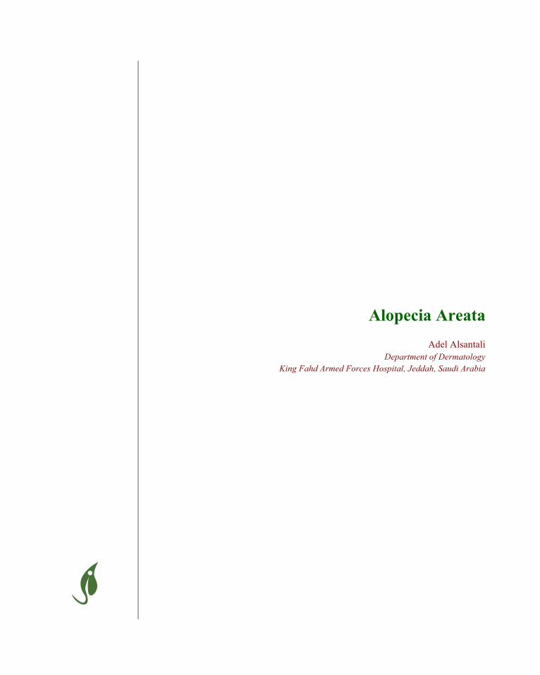
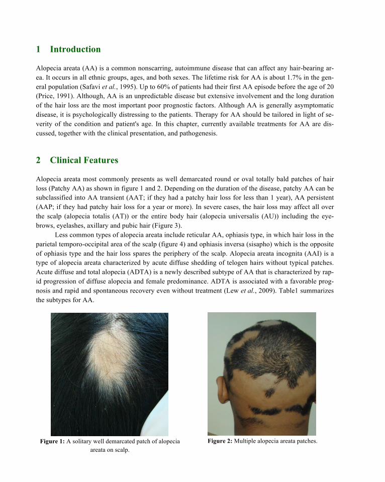
Alopecia areata most commonly presents as well demarcated round or oval totally bald patches of hair loss (Patchy AA) as shown in figure 1 and 2. Depending on the duration of the disease, patchy AA can be subclassified into AA transient (AAT; if they had a patchy hair loss for less than 1 year), AA persistent (AAP; if they had patchy hair loss for a year or more). In severe cases, the hair loss may affect all over the scalp (alopecia totalis (AT)) or the entire body hair (alopecia universalis (AU)) including the eye-brows, eyelashes, axillary and pubic hair (Figure 3).
Less common types of alopecia areata include reticular AA, ophiasis type, in which hair loss in the parietal temporo-occipital area of the scalp (figure 4) and ophiasis inversa (sisapho) which is the opposite of ophiasis type and the hair loss spares the periphery of the scalp. Alopecia areata incognita (AAI) is a type of alopecia areata characterized by acute diffuse shedding of telogen hairs without typical patches. Acute diffuse and total alopecia (ADTA) is a newly described subtype of AA that is characterized by rap-id progression of diffuse alopecia and female predominance. ADTA is associated with a favorable prog-nosis and rapid and spontaneous recovery even without treatment (Lew et al., 2009). Table1 summarizes the subtypes for AA.
Figure 1: A solitary well demarcated patch of alopecia
areata on scalp.
Figure 2: Multiple alopecia areata patches.
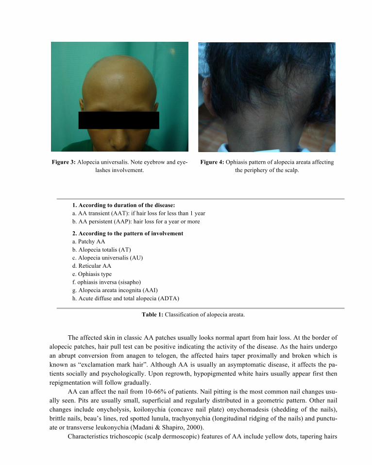
Figure 3: Alopecia universalis. Note eyebrow and eye-lashes involvement.
Figure 4: Ophiasis pattern of alopecia areata affecting the periphery of the scalp.
1. According to duration of the disease: a. AA transient (AAT): if hair loss for less than 1 year b. AA persistent (AAP): hair loss for a year or more
2. According to the pattern of involvement a. Patchy AA b. Alopecia totalis (AT) c. Alopecia universalis (AU) d. Reticular AA e. Ophiasis type f. ophiasis inversa (sisapho) g. Alopecia areata incognita (AAI) h. Acute diffuse and total alopecia (ADTA)
Table 1: Classification of alopecia areata.
The affected skin in classic AA patches usually looks normal apart from hair loss. At the border of alopecic patches, hair pull test can be positive indicating the activity of the disease. As the hairs undergo an abrupt conversion from anagen to telogen, the affected hairs taper proximally and broken which is known as “exclamation mark hair”. Although AA is usually an asymptomatic disease, it affects the pa-tients socially and psychologically. Upon regrowth, hypopigmented white hairs usually appear first then repigmentation will follow gradually.
AA can affect the nail from 10-66% of patients. Nail pitting is the most common nail changes usu-ally seen. Pits are usually small, superficial and regularly distributed in a geometric pattern. Other nail changes include onycholysis, koilonychia (concave nail plate) onychomadesis (shedding of the nails), brittle nails, beau’s lines, red spotted lunula, trachyonychia (longitudinal ridging of the nails) and punctu-ate or transverse leukonychia (Madani & Shapiro, 2000).
Characteristics trichoscopic (scalp dermoscopic) features of AA include yellow dots, tapering hairs
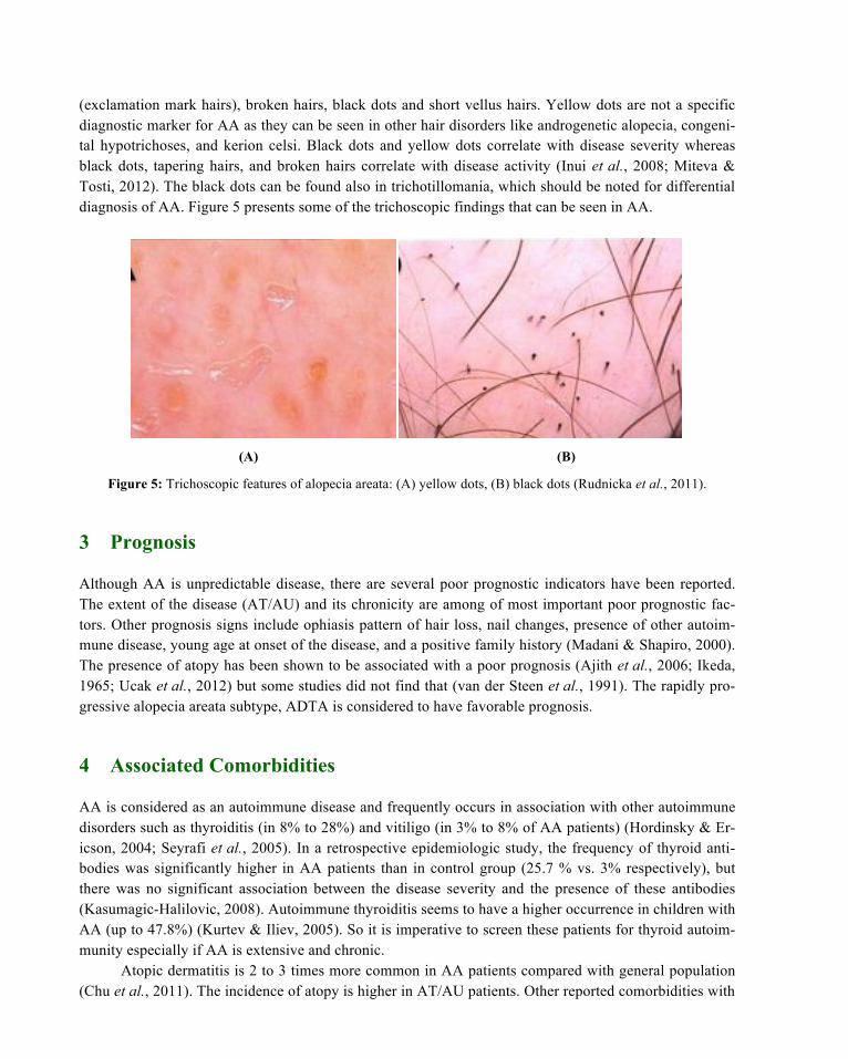
(exclamation mark hairs), broken hairs, black dots and short vellus hairs. Yellow dots are not a specific diagnostic marker for AA as they can be seen in other hair disorders like androgenetic alopecia, congeni-tal hypotrichoses, and kerion celsi. Black dots and yellow dots correlate with disease severity whereas black dots, tapering hairs, and broken hairs correlate with disease activity (Inui et al., 2008; Miteva & Tosti, 2012). The black dots can be found also in trichotillomania, which should be noted for differential diagnosis of AA. Figure 5 presents some of the trichoscopic findings that can be seen in AA.
(A) (B)
Figure 5: Trichoscopic features of alopecia areata: (A) yellow dots, (B) black dots (Rudnicka et al., 2011).
3 Prognosis
Although AA is unpredictable disease, there are several poor prognostic indicators have been reported. The extent of the disease (AT/AU) and its chronicity are among of most important poor prognostic fac-tors. Other prognosis signs include ophiasis pattern of hair loss, nail changes, presence of other autoim-mune disease, young age at onset of the disease, and a positive family history (Madani & Shapiro, 2000). The presence of atopy has been shown to be associated with a poor prognosis (Ajith et al., 2006; Ikeda, 1965; Ucak et al., 2012) but some studies did not find that (van der Steen et al., 1991). The rapidly pro-gressive alopecia areata subtype, ADTA is considered to have favorable prognosis.
4 Associated Comorbidities
AA is considered as an autoimmune disease and frequently occurs in association with other autoimmune disorders such as thyroiditis (in 8% to 28%) and vitiligo (in 3% to 8% of AA patients) (Hordinsky & Er-icson, 2004; Seyrafi et al., 2005). In a retrospective epidemiologic study, the frequency of thyroid anti-bodies was significantly higher in AA patients than in control group (25.7 % vs. 3% respectively), but there was no significant association between the disease severity and the presence of these antibodies (Kasumagic-Halilovic, 2008). Autoimmune thyroiditis seems to have a higher occurrence in children with AA (up to 47.8%) (Kurtev & Iliev, 2005). So it is imperative to screen these patients for thyroid autoim-munity especially if AA is extensive and chronic.
Atopic dermatitis is 2 to 3 times more common in AA patients compared with general population (Chu et al., 2011). The incidence of atopy is higher in AT/AU patients. Other reported comorbidities with
AA include Down’s syndrome, autosomal recessive autoimmune polyglandular syndrome (APS-1), per-nicious anemia, celiac disease, multiple sclerosis, Addison disease, ulcerative colitis, lupus erythematosus, psoriasis, and Turner’s syndrome. Ocular abnormalities have been reported in AA patients. Lenticular changes in the form of punctuate opacities and cataracts were found in 40% of AA patients (Pandhi et al., 2009). In another study these asymptomatic lens opacities were observed in 51% of patients (Recupero et al., 1999). Also, retinal alterations were found in 41% AA subjects.
5 Differential Diagnosis
In children, AA should be differentiated from tinea capitis, trichotillomania and congenital atrichia. The alopecic patches in tinea capitis are usually associated with scaling and inflammation that may progress to severely inflame deep abscesses termed kerion. Trichotillomania is a compulsive disorder that often presents as solitary or multiple alopecic patches with broken hair of varying length that resulted from hair pulling by the patient him/herself. The other differential diagnoses that may also be considered are sec-ondary syphilis, lupus erythematosus, telogen effluvium and traction alopecia (Table 3).
1. Autoimmune thyroid diseases 2. Vitiligo 3. Atopy (atopic dermatitis, allergic rhinitis) 4. Psychiatric morbidity (anxiety, depression and mood disturbance) 5. Ophthalmologic changes (lenticular and retinal alterations) 6. Down syndrome 7. Addison disease 8. Pernicious anemia 9. Psoriasis 10. Lupus erythematosus 11. Celiac disease 12. Ulcerative colitis 13. Multiple sclerosis 14. Autosomal recessive autoimmune polyglandular syndrome (APS-1)
Table 2: Comorbidities associated with AA
● Tinea capitis ● Trichotillomania ● Secondary syphilis ● Discoid lupus erythematosus (DLE) ● Traction alopecia ● Lichen planopilaris ● Pseudopelade of Brocq ● Alopecia mucinosa ● Congenital atrichia ● Telogen effluvium
Table 3: AA differential diagnosis
6 Investigation
In most cases, the diagnosis of AA is straightforward and based on the clinical presentation. Skin biopsy and fungal cultures may be considered in difficult cases. Routine screening tests for the associated auto-immune diseases is not generally recommended. But as stated before it is wise to screen for autoimmune thyroid disease particularly in chronic extensive AA (AT/AU).
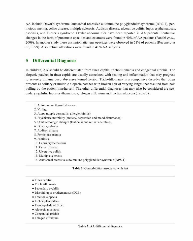
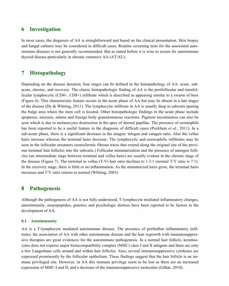
7 Histopathology
Depending on the disease duration, four stages can be defined in the histopathology of AA: acute, sub-acute, chronic, and recovery. The classic histopathologic finding of AA is the perifollicular and intrafol-licular lymphocytic (CD4+, CD8+) infiltrate which is described as appearing similar to a swarm of bees (Figure 6). This characteristic feature occurs in the acute phase of AA but may be absent in a late stages of the disease (Dy & Whiting, 2011). The lymphocytic infiltrate in AA is usually deep in subcutis sparing the bulge area where the stem cell is located. Other histopathologic findings in the acute phase include apoptosis, necrosis, edema and foreign body granulomatous reactions. Pigment incontinence can also be seen which is due to melanocytes destruction in the apex of dermal papillae. The presence of eosinophils has been reported to be a useful feature in the diagnosis of difficult cases (Peckham et al., 2011). In a sub-acute phase, there is a significant decrease in the anagen: telogen and catagen ratio. Also the vellus hairs increase whereas the terminal hairs decrease. The lymphocytic and eosinophilic infiltrates may be seen in the follicular streamers (nonsclerotic fibrous tracts that extend along the original site of the previ-ous terminal hair follicles into the subcutis.) Follicular miniaturization and the presence of nanogen folli-cles (an intermediate stage between terminal and vellus hairs) are usually evident in the chronic stage of the disease (Figure 7). The terminal to vellus (T:V) hair ratio declines to 1.3:1 (normal T:V ratio is 7:1). In the recovery stage, there is little or no inflammation. As the miniaturized hairs grow, the terminal hairs increases and T:V ratio returns to normal (Whiting, 2003).
8 Pathogenesis
Although the pathogenesis of AA is not fully understood, T-lymphocyte mediated inflammatory changes, autoimmunity, neuropeptides, genetics and pyschologic distress have been reported to be factors in the development of AA.
8.1 Autoimmunity
AA is a T-lymphocyte mediated autoimmune disease. The presence of peribulbar inflammatory infil-trates, the association of AA with other autoimmune disease and the hair regrowth with immunosuppres-sive therapies are great evidences for the autoimmune pathogenesis. In a normal hair follicle, keratino-cytes does not express major histocompatibility complex (MHC) class I and II antigens and there are only a few Langerhans cells around and within hair follicles. Also, several immunosuppressive cytokines are expressed prominently by the follicular epithelium. These findings suggest that the hair follicle is an im-mune privileged site. However, in AA this immune privilege seem to be lost as there are an increased expression of MHC-I and II, and a decrease of the immunosuppressive molecules (Gilhar, 2010).
Figure 6: Acute alopecia areata. Horizontal section: classic peribulbar lymphocytic infiltrate (Hematoxylin-eosin stain; original magnification:×20).
Figure 7: Chronic alopecia areata. Horizontal section: marked miniaturization and on inflam-mation. (Hematoxylin-eosin stain; original magnification:×4).
Moreover, the expression of adhesion molecules (ICAM-2 and ELAM-1) have been shown to be increased in the peribulbar and perivascular areas of AA affected skin. These changes lead to targeting the hair follicles by the inflammatory cells and the development of AA. Activated cytotoxic T-lymphocytes produce tumor necrosis factors, Fas ligands, and granzymes which lead to hair follicle apop-tosis (Kalkan et al., 2013). CD8+ T-lymphocytes seem to have a primary role in hair follicle damage whereas CD4+ cells have a helper role in the process (McElwee et al., 2005). Why the immune privilege for the hair follicle is lost in AA patients and what are the initial events leading to the development of AA? The cause or causes for the collapse of immune privilege are not clear but, it has been suggested that events such as stress, infection, or microtrauma might lead to downregulation of the immunosuppressive cytokines. The onset of AA may be due to abnormal regression of catagen hair leading to hair follicle antigen uptake and presentation to lymphocytes (Botchkareva et al., 2006). From mice studies, proin-flammatory changes started in skin draining lymph nodes several weeks before the onset of alopecia or even before lymphocytic infiltration of the skin (Zoller et al., 2002).
Recently oxidative stress has come to light as a possible triggering factor for autoimmune diseases. In a case-control study, the mean serum total oxidant capacity, malondialdehyde, and the oxidative stress index were found to be significantly higher in AA patients than in the control group and correlate with disease severity (Bakry et al., 2013). Other studies showed an increase in lipid peroxidation and defective antioxidant activity of superoxide dismutase in patients with AA (Abdel Fattah et al., 2011; Koca et al., 2005). Whether these changes play a role in disease pathogenesis or result from the inflammatory process needs further investigation. Also, the role of antioxidants in AA treatment and prevention warrant further clinical studies.
8.1.1 Humoral Immunity
Hair follicle specific IgG antibody has been shown to be increased in the blood of AA patients. However, the injections of these autoantibodies in different models did not prove a significant role of these autoan-tibodies in the development of AA. Of notes, antithyroid and anti nuclear antibodies can be found more in AA patients than in normal population (Grandolfo et al., 2008; Yano et al., 1999).
8.2 Genetics
AA is considered an autoimmune disease with genetic background due to the following:
1. High frequency of a family history of AA in affected people particularly in patients with early onset AA (range from 10% to 42%).
2. Concordance rate in identical twins was repeated up to 55%.
3. Although the lifetime risk for AA is about 1.7% in the general population, the estimated lifetime risk of the disease is 7.1% in siblings, 7.8% in parents, and 5.7% in offsprings of AA patients. The first genome-wide association study (GWAS) identified at least eight regions in the genome (Petukhova et al., 2010). These eight regions implicated genes of the immune system, as well as genes that are unique to the hair follicle itself.
(i) Chromosome 2q33.2 containing the CTLA4 gene;
(ii) Chromosome 4q27 containing the IL2/IL21 locus;
(iii) Chromosome 6p21.32 containing the HLA class II region;
(iv) Chromosome 6q25.1 which harbors the cytomegalovirus UL16-binding protein (ULBP) gene cluster;
(v) Chromosome 9q31.1 containing syntaxin 17 (STX17);
(vi) Chromosome 10p15.1 containing IL-2 receptor A (IL-2RA; CD25;
(vii) Chromosome 11q13 containing peroxiredoxin 5 (PRDX5);
(viii) Chromosome 12q13 containing Eos (also known as Ikaros family zinc finger 4; IKZF4).
These genomic region contain several genes that control the activation and proliferation of regula-tory T cells, cytotoxic T-lymphocyte – associated antigen 4 (CTL4), interleukin (IL)-2/IL-21, IL-2 recep-tor A (IL-2RA; CD25) and Eos, as well as the human leukocyte antigen (HLA) region. A region of strong association was found within ULBP (cytomegalovirus UL16 binding protein) gene cluster in chromo-some 6q25.1. ULBP3 and ULBP6 genes make the natural killer cell receptor NKG2D activating ligands or signal that can trigger the NKG2D receptor initiating an autoimmune response. NKG2DLs are stress-induced molecules that act as “danger signals” to alert NK and CD8T lymphocytes through the engage-ment of the activating receptor, NKG2D. AA hair follicle dermal sheath showed a greater expression of ULBP3. Another two GWASs confirm these findings and add new susceptibility loci for AA. These loci are for IL-13, KIAA0350/CLEC16A and SPATA5 (spermatogenesis-associated protein 5) gene on chro-mosome 4 (Forstbauer et al., 2012; Jagielska et al., 2012). The GWASs implicated both innate and adap-tive immunity in the pathogenesis of AA. These findings suggest new approaches of exploration for ther-apy based on the underlying mechanisms of AA with a focus not only on T cells, but on cells that express the NKG2D receptor as well.
8.3 Stress
Psychosocial stress has been reported to play a role in the onset and the progression of alopecia areata but controlled clinical studies were not conclusive. Some studies showed a positive relationship between the stress and AA (Manolache & Benea, 2007; Taheri et al., 2012). In the other hand, some did not find a significant relation between AA and stressful events (Brajac et al., 2003). Some studies have shown that AA patients have a higher reactivity to stress and higher scores for depression than normal population (Chu et al., 2012). Experimental studies in mice with alopecia areata showed a marked increase in the hypothalamic-pituitary-adrenal (HPA) axis tone and activity centrally, and peripherally in the skin and lymph nodes. Stress further exacerbated changes in AA mouse HPA activity. The positive correlation of HPA hormone levels with skin Th1 cytokines suggests that altered HPA activity may occur as a conse-quence of the immune response associated with AA (Zhang et al., 2009).
AA itself can cause a severe psychological distress, depression and anxiety. Also, there is a high psychiatric comorbidity (up to 78%) in AA (mainly as generalized anxiety disorder, depression, and mood disorders) requiring systematic psychiatric evaluation and treatments. In alopecia areata, like in some other diseases, psychosomatics and immunology are not opposed because immune cells are con-trolled by the nervous system through neurotransmitters. Stress may increase the production of neuropep-tides such as substance P (SP) which induces accumulation of CD8+ cells and induce these cells to pro-duce large amounts of IFN-γ. Also, SP may stimulate the production of nerve growth factor, which in turn induces mast cells accumulation and degranulation around hair follicles leading to hair follicle re-gression.
8.4 The effect of AA on hair follicle growth cycle
Normal hair follicles pass through 3 phases: the growth phase (anagen), the regression phase (catagen), and the resting phase (telogen). Several possible pathologic patterns of hair cycle can be seen in AA which include, dystrophic anagen in which the hair follicle are unable to produce hair fibers, truncated cycling (hairs cycle through multiple anagen – telogen phases of brief duration) and prolonged telogen phase without attempt to produce hair follicle (in chronic AA) (Freyschmidt-Paul et al., 2008). The sever-ity of inflammation and the chronicity of the disease may play a role in determining the nature and the pattern of the hair cycle derangement (Whiting, 2003). More than one hair cycle pattern can occur in the same AA patient.
9 Treatment
Many therapeutic modalities have been used to treat alopecia areata, with variable efficacy and safety profiles. Unfortunately, none of these agents is curative or preventive. Also, many of these therapeutic agents have not been subjected to randomized, controlled trials, and, except for topical immunotherapy, there are few published studies on long-term outcomes. In the view of the author, the therapeutic agents are organized according to their efficacy and safety profiles into first-line, second-line, and third-line op-tions. Usually, there is a lag time from initiation of therapy to hair regrowth. This lag time varies from one therapeutic agent to another and it is usually about 3-6 months. This lag time should be kept in mind, so as not abandon therapy before having allowed a minimum time for response. Of note, the lag time is shorter with intralesional and systemic corticosteroids than other agents (Chartaux & Joly, 2010).
9.1 First-line therapies
9.1.1 Intralesional corticosteroids
Several studies have shown the efficacy of intralesional corticosteroid injections. Abell and Munro re-ported hair regrowth in 71% of patients with subtotal alopecia areata treated by triamcinolone acetonide injections and in 7% of a placebo group (Abell & Munro, 1973). For limited scalp alopecia areata, in-tralesional corticosteroid therapy is considered as the drug of choice by many experts. The most widely used agent is triamcinolone acetonide. Different concentrations of triamcinolone acetonide are used, in the range of 2.5–10 mg/ml, but 5 mg/ml is the preferred concentration for the scalp and face. A maximum volume of 3 ml on the scalp in one visit is recommended. Corticosteroid is injected into the deep dermis level or just beneath the dermis in the upper subcutis. The injections can be repeated at 4–6 weekly inter-vals. The use of mesotherapy multi-injectors with 5–7 needles is an alternative approach to decrease in-jection pain and to make the procedure more homogenous. Side effects include skin atrophy and telangi-ectasia which can be minimized by the use of smaller volumes and avoiding superficial injections. To alleviate injection pain, topical anesthetic may be applied 30–60 minutes before the treatment. Although the effect of a single intralesional corticosteroid injection has been observed to persist for up to 9 months, reported relapse rates were 29% in limited alopecia areata and 72% in alopecia totalis during a 3-month follow-up period. Patients who received intralesional triamcinolone acetonide therapy for long time (cu-mulative dose greater than 500 mg) should be screened for osteoporosis and monitored for effects on bone mineral density (Samrao et al., 2013).
9.1.2 Topical corticosteroids
Many forms of topical corticosteroids have been prescribed for alopecia areata, including creams, gels, ointments, lotions, and foams. Sixty-one percent of patients using 0.1% betamethasone valerate foam achieved more than 75% hair regrowth in comparison with 27% in the 0.05% betamethasone dipropio-nate lotion group (Mancuso et al., 2003). Topical corticosteroids are far less effective in alopecia totalis and alopecia universalis. A highly potent topical corticosteroid under occlusion is the preferred method when using topical corticosteroids. Folliculitis is a common side effect to topical corticosteroids. Telangi-ectasia and atrophy may develop rarely. The reported relapse rate is 37%–63% (Tosti et al., 2003).
9.1.3 Minoxidil
In a placebo-controlled, double-blind study, hair regrowth was observed in 63.6% and 35.7% of the minoxidil-treated and placebo groups, respectively (Price, 1987a). However, only 27% of the minoxidil-treated patients showed cosmetically acceptable hair regrowth. In another study, hair regrowth was achieved in 38% and 81% of patients treated with 1% and 5% topical minoxidil, respectively (Fiedler-Weiss, 1987). Most studies have shown no beneficial effect of topical minoxidil in alopecia totalis and alopecia universalis (Price, 1987b). Minoxidil 5% solution or foam is frequently used with other thera-peutic agents as an adjuvant therapy. The adverse effects of topical minoxidil include contact dermatitis and facial hypertrichosis.
9.1.4 Anthralin
A few controlled trials have assessed the efficacy of topical anthralin in the treatment of alopecia areata. In an open study, a cosmetic response was seen in 25% of patients with severe alopecia areata treated using 0.5%–1.0% anthralin cream (Fiedler-Weiss & Buys, 1987). In another trial, combination therapy of 5% minoxidil and 0.5% anthralin was used to treat 51 patients with severe alopecia areata; only 11% of patients achieved cosmetically acceptable hair regrowth (Fiedler et al., 1990). Anthralin needs to be ap-plied in a high enough concentration (0.5%–1%) and sufficiently frequently (daily) to produce a mild irritant reaction in order to be effective. Severe irritation and staining of skin and clothes are some of the possible adverse events with anthralin.
9.1.5 Topical immunotherapy
Topical sensitizers that have been used in the treatment of alopecia areata include diphenylcycloprope-none (DPCP), squaric acid dibutylester (SADBE), and dinitrochlorobenzene. Dinitrochlorobenzene is no longer used because it was shown to be mutagenic in the Ames test (Strobel & Rohrborn, 1980). DPCP is the topical sensitizer of choice. SADBE is expensive and not stable in ace-tone. DPCP is light sensitive and should be protected from light. Initially the patient is sensitized using a 2% solution of DPCP applied to a 4×4 cm area of the scalp. After two weeks, 0.001% DPCP solution is applied to the same half of the scalp. The DPCP concentration is increased gradually every week until mild dermatitis is observed (Orecchia & Perfetti, 1991). The solution should be on the scalp for 48 hours. The scalp should be pro-tected from the sun during this time. Once hair regrowth is obtained on the treated half of the scalp, both sides are treated. Both sides of the scalp can be treated from the start also. DPCP is applied on a weekly basis by a trained nurse. If there is no response after 6 months of treatment, DPCP can be discontinued. SADBE may be tried in poor responders to DPCP or in those who do not develop a sensitization to 2% DPCP. SADBE is applied once or twice per week. The adverse effects to topical sensitizers include cer-
vical lymphadenopathy, a severe eczematous reaction, urticaria, and postinflammatory pigment changes. The response rate of alopecia totalis/alopecia universalis patients to DPCP was 17.4% in the largest re-ported diphenylcyclopropenone study, whereas the cumulative patient response was 77% (Wiseman et al., 2001). Several negative prognostic factors in the treatment of alopecia areata with DPCP have been suggested, including long duration of disease, alopecia totalis/alopecia universalis, nail changes, atopy, and family history of alopecia areata. Recurrence of alopecia areata after achieving significant hair re-growth developed in 62.6% of patients. In a retrospective study of 121 patients with extensive alopecia areata, fexofenadine hydrochloride has been shown to enhance the efficacy of topical immunotherapy (Inui et al., 2009). The mechanism of action of topical sensitizers could be due to perifollicular lympho-cyte apoptosis, changes in the peribulbar CD4/CD8 lymphocyte ratio, and antigenic competition (Wasylyszyn et al., 2007).
9.1.6 Prostaglandin analogs
Eyelash hypertrichosis is a common adverse effect to the use of these antiglaucoma eye drops (Hart & Shafranov, 2004). Some case series did not show an effect in the treatment of eyelashes in patients with alopecia areata (Roseborough et al., 2009). In a nonrandomized, controlled study of latanoprost (a prosta-glandin F2 α analog) eye drops in patients with alopecia universalis, acceptable results (total and moder-ate hair regrowth) were achieved in 45% of patients (Coronel-Perez et al., 2010). In another retrospec-tive trial, 0.03% bimatoprost eye drops were used once a day for one year. Complete regrowth of the eye-lashes was noted in 24.3% of patients and moderate growth in 18.9% of treated subjects (Vila et al., 2010). Relapses were observed in 17.5% of the patients, mainly in the slight response group.
9.1.7 Topical retinoids
In a comparative study of topical tretinoin 0.05%, topical betamethasone dipropionate lotion, and dithranol paste 0.25%, a good response has been seen in 55% of patients treated with topical tretinoin in comparison with 70% and 35% in the topical steroid and dithranol groups, respectively (Das et al., 2010). Although the mechanism for its action in alopecia areata is not completely understood, the associated tretinoin-induced dermatitis might contribute to regrowth in alopecia areata. Larger, double-blind, place-bo-controlled trials are needed.
9.1.8 Bexarotene
In a randomized bilateral half-head study, hair regrowth of at least 50% on treated sites was noticed in only 26% of patients treated with 1% bexarotene gel (Talpur et al., 2009). Mild irritation is a common side effect.
9.1.9 Capsaicin
In a nonblinded randomized study, 9.5% of patients with alopecia areata showed cosmetically acceptable hair regrowth after 12 weeks of applying capsaicin ointment (Ehsani et al., 2009).
9.2 Second-line therapies
9.2.1 Sulfasalazine
Sulfasalazine is a combination of sulfapyridine and 5-aminosalicylic acid linked by a diazo bond. Sul-
fasalazine has both immunomodulatory and immunosuppressive actions that include suppression of T cell proliferation and reducing the synthesis of cytokines, including interleukin (IL) 6, 1, and 12, tumor necro-sis factor alpha, and antibody production (Ranganath & Furst, 2007). Sulfasalazine has been used safely as a long-term treatment of various inflammatory and autoimmune diseases, including inflammatory bowel disease and rheumatoid arthritis. Several case reports and case series showed good hair regrowth with sulfasalazine in the treatment of alopecia areata. In an uncontrolled prospective trial of sulfasalazine in 39 patients with persistent alopecia areata, hair regrowth of more than 60% was achieved in 25.6% of patients. A moderate response was seen in 30.7% of patients (Rashidi & Mahd, 2008). Also, in another uncontrolled open-label study, complete hair regrowth was reported in 27.3% of subjects (Aghaei, 2008). Sulfasalazine was started at 500 mg twice daily for one month, 1 g twice daily for one month, and then 1 g three times daily (Ellis et al., 2002). Side effects to sulfasalazine include gastrointestinal distress, dizzi-ness, and headache. Gastrointestinal symptoms can be minimized by using enteric-coated tablets, taking the medication with food, and starting at lower doses. Initially, patients should have a complete blood count, liver function tests, creatinine, and glucose-6-phosphate dehydrogenase level measurement. Com-plete blood counts and liver function tests should be performed at 2–4-week intervals during the first three months of therapy. The reported relapse rates are 22.7%–45.5%.
9.2.2 Photochemotherapy
The success rate for oral and topical psoralen plus ultraviolet A (PUVA) ranged from 15% to more than 70% (Mohamed et al., 2005; Taylor & Hawk, 1995). PUVA turban is a method of administering a dilute psoralen solution (8-methoxypsoralen 0.0001%) selectively to the scalp for 20 minutes using a cotton towel as a turban. The patient’s scalp is then exposed to ultraviolet A radiation. Treatment sessions are performed two or three times per week. PUVA- turban has been shown to be effective in about 70% of treated patients (Behrens-Williams et al., 2001; Broniarczyk-Dyla et al., 2006). During a follow-up peri-od of 15 months after PUVA-turban therapy, recurrences of alopecia areata were observed in 26% of re-sponders. PUVA-turban therapy lacks the systemic side effects of oral PUVA and can be considered as alternative therapy for patients with alopecia areata.
9.2.3 Excimer laser
In a treatment of 42 alopecia areata patches with the 308 nm excimer laser, hair regrowth was observed in 41.5% of treated areas (Al-Mutairi, 2007). Hair regrowth was noticed to begin to appear during the se-cond month of therapy. No regrowth of hair was noted on the control patches. Laser therapy was adminis-tered twice a week for a maximum of 24 sessions. Apart from erythema at the treated sites, there were no significant adverse effects. Relapses of alopecia areata were observed in two patients with patchy alope-cia areata of the scalp who had shown complete regrowth earlier. Also, the use of excimer laser in chil-dren with alopecia areata has been reported to have a good success rate (Al-Mutairi, 2009).
9.2.4 Fractional photothermolysis laser
Good hair regrowth was achieved with fractional Er: Glass laser in a single case report (Yoo et al., 2010). Randomized controlled trials in a larger number of patients are required to confirm the efficacy of this modality of treatment.
9.3 Third-line therapies
9.3.1 Systemic corticosteroids
Systemic corticosteroids are one of the commonly prescribed therapies in patients with extensive alopecia areata. Various forms of corticosteroids have been used in different regimens. In one study, a once-monthly oral pulse of 300 mg prednisone induced a complete response in 41% of patients (Ait Ourhroui et al., 2010). A similar effect has been reported in a placebo-controlled trial of oral prednisolone 200 mg once weekly in the treatment of extensive alopecia areata (Kar et al., 2005). The relapse rate was 25%, and side effects of the therapy were noted in 55% of patients. In a comparative trial, the response rate was better in patients treated with intramuscular triamcinolone acetonide 40 mg once monthly than in those treated with oral dexamethasone 0.5 mg/day (Kurosawa et al., 2006). In the same study, impairment of adrenocortical reserve was seen in 23% of the intramuscular triamcinolone acetonide group and in 7% of patients treated with oral prednisolone pulse therapy of 80 mg for 3 consecutive days once every 3 months. In a study of 139 patients treated with pulse corticosteroid therapy, a good response was achieved in 59.4% of patients with recent-onset disease (duration of alopecia areata up to 6 months) in comparison with 15.8% of subjects who had had alopecia areata for more than 6 months (Nakajima et al., 2007). Alopecia totalis and alopecia universalis are far less responsive to this therapy than patchy alope-cia areata (Friedli et al., 1998). The use of systemic corticosteroids is limited by their side effects (hyper-glycemia, weight gain, hypertension, adrenal suppression, dysmenorrhea, immunosuppression, and acnei-form eruption) (Lester et al., 1998) and the high relapse rate (14%–100%).
9.3.2 Methotrexate
In a long-term follow-up study of methotrexate in 33 patients with alopecia areata, complete hair re-growth was achieved in 57% and 63% of patients who used methotrexate alone or with low doses of oral corticosteroids (prednisone 10–20 mg/day), respectively (Chartaux & Joly, 2010). Thirty percent of pa-tients had partial hair regrowth. The weekly dosages of methotrexate were 15–25 mg. The onset of hair regrowth was seen after a median delay of three months. Recurrences of alopecia areata after a decrease of the methotrexate dose or after stopping treatment were observed in 57% (8/14 cases) of responders. In a retrospective trial of methotrexate in 14 children with alopecia areata, approximately one third of pa-tients experienced a clinically relevant therapeutic response (Royer et al., 2011). The mean age of the patients was 14.7 (range 8–18) years. Adverse effects to methotrexate include persistent nausea, transient elevation of hepatic enzymes, and leucopenia.
9.3.3 Cyclosporine
The success rate with oral cyclosporine is 25%–76.6% (Kim et al., 2008; Shapiro et al., 1997). A recent study showed that a good response to oral cyclosporine can be predicted if the serum level of IL-18 is elevated and the level of soluble IL-2 receptor is low (Lee et al., 2010). The use of oral cyclosporine in patients with alopecia areata is not generally favored due to its adverse event profile (nephrotoxicity, im-mune suppression, and hypertension) and a high relapse rate (up to 100%).(Gupta et al., 1990). Also, alo-pecia areata incidence has been reported in several organ transplant patients receiving cyclosporine (Ce-rottini et al., 1999; Phillips et al., 2005). Although hypertrichosis is a documented side effect of oral cy-closporine, a good response has not been achieved by using topical cyclosporine in humans (Gilhar et al., 1989).
9.3.4 Azathioprine
Azathioprine, a thiopurine analog immunosuppressive drug, has been used to treat a vast array of auto-immune diseases. It inhibits DNA synthesis and thus decreases proliferation of cells, especially T and B lymphocytes. Azathioprine also decreases the number of Langerhans cells and other antigen-presenting cells in the skin. In a recent pilot study of 20 patients treated with azathioprine 2 mg/kg/day as monother-apy, mean hair regrowth was 52.3% (Farshi et al., 2010). These results need to be confirmed in large-scale, randomized, controlled studies.
9.3.5 Biologics
Although tumor necrosis factor alpha is implicated in the pathogenesis of alopecia areata, there are sever-al reported cases that have shown either development of alopecia areata or complete failure to respond to different tumor necrosis factor alpha inhibitors, including adalimumab (Garcia Bartels et al., 2006; Kirshen & Kanigsberg, 2009), infliximab (Ettefagh et al., 2004; Fabre & Dereure, 2008), and etanercept (Pan & Rao, 2009; Posten & Swan, 2005). In a prospective trial of 17 patients with alopecia areata, Strober et al. concluded that etanercept does not effectively treat moderate to severe alopecia areata (Strober et al., 2005). Also, in a placebo-controlled study, Price et al showed that efalizumab, an anti-CD11a antibody, is not effective in the treatment of alopecia areata (Price et al., 2008). Some clinical trials are ongoing to evaluate the efficacy of the newer biologic therapies in the treatment of alopecia ar-eata.
9.4 Psychological support
Alopecia areata is considered to be an example of a psychosomatic disorder, leading to dramatic and dev-astating emotions which can negatively impact patient self-esteem, body image, and self-confidence (Ruiz-Doblado, Carrizosa, & Garcia-Hernandez, 2003). One important step that should not be overlooked during the course of management of alopecia areata is offering psychological support to foster increased self-esteem and adaptation to this disease. Helping patients with alopecia areata cope with depression and an unpredictable disease like alopecia areata can be achieved by several ways, including education of the patient about the nature of disease, psychotherapy, hypnotherapy, antidepressants, and support groups (Abedini, Farshi, Mirabzadeh, & Keshavarz, 2013). Hypnotherapy may significantly improve depression, anxiety, and quality of life, but not hair regrowth (Willemsen, Haentjens, Roseeuw, & Vanderlinden, 2010). Patients with extensive disease may wear scalp prostheses, such as wigs, hairpieces, or other scalp coverings.
9.5 Other therapies
Other therapeutic agents have been tried, with some degree of success. These modalities include aroma-therapy (Hay et al., 1998), a combination of topical garlic gel and betamethasone valerate cream (Hajheydari et al., 2007), topical azelic acid (Sasmaz & Arican, 2005), oral zinc supplementation (Bhat et al., 2009; H. Park et al.,2009), topical onion juice (Sharquie & Al-Obaidi, 2002), a simvastatin-ezetimibe combination (Ali & Martin, 2010; Robins, 2007), inosiplex (Georgala et al., 2006), and intralesional in-jections of candida antigen (Rosenberg & Skinner, 2006). These treatment modalities need to be con-firmed in large scale, double-blind, placebo-controlled trials. There are other modalities of therapy that have not shown good efficacy. These agents include imiquimod (D'Ovidio et al., 2002; Koc et al., 2008), topical calcineurin inhibitors (S. W. Park et al., 2002; Price et al., 2005; Rigopoulos et al., 2007), botuli-
num toxin type A (Cho et al., 2010), topical tri-iodothyronine ointment (Nasiri et al., 2012), photo-dynamic therapy (Yoo et al., 2010), and topical 5-Fluorouracil (Kaplan & Olsen, 2004).
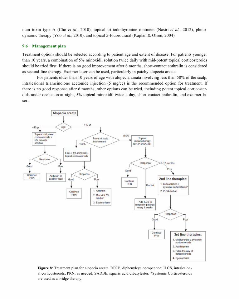
9.6 Management plan
Treatment options should be selected according to patient age and extent of disease. For patients younger than 10 years, a combination of 5% minoxidil solution twice daily with mid-potent topical corticosteroids should be tried first. If there is no good improvement after 6 months, short-contact anthralin is considered as second-line therapy. Excimer laser can be used, particularly in patchy alopecia areata.
For patients older than 10 years of age with alopecia areata involving less than 50% of the scalp, intralesional triamcinolone acetonide injection (5 mg/cc) is the recommended option for treatment. If there is no good response after 6 months, other options can be tried, including potent topical corticoster-oids under occlusion at night, 5% topical minoxidil twice a day, short-contact anthralin, and excimer la-ser.
Figure 8: Treatment plan for alopecia areata. DPCP, diphenylcyclopropenone; ILCS, intralesion-al corticosteroids; PRN, as needed; SADBE, squaric acid dibutylester. *Systemic Corticosteroids are used as a bridge therapy.
If alopecia areata involves more than 50% of the scalp, topical immunotherapy with DPCP is the first therapeutic option recommended by many experts in hair diseases. Intralesional injections of tri-amcinolone acetonide are used to treat persistent alopecic patches.
For patients who respond poorly to DPCP and those who cannot use it, second-line therapies can be used. Several review articles of alopecia areata therapy suggest topical minoxidil and topical cortico-steroids (Alkhalifah et al., 2010; Madani & Shapiro, 2000; Ross & Shapiro, 2005; Wasserman et al., 2007) but, as discussed earlier, the yield of these topical agents in the treatment of extensive alopecia ar-eata is limited. Therefore, we suggest that patients with extensive resistant disease can use sulfasalazine with or without systemic corticosteroids. Systemic steroids are used as bridge therapy until the sulfasala-zine takes effect. Treatment with sulfasalazine is generally well tolerated and characterized by a lower incidence of serious side effects in comparison with other systemic therapies like corticosteroids and methotrexate. The other second-line therapy is PUVA turban. It is a well tolerated therapy with minimal local phototoxic side effects and without the systemic side effects of PUVA. These options are selected based on a balance between the efficacy and safety of these therapeutic agents.
If these therapies fail or are not tolerated, third-line therapeutic options can be discussed with pa-tients in terms of the expected outcome of therapy and possible side effects. These agents include metho-trexate with or without a systemic corticosteroid, azathioprine, cyclosporine, and pulse therapy of cortico-steroids. While using these drugs, close monitoring of patients is important to avoid possible side effects. A summary of an alopecia areata treatment plan is shown as an algorithmic approach in Figure 8.
References
Abdel Fattah, N. S., Ebrahim, A. A., & El Okda, E. S. (2011). Lipid peroxidation/antioxidant activity in patients with alo-pecia areata. J Eur Acad Dermatol Venereol, 25(4), 403-408.
Abedini, H., Farshi, S., Mirabzadeh, A., & Keshavarz, S. (2013). Antidepressant effects of citalopram on treatment of alo-pecia areata in patients with major depressive disorder. J Dermatolog Treat.
Abell, E., & Munro, D. D. (1973). Intralesional treatment of alopecia areata with triamcinolone acetonide by jet injector. Br J Dermatol, 88(1), 55-59.
Aghaei, S. (2008). An uncontrolled, open label study of sulfasalazine in severe alopecia areata. Indian J Dermatol Venere-ol Leprol, 74(6), 611-613.
Ait Ourhroui, M., Hassam, B., & Khoudri, I. (2010). [Treatment of alopecia areata with prednisone in a once-monthly oral pulse]. Ann Dermatol Venereol, 137(8-9), 514-518.
Ajith, C., Gupta, S., & Kanwar, A. J. (2006). Efficacy and safety of the topical sensitizer squaric acid dibutyl ester in Alo-pecia areata and factors influencing the outcome. J Drugs Dermatol, 5(3), 262-266.
Al-Mutairi, N. (2007). 308-nm excimer laser for the treatment of alopecia areata. Dermatol Surg, 33(12), 1483-1487.
Al-Mutairi, N. (2009). 308-nm excimer laser for the treatment of alopecia areata in children. Pediatr Dermatol, 26(5), 547-550.
Ali, A., & Martin, J. M. t. (2010). Hair growth in patients alopecia areata totalis after treatment with simvastatin and ezetimibe. J Drugs Dermatol, 9(1), 62-64.
Alkhalifah, A., Alsantali, A., Wang, E., McElwee, K. J., & Shapiro, J. (2010). Alopecia areata update: part II. Treatment. J Am Acad Dermatol, 62(2), 191-202, quiz 203-194.
Bakry, O. A., Elshazly, R. M., Shoeib, M. A., & Gooda, A. (2013). Oxidative Stress in Alopecia Areata: A Case-Control Study. Am J Clin Dermatol.
Behrens-Williams, S. C., Leiter, U., Schiener, R., Weidmann, M., Peter, R. U., & Kerscher, M. (2001). The PUVA-turban as a new option of applying a dilute psoralen solution selectively to the scalp of patients with alopecia areata. J Am Acad Dermatol, 44(2), 248-252.
Bhat, Y. J., Manzoor, S., Khan, A. R., & Qayoom, S. (2009). Trace element levels in alopecia areata. Indian J Dermatol Venereol Leprol, 75(1), 29-31.
Botchkareva, N. V., Ahluwalia, G., & Shander, D. (2006). Apoptosis in the hair follicle. J Invest Dermatol, 126(2), 258-264.
Brajac, I., Tkalcic, M., Dragojevic, D. M., & Gruber, F. (2003). Roles of stress, stress perception and trait-anxiety in the onset and course of alopecia areata. J Dermatol, 30(12), 871-878.
Broniarczyk-Dyla, G., Wawrzycka-Kaflik, A., Dubla-Berner, M., & Prusinska-Bratos, M. (2006). Effects of psoralen-UV-A-Turban in alopecia areata. Skinmed, 5(2), 64-68.
Cerottini, J. P., Panizzon, R. G., & de Viragh, P. A. (1999). Multifocal alopecia areata during systemic cyclosporine A therapy. Dermatology, 198(4), 415-417.
Chartaux, E., & Joly, P. (2010). [Long-term follow-up of the efficacy of methotrexate alone or in combination with low doses of oral corticosteroids in the treatment of alopecia areata totalis or universalis]. Ann Dermatol Venereol, 137(8-9), 507-513.
Cho, H. R., Lew, B. L., Lew, H., & Sim, W. Y. (2010). Treatment effects of intradermal botulinum toxin type A injection on alopecia areata. Dermatol Surg, 36 Suppl 4, 2175-2181.
Chu, S. Y., Chen, Y. J., Tseng, W. C., Lin, M. W., Chen, T. J., Hwang, C. Y., et al. (2011). Comorbidity profiles among pa-tients with alopecia areata: the importance of onset age, a nationwide population-based study. J Am Acad Der-matol, 65(5), 949-956.
Chu, S. Y., Chen, Y. J., Tseng, W. C., Lin, M. W., Chen, T. J., Hwang, C. Y., et al. (2012). Psychiatric comorbidities in pa-tients with alopecia areata in Taiwan: a case-control study. Br J Dermatol, 166(3), 525-531.
Coronel-Perez, I. M., Rodriguez-Rey, E. M., & Camacho-Martinez, F. M. (2010). Latanoprost in the treatment of eyelash alopecia in alopecia areata universalis. J Eur Acad Dermatol Venereol, 24(4), 481-485.
D'Ovidio, R., Claudatus, J., & Di Prima, T. (2002). Ineffectiveness of imiquimod therapy for Alopecia Totalis/Universalis. J Eur Acad Dermatol Venereol, 16(4), 416-417.
Das, S., Ghorami, R. C., Chatterjee, T., & Banerjee, G. (2010). Comparative assessment of topical steroids, topical trete-noin (0.05%) and dithranol paste in alopecia areata. Indian J Dermatol, 55(2), 148-149.
Dy, L. C., & Whiting, D. A. (2011). Histopathology of alopecia areata, acute and chronic: Why is it important to the clini-cian? Dermatol Ther, 24(3), 369-374.
Ehsani, A. H., Toosi, S., Seirafi, H., Akhyani, M., Hosseini, M., Azadi, R., et al. (2009). Capsaicin vs. clobetasol for the treatment of localized alopecia areata. J Eur Acad Dermatol Venereol, 23(12), 1451-1453.
Ellis, C. N., Brown, M. F., & Voorhees, J. J. (2002). Sulfasalazine for alopecia areata. J Am Acad Dermatol, 46(4), 541-544.
Ettefagh, L., Nedorost, S., & Mirmirani, P. (2004). Alopecia areata in a patient using infliximab: new insights into the role of tumor necrosis factor on human hair follicles. Arch Dermatol, 140(8), 1012.
Fabre, C., & Dereure, O. (2008). Worsening alopecia areata and de novo occurrence of multiple halo nevi in a patient re-ceiving infliximab. Dermatology, 216(2), 185-186.
Farshi, S., Mansouri, P., Safar, F., & Khiabanloo, S. R. (2010). Could azathioprine be considered as a therapeutic alterna-tive in the treatment of alopecia areata? A pilot study. Int J Dermatol, 49(10), 1188-1193.
Fiedler-Weiss, V. C. (1987). Topical minoxidil solution (1% and 5%) in the treatment of alopecia areata. J Am Acad Der-matol, 16(3 Pt 2), 745-748.
Fiedler-Weiss, V. C., & Buys, C. M. (1987). Evaluation of anthralin in the treatment of alopecia areata. Arch Dermatol, 123(11), 1491-1493.
Fiedler, V. C., Wendrow, A., Szpunar, G. J., Metzler, C., & DeVillez, R. L. (1990). Treatment-resistant alopecia areata. Response to combination therapy with minoxidil plus anthralin. Arch Dermatol, 126(6), 756-759.
Forstbauer, L. M., Brockschmidt, F. F., Moskvina, V., Herold, C., Redler, S., Herzog, A., et al. (2012). Genome-wide pool-ing approach identifies SPATA5 as a new susceptibility locus for alopecia areata. Eur J Hum Genet, 20(3), 326-332.
Freyschmidt-Paul P, McElwee KJ, Hoffmann R. (2008). Alopecia areata. In: Whiting DA, Blume-Peytavi U, Tosti A, edi-tors. Hair growth and disorders. Berlin: Springer; pp. 311-332.
Friedli, A., Labarthe, M. P., Engelhardt, E., Feldmann, R., Salomon, D., & Saurat, J. H. (1998). Pulse methylprednisolone therapy for severe alopecia areata: an open prospective study of 45 patients. J Am Acad Dermatol, 39(4 Pt 1), 597-602.
Garcia Bartels, N., Lee, H. H., Worm, M., Burmester, G. R., Sterry, W., & Blume-Peytavi, U. (2006). Development of alo-pecia areata universalis in a patient receiving adalimumab. Arch Dermatol, 142(12), 1654-1655.
Georgala, S., Katoulis, A. C., Befon, A., Georgala, K., & Stavropoulos, P. G. (2006). Inosiplex for treatment of alopecia areata: a randomized placebo-controlled study. Acta Derm Venereol, 86(5), 422-424.
Gilhar, A. (2010). Collapse of immune privilege in alopecia areata: coincidental or substantial? J Invest Dermatol, 130(11), 2535-2537.
Gilhar, A., Pillar, T., & Etzioni, A. (1989). Topical cyclosporin A in alopecia areata. Acta Derm Venereol, 69(3), 252-253.
Grandolfo, M., Biscazzi, A. M., & Pipoli, M. (2008). Alopecia areata and autoimmunity. G Ital Dermatol Venereol, 143(5), 277-281.
Gupta, A. K., Ellis, C. N., Cooper, K. D., Nickoloff, B. J., Ho, V. C., Chan, L. S., et al. (1990). Oral cyclosporine for the treatment of alopecia areata. A clinical and immunohistochemical analysis. J Am Acad Dermatol, 22(2 Pt 1), 242-250.
Hajheydari, Z., Jamshidi, M., Akbari, J., & Mohammadpour, R. (2007). Combination of topical garlic gel and be-tamethasone valerate cream in the treatment of localized alopecia areata: a double-blind randomized controlled study. Indian J Dermatol Venereol Leprol, 73(1), 29-32.
Hart, J., & Shafranov, G. (2004). Hypertrichosis of vellus hairs of the malar region after unilateral treatment with bimato-prost. Am J Ophthalmol, 137(4), 756-757.
Hay, I. C., Jamieson, M., & Ormerod, A. D. (1998). Randomized trial of aromatherapy. Successful treatment for alopecia areata. Arch Dermatol, 134(11), 1349-1352.
Hordinsky, M., & Ericson, M. (2004). Autoimmunity: alopecia areata. J Investig Dermatol Symp Proc, 9(1), 73-78.
Ikeda, T. (1965). A new classification of alopecia areata. Dermatologica, 131(6), 421-445.
Inui, S., Nakajima, T., Nakagawa, K., & Itami, S. (2008). Clinical significance of dermoscopy in alopecia areata: analysis of 300 cases. Int J Dermatol, 47(7), 688-693.
Inui, S., Nakajima, T., Toda, N., & Itami, S. (2009). Fexofenadine hydrochloride enhances the efficacy of contact immuno-therapy for extensive alopecia areata: Retrospective analysis of 121 cases. J Dermatol, 36(6), 323-327.
Jagielska, D., Redler, S., Brockschmidt, F. F., Herold, C., Pasternack, S. M., Garcia Bartels, N., et al. (2012). Follow-up study of the first genome-wide association scan in alopecia areata: IL13 and KIAA0350 as susceptibility loci sup-ported with genome-wide significance. J Invest Dermatol, 132(9), 2192-2197.
Kalkan, G., Ates, O., Karakus, N., & Sezer, S. (2013). Functional polymorphisms in cell death pathway genes FAS and FAS ligand and risk of alopecia areata. Arch Dermatol Res.
Kaplan, A. L., & Olsen, E. A. (2004). Topical 5-fluorouracil is ineffective in the treatment of extensive alopecia areata. J Am Acad Dermatol, 50(6), 941-943.
Kar, B. R., Handa, S., Dogra, S., & Kumar, B. (2005). Placebo-controlled oral pulse prednisolone therapy in alopecia areata. J Am Acad Dermatol, 52(2), 287-290.
Kasumagic-Halilovic, E. (2008). Thyroid autoimmunity in patients with alopecia areata. Acta Dermatovenerol Croat, 16(3), 123-125.
Kim, B. J., Min, S. U., Park, K. Y., Choi, J. W., Park, S. W., Youn, S. W., et al. (2008). Combination therapy of cyc-losporine and methylprednisolone on severe alopecia areata. J Dermatolog Treat, 19(4), 216-220.
Kirshen, C., & Kanigsberg, N. (2009). Alopecia areata following adalimumab. J Cutan Med Surg, 13(1), 48-50.
Koc, E., Tunca, M., Akar, A., & Kurumlu, Z. (2008). Lack of efficacy of topical imiquimod in the treatment of patchy alo-pecia areata. Int J Dermatol, 47(10), 1088-1089.
Koca, R., Armutcu, F., Altinyazar, C., & Gurel, A. (2005). Evaluation of lipid peroxidation, oxidant/antioxidant status, and serum nitric oxide levels in alopecia areata. Med Sci Monit, 11(6), CR296-299.
Kurosawa, M., Nakagawa, S., Mizuashi, M., Sasaki, Y., Kawamura, M., Saito, M., et al. (2006). A comparison of the effi-cacy, relapse rate and side effects among three modalities of systemic corticosteroid therapy for alopecia areata. Dermatology, 212(4), 361-365.
Kurtev, A., & Iliev, E. (2005). Thyroid autoimmunity in children and adolescents with alopecia areata. Int J Dermatol, 44(6), 457-461.
Lee, D., Hong, S. K., Park, S. W., Hur, D. Y., Shon, J. H., Shin, J. G., et al. (2010). Serum levels of IL-18 and sIL-2R in patients with alopecia areata receiving combined therapy with oral cyclosporine and steroids. Exp Dermatol, 19(2), 145-147.
Lester, R. S., Knowles, S. R., & Shear, N. H. (1998). The risks of systemic corticosteroid use. Dermatol Clin, 16(2), 277-288.
Lew, B. L., Shin, M. K., & Sim, W. Y. (2009). Acute diffuse and total alopecia: A new subtype of alopecia areata with a favorable prognosis. J Am Acad Dermatol, 60(1), 85-93.
Madani, S., & Shapiro, J. (2000). Alopecia areata update. J Am Acad Dermatol, 42(4), 549-566; quiz 567-570.
Mancuso, G., Balducci, A., Casadio, C., Farina, P., Staffa, M., Valenti, L., et al. (2003). Efficacy of betamethasone val-erate foam formulation in comparison with betamethasone dipropionate lotion in the treatment of mild-to-moderate alopecia areata: a multicenter, prospective, randomized, controlled, investigator-blinded trial. Int J Dermatol, 42(7), 572-575.
Manolache, L., & Benea, V. (2007). Stress in patients with alopecia areata and vitiligo. J Eur Acad Dermatol Venereol, 21(7), 921-928.
McElwee, K. J., Freyschmidt-Paul, P., Hoffmann, R., Kissling, S., Hummel, S., Vitacolonna, M., et al. (2005). Transfer of CD8(+) cells induces localized hair loss whereas CD4(+)/CD25(-) cells promote systemic alopecia areata and CD4(+)/CD25(+) cells blockade disease onset in the C3H/HeJ mouse model. J Invest Dermatol, 124(5), 947-957.
Miteva, M., & Tosti, A. (2012). Hair and scalp dermatoscopy. J Am Acad Dermatol, 67(5), 1040-1048.
Mohamed, Z., Bhouri, A., Jallouli, A., Fazaa, B., Kamoun, M. R., & Mokhtar, I. (2005). Alopecia areata treatment with a phototoxic dose of UVA and topical 8-methoxypsoralen. J Eur Acad Dermatol Venereol, 19(5), 552-555.
Nakajima, T., Inui, S., & Itami, S. (2007). Pulse corticosteroid therapy for alopecia areata: study of 139 patients. Derma-tology, 215(4), 320-324.
Nasiri, S., Haghpanah, V., Taheri, E., Heshmat, R., Larijani, B., & Saeedi, M. (2012). Hair regrowth with topical triiodo-thyronine ointment in patients with alopecia areata: a double-blind, randomized pilot clinical trial of efficacy. J Eur Acad Dermatol Venereol, 26(5), 654-656.
Orecchia, G., & Perfetti, L. (1991). Alopecia areata and topical sensitizers: allergic response is necessary but irritation is not. Br J Dermatol, 124(5), 509.
Pan, Y., & Rao, N. A. (2009). Alopecia areata during etanercept therapy. Ocul Immunol Inflamm, 17(2), 127-129.
Pandhi, D., Singal, A., Gupta, R., & Das, G. (2009). Ocular alterations in patients of alopecia areata. J Dermatol, 36(5), 262-268.
Park, H., Kim, C. W., Kim, S. S., & Park, C. W. (2009). The therapeutic effect and the changed serum zinc level after zinc supplementation in alopecia areata patients who had a low serum zinc level. Ann Dermatol, 21(2), 142-146.
Park, S. W., Kim, J. W., & Wang, H. Y. (2002). Topical tacrolimus (FK506): treatment failure in four cases of alopecia universalis. Acta Derm Venereol, 82(5), 387-388.
Peckham, S. J., Sloan, S. B., & Elston, D. M. (2011). Histologic features of alopecia areata other than peribulbar lympho-cytic infiltrates. J Am Acad Dermatol, 65(3), 615-620.
Petukhova, L., Duvic, M., Hordinsky, M., Norris, D., Price, V., Shimomura, Y., et al. (2010). Genome-wide association study in alopecia areata implicates both innate and adaptive immunity. Nature, 466(7302), 113-117.
Phillips, M. A., Graves, J. E., & Nunley, J. R. (2005). Alopecia areata presenting in 2 kidney-pancreas transplant recipi-ents taking cyclosporine. J Am Acad Dermatol, 53(5 Suppl 1), S252-255.
Posten, W., & Swan, J. (2005). Recurrence of alopecia areata in a patient receiving etanercept injections. Arch Dermatol, 141(6), 759-760.
Price, V. H. (1987a). Double-blind, placebo-controlled evaluation of topical minoxidil in extensive alopecia areata. J Am Acad Dermatol, 16(3 Pt 2), 730-736.
Price, V. H. (1987b). Topical minoxidil (3%) in extensive alopecia areata, including long-term efficacy. J Am Acad Derma-tol, 16(3 Pt 2), 737-744.
Price, V. H. (1991). Alopecia areata: clinical aspects. J Invest Dermatol, 96(5), 68S.
Price, V. H., Hordinsky, M. K., Olsen, E. A., Roberts, J. L., Siegfried, E. C., Rafal, E. S., et al. (2008). Subcutaneous efali-zumab is not effective in the treatment of alopecia areata. J Am Acad Dermatol, 58(3), 395-402.
Price, V. H., Willey, A., & Chen, B. K. (2005). Topical tacrolimus in alopecia areata. J Am Acad Dermatol, 52(1), 138-139.
Ranganath, V. K., & Furst, D. E. (2007). Disease-modifying antirheumatic drug use in the elderly rheumatoid arthritis pa-tient. Rheum Dis Clin North Am, 33(1), 197-217.
Rashidi, T., & Mahd, A. A. (2008). Treatment of persistent alopecia areata with sulfasalazine. Int J Dermatol, 47(8), 850-852.
Recupero, S. M., Abdolrahimzadeh, S., De Dominicis, M., Mollo, R., Carboni, I., Rota, L., et al. (1999). Ocular alterations in alopecia areata. Eye (Lond), 13 ( Pt 5), 643-646.
Rigopoulos, D., Gregoriou, S., Korfitis, C., Gintzou, C., Vergou, T., Katrinaki, A., et al. (2007). Lack of response of alope-cia areata to pimecrolimus cream. Clin Exp Dermatol, 32(4), 456-457.
Robins, D. N. (2007). Case reports: alopecia universalis: hair growth following initiation of simvastatin and ezetimibe ther-apy. J Drugs Dermatol, 6(9), 946-947.
Roseborough, I., Lee, H., Chwalek, J., Stamper, R. L., & Price, V. H. (2009). Lack of efficacy of topical latanoprost and bimatoprost ophthalmic solutions in promoting eyelash growth in patients with alopecia areata. J Am Acad Der-matol, 60(4), 705-706.
Rosenberg, E. W., & Skinner, R. B., Jr. (2006). Immunotherapy of alopecia areata with intralesional Candida antigen. Pe-diatr Dermatol, 23(3), 299.
Ross, E. K., & Shapiro, J. (2005). Management of hair loss. Dermatol Clin, 23(2), 227-243.
Royer, M., Bodemer, C., Vabres, P., Pajot, C., Barbarot, S., Paul, C., et al. (2011). Efficacy and tolerability of methotrex-ate in severe childhood alopecia areata. Br J Dermatol, 165(2), 407-410.
Rudnicka, L., Olszewska, M., Rakowska, A., & Slowinska, M. (2011). Trichoscopy update 2011. J Dermatol Case Rep, 5(4), 82-88.
Ruiz-Doblado, S., Carrizosa, A., & Garcia-Hernandez, M. J. (2003). Alopecia areata: psychiatric comorbidity and adjust-ment to illness. Int J Dermatol, 42(6), 434-437.
Safavi, K. H., Muller, S. A., Suman, V. J., Moshell, A. N., & Melton, L. J., 3rd. (1995). Incidence of alopecia areata in Olmsted County, Minnesota, 1975 through 1989. Mayo Clin Proc, 70(7), 628-633.
Samrao, A., Fu, J. M., Harris, S. T., & Price, V. H. (2013). Bone mineral density in patients with alopecia areata treated with long-term intralesional corticosteroids. J Drugs Dermatol, 12(2), e36-40.
Sasmaz, S., & Arican, O. (2005). Comparison of azelaic acid and anthralin for the therapy of patchy alopecia areata: a pilot study. Am J Clin Dermatol, 6(6), 403-406.
Seyrafi, H., Akhiani, M., Abbasi, H., Mirpour, S., & Gholamrezanezhad, A. (2005). Evaluation of the profile of alopecia areata and the prevalence of thyroid function test abnormalities and serum autoantibodies in Iranian patients. BMC Dermatol, 5, 11.
Shapiro, J., Lui, H., Tron, V., & Ho, V. (1997). Systemic cyclosporine and low-dose prednisone in the treatment of chronic severe alopecia areata: a clinical and immunopathologic evaluation. J Am Acad Dermatol, 36(1), 114-117.
Sharquie, K. E., & Al-Obaidi, H. K. (2002). Onion juice (Allium cepa L.), a new topical treatment for alopecia areata. J Dermatol, 29(6), 343-346.
Strobel, R., & Rohrborn, G. (1980). Mutagenic and cell transforming activities of 1-chlor-2,4-dinitrobenzene (DNCB) and squaric-acid-dibutylester (SADBE). Arch Toxicol, 45(4), 307-314.
Strober, B. E., Siu, K., Alexis, A. F., Kim, G., Washenik, K., Sinha, A., et al. (2005). Etanercept does not effectively treat moderate to severe alopecia areata: an open-label study. J Am Acad Dermatol, 52(6), 1082-1084.
Taheri, R., Behnam, B., Tousi, J. A., Azizzade, M., & Sheikhvatan, M. R. (2012). Triggering role of stressful life events in patients with alopecia areata. Acta Dermatovenerol Croat, 20(4), 246-250.
Talpur, R., Vu, J., Bassett, R., Stevens, V., & Duvic, M. (2009). Phase I/II randomized bilateral half-head comparison of topical bexarotene 1% gel for alopecia areata. J Am Acad Dermatol, 61(4), 592 e591-599.
Taylor, C. R., & Hawk, J. L. (1995). PUVA treatment of alopecia areata partialis, totalis and universalis: audit of 10 years' experience at St John's Institute of Dermatology. Br J Dermatol, 133(6), 914-918.
Tosti, A., Piraccini, B. M., Pazzaglia, M., & Vincenzi, C. (2003). Clobetasol propionate 0.05% under occlusion in the treatment of alopecia totalis/universalis. J Am Acad Dermatol, 49(1), 96-98.
Ucak, H., Cicek, D., Demir, B., Erden, I., & Ozturk, S. (2012). Prognostic factors that affect the response to topical treat-ment in patchy alopecia areata. J Eur Acad Dermatol Venereol.
van der Steen, P. H., van Baar, H. M., Happle, R., Boezeman, J. B., & Perret, C. M. (1991). Prognostic factors in the treat-ment of alopecia areata with diphenylcyclopropenone. J Am Acad Dermatol, 24(2 Pt 1), 227-230.
Vila, O. T., & Camacho Martinez, F. M. (2010). Bimatoprost in the treatment of eyelash universalis alopecia areata. Int J Trichology, 2 (2), 86-88.
Wasserman, D., Guzman-Sanchez, D. A., Scott, K., & McMichael, A. (2007). Alopecia areata. Int J Dermatol, 46(2), 121-131.
Wasylyszyn, T., Kozlowski, W., & Zabielski, S. L. (2007). Changes in distribution pattern of CD8 lymphocytes in the scalp in alopecia areata during treatment with diphencyprone. Arch Dermatol Res, 299(5-6), 231-237.
Whiting, D. A. (2003). Histopathologic features of alopecia areata: a new look. Arch Dermatol, 139(12), 1555-1559.
Willemsen, R., Haentjens, P., Roseeuw, D., & Vanderlinden, J. (2010). Hypnosis in refractory alopecia areata significantly improves depression, anxiety, and life quality but not hair regrowth. J Am Acad Dermatol, 62(3), 517-518.
Wiseman, M. C., Shapiro, J., MacDonald, N., & Lui, H. (2001). Predictive model for immunotherapy of alopecia areata
with diphencyprone. Arch Dermatol, 137(8), 1063-1068.
Yano, S., Ihn, H., Nakamura, K., Okochi, H., & Tamaki, K. (1999). Antinuclear and antithyroid antibodies in 68 Japanese patients with alopecia areata. Dermatology, 199(2), 191.
Yoo, K. H., Kim, M. N., Kim, B. J., & Kim, C. W. (2010). Treatment of alopecia areata with fractional photothermolysis laser. Int J Dermatol, 49(7), 845-847.
Yoo, K. H., Lee, J. W., Li, K., Kim, B. J., & Kim, M. N. (2010). Photodynamic therapy with methyl 5-aminolevulinate acid might be ineffective in recalcitrant alopecia totalis regardless of using a microneedle roller to increase skin penetra-tion. Dermatol Surg, 36(5), 618-622.
Zhang, X., Yu, M., Yu, W., Weinberg, J., Shapiro, J., & McElwee, K. J. (2009). Development of alopecia areata is associ-ated with higher central and peripheral hypothalamic-pituitary-adrenal tone in the skin graft induced C3H/HeJ mouse model. J Invest Dermatol, 129(6), 1527-1538.
Zoller, M., McElwee, K. J., Engel, P., & Hoffmann, R. (2002). Transient CD44 variant isoform expression and reduction in CD4(+)/CD25(+) regulatory T cells in C3H/HeJ mice with alopecia areata. J Invest Dermatol, 118(6), 983-992.