aloe vera oral administration accelerates acute radiation-delayed wound healing
DESCRIPTION
lidah buaya mempercepat penyembuhan lukaTRANSCRIPT
H
G
cii
2
0d
The American Journal of Surgery (2011) 201, 809–818
Scientific (Exp)/Research
Aloe vera oral administration accelerates acute radiation-delayed wound healing by stimulating transforminggrowth factor-� and fibroblast growth factor production
Ayman Atiba, M.V.Sc.a,b,*, Mayumi Nishimura, Ph.D.c, Shizuko Kakinuma, Ph.D.c,Takeshi Hiraoka, Ph.D.c, Masanobu Goryo, Ph.D.d, Yoshiya Shimada, Ph.D.c,
iroshi Ueno, Ph.D.e, Yuji Uzuka, Ph.D.a,b
aLaboratory of Small Animal Surgery, Department of Veterinary Medicine, Iwate University, 3-18-8 Ueda, Morioka, Iwate 020-8550,Japan; bDepartment of veterinary Clinical Science, The United Graduate School of Veterinary Science, Gifu University, 1-1 Yanagido,
ifu, Japan; cDepartment of Experimental Radiobiology for Children’s Health Research, National Institute of Radiological Sciences,Chiba, Japan; dDepartment of Veterinary Pathology, Faculty of Agriculture, Iwate University, 3-18-8 Ueda, Morioka, Iwate 020-8550,
Japan; eDepartment of Small Animal Clinical Sciences, Rakuno Gakuen University, Hokkaido 069-8501, JapanAbstractBACKGROUND: Delayed wound healing is a significant clinical problem in patients who have had
previous irradiation. This study investigated the effectiveness of Aloe vera (Av) on acute radiation-delayedwound healing.
METHODS: The effect of Av was studied in radiation-exposed rats compared with radiation-only andcontrol rats. Skin wounds were excised on the back of rats after 3 days of local radiation. Wound size wasmeasured on days 0, 3, 6, 9, and 12 after wounding. Wound tissues were examined histologically and theexpressions of transforming growth factor �-1 (TGF-�-1) and basic fibroblast growth factor (bFGF) wereexamined by immunohistochemistry and reverse-transcription polymerase chain reaction.
RESULTS: Wound contraction was accelerated significantly by Av on days 6 and 12 after wounding.Furthermore, the inflammatory cell infiltration, fibroblast proliferation, collagen deposition, angiogenesis,and the expression levels of TGF-�-1 and bFGF were significantly higher in the radiation plus Av groupcompared with the radiation-only group.
CONCLUSIONS: These data showed the potential application of Av to improve the acute radiation-delayed wound healing by increasing TGF-�-1 and bFGF production.© 2011 Elsevier Inc. All rights reserved.
KEYWORDS:Aloe vera;Radiation wound;TGF-�;FGF
Wound healing is a highly complex but orchestrated cas-ade of cellular and molecular events. Successful wound heal-ng is the result of a sequence of several basic processes,ncluding inflammation, cell proliferation, angiogenesis,
* Corresponding author. Tel.: �81-19-621-6265; fax: �81-19-621-6265.E-mail address: [email protected] received February 19, 2010; revised manuscript June 21,
010
002-9610/$ - see front matter © 2011 Elsevier Inc. All rights reserved.oi:10.1016/j.amjsurg.2010.06.017
wound contraction, epithelialization, and matrix remodeling.These processes, which depend on the appropriate and inte-grated functions of neutrophils, macrophages, fibroblasts, andendothelial cells, are thought to be organized by interactionsamong cells, extracellular matrix proteins, and growth factors.Any alteration of these healing processes will result in abnor-mal wound healing and adversely affect the surgical outcome.1
Combined radiotherapy and surgery have resulted in
improved functional and cosmetic outcomes in cancer pa-
riat
g
cplt
p
Ad
i
hcb
810 The American Journal of Surgery, Vol 201, No 6, June 2011
tients.2 However, preoperative radiotherapy carries a goodesponse rate; it is associated with significant morbidityncluding wound infection and delayed healing.3 Radiother-py has been shown to affect nearly every cell type involved inhe process of wound healing.4 It declines neovascularization,
fibroblast proliferation, collagen deposition, and varying levelsof regulatory growth factors of wound healing.5–7
Various growth factors are found to be down-regulatedafter acute radiation exposure.3,4,7,8 Among the manyrowth factors, transforming growth factor �-1(TGF-�-1)
and basic fibroblast growth factor (bFGF) are stronglydown-regulated after acute radiation exposure.4,7,9 Con-versely, TGF-�-1 has been found to be increased in thehronic phase of irradiated wound healing.3,4 Fibroblastroliferation, transformation to myofibroblasts, and stimu-ation of extracellular matrix (ECM) production are all con-rolled by growth factors, primarily TGF-�-1.10 Another
important growth factor in the wound-healing process isbFGF. bFGF can regulate the migration and replication ofendothelial and epithelial cells as well as fibroblasts, whichare responsible for neovascularization, epithelialization, andcollagen production, respectively.7
Several trials have been performed to identify possibletherapeutic approaches to increase healing after radiation. Inrecent years, the use of natural/herbal products has beenincreased dramatically in wound healing because they con-tain a wealth of interesting and possibly beneficial pharma-cologically active compounds. Moreover, they have wideacceptability and better tolerance, are inexpensive, and canbe manipulated safely for human use.11 The Aloe vera (Av)lant has been known as “the healing plant.”12 Av has been
used to treat wounds and burns for centuries.11 The oral andtopical application of Av gel has been reported to be effec-tive in wound repair in normal and/or diabetic condi-tions.13–15 Recent studies have shown that treatment witheither Av crude gel or its extracts, such as acemannan,�-sitosterol, and others, resulted in faster healing of woundsby stimulating fibroblast proliferation, collagen deposition,angiogenesis, and production of growth factors.13–19 Clini-cal trials also have examined the effectiveness of Av gel inthe treatment of wound healing and burn wound healing.16
Fulton20 documented stimulation of wound healing of full-face dermabrasion in 18 patients treated with a polyethyleneoxide gel dressing saturated with stabilized Av comparedwith a polyethylene oxide gel dressing. Khorasani et al21
clinically reported that healing of partial-thickness burns inpatients was significantly faster in sites treated with aloethan in sites treated with silver sulfadiazine. Based on themeta-analysis of 2 studies using duration of wound healingas an outcome measure, the Av group was quicker to healburn wounds: 8.79 days shorter than those in the controlgroup (CG).22 Despite reported radioprotective effects of
v,23,24 its influence on the early phase of acute radiated-elayed wound healing has not been studied.
The purposes of the current study were to investigate the
nfluence of Av oral administration on cutaneous woundealing in an irradiated rat model and to correlate the out-ome of healing with the expression levels of TGF-�-1 andFGF in the wound.
Methods
Animals
Thirty-six male Wistar rats, 6 weeks old, weighing 200to 250 g (Nihon CLEA, Inc, Tokyo, Japan) were assignedrandomly to 3 groups (n � 12 each) as follows: CG (sham-radiation), radiation-only group (RG), and radiation plus Avtreatment group (RAG). Animals were housed individuallyfor 1 week before the experiment for accommodation. Ratswere given free access to commercial rat food and water adlibitum and were subjected to a 12-hour light–dark cycle.This study was approved by the ethics committee of theNational Institute of Radiological Sciences in accordancewith the guidelines of the National Institutes of Health.
Radiation
RG and RAG rats were anesthetized with pentobarbitalsodium (Nembutal; Dainippon Sumitomo Pharma, Co, Ltd,Osaka, Japan), at a dose rate of 40 mg/kg of body weight,administered intraperitoneally (IP). The dorsal hair wasclipped and the corners of a 3 � 3-cm area were tattooed onthe back of each animal. A sheet of 3-mm lead with 3 �3-cm cut out to accommodate the skin was used to shield therats’ bodies. A single dose of 9 Gy soft x-rays, generated bya M-70-WE machine (SOFTEX Cooperation, Kanagawa,Japan), was administered to the selected area on each rat.The radiation conditions were as follows: 30 kV, 30 mA, .1mm Al Filter, and a 46-cm focus-surface distance. The doserate in this condition was about 1.1 Gy/min, and exposuretime was 8 minutes and 55 seconds.
Wound creation and assessment of wound size
Three days postradiation, all rats were anesthetized withpentobarbital (40 mg/kg) IP. The back hair was shaved anddisinfected with 70% ethanol. A full-thickness skin woundexcision measuring 1.5 � 1.5 cm was made on the back ofeach animal in the center of the irradiated skin area. Thewounds were photographed on days 0, 3, 6, 9, and 12 afterwounding by a digital camera. All wound images weretransferred to the computer, changed to a tagged informa-tion file format extension using Adobe Photoshop Elements4.0 (Adobe Systems, Tokyo, Japan). The wound size areawas measured with special size analysis software, (NationalInstitutes of Health ImageJ software, downloaded fromhttp://www.rsb.info.nih.gov/ij). The change in wound size
was expressed as a percentage of the original wound size
ow
dagiepw(a
tsitnti4
bessTJEi
tewr
tppfisesefi
TiwittU
a
811A. Atiba et al. Aloe promotes radiation-delayed wound healing
(day 0). Wound dressing was performed with a BAND-AIDKizu Power Pad (Johnson & Johnson, Tokyo, Japan).
Aloe vera administration
RAG rats were administered lyophilized Av powder(Coral Vegetable, Miyakojima, Japan) at a dose of 30 mg/head,14 dissolved in 1.5 mL purified water. Av was givenrally by an oral tube on days 0, 3, 6, 9, and 12 afterounding.Five rats from each group were euthanized by an over-
ose of pentobarbital anesthesia (500 mg/kg, IP) on days 6nd 15 after wounding. The entire wound including a mar-in of approximately 5 mm of unwounded skin was excisedn-depth to include underlying connective tissues above thexternal fascia of the dorsal muscles. The skin wound sam-les of day 6 postwounding were divided into 2 halves; 1as fixed for 48 hours in a 10% buffered formalin solution
pH 7.4) and embedded in paraffin, the other half was frozent �80°C.
Histopathologic examination
Skin wound tissues were cut 4-�m thick perpendicular tohe wound and stained with hematoxylin-eosin and Mas-on’s trichrome stain (for scoring of collagen). The follow-ng parameters were assessed individually: the epithelializa-ion, inflammatory cells infiltration, fibroblast proliferation,eovascularization, and collagen deposition. Epithelializa-ion and collagen deposition were graded histologically us-ng a modified Ehrlich and Hunt numeric scale from 0 to.25 The inflammatory cell infiltration and fibroblast prolif-
eration were evaluated by counting the inflammatory cells(neutrophils, macrophages, and lymphocytes) and fibro-blasts. The counting of inflammatory cells and fibroblastswere assessed by capture 8 (day 6) and 4 (day 15) randomlyselected high power-fields (magnification, 400�) imagesand counting using a manual cell counter in ImageJ soft-ware. To evaluate the neovascularization, the number ofblood vessels was counted in 4 randomly selected fields(magnification, 100�) 3 times and the mean number ofblood vessels in each group was compared. All the woundtissue samples were graded histologically in a blind fashionon 2 slides per animal.
Immunohistochemical staining
Immunohistochemistry was used to detect TGF-�-1 andFGF protein in rat skin wounds. Two slides obtained fromach specimen were used for immunohistochemistry. Theections were deparaffinized in xylene and rehydrated. Thelides were immersed in a covered plastic container witharget Retrieval Solution pH 9 (DAKO Corporation, Kyoto,apan) and placed in an autoclave at 121°C for 10 minutes.ndogenous peroxidase activity was quenched with perox-
dase blocking solution (DAKO) for 10 minutes at room c
emperature. After a thorough washing in phosphate-buff-red saline (PBS), the protein block serum-free (DAKO)as applied to block nonspecific binding for 10 minutes at
oom temperature. The primary antibody (TGF-�-1 [V],Sc-146 [dilution 1:500], and bFGF [147] Sc-79 [dilution1:500]; Santa Cruz Biotechnologies, Santa Cruz, CA) wereapplied to slides and incubated in a moist chamber at 4°Covernight. After washing in PBS, the sections were incu-bated with a biotinylated secondary antibody (DAKO) for30 minutes. After a second washing in PBS, the sectionswere incubated with avidin-biotin complex with horseradishperoxidase solution (Vectastain ABC kit, catalog # SP-2001; Vector Laboratories, Burlingham, CA) for 30 min-utes. After final PBS washing was performed. Diaminoben-zidine tetrahydrochloride (DAKO) was treated as achromogen. All slides were counterstained with hematoxy-lin, rinsed in distilled water again, dehydrated, andmounted. The sections were examined qualitatively under abright field microscope at 100 to 400� magnification forchanges in the expression of TGF-�-1 and bFGF in woundissue among the groups. For quantitative analysis of ex-ression labeling indexes were determined as the ratio ofositively stained cells to the total number of cells per visualeld. For this study, only cell-associated staining was con-idered. The criteria for a positively expressing cell were thexistence of a clear cell structure with nucleolus and clearpecific staining of the cytoplasm. The overall number ofvaluated cells per section was 900 � 100 cells (at least 3elds per section were examined with 400� magnification).
Total RNA extraction and reverse-transcriptionpolymerase chain reaction
A Mixer Mill MM 301 (Retsch, Inc, Pittsburgh, PA) wasused to homogenize granulation tissue specimens. TotalRNA was extracted from the homogenized granulation tis-sue using the Maxwell 16 Total RNA Purification Kit (Pro-mega Corporation, Madison, WI) according to the manu-facturer’s protocol. The A260/A280 ratio was used todetermine the quantity of total RNA. The ratio of A260/A280 was from 1.8 to 2.0. First-strand complementaryDNA was synthesized by reverse transcription of total RNA(1 �g/�L) using ReverTra Ace kit (ReverTra Ace-�;
oyobo, Osaka, Japan) according to the manufacturer’snstructions. The polymerase chain reaction (PCR) reactionas performed with a diluted complementary DNA sample
n a 50-�L reaction volume. The final reaction concentra-ions were 20 �mol/L primers, 10 mmol/L deoxynucleosideriphosphates (dNTPs) mixture, 10 � PCR buffer II, and .5/�L Takara Ex Taq HS polymerase (Takara Bio, Inc,
Shiga, Japan) per reaction. Rat-specific primers for glycer-aldehyde-3-phosphate dehydrogenase (GAPDH), TGF-�-1,and bFGF were designed and tested elsewhere (Table1).26,27 The cycling conditions for PCR amplification weres follows: 1 cycle of 95°C for 3 minutes followed by 30
ycles of 94°C (45 s), 60°C (45 s), and 72°C (75 s) for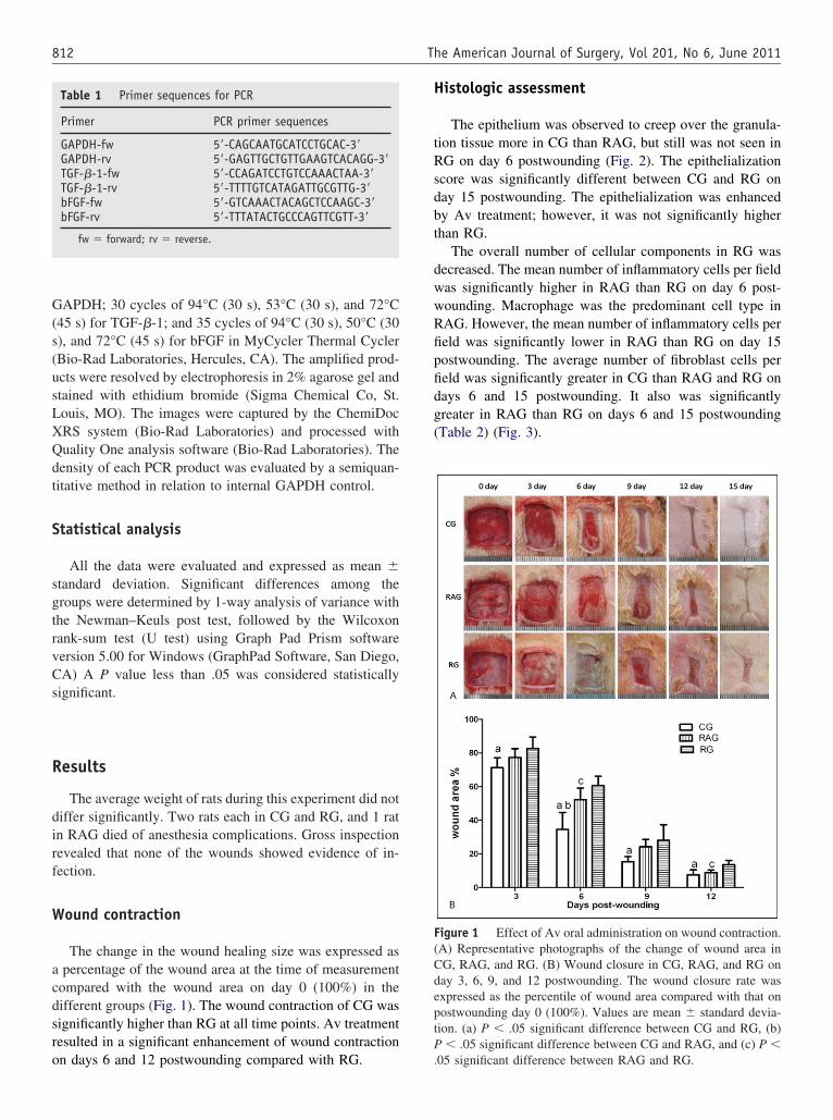
s(usLXQdt
sgtrvCs
tP
812 The American Journal of Surgery, Vol 201, No 6, June 2011
GAPDH; 30 cycles of 94°C (30 s), 53°C (30 s), and 72°C(45 s) for TGF-�-1; and 35 cycles of 94°C (30 s), 50°C (30), and 72°C (45 s) for bFGF in MyCycler Thermal CyclerBio-Rad Laboratories, Hercules, CA). The amplified prod-cts were resolved by electrophoresis in 2% agarose gel andtained with ethidium bromide (Sigma Chemical Co, St.ouis, MO). The images were captured by the ChemiDocRS system (Bio-Rad Laboratories) and processed withuality One analysis software (Bio-Rad Laboratories). Theensity of each PCR product was evaluated by a semiquan-itative method in relation to internal GAPDH control.
Statistical analysis
All the data were evaluated and expressed as mean �tandard deviation. Significant differences among theroups were determined by 1-way analysis of variance withhe Newman–Keuls post test, followed by the Wilcoxonank-sum test (U test) using Graph Pad Prism softwareersion 5.00 for Windows (GraphPad Software, San Diego,A) A P value less than .05 was considered statistically
ignificant.
Results
The average weight of rats during this experiment did notdiffer significantly. Two rats each in CG and RG, and 1 ratin RAG died of anesthesia complications. Gross inspectionrevealed that none of the wounds showed evidence of in-fection.
Wound contraction
The change in the wound healing size was expressed asa percentage of the wound area at the time of measurementcompared with the wound area on day 0 (100%) in thedifferent groups (Fig. 1). The wound contraction of CG wassignificantly higher than RG at all time points. Av treatmentresulted in a significant enhancement of wound contraction
Table 1 Primer sequences for PCR
Primer PCR primer sequences
GAPDH-fw 5=-CAGCAATGCATCCTGCAC-3=GAPDH-rv 5=-GAGTTGCTGTTGAAGTCACAGG-3=TGF-�-1-fw 5=-CCAGATCCTGTCCAAACTAA-3=TGF-�-1-rv 5=-TTTTGTCATAGATTGCGTTG-3=bFGF-fw 5=-GTCAAACTACAGCTCCAAGC-3=bFGF-rv 5=-TTTATACTGCCCAGTTCGTT-3=
fw � forward; rv � reverse.
on days 6 and 12 postwounding compared with RG. .
Histologic assessment
The epithelium was observed to creep over the granula-tion tissue more in CG than RAG, but still was not seen inRG on day 6 postwounding (Fig. 2). The epithelializationscore was significantly different between CG and RG onday 15 postwounding. The epithelialization was enhancedby Av treatment; however, it was not significantly higherthan RG.
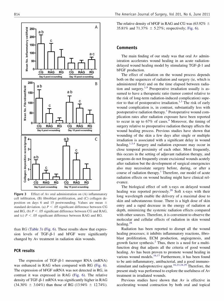
The overall number of cellular components in RG wasdecreased. The mean number of inflammatory cells per fieldwas significantly higher in RAG than RG on day 6 post-wounding. Macrophage was the predominant cell type inRAG. However, the mean number of inflammatory cells perfield was significantly lower in RAG than RG on day 15postwounding. The average number of fibroblast cells perfield was significantly greater in CG than RAG and RG ondays 6 and 15 postwounding. It also was significantlygreater in RAG than RG on days 6 and 15 postwounding(Table 2) (Fig. 3).
Figure 1 Effect of Av oral administration on wound contraction.(A) Representative photographs of the change of wound area inCG, RAG, and RG. (B) Wound closure in CG, RAG, and RG onday 3, 6, 9, and 12 postwounding. The wound closure rate wasexpressed as the percentile of wound area compared with that onpostwounding day 0 (100%). Values are mean � standard devia-ion. (a) P � .05 significant difference between CG and RG, (b)
� .05 significant difference between CG and RAG, and (c) P �
05 significant difference between RAG and RG.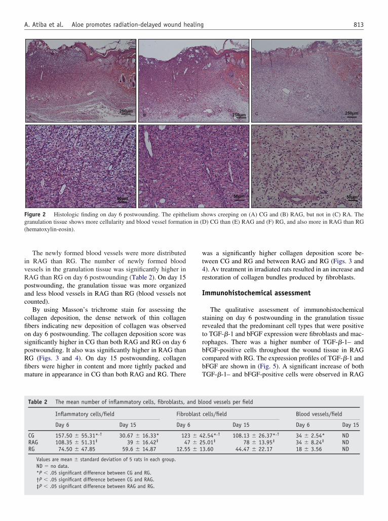
fim
rbc
813A. Atiba et al. Aloe promotes radiation-delayed wound healing
The newly formed blood vessels were more distributedin RAG than RG. The number of newly formed bloodvessels in the granulation tissue was significantly higher inRAG than RG on day 6 postwounding (Table 2). On day 15postwounding, the granulation tissue was more organizedand less blood vessels in RAG than RG (blood vessels notcounted).
By using Masson’s trichrome stain for assessing thecollagen deposition, the dense network of thin collagenfibers indicating new deposition of collagen was observedon day 6 postwounding. The collagen deposition score wassignificantly higher in CG than both RAG and RG on day 6postwounding. It also was significantly higher in RAG thanRG (Figs. 3 and 4). On day 15 postwounding, collagen
bers were higher in content and more tightly packed andature in appearance in CG than both RAG and RG. There
Figure 2 Histologic finding on day 6 postwounding. The epithegranulation tissue shows more cellularity and blood vessel formati(hematoxylin-eosin).
Table 2 The mean number of inflammatory cells, fibroblasts,
Inflammatory cells/field Fibro
Day 6 Day 15 Day
CG 157.50 � 55.31*,† 30.67 � 16.33* 12RAG 108.35 � 51.31‡ 39 � 16.42‡ 4RG 74.50 � 47.85 59.6 � 14.87 12.5
Values are mean � standard deviation of 5 rats in each group.ND � no data.*P � .05 significant difference between CG and RG.†P � .05 significant difference between CG and RAG.‡P � .05 significant difference between RAG and RG.
was a significantly higher collagen deposition score be-tween CG and RG and between RAG and RG (Figs. 3 and4). Av treatment in irradiated rats resulted in an increase andrestoration of collagen bundles produced by fibroblasts.
Immunohistochemical assessment
The qualitative assessment of immunohistochemicalstaining on day 6 postwounding in the granulation tissuerevealed that the predominant cell types that were positiveto TGF-�-1 and bFGF expression were fibroblasts and mac-ophages. There was a higher number of TGF-�-1– andFGF-positive cells throughout the wound tissue in RAGompared with RG. The expression profiles of TGF-�-1 and
bFGF are shown in (Fig. 5). A significant increase of bothTGF-�-1– and bFGF-positive cells were observed in RAG
hows creeping on (A) CG and (B) RAG, but not in (C) RA. The) CG than (E) RAG and (F) RG, and also more in RAG than RG
ood vessels per field
cells/field Blood vessels/field
Day 15 Day 6 Day 15
2.54*,† 108.13 � 26.37*,† 34 � 2.54* ND5.01‡ 78 � 13.95‡ 34 � 8.24‡ ND3.60 44.47 � 22.17 18 � 3.56 ND
lium son in (D
and bl
blast
6
3 � 47 � 25 � 1
c
(
T3
swwihctsaac
lsedwmh
fhv
814 The American Journal of Surgery, Vol 201, No 6, June 2011
than RG (Table 3) (Fig. 6). These results show that expres-sion levels of TGF-�-1 and bFGF were significantlyhanged by Av treatment in radiation skin wounds.
PCR results
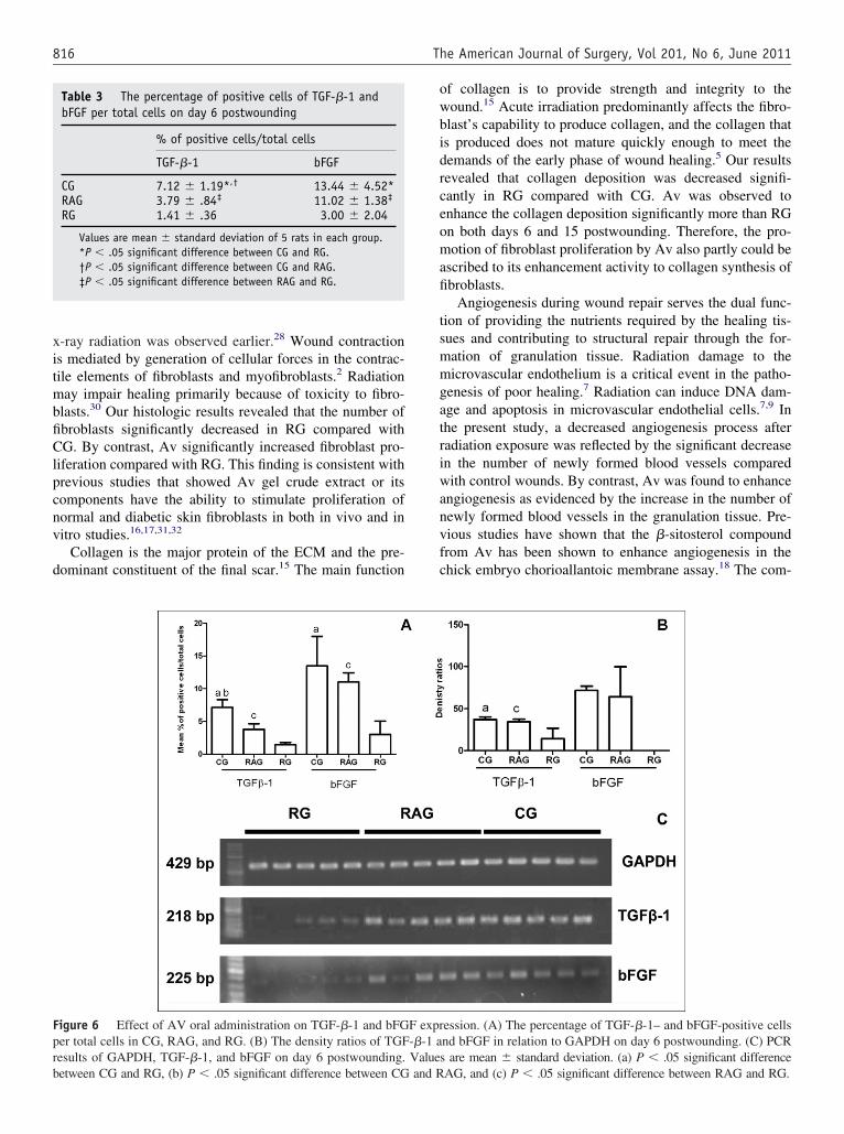
The expression of TGF-�-1 messenger RNA (mRNA)was enhanced in RAG when compared with RG (Fig. 6).The expression of bFGF mRNA was not detected in RG, incontrast it was expressed in RAG (Fig. 6). The relativedensity of TGF-�-1 mRNA was significantly higher in RAG
Figure 3 Effect of Av oral administration on (A) inflammatorycell infiltration, (B) fibroblast proliferation, and (C) collagen de-position on days 6 and 15 postwounding. Values are mean �standard deviation. (a) P � .05 significant difference between CGand RG, (b) P � .05 significant difference between CG and RAG,and (c) P � .05 significant difference between RAG and RG.
34.30% � 3.04%) than those of RG (13.94% � 12.74%).
he relative density of bFGF in RAG and CG was (63.92% �5.81% and 71.37% � 5.27%; respectively; Fig. 6).
Comments
The main finding of our study was that oral Av admin-istration accelerates wound healing in an acute radiation-delayed wound healing model by stimulating TGF-�-1 andbFGF production.
The effect of radiation on the wound process dependsboth on the sequences of radiation and surgery (ie, which isadministered first) and on the time elapsed between radia-tion and surgery.1,9 Preoperative irradiation usually is as-sumed to have a therapeutic ratio (tumor control relative tothe risk of long-term radiation-induced complication) supe-rior to that of postoperative irradiation.1,3 The risk of earlywound complication is, in contrast, substantially less withpostoperative radiation therapy.1 Postoperative wound com-plication rates after radiation exposure have been reportedto occur in up to 67% of cases.4 Moreover, the timing ofurgery relative to preoperative radiation therapy affects theound healing process. Previous studies have shown thatounding of the skin a few days after single or multiple
rradiation is associated with a significant delay in woundealing.1,3,4 Surgery and radiation exposure may occur inlose temporal proximity of each other. Most frequently,his occurs in the setting of adjuvant radiation therapy, andurgeons do not frequently create excisional wounds acutelyfter radiation but the development of surgical emergencieslso may necessitate surgery before, during, or after aourse of radiation therapy.1 Therefore, our model of acute
radiation effects on wound healing might have clinical rel-evance.
The biological effect of soft x-rays on delayed woundhealing was reported previously.28 Soft x-rays with theirong wavelength enable the delivery of a maximal dose tokin and subcutaneous tissue. There is a high dose of skinntry and a rapid decrease in the energy of radiation atepth, minimizing the systemic radiation effects comparedith other sources. Therefore, it is convenient to observe theolecular and cellular effects of radiation in skin wound
ealing.28
Radiation has been reported to disrupt all the woundhealing processes; it inhibits inflammatory reactions, fibro-blast proliferation, ECM production, angiogenesis, andgrowth factor synthesis.2 Thus, there is a need for a multi-unction drug that adjusts all the criteria of good woundealing. Av has been proven to promote wound healing inarious wound models.16,17 Furthermore, it has been found
to be anti-inflammatory, antibacterial, and a good immuno-stimulant and radioprotective agent.16,23,24,29 Therefore, thepresent study was performed to explore the usefulness of Avtreatment in irradiated wounds.
Previous studies have shown that Av is effective in
accelerating wound contraction by both oral and topical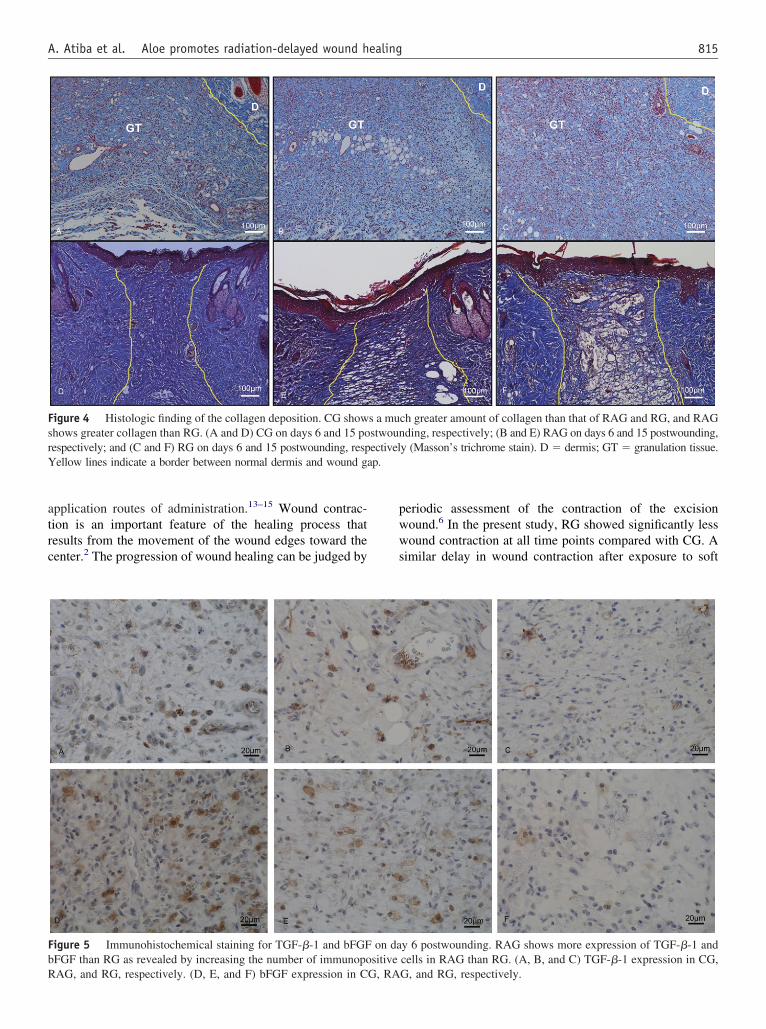
trc
pwws
s
ap.
815A. Atiba et al. Aloe promotes radiation-delayed wound healing
application routes of administration.13–15 Wound contrac-ion is an important feature of the healing process thatesults from the movement of the wound edges toward theenter.2 The progression of wound healing can be judged by
Figure 4 Histologic finding of the collagen deposition. CG showhows greater collagen than RG. (A and D) CG on days 6 and 15 po
respectively; and (C and F) RG on days 6 and 15 postwounding, respYellow lines indicate a border between normal dermis and wound g
Figure 5 Immunohistochemical staining for TGF-�-1 and bFGFbFGF than RG as revealed by increasing the number of immunopo
RAG, and RG, respectively. (D, E, and F) bFGF expression in CG, RAeriodic assessment of the contraction of the excisionound.6 In the present study, RG showed significantly lessound contraction at all time points compared with CG. A
imilar delay in wound contraction after exposure to soft
ch greater amount of collagen than that of RAG and RG, and RAGding, respectively; (B and E) RAG on days 6 and 15 postwounding,
y (Masson’s trichrome stain). D � dermis; GT � granulation tissue.
y 6 postwounding. RAG shows more expression of TGF-�-1 andcells in RAG than RG. (A, B, and C) TGF-�-1 expression in CG,
s a mustwounectivel
on dasitive
G, and RG, respectively.
it
ow
rceomafi
tsmmgatriwanvfc
r
816 The American Journal of Surgery, Vol 201, No 6, June 2011
x-ray radiation was observed earlier.28 Wound contractions mediated by generation of cellular forces in the contrac-ile elements of fibroblasts and myofibroblasts.2 Radiation
may impair healing primarily because of toxicity to fibro-blasts.30 Our histologic results revealed that the number offibroblasts significantly decreased in RG compared withCG. By contrast, Av significantly increased fibroblast pro-liferation compared with RG. This finding is consistent withprevious studies that showed Av gel crude extract or itscomponents have the ability to stimulate proliferation ofnormal and diabetic skin fibroblasts in both in vivo and invitro studies.16,17,31,32
Collagen is the major protein of the ECM and the pre-dominant constituent of the final scar.15 The main function
Table 3 The percentage of positive cells of TGF-�-1 andbFGF per total cells on day 6 postwounding
% of positive cells/total cells
TGF-�-1 bFGF
CG 7.12 � 1.19*,† 13.44 � 4.52*RAG 3.79 � .84‡ 11.02 � 1.38‡
RG 1.41 � .36 3.00 � 2.04
Values are mean � standard deviation of 5 rats in each group.*P � .05 significant difference between CG and RG.†P � .05 significant difference between CG and RAG.‡P � .05 significant difference between RAG and RG.
Figure 6 Effect of AV oral administration on TGF-�-1 and bFGper total cells in CG, RAG, and RG. (B) The density ratios of TGFesults of GAPDH, TGF-�-1, and bFGF on day 6 postwounding.
between CG and RG, (b) P � .05 significant difference between CG and R
f collagen is to provide strength and integrity to theound.15 Acute irradiation predominantly affects the fibro-
blast’s capability to produce collagen, and the collagen thatis produced does not mature quickly enough to meet thedemands of the early phase of wound healing.5 Our resultsevealed that collagen deposition was decreased signifi-antly in RG compared with CG. Av was observed tonhance the collagen deposition significantly more than RGn both days 6 and 15 postwounding. Therefore, the pro-otion of fibroblast proliferation by Av also partly could be
scribed to its enhancement activity to collagen synthesis ofbroblasts.
Angiogenesis during wound repair serves the dual func-ion of providing the nutrients required by the healing tis-ues and contributing to structural repair through the for-ation of granulation tissue. Radiation damage to theicrovascular endothelium is a critical event in the patho-
enesis of poor healing.7 Radiation can induce DNA dam-ge and apoptosis in microvascular endothelial cells.7,9 Inhe present study, a decreased angiogenesis process afteradiation exposure was reflected by the significant decreasen the number of newly formed blood vessels comparedith control wounds. By contrast, Av was found to enhance
ngiogenesis as evidenced by the increase in the number ofewly formed blood vessels in the granulation tissue. Pre-ious studies have shown that the �-sitosterol compoundrom Av has been shown to enhance angiogenesis in thehick embryo chorioallantoic membrane assay.18 The com-
ession. (A) The percentage of TGF-�-1– and bFGF-positive cellsnd bFGF in relation to GAPDH on day 6 postwounding. (C) PCRs are mean � standard deviation. (a) P � .05 significant difference
F expr-�-1 aValue
AG, and (c) P � .05 significant difference between RAG and RG.

slVl
bp
ew
lc
ri
r
d
1
1
1
1
1
1
1
817A. Atiba et al. Aloe promotes radiation-delayed wound healing
pound also showed positive results in the human umbilicalvein endothelial cell motility assay.11 In another experi-ment, �-sitosterol was shown to enhance the expression ofeveral proteins related to angiogenesis, namely Von Wil-ebrand factors, vascular endothelial growth factor (VEGF),EGF-receptor fetal liver kinase-1, and blood vessel matrix
aminin in the brain of Mongolian gerbils.19
Growth factors are critical regulators of the wound-heal-ing process.33 Among many growth factors, TGF-�-1 andFGF are strongly down-regulated after acute radiation ex-osure.4,7,9 Although nearly all cell types produce TGF-�-1,
its main sources of production in wound healing are plate-lets, monocytes/macrophages, and fibroblasts.10 Our datarevealed that all of those cells are decreased and TGF-�-1xpression was decreased significantly in RG comparedith CG. Similarly, Johnson et al3 reported that acute radi-
ation combined with surgery leads to marked depression inlocal collagen deposition and TGF-�-1 expression, whereasate effects of radiotherapy tend to be associated with in-reased collagen deposition and TGF-�-1 expression lev-
els.3–5 In our model, only the early phase after radiation wasstudied.
Another important growth factor in the wound-healingprocess is bFGF. bFGF is one of the most potent angiogenicgrowth factors involved in soft-tissue healing.7 Our dataevealed that bFGF expression was decreased significantlyn RG compared with CG. Tattini et al4 reported that the
combination of TGF-�-1 and bFGF improved wound heal-ing in an acute postirradiation impaired wound-healingmodel. Av treatment was shown to have similar effects byincreasing the expression of both TGF-�-1 and bFGF com-pared with RG. We suggested that Av with its componentsstimulated the production of TGF-�-1 and bFGF, whichenhanced the fibroblast proliferation, collagen deposition,and angiogenesis. Previous reports have stated that aceman-nan and other polysaccharides extracted from Av exert animmunostimulative effect by activating macrophages.Those stimulated macrophages activate phagocytosis, ni-trous oxide, cytokines, and growth factor production.16,34–36
In the present study, Av accelerated infiltration of macro-phages in the early phases of wound healing even underirradiated conditions. The macrophage is the prime sourceof several growth factors in wound healing.37 Moreover,acemannan was found to stimulate fibroblasts in vitro toproduce keratinocyte growth factor-1 and VEGF.17 �-sitos-terol also was shown to enhance the expression of VEGF.19
The exact mechanism whereby Av stimulates cell pro-liferation and growth factor production in the macrophageand/or fibroblast still is unclear. Previous reports speculatedthat Av active components such as mannose-6-phosphateand/or acemannan bind with special ligands to the mannosereceptor on the cell surface of macrophages and fibroblasts.After this binding the cells will be stimulated to producegrowth factors and proliferate.17,34,35 Further study still is
equired to confirm this speculation.The cellular and molecular bases of wound healing areifferent between acute and chronic radiation exposure.3,4
The present study investigated the effect of Av on woundhealing in the early phase after single-dose acute radiationexposure. Further studies are needed to determine the effectof Av on wound healing after chronic radiation exposureand multiple doses of radiation exposure.
In conclusion, Av oral administration showed woundhealing enhancement in the early phase after single-doseacute radiation exposure. The improved wound activitymight be attributed to its stimulating effect on increasinginflammatory cell infiltration, fibroblast proliferation, angio-genesis, and growth factor production.
References
1. Wang J, Boerma M, Fu Q, et al. Radiation responses in skin andconnective tissues: effect on wound healing and surgical outcome.Hernia 2006;10:502–6.
2. Ozbek N, Guneren E, Yildiz L, et al. The effect of pre-operativeconventional and hyperfractionated radiotherapy schedules on woundhealing and tensile strength in rats: an experimental study. Int J OralMaxillofac Surg 2005;34:185–92.
3. Johnson LB, Adawi D, Agren MS, et al. Combination of pre-operativeradiotherapy and surgery suppresses local accumulation of collagenand TGF-[beta]1 in rats. J Surg Res 2006;133:136–42.
4. Tattini C, Manchio J, Zaporojan V, et al. Role of TGF-[beta] and FGFin the treatment of radiation-impaired wounds using a novel drugdelivery system. Plast Reconstr Surg 2008;122:1036–45.
5. Tibbs MK. Wound healing following radiation therapy: a review.Radiother Oncol 1997;42:99–106.
6. Jagetia GC, Rajanikant GK, Mallikarjun Rao KVN. Ascorbic acidincreases healing of excision wounds of mice whole body exposed todifferent doses of [gamma]-radiation. Burns 2007;33:484–94.
7. David BH, Gretchen MU, Kerri JP, et al. Improving surgical woundhealing with basic fibroblast growth factor after radiation. Laryngo-scope 2005;115:412–22.
8. Wehrhan F, Grabenbauer GG, Rödel F, et al. Exogenous modulation ofTGF-�-1 influences TGF-�R-III-associated vascularization during woundhealing in irradiated tissue. Strahlenther Onkol 2004;180:526–33.
9. Riedel F, Philipp K, Sadick H, et al. Immunohistochemical analysis ofradiation-induced non-healing dermal wounds of the head and neck. InVivo 2005;19:343–50.
0. Faler BJ, Macsata RA, Plummer D, et al. Transforming growth factor-{beta} and wound healing. Perspect Vasc Surg Endovasc Ther 2006;18:55–62.
1. Davis SC, Perez R. Cosmeceuticals and natural products: woundhealing. Clin Dermatol 2009;27:502–6.
2. Choi SW, Son BW, Son YS, et al. The wound-healing effect of aglycoprotein fraction isolated from Aloe vera. Br J Dermatol 2001;145:535–45.
3. Davis RH, Leitner MG, Russo JM, et al. Wound healing. Oral and topicalactivity of Aloe vera. J Am Podiatr Med Assoc 1989;79:559–62.
4. Chithra P, Sajithlal GB, Chandrakasan G. Influence of aloe vera on thehealing of dermal wounds in diabetic rats. J Ethnopharmacol 1998;59:195–201.
5. Chithra P, Sajithlal GB, Chandrakasan G. Influence of Aloe vera oncollagen characteristics in healing dermal wounds in rats. Mol CellBiochem 1998;181:71–6.
6. Boudreau MD, Beland FA. An evaluation of the biological and toxi-cological properties of Aloe barbadensis (Miller), Aloe vera. J Environ
Sci Healthc C Environ Carcinog Ecotoxicol Rev 2006;24:103–54.
818 The American Journal of Surgery, Vol 201, No 6, June 2011
17. Jettanacheawchankit S, Sasithanasate S, Sangvanich P, et al. Aceman-nan stimulates gingival fibroblast proliferation; expressions of kera-tinocyte growth factor-1, vascular endothelial growth factor, and typeI collagen; and wound healing. J Pharmacol Sci 2009;109:525–31.
18. Moon E-J, Lee Y, Lee O-H, et al. A novel angiogenic factor derivedfrom Aloe vera gel: �-sitosterol, a plant sterol. Angiogenesis 1999;3:117–23.
19. Choi S, Kim K-W, Choi J-S, et al. Angiogenic activity of beta-sitosterol in the ischaemia/reperfusion-damaged brain of Mongoliangerbil. Planta Med 2002;68:330–5.
20. Fulton JE. The stimulation of postdermabrasion wound healing withstabilized aloe vera gel-polyethylene oxide dressing. J Dermatol SurgOncol 1990;16:460–7.
21. Khorasani G, Hosseinimehr SJ, Azabdakht M, et al. Aloe versus silversulfadiazine creams for second-degree burns: a randomized controlledstudy. Surg Today 2009;39:587–91.
22. Maenthaisong R, Chaiyakunapruk N, Niruntraporn S, et al. The effi-cacy of aloe vera used for burn wound healing: a systematic review.Burns 2007;33:713–8.
23. Roberts DB, Travis EL. Acemannan-containing wound dressing gelreduces radiation-induced skin reactions in C3H mice. Int J RadiatOncol Biol Phys 1995;32:1047–52.
24. Wang ZW, Zhou JM, Huang ZS, et al. Aloe polysaccharides mediatedradioprotective effect through the inhibition of apoptosis. J Radiat Res(Tokyo) 2004;45:447–54.
25. Turan M, Saraydyn S, Bulut H, et al. Do vascular endothelial growthfactor promote phenytoin’s wound healing effect in rat? An immunohis-tochemical and histopathologic study. Dermatol Surg 2004;30:1303–9.
26. Nakahara T, Hashimoto K, Hirano M, et al. Acute and chronic effectsof alcohol exposure on skeletal muscle c-myc, p53, and Bcl-2 mRNA
expression. Am J Physiol Endocrinol Metab 2003;285:1273–81.27. Zhu Y, Shi B, Xu Z, et al. Are TGF-� 1 and bFGF correlated withbladder underactivity induced by bladder outlet obstruction? Urol Int2008;81:222–7.
28. Liu X, Liu J-Z, Zhang E, et al. Impaired wound healing after local softx-ray irradiation in rat skin: time course study of pathology, prolifer-ation, cell cycle, and apoptosis. J Trauma 2005;59:682–90.
29. Yun N, Lee C-H, Lee S-M. Protective effect of Aloe vera on polymi-crobial sepsis in mice. Food Chem Toxicol 2009;47:1341–8.
30. Bernstein EF, Harisiadis L, Salomon G, et al. Transforming growthfactor-[beta] improves healing of radiation-impaired wounds. J Inves-tig Dermatol 1991;97:430–4.
31. Yao H, Chen Y, Li S, et al. Promotion proliferation effect of apolysaccharide from Aloe barbadensis Miller on human fibroblasts invitro. Int J Biol Macromol 2009;45:152–6.
32. Takzare N, Hosseini M-J, Hasanzadeh G, et al. Influence of Aloe veragel on dermal wound healing process in rat. Toxicol Mech Methods2009;19:73–7.
33. Ni Y, Turner D, Yates K, et al. Stabilization of growth factors Relevantto wound healing by a plant cell wall biomaterial. Planta Med 2007;73:1260–6.
34. Zhang L, Tizard IR. Activation of a mouse macrophage cell line byacemannan: the major carbohydrate fraction from Aloe vera gel. J Im-munopharmacol 1996;35:119–28.
35. Djeraba A, Quere P. In vivo macrophage activation in chickens withacemannan, a complex carbohydrate extracted from Aloe vera. IntJ Immunopharmacol 2000;22:365–72.
36. Liu C, Leung MY, Koon JC, et al. Macrophage activation by poly-saccharide biological response modifier isolated from Aloe vera L. var.chinensis (Haw.) Berg. Int J Immunopharmacol 2006;6:1634–41.
37. Saaristo A, Tammela T, Farkkila A, et al. Vascular endothelial growthfactor-C accelerates diabetic wound healing. Am J Pathol 2006;169:
1080–7.