alleviation of chronic pain following rat spinal cord … of chronic pain following rat spinal cord...
TRANSCRIPT
Alleviation of chronic pain following rat spinalcord compression injury with multimodalactions of huperzine ADou Yua,b,1, Devang K. Thakora,b,1, Inbo Hana,b, Alexander E. Roppera,b, Hariprakash Haragopala,b, Richard L. Sidmanc,2,Ross Zafonted, Steven C. Schachterc,e, and Yang D. Tenga,b,d,2
aDepartment of Neurosurgery, Harvard Medical School/Brigham and Women’s Hospital/Boston Children’s Hospital, Boston, MA 02115; bDivision of SCIResearch, Veteran Affairs Boston Healthcare System, Boston, MA 02130; cDepartment of Neurology, Harvard Medical School/Beth Israel Deaconess MedicalCenter, Boston, MA 02215; dDepartment of Physical Medicine and Rehabilitation, Harvard Medical School/Spaulding Rehabilitation Hospital, Boston, MA02114; and eCenter for Integration of Medicine and Innovative Technology, Boston, MA 02114
Contributed by Richard L. Sidman, January 2, 2013 (sent for review October 11, 2012)
Diverse mechanisms including activation of NMDA receptors,microglial activation, reactive astrogliosis, loss of descendinginhibition, and spasticity are responsible for ∼40% of cases of in-tractable neuropathic pain after spinal cord injury (SCI). Becauseconventional treatments blocking individual mechanisms elicitonly short-term effectiveness, a multimodal approach with simul-taneous actions against major pain-related pathways may havevalue for clinical management of chronic pain. We hypothesizethat [-]-huperzine A (HUP-A), an alkaloid isolated from the clubmoss Huperzia serrata, that is a potent reversible inhibitor of ace-tylcholinesterase andNMDA receptors, couldmitigate painwithoutinvoking drug tolerance or dependence by stimulating cholinergicinterneurons to impede pain signaling, inhibiting inflammation viamicroglial cholinergic activation, and blocking NMDA-mediatedcentral hypersensitization. We tested our hypothesis by adminis-tering HUP-A i.p. or intrathecally to female Sprague–Dawley rats(200–235 g body weight) after moderate static compression (35 gfor 5 min) of T10 spinal cord. Compared with controls, HUP-A treat-ment demonstrates significant analgesic effects in both regimens.SCI rats manifested no drug tolerance following repeated bolus i.p.or chronic intrathecal HUP-A dosing. The pain-ameliorating effectof HUP-A is cholinergic dependent. Relative to vehicle treatment,HUP-A administration also reduced neural inflammation, retainedhigher numbers of calcium-impermeable GluR2-containing AMPAreceptors, and prevented Homer1a up-regulation in dorsal hornsensory neurons. Therefore, HUP-A may provide safe and effectivemanagement for chronic postneurotrauma pain by reestablishinghomeostasis of sensory circuits.
botanical medicine | hyperalgesia
Neuropathic pain is one of the most debilitating sequelae ofneurotrauma and is an unmet clinical need for at least 40%
of patients with spinal cord injury (SCI) (1). Administration ofconventional drugs has shown only various degrees of short-termefficacy in reducing at- and/or below-injury-level hypersensitivityafter SCI by acting on individual pathways to inhibit descendingfacilitation of pain-transmission neurons (2), activate inhibitoryinterneurons in the spinal cord (2, 3), mitigate firing of pain-transmission neurons (3–7), or impede inflammation triggered bythe activation of astrocytes and microglial cells (8). The diversityof mechanisms and potential targets for intervention may bepartially responsible for the difficulty in developing therapeuticsthat can provide long-term efficacy. Therefore we postulated thatdevising a multimodal treatment with potent, defined simulta-neous effects on multiple pain-related pathways might be aneffective concept for clinical management of neuropathic pain.Given the potential roles of cholinergic agonists (9, 10) and
antagonists of NMDA-subtype glutamate receptors (10, 11) in thetreatment of neuropathic pain, we hypothesized that [-]-huperzineA (HUP-A) (Fig. 1A), a naturally occurring Lycopodium alkaloid
isolated from the Chinese club moss, Huperzia serrata (Fig. 1B)that has potent reversible inhibitory action on acetylcholinester-ase (AChE) (Fig. 1C) (10, 12) and NMDA receptors (Fig. 1D)(13), might be an exceptional prospect for multimodal treatmentof SCI-induced neuropathic pain. We tested whether HUP-A–
derived NMDA blockade and the overall HUP-A–derived aug-mentation of cholinergic neurotransmission arising from AChEinhibition might be harnessed specifically to stage a multifocalintervention in post-SCI pain pathways because (i) antagonism ofNMDA receptor prevents the post-SCI hyperexcitability of neu-rons in the dorsal horn (DH) of the spinal cord that feature a wide,dynamic range of pain transmission (7); (ii) activation of pre-synaptic α3β2 nicotinic ACh receptors (nAChR) minimizes re-lease of glutamate from C-fiber terminals in the DH that areinvolved in nociceptive neurotransmission (14); (iii) activation ofGABAergic interneurons via stimulation of their M2 and M4muscarinic ACh receptors (mAChR), in turn, inhibits presynapticrelease of glutamate from primary afferent axons (15, 16); (iv)activation of the α4β2 nAChR on GABAergic inhibitory inter-neurons mitigates the firing of secondary spinothalamic paintransmission neurons (17); and (v) stimulation of the α7 nAChRon microglial cells blocks their activation, ameliorating neuro-inflammation (18, 19). The primary goal of our study is to developa class of therapeutics for themanagement of chronic neuropathic
Significance
Neuropathic pain, one of the most debilitating sequelae ofneurotrauma, is an unmet clinical need for at least 40% ofpatients with spinal cord injury (SCI). We demonstrate that[-]-huperzine A (HUP-A), a naturally occurring Lycopodium al-kaloid isolated from the Chinese club moss, Huperzia serrata,with potent reversible inhibitory action on acetylcholinesteraseand N-methyl-D-aspartate glutamate receptors, offers an ex-ceptional prospect for multimodal treatment of SCI-inducedneuropathic pain in rats. HUP-A restores homeostasis of centralsensory neurocircuitry without invoking drug tolerance anddependence or respiratory suppression. We therefore concludethat multimodal actions provide a fresh translational approachto reduce chronic pain.
Author contributions: D.K.T., R.Z., S.C.S., and Y.D.T. designed research; D.Y., D.K.T., I.H.,A.E.R., H.H., and Y.D.T. performed research; D.Y., D.K.T., R.L.S., R.Z., S.C.S., and Y.D.T.analyzed data; and R.L.S., R.Z., S.C.S., and Y.D.T. wrote the paper.
Conflict of interest statement: S.C.S. is an inventor on a patent for the use of huperzinefor treatment of neuropathic pain, which is licensed by Harvard Medical School to InseroHealth, Inc., in which S.C.S. holds less than 5% equity and for which he serves as chair ofthe scientific advisory board.
Freely available online through the PNAS open access option.1D.Y. and D.K.T. contributed equally to this work.2To whom correspondence may be addressed. E-mail: [email protected] [email protected].
E746–E755 | PNAS | Published online February 5, 2013 www.pnas.org/cgi/doi/10.1073/pnas.1300083110
pain that aims to restore homeostasis of the sensory neurocircuitryby simultaneous multimodal mechanisms without invoking drugtolerance and dependence or respiratory suppression. HUP-A hasbeen used for centuries in herbal medicine to treat inflammatoryand other diseases (10, 12), has demonstrated effectiveness ininhibiting hypersensitivity triggered by peripheral neuropathy (20),and has been studied in phase II clinical trials for Alzheimer’s dis-ease in theUnited States (11).We testedwhetherHUP-A regimensmight translate into an effective and safe therapy for neuropathicpain following SCI in adult female rats (21–23).
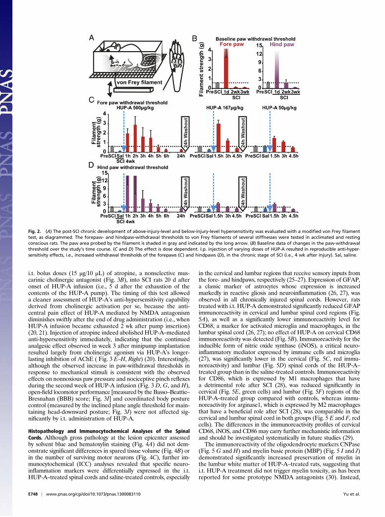
ResultsPost-SCI Hypersensitivity to Mechanical Stimuli. To assess the po-tential therapeutic effect of HUP-A, we first used our clinicallyrelevant model of moderate, static-compression SCI (Fig. 1E) toprofile reproducible post-SCI mechanical hypersensitivity in boththe fore- and hindpaws of experimental rats. In a modified vonFrey filament assay (Fig. 2A), the pre-SCI paw-withdrawal sen-sitivity threshold was 0.63 ± 0.06 g for the forepaws and 2.00 ±0.33 g for the hindpaws (n = 4; Fig. 2B). In the acute phase afterSCI (i.e., 24 h after lesion, without treatment), the force requiredto trigger a withdrawal response increased drastically, to >3 g forthe forepaws and >50 g for the hindpaws (day 1 after SCI, Fig.2B). The lack of response in the acute phase is attributable to“spinal shock” associated with the transient shutdown of senso-rimotor function in the spinal cord acutely after SCI (24). In thesubacute and chronic phases after injury, the withdrawalthreshold decreased gradually to below the pre-SCI level in theforepaws (0.11 ± 0.02 g in week 2, P < 0.01, and 0.15 ± 0.01 g inweek 3, P < 0.03; n = 4). The hindpaws took a week longer todevelop hypersensitivity (1.97 ± 1.17 g in week 2, P > 0.05, and0.47 ± 0.10 g in week 3, P < 0.05; n = 4); the results in both pawsindicate the development of persistent hypersensitivity to me-chanical stimuli (Fig. 2B).To evaluate pharmacological specificity and potential side
effects of systemic HUP-A administration, following a sequentialdesign with adequate drug washout intervals (i.e., >5× t1/2), weinjected three doses of HUP-A i.p. following preinjection of anequivolume of saline in the same rats as self-control in week 4after SCI (10). HUP-A (500, 167, 50 μg/kg, i.p.; n = 4 rats pergroup) induced dose-dependent anti-hypersensitivity (Fig. 2 C
and D). Additionally, after drug administration fine muscletremors, primarily in the forepaw digits, were observed, in-dicating systemic HUP-A–mediated AChE inhibition at theneuromuscular junction (10, 12); however, we did not observeany worsening of the hindlimb muscle spasticity that typicallydevelops 3–4 wk after lower thoracic SCI (24). This sign per-sisted for ∼0.2–2 h in the group with the highest dose of HUP-A.In contrast, the analgesic effects of HUP-A lasted much longer,averaging up to 6 h with bolus i.p. delivery of HUP-A (500 μg/kg;Fig. 2 C and D). We observed no other isolated or cumulativeadverse effects and particularly no effects representing gastro-intestinal hyperactivity (23).
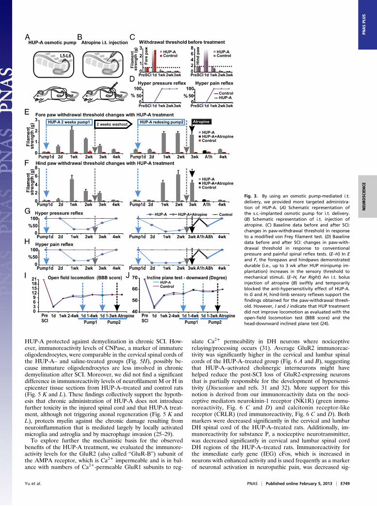
Continuous Intrathecal Administration of HUP-A Induces SustainableSuppression of Post-SCI Hypersensitivity. We next tested a contin-uous drug-delivery regimen with a s.c.-implanted osmotic pumpthat delivered HUP-A directly and continuously into the in-trathecal (i.t.) space at L5–L6 for 2 wk (0.5 μL/h × 24 h × 14 d;total dose: 90 μg/168 μL) (Fig. 3A), with the aim of maintainingthe long-term effect of central anti-hypersensitivity and pre-venting systemic side effects (20). Continuous i.t. delivery ofHUP-A via the osmotic pump effectively eliminated the forepawand hindpaw hypersensitivity to mechanical stimuli. A stronganalgesic effect was noted between weeks 1 and 3 after pumpimplantation: The post–HUP-A treatment sensitivity thresholdwas significantly higher (Fig. 3 E–H) (n = 7 per group) than thepretreatment/post-SCI baseline sensitivity thresholds of 0.60 ±0.04 g and 2.00 ± 0.18 g for the fore- and hindpaws, respectively(Fig. 3 C and D). The anti-hypersensitivity and analgesic effectswere HUP-A dependent, because the effect was first spotted∼24 h after pump implantation (Fig. 3 E–H), reflecting the timeneeded for HUP-A to course through the microtubing beforeentering the i.t. space (additional details are given in Materialsand Methods), and a return of hypersensitivity was observed 2 wkafter depletion of HUP-A from the osmotic pump (i.e., 4 wkafter s.c. pump insertion). However, readministration of thesame dose reestablished the anti-hypersensitivity effect of HUP-A without apparent tolerance or adverse effects (Fig. 3 E and F;note: the effectiveness of the second HUP-A pumping also lastedfor ∼3 wk). To check whether HUP-A’s augmentation of centralmuscarinic activation of cholinergic transmission indeed playeda definitive role in managing hypersensitivity, we administered
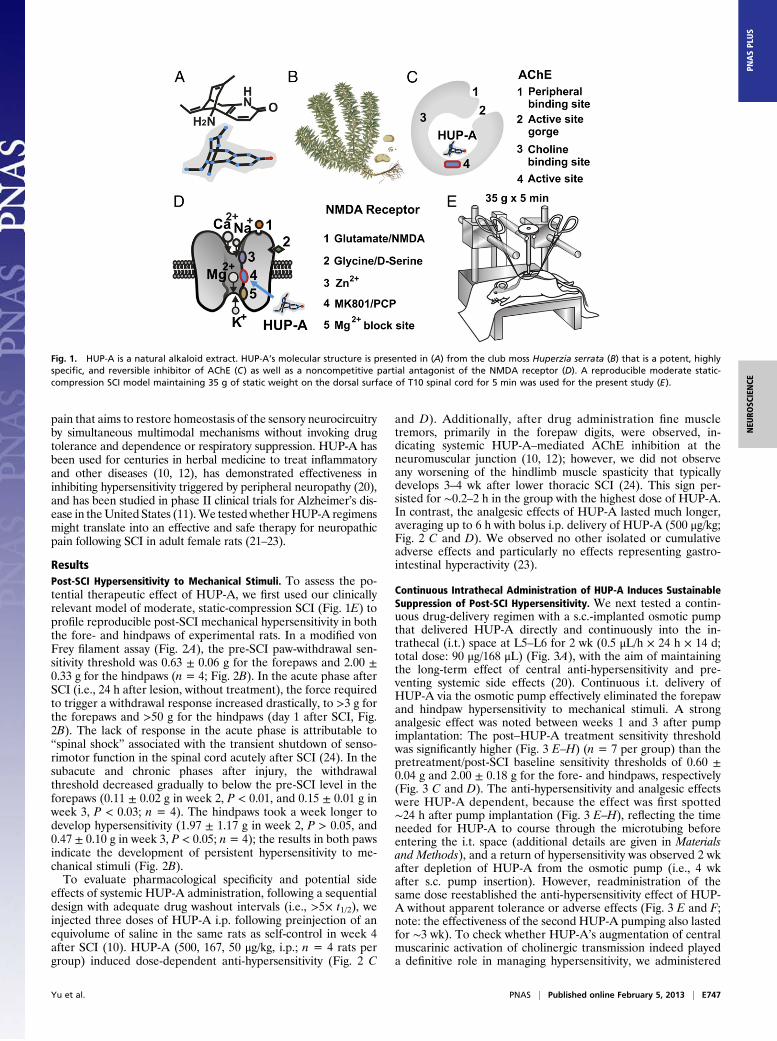
Fig. 1. HUP-A is a natural alkaloid extract. HUP-A’s molecular structure is presented in (A) from the club moss Huperzia serrata (B) that is a potent, highlyspecific, and reversible inhibitor of AChE (C) as well as a noncompetitive partial antagonist of the NMDA receptor (D). A reproducible moderate static-compression SCI model maintaining 35 g of static weight on the dorsal surface of T10 spinal cord for 5 min was used for the present study (E).
Yu et al. PNAS | Published online February 5, 2013 | E747
NEU
ROSC
IENCE
PNASPL
US
i.t. bolus doses (15 μg/10 μL) of atropine, a nonselective mus-carinic cholinergic antagonist (Fig. 3B), into SCI rats 20 d afteronset of HUP-A infusion (i.e., 5 d after the exhaustion of thecontents of the HUP-A pump). The timing of this test alloweda cleaner assessment of HUP-A’s anti-hypersensitivity capabilityderived from cholinergic activation per se, because the anti-central pain effect of HUP-A mediated by NMDA antagonismdiminishes swiftly after the end of drug administration (i.e., whenHUP-A infusion became exhausted 2 wk after pump insertion)(20, 21). Injection of atropine indeed abolished HUP-A–mediatedanti-hypersensitivity immediately, indicating that the continuedanalgesic effect observed in week 3 after minipump implantationresulted largely from cholinergic agonism via HUP-A’s longer-lasting inhibition of AChE ( Fig. 3 E–H, Right) (20). Interestingly,although the observed increase in paw-withdrawal thresholds inresponse to mechanical stimuli is consistent with the observedeffects on nonnoxious paw pressure and nociceptive pinch reflexesduring the second week of HUP-A infusion (Fig. 3 D, G, and H),open-field locomotor performance [measured by the Basso–Beattie–Bresnahan (BBB) score; Fig. 3I] and coordinated body posturecontrol (measured by the inclined plane angle threshold for main-taining head-downward posture; Fig. 3J) were not affected sig-nificantly by i.t. administration of HUP-A.
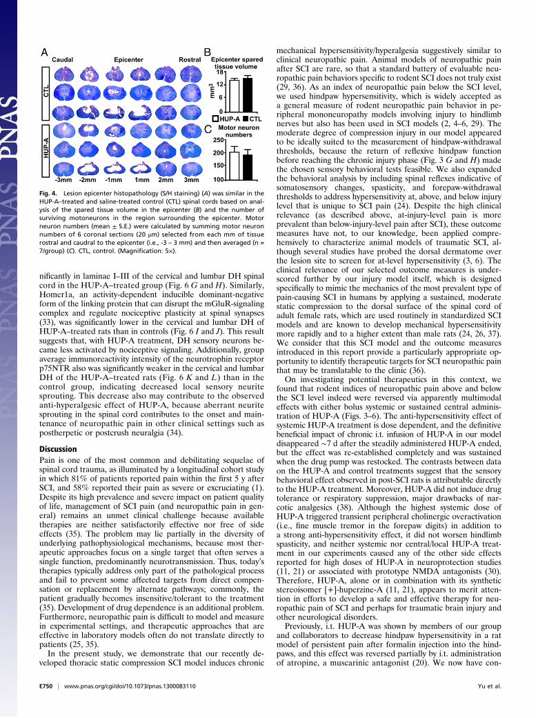
Histopathology and Immunocytochemical Analyses of the SpinalCords. Although gross pathology at the lesion epicenter assessedby solvent blue and hematoxylin staining (Fig. 4A) did not dem-onstrate significant differences in spared tissue volume (Fig. 4B) orin the number of surviving motor neurons (Fig. 4C), further im-munocytochemical (ICC) analyses revealed that specific neuro-inflammation markers were differentially expressed in the i.t.HUP-A–treated spinal cords and saline-treated controls, especially
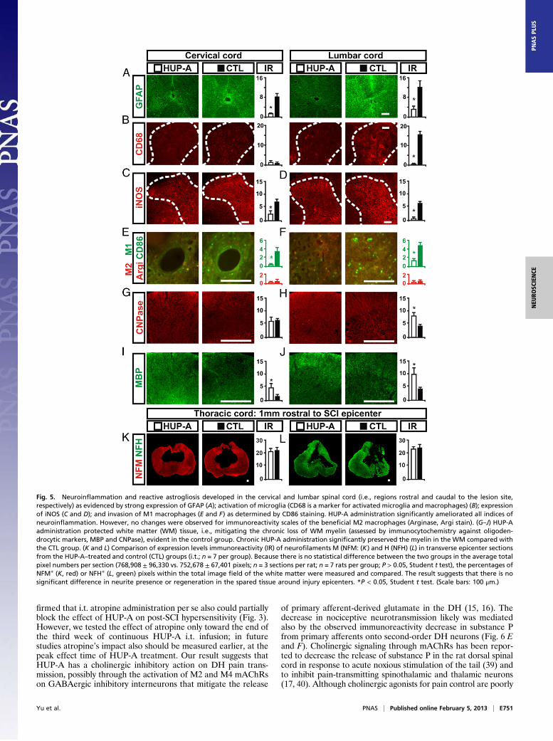
in the cervical and lumbar regions that receive sensory inputs fromthe fore- and hindpaws, respectively (25–27). Expression of GFAP,a classic marker of astrocytes whose expression is increasedmarkedly in reactive gliosis and neuroinflammation (26, 27), wasobserved in all chronically injured spinal cords. However, ratstreated with i.t. HUP-A demonstrated significantly reduced GFAPimmunoreactivity in cervical and lumbar spinal cord regions (Fig.5A), as well as a significantly lower immunoreactivity level forCD68, a marker for activated microglia and macrophages, in thelumbar spinal cord (26, 27); no effect of HUP-A on cervical CD68immunoreactivity was detected (Fig. 5B). Immunoreactivity for theinducible form of nitric oxide synthase (iNOS), a critical neuro-inflammatory mediator expressed by immune cells and microglia(27), was significantly lower in the cervical (Fig. 5C, red immu-noreactivity) and lumbar (Fig. 5D) spinal cords of the HUP-A–treated group than in the saline-treated controls. Immunoreactivityfor CD86, which is expressed by M1 macrophages that havea detrimental role after SCI (28), was reduced significantly incervical (Fig. 5E, green cells) and lumbar (Fig. 5F) regions of theHUP-A–treated group compared with controls, whereas immu-noreactivity for arginase1, which is expressed by M2 macrophagesthat have a beneficial role after SCI (28), was comparable in thecervical and lumbar spinal cord in both groups (Fig. 5 E and F, redcells). The differences in the immunoreactivity profiles of cervicalCD68, iNOS, and CD86 may carry further mechanistic informationand should be investigated systematically in future studies (29).The immunoreactivity of the oligodendrocyte markers CNPase
(Fig. 5 G and H) and myelin basic protein (MBP) (Fig. 5 I and J)demonstrated significantly increased preservation of myelin inthe lumbar white matter of HUP-A–treated rats, suggesting thati.t. HUP-A treatment did not trigger myelin toxicity, as has beenreported for some prototype NMDA antagonists (30). Instead,
Fig. 2. (A) The post-SCI chronic development of above-injury-level and below-injury-level hypersensitivity was evaluated with a modified von Frey filamenttest, as diagrammed. The forepaw- and hindpaw-withdrawal thresholds to von Frey filaments of several stiffnesses were tested in acclimated and restingconscious rats. The paw area probed by the filament is shaded in gray and indicated by the long arrow. (B) Baseline data of changes in the paw-withdrawalthreshold over the study’s time course. (C and D) The effect is dose dependent. I.p. injection of varying doses of HUP-A resulted in reproducible anti-hyper-sensitivity effects, i.e., increased withdrawal thresholds of the forepaws (C) and hindpaws (D), in the chronic stage of SCI (i.e., 4 wk after injury). Sal, saline.
E748 | www.pnas.org/cgi/doi/10.1073/pnas.1300083110 Yu et al.
HUP-A protected against demyelination in chronic SCI. How-ever, immunoreactivity levels of CNPase, a marker of immatureoligodendrocytes, were comparable in the cervical spinal cords ofthe HUP-A– and saline-treated groups (Fig. 5H), possibly be-cause immature oligodendrocytes are less involved in chronicdemyelination after SCI. Moreover, we did not find a significantdifference in immunoreactivity levels of neurofilament M or H inepicenter tissue sections from HUP-A–treated and control rats(Fig. 5 K and L). These findings collectively support the hypoth-esis that chronic administration of HUP-A does not introducefurther toxicity in the injured spinal cord and that HUP-A treat-ment, although not triggering axonal regeneration (Fig. 5 K andL), protects myelin against the chronic damage resulting fromneuroinflammation that is mediated largely by locally activatedmicroglia and astroglia and by macrophage invasion (25–29).To explore further the mechanistic basis for the observed
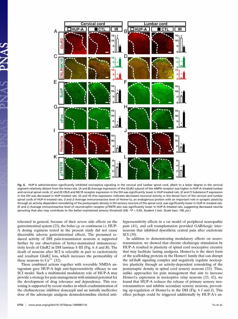
benefits of the HUP-A treatment, we evaluated the immunore-activity levels for the GluR2 (also called “GluR-B”) subunit ofthe AMPA receptor, which is Ca2+ impermeable and is in bal-ance with numbers of Ca2+-permeable GluR1 subunits to reg-
ulate Ca2+ permeability in DH neurons where nociceptiverelaying/processing occurs (31). Average GluR2 immunoreac-tivity was significantly higher in the cervical and lumbar spinalcords of the HUP-A–treated group (Fig. 6 A and B), suggestingthat HUP-A–activated cholinergic interneurons might havehelped reduce the post-SCI loss of GluR2-expressing neuronsthat is partially responsible for the development of hypersensi-tivity (Discussion and refs. 31 and 32). More support for thisnotion is derived from our immunoreactivity data on the noci-ceptive mediators neurokinin-1 receptor (NK1R) (green immu-noreactivity, Fig. 6 C and D) and calcitonin receptor-likereceptor (CRLR) (red immunoreactivity, Fig. 6 C and D). Bothmarkers were decreased significantly in the cervical and lumbarDH spinal cord of the HUP-A–treated rats. Additionally, im-munoreactivity for substance P, a nociceptive neurotransmitter,was decreased significantly in cervical and lumbar spinal cordDH regions of the HUP-A–treated rats. Immunoreactivity forthe immediate early gene (IEG) cFos, which is increased inneurons with enhanced activity and is used frequently as a markerof neuronal activation in neuropathic pain, was decreased sig-
Fig. 3. By using an osmotic pump-mediated i.t.delivery, we provided more targeted administra-tion of HUP-A. (A) Schematic representation ofthe s.c.-implanted osmotic pump for i.t. delivery.(B) Schematic representation of i.t. injection ofatropine. (C) Baseline data before and after SCI:changes in paw-withdrawal threshold in responseto a modified von Frey filament test. (D) Baselinedata before and after SCI: changes in paw-with-drawal threshold in response to conventionalpressure and painful spinal reflex tests. (E–H) In Eand F, the forepaws and hindpaws demonstrateddurable (i.e., up to 3 wk after HUP minipump im-plantation) increases in the sensory threshold tomechanical stimuli. (E–H, Far Right) An i.t. bolusinjection of atropine (B) swiftly and temporarilyblocked the anti-hypersensitivity effect of HUP-A.In G and H, hind-limb sensory reflexes support thefindings obtained for the paw-withdrawal thresh-old. However, I and J indicate that HUP treatmentdid not improve locomotion as evaluated with theopen-field locomotion test (BBB score) and thehead-downward inclined plane test (24).
Yu et al. PNAS | Published online February 5, 2013 | E749
NEU
ROSC
IENCE
PNASPL
US
nificantly in laminae I–III of the cervical and lumbar DH spinalcord in the HUP-A–treated group (Fig. 6 G and H). Similarly,Homer1a, an activity-dependent inducible dominant-negativeform of the linking protein that can disrupt the mGluR-signalingcomplex and regulate nociceptive plasticity at spinal synapses(33), was significantly lower in the cervical and lumbar DH ofHUP-A–treated rats than in controls (Fig. 6 I and J). This resultsuggests that, with HUP-A treatment, DH sensory neurons be-came less activated by nociceptive signaling. Additionally, groupaverage immunoreactivity intensity of the neurotrophin receptorp75NTR also was significantly weaker in the cervical and lumbarDH of the HUP-A–treated rats (Fig. 6 K and L) than in thecontrol group, indicating decreased local sensory neuritesprouting. This decrease also may contribute to the observedanti-hyperalgesic effect of HUP-A, because aberrant neuritesprouting in the spinal cord contributes to the onset and main-tenance of neuropathic pain in other clinical settings such aspostherpetic or postcrush neuralgia (34).
DiscussionPain is one of the most common and debilitating sequelae ofspinal cord trauma, as illuminated by a longitudinal cohort studyin which 81% of patients reported pain within the first 5 y afterSCI, and 58% reported their pain as severe or excruciating (1).Despite its high prevalence and severe impact on patient qualityof life, management of SCI pain (and neuropathic pain in gen-eral) remains an unmet clinical challenge because availabletherapies are neither satisfactorily effective nor free of sideeffects (35). The problem may lie partially in the diversity ofunderlying pathophysiological mechanisms, because most ther-apeutic approaches focus on a single target that often serves asingle function, predominantly neurotransmission. Thus, today’stherapies typically address only part of the pathological processand fail to prevent some affected targets from direct compen-sation or replacement by alternate pathways; commonly, thepatient gradually becomes insensitive/tolerant to the treatment(35). Development of drug dependence is an additional problem.Furthermore, neuropathic pain is difficult to model and measurein experimental settings, and therapeutic approaches that areeffective in laboratory models often do not translate directly topatients (25, 35).In the present study, we demonstrate that our recently de-
veloped thoracic static compression SCI model induces chronic
mechanical hypersensitivity/hyperalgesia suggestively similar toclinical neuropathic pain. Animal models of neuropathic painafter SCI are rare, so that a standard battery of evaluable neu-ropathic pain behaviors specific to rodent SCI does not truly exist(29, 36). As an index of neuropathic pain below the SCI level,we used hindpaw hypersensitivity, which is widely accepted asa general measure of rodent neuropathic pain behavior in pe-ripheral mononeuropathy models involving injury to hindlimbnerves but also has been used in SCI models (2, 4–6, 29). Themoderate degree of compression injury in our model appearedto be ideally suited to the measurement of hindpaw-withdrawalthresholds, because the return of reflexive hindpaw functionbefore reaching the chronic injury phase (Fig. 3 G and H) madethe chosen sensory behavioral tests feasible. We also expandedthe behavioral analysis by including spinal reflexes indicative ofsomatosensory changes, spasticity, and forepaw-withdrawalthresholds to address hypersensitivity at, above, and below injurylevel that is unique to SCI pain (24). Despite the high clinicalrelevance (as described above, at-injury-level pain is moreprevalent than below-injury-level pain after SCI), these outcomemeasures have not, to our knowledge, been applied compre-hensively to characterize animal models of traumatic SCI, al-though several studies have probed the dorsal dermatome overthe lesion site to screen for at-level hypersensitivity (3, 6). Theclinical relevance of our selected outcome measures is under-scored further by our injury model itself, which is designedspecifically to mimic the mechanics of the most prevalent type ofpain-causing SCI in humans by applying a sustained, moderatestatic compression to the dorsal surface of the spinal cord ofadult female rats, which are used routinely in standardized SCImodels and are known to develop mechanical hypersensitivitymore rapidly and to a higher extent than male rats (24, 26, 37).We consider that this SCI model and the outcome measuresintroduced in this report provide a particularly appropriate op-portunity to identify therapeutic targets for SCI neuropathic painthat may be translatable to the clinic (36).On investigating potential therapeutics in this context, we
found that rodent indices of neuropathic pain above and belowthe SCI level indeed were reversed via apparently multimodaleffects with either bolus systemic or sustained central adminis-tration of HUP-A (Figs. 3–6). The anti-hypersensitivity effect ofsystemic HUP-A treatment is dose dependent, and the definitivebeneficial impact of chronic i.t. infusion of HUP-A in our modeldisappeared ∼7 d after the steadily administered HUP-A ended,but the effect was re-established completely and was sustainedwhen the drug pump was restocked. The contrasts between dataon the HUP-A and control treatments suggest that the sensorybehavioral effect observed in post-SCI rats is attributable directlyto the HUP-A treatment. Moreover, HUP-A did not induce drugtolerance or respiratory suppression, major drawbacks of nar-cotic analgesics (38). Although the highest systemic dose ofHUP-A triggered transient peripheral cholinergic overactivation(i.e., fine muscle tremor in the forepaw digits) in addition toa strong anti-hypersensitivity effect, it did not worsen hindlimbspasticity, and neither systemic nor central/local HUP-A treat-ment in our experiments caused any of the other side effectsreported for high doses of HUP-A in neuroprotection studies(11, 21) or associated with prototype NMDA antagonists (30).Therefore, HUP-A, alone or in combination with its syntheticstereoisomer [+]-huperzine-A (11, 21), appears to merit atten-tion in efforts to develop a safe and effective therapy for neu-ropathic pain of SCI and perhaps for traumatic brain injury andother neurological disorders.Previously, i.t. HUP-A was shown by members of our group
and collaborators to decrease hindpaw hypersensitivity in a ratmodel of persistent pain after formalin injection into the hind-paws, and this effect was reversed partially by i.t. administrationof atropine, a muscarinic antagonist (20). We now have con-
Fig. 4. Lesion epicenter histopathology (S/H staining) (A) was similar in theHUP-A–treated and saline-treated control (CTL) spinal cords based on anal-ysis of the spared tissue volume in the epicenter (B) and the number ofsurviving motoneurons in the region surrounding the epicenter. Motorneuron numbers (mean ± S.E.) were calculated by summing motor neuronnumbers of 6 coronal sections (20 μm) selected from each mm of tissuerostral and caudral to the epicenter (i.e., -3 – 3 mm) and then averaged (n =7/group) (C). CTL, control. (Magnification: 5×).
E750 | www.pnas.org/cgi/doi/10.1073/pnas.1300083110 Yu et al.
firmed that i.t. atropine administration per se also could partiallyblock the effect of HUP-A on post-SCI hypersensitivity (Fig. 3).However, we tested the effect of atropine only toward the end ofthe third week of continuous HUP-A i.t. infusion; in futurestudies atropine’s impact also should be measured earlier, at thepeak effect time of HUP-A treatment. Our result suggests thatHUP-A has a cholinergic inhibitory action on DH pain trans-mission, possibly through the activation of M2 and M4 mAChRson GABAergic inhibitory interneurons that mitigate the release
of primary afferent-derived glutamate in the DH (15, 16). Thedecrease in nociceptive neurotransmission likely was mediatedalso by the observed immunoreactivity decrease in substance Pfrom primary afferents onto second-order DH neurons (Fig. 6 Eand F). Cholinergic signaling through mAChRs has been repor-ted to decrease the release of substance P in the rat dorsal spinalcord in response to acute noxious stimulation of the tail (39) andto inhibit pain-transmitting spinothalamic and thalamic neurons(17, 40). Although cholinergic agonists for pain control are poorly
Fig. 5. Neuroinflammation and reactive astrogliosis developed in the cervical and lumbar spinal cord (i.e., regions rostral and caudal to the lesion site,respectively) as evidenced by strong expression of GFAP (A); activation of microglia (CD68 is a marker for activated microglia and macrophages) (B); expressionof iNOS (C and D); and invasion of M1 macrophages (E and F) as determined by CD86 staining. HUP-A administration significantly ameliorated all indices ofneuroinflammation. However, no changes were observed for immunoreactivity scales of the beneficial M2 macrophages (Arginase, Argi stain). (G–J) HUP-Aadministration protected white matter (WM) tissue, i.e., mitigating the chronic loss of WM myelin (assessed by immunocytochemistry against oligoden-drocytic markers, MBP and CNPase), evident in the control group. Chronic HUP-A administration significantly preserved the myelin in the WM compared withthe CTL group. (K and L) Comparison of expression levels immunoreactivity (IR) of neurofilaments M (NFM: (K) and H (NFH) (L) in transverse epicenter sectionsfrom the HUP-A–treated and control (CTL) groups (i.t.; n = 7 per group). Because there is no statistical difference between the two groups in the average totalpixel numbers per section (768,908 ± 96,330 vs. 752,678 ± 67,401 pixels; n = 3 sections per rat; n = 7 rats per group; P > 0.05, Student t test), the percentages ofNFM+ (K, red) or NFH+ (L, green) pixels within the total image field of the white matter were measured and compared. The result suggests that there is nosignificant difference in neurite presence or regeneration in the spared tissue around injury epicenters. *P < 0.05, Student t test. (Scale bars: 100 μm.)
Yu et al. PNAS | Published online February 5, 2013 | E751
NEU
ROSC
IENCE
PNASPL
US
tolerated in general, because of their severe side effects on thegastrointestinal system (23), the bolus i.p. or continuous i.t. HUP-A dosing regimens tested in the present study did not causediscernible adverse gastrointestinal effects. The presumed re-duced activity of DH pain-transmission neurons is supportedfurther by our observation of better-maintained immunoreac-tivity levels of GluR2 in DH laminae I–III (Fig. 6 A and B). Thedeath of neurons after SCI is referable in part to excitotoxicityand resultant GluR2 loss, which increases the permeability ofthese neurons to Ca2+ (32).These combined actions together with reversible NMDA an-
tagonism gave HUP-A high anti-hypersensitivity efficacy in ourSCI model. Such a multimodal modulatory role of HUP-A mayprovide a strategy for painmanagement with minimal potential forthe development of drug tolerance and dependence. This rea-soning is supported by recent studies in which coadministration ofthe cholinesterase inhibitor donezepil and an initially ineffectivedose of the adrenergic analgesic dexmedetomidine elicited anti-
hypersensitivity effects in a rat model of peripheral neuropathicpain (41), and cell transplantation provided GABAergic inter-neurons that inhibited dysesthetic central pain after excitotoxicSCI (39).In addition to demonstrating modulatory effects on neuro-
transmission, we showed that chronic cholinergic stimulation byHUP-A resulted in plasticity of spinal cord nociceptive circuitrythat may facilitate lasting analgesia. Homer1a is the short formof the scaffolding proteins in the Homer1 family that can disruptthe mGluR signaling complex and negatively regulate nocicep-tive plasticity through an activity-dependent remodeling of thepostsynaptic density in spinal cord sensory neurons (33). Thus,unlike approaches for pain management that aim to increaseHomer1a expression in nociceptive relay neurons (33, 42), wefound that HUP-A reduces the release of primary sensory neu-rotransmitters and inhibits secondary sensory neurons, prevent-ing up-regulation of Homer1a in the DH (Fig. 6 I and J). Thiseffect perhaps could be triggered additionally by HUP-A’s an-
Fig. 6. HUP-A administration significantly inhibited nociceptive signaling in the cervical and lumbar spinal cord, albeit to a lesser degree in the cervicalsegment relatively distant from the lesion site. (A and B) Average expression of the GluR2 subunit of the AMPA receptor was higher in HUP-A–treated lumbarand cervical spinal cords. (C and D) CRLR and NK1R receptor expression in the DH was significantly lower in HUP-treated rats. (E and F) Substance P expressionin the DH was decreased in HUP-treated rats. (G and H) cFos expression indicates decreased neuronal activity in the dorsal horn of the cervical and lumbarspinal cords of HUP-A-treated rats. (I and J) Average immunoreactive level of Homer1a, an endogenous protein with an important role in synaptic plasticitythrough an activity-dependent remodeling of the postsynaptic density in DH sensory neurons of the spinal cord, was significantly lower in HUP-A–treated rats.(K and L) Average immunoreactive level of neurotrophin receptor p75NTR also was significantly lower in HUP-A–treated rats, suggesting decreased neuritesprouting that also may contribute to the better-maintained sensory threshold (34). *P < 0.05, Student t test. (Scale bars: 100 μm.)
E752 | www.pnas.org/cgi/doi/10.1073/pnas.1300083110 Yu et al.
tagonist action on the NMDA receptor, similar to the inhibitoryaction of the NMDA receptor antagonist MK-801 on Homer1aobserved in rat peripheral neuropathic pain and thermal hyper-algesia (42). HUP-A–mediated reduction of DH neuronal ac-tivity also is evidenced by the reduced expression of IEG cFos(Fig. 6 G and H) and p75 neurotrophin receptor (i.e., reducedneurite sprouting) in DH laminae I–III (Fig. 6 K and L), both ofwhich are largely dependent on neuronal activity (43). Likewise,HUP-A–mediated postsynaptic inhibition (44) may be respon-sible for the observed down-regulation of the CRLR and theNK1R in DH sensory neurons (Fig. 6 C and D). These two typesof receptors mediate pain-signal transmission for calcitoningene-related peptide and substance P, respectively, suggestingthat HUP-A triggers activity-dependent plasticity events in thedorsal spinal cord (44).We hypothesized that HUP-A treatment also would impede
post-SCI development of hypersensitivity through an anti-in-flammatory effect, because acetylcholine inhibits the activationof macrophages/microglia (45–47) and astrocytes (48) via the α7nAChR. Onset and chronic maintenance of neuropathic painbehavior depends, at least in part, on persistent microglial acti-vation and secretion of inflammatory mediators (25, 49, 50). In-deed, we observed decreased ICC staining for activated microgliaand CD86+ M1 macrophages, decreased astrogliosis, and de-creased expression of iNOS [a key enzyme involved in the gen-eration of reactive oxidative species during inflammation (28)] inrats treated with HUP-A (Fig. 5 C and D). Additionally, our datasuggest that the different effects of HUP-A on immunoreactivityof CD68, iNOS, and CD86 in the cervical compared with thelumbar segments may reflect varying degrees of involvement ofthe several cell types and reactive molecules in triggering above-
injury-level or below-injury-level hyperalgesia (Fig. 5) (29). Thesefindings, in addition to underlying the continued analgesic effectobserved during the third week of HUP-A i.t. treatment (i.e., 1 wkafter drug pump exhaustion), provide a perspective on thetreatment of reactive astroglial neuropathic pain, because pre-vious studies on cholinergic analgesia focused on acute effects onneurotransmission by nociceptive neurons and did not address thepathophysiological modulation of other spinal cord cell types thatcontribute to neuropathic hypersensitivity (15, 16, 20, 41). Inparallel with the decrease in neuroinflammation, we observedmitigation of chronic demyelination in the HUP-A–treated spinalcord (Fig. 5 G–J). Axonal demyelination occurs simultaneouslywith disruption of gene expression for several types of sodiumchannels and contributes to chronic pain (51). Conversely, ourHUP-A treatment regimen did not reduce significantly the loss oftissue volume (Fig. 4B) and motoneurons (Fig. 4C), nor did itimprove axonal regeneration or motor function indices such asopen-field locomotion (Fig. 5 K and L) or coordinated bodyposture control (Fig. 3 E–H). Because NMDA-mediated excito-toxicity plays primary roles in acute SCI, future studies should testif HUP-A administration in the early phase of SCI might reducesecondary tissue injury and motor dysfunction (24).In summary, our findings suggest that HUP-A represents a
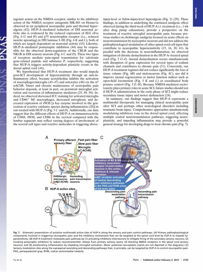
multimodal therapeutic for managing clinical neuropathic painafter SCI and perhaps other neurological disorders includingtraumatic brain injury. Comprehensive approaches simultaneouslymodulating inhibitory tone in the dorsal spinal cord, affectingmultiple central neurotransmission pathways, triggering neuro-plasticity, and impeding inflammation may provide a powerfulgeneral strategy for developing drugs to treat chronic pain (Fig. 7).
Fig. 7. Schematic presentation of putative multimodal action sites of HUP-A along the sensory and pain control pathways. (A) Primary pathophysiologicalcomponents involved in triggering neuropathic pain and the inhibitory mechanisms that can be targeted at the spinal cord level by HUP-A to impede hy-persensitivity. (B) HUP-A treatment modulates pain pathway by (1) activating inhibitory interneurons to mitigate firing of the secondary sensory neurons; (2)invoking presynaptic inhibition to reduce neurotransmitter release from primary sensory axons; (3) blocking NMDA receptors in the spinal cord sensoryneurons; and (4) ameliorating inflammation by impeding microglial activation. (Note: potential neuroplastic events are not depicted in the diagrams.) (C)Sensory modulation sites along the supraspinal ascending and descending pathways that, in principle, can be targeted by HUP-A to control neuropathic pain.PAG, periaqueductal gray; RVM, rostral ventromedial medulla.
Yu et al. PNAS | Published online February 5, 2013 | E753
NEU
ROSC
IENCE
PNASPL
US
Materials and MethodsSurgical Model of Moderate Static Compression SCI. All procedures wereevaluated and approved by the Institutional Animal Care and Use Committeeof Harvard Medical School. Young adult female Sprague–Dawley rats (200–235 g body weight, n = 20; Taconic) were anesthetized with ketamine (75mg/kg i.p.) and xylazine (10 mg/kg i.p.) before operation. Under a surgicalstereomicroscope, dorsal laminectomy at thoracic vertebra 10 (T10) wasperformed to expose the spinal cord. After hemostasis was induced withGelfoam (Pfizer), the rat body was suspended with a Teng Laboratory spinestabilization frame (fabricated at the Scientific Instrument Shop, HarvardUniversity School of Engineering and Applied Sciences, Cambridge, MA),with 1-cm clearance between the ventral side of the rat and the frame baseplatform. A 35-g stainless steel impounder was lowered gently upon thedura surface via a micromanipulator, and the entire weight was allowed tocompress the cord for 5 min (Fig. 1E). The lesion zone then was closed withsutures and stainless wound clips (Fine Science Tools Inc.), and the rat wasallowed to recover in a clean, heated cage. Lactated Ringer’s solution (10mL/d, s.c.) was given daily for 5 d postoperation, and the urinary bladder wasevacuated manually without urinary catheterization twice daily until reflexbladder function became reestablished (24).
HUP-A Preparation and Treatment. HUP-A [5R(5a,9b,11E)]-5-amino11ethlidene-5,6,9,10-tetrahydro-7-methyl-5,9-methanocycloocta[b]pyridin-2(1H)-one(molecular weight: 242.32; HPLC/UV purity: 98%; specific rotation: −152.8°;appearance: white powder] (Fig. 1A) was purchased from ChromaDex. Threeweeks after SCI, HUP-A was administered as follows: (i) for the i.p. study (bolusdose treatment, n = 4): After sterile saline (2 mL per rat, i.p.) administrationtogether with subsequent behavioral evaluation to obtain vehicle-control out-come measures, HUP-A, dissolved in 10% beta cyclodextrin in sterile saline(Sigma-Aldrich) at a concentration of 10 μg/μL before saline dilution to the finaldose of 500, 167, or 50 μg/kg in a 2-mL bolus volume per rat for three treatedgroups, respectively (20), was administered. The injection was done with syringeneedle passing 5 mm upward in the subdermal layer before penetratingthrough the muscle and peritoneal membrane layer to prevent postinjectionleak and a brief withdrawal of the plunger to ensure no blood vessel piercing.After each dosing of saline followed with HUP-A (i.e., 500, 167, and 50 μg/kgwith intervals of 24 h and 48 h, respectively), tests were performed usingstandard behavior batteries (see below). (ii) Implantation of the i.t. osmoticpump for chronic 2-wk treatment: HUP-A dissolved in 10% beta cyclodextrinin sterile saline (Sigma-Aldrich) was loaded in Alzet osmotic pumps (DurectCorporation) at a final dose per pump of 90 μg/168 μL to provide a 14-d i.t.continuous supply (pumping rate: 0.5 μL/h; total dose: 12 μL/d × 14 d = 168 μL).After reanesthetization and a small incision to expose the L5–L6 space, a durapuncture was made by an 18-G needle. A PE20 sterile catheter was insertedthrough the puncture with the beveled tip facing rostrally andwas securedwitha suture after a brief sign of tail flick was elicited by a light touch of nerve rootsin the cauda equina to confirm proper i.t. penetration. The catheter was pre-filled with 12 μL of sterile saline before the insertion and was connected to anosmotic pump filled with either HUP-A (treatment group; n = 7) or sterile saline
containing 10% beta cyclodextrin (control group, n = 7). The tubing also wassutured to the fascia and musculature near the connection site with the s.c.embedded pump. The skin incision was closed with stainless steel wound clips,and standard postprocedure care was given (52). It took ∼24 h at a pumpingrate of 0.5 μL/h, for HUP-A or saline to start entering the i.t. space. We firstvalidated the effectiveness of the i.t. delivery by microinjection of 10 μL of 0.2%trypan blue via PE20 tubing in a pilot experiment (n = 2). (iii) I.t. redosing andatropine administration: The first round of HUP-A i.t. delivery was followed bya 2-wk “wash-out” period to permit the HUP-A effect to subside. We thenstarted a second cycle of HUP-A treatment by replacing the HUP-A pump. Thesame sets of behavioral assays were performed, and 20 d later we administereda bolus dose of atropine (15 μg/10 μL, i.t. at L5–L6) to determine the effect ofmuscarinic antagonism on analgesic impact of HUP-A.
Behavioral Monitoring. A battery of behavioral tests was performed byobservers blinded to the treatments on the day before the surgery, 1 d afterSCI, and weekly thereafter to assess functional deficits and nociceptivechanges. These tests evaluated open-field locomotion based on the BBBscale, the ability to maintain coordinated posture control on an inclinedplane, the contact-righting reflex, the hindpaw-placing reflex, and thewithdrawal reflex in response to pinch and pressure. In addition, after 30 minacclimation, the rats were subjected to a paw hyperalgesia test (modified vonFrey filament test) to compare the withdrawal threshold before and after SCIor treatments, followed by hourly testing for up to 8 h after i.p. HUP-A in-jection and for 1 d, 2 d, and weekly thereafter up to 4 wk posttreatmentfor the i.t. HUP-A delivery study. Using the data obtained, we determinedthat with the α value (probability of rejecting H0 when H0 is true) beingset at 5%, an average sample value of 0.41 g (post-SCI baseline sensorythreshold), mean test value (posttreatment) of 3.1 g, and group size of fourrats, the statistical power (i.e., 1-β) equals ∼100%, indicating that the β value(i.e., the probability of not rejecting H0 when H0 is false) = ∼0. The BBB andpaw hyperalgesia test data were analyzed by repeated-measures ANOVAand by Bonferroni post hoc tests for multiple group comparisons with An-alyze-it Statistics (Analyze-it Software Ltd. on Excel (Microsoft) (24, 52).
Histopathological Analysis. Rats were killed with an overdose i.p. injection ofanesthetics followed by intracardiac perfusion with 4% (wt/vol) paraform-aldehyde (PFA) in 0.1 M phosphate buffer at pH 7.4. Brain and spinal cordwere dissected carefully, postfixed in 4% (wt/vol) PFA overnight at roomtemperature, dehydrated in 30% (wt/vol) sucrose at 4 °C for 24 h, and frozenin isopentane (Sigma-Aldrich) at −45 °C. The tissue then was encased in OCTcompound (Sakura Finetek) and cryosectioned transversely at 20 μm. Sectionsrepresentative of each millimeter of the spinal cord at and around the injuryepicenter were chosen for solvent blue (a myelin stain, similar to luxol fastblue)/hematoxylin (S/H; both from Sigma) staining for analysis of lesion vol-ume and counting of motor neurons. Lesion volume was measured based onthe S/H staining from the area of the visualized lesion region in each repre-sentative section per millimeter of tissue and was calculated based on totallesion length. The motor neuron count was carried out as previously described
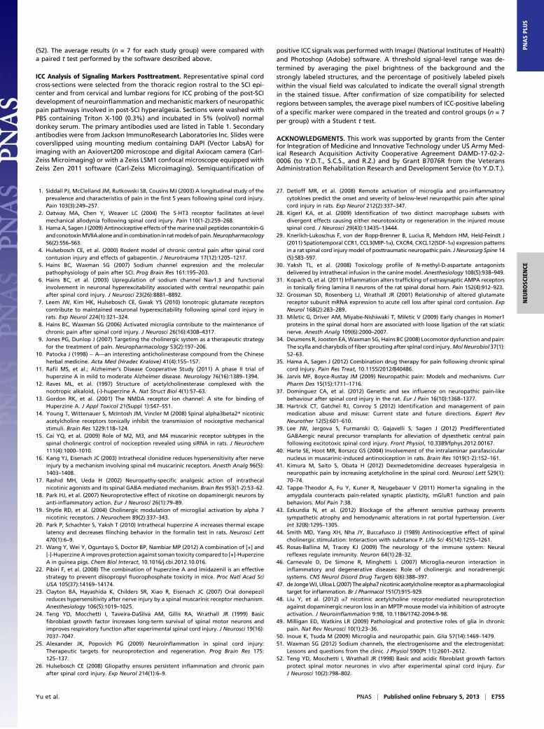
Table 1. Antibodies used in immunocytochemical assays
Antibody against Marker for Species Dilution Source Location
GFAP Intermediate filament of astrocytes Rabbit 1:2,000 Dakocytomation Carpentaria, CACD68 Microglia activation Mouse 1:250 EMD Millipore Carlsbad, CAiNOS Inducible NOS expression Rabbit 1:500 Santa Cruz Biotechnology Santa Cruz, CAArginase1 M2 Macrophage Mouse 1:250 Santa Cruz Biotechnology Santa Cruz, CACD86 M1 Macrophage Rabbit 1:250 Abcam Cambridge, MACNPase Developing and adult myelin Mouse 1:400 Sigma-Aldrich St. Louis, MOMBP Mature myelin Chicken 1:100 EMD Millipore Carlsbad, CAGluR2 Ca2+ impermeable subunit of AMPA-R Mouse 1:250 EMD Millipore Carlsbad, CAHomer1a Dominant negative linking protein of
mGluR-signaling complexGoat 1:250 Santa Cruz Biotechnology Santa Cruz, CA
NK1R Neurokinin-1 receptor Goat 1:250 Santa Cruz Biotechnology Santa Cruz, CACRLR Calcitonin receptor-like receptor Rabbit 1:250 Santa Cruz Biotechnology Santa Cruz, CASubstance P Neurotransmitter related to pain Rabbit 1:250 EMD Millipore Carlsbad, CANeurofila-ment M Marker for neuronal axons Rabbit 1:200 EMD Millipore Carlsbad, CANeurofila-ment H Marker for mature neuronal axons Mouse 1:200 EMD Millipore Carlsbad, CAcFos Activated by strong neural activity Rabbit 1:250 Santa Cruz Biotech. Santa Cruz, CAp75NTR Low-affinity NGF receptor Mouse 1:2 Gift from Mary B. Bunge University of Miami,
Miami, FL
E754 | www.pnas.org/cgi/doi/10.1073/pnas.1300083110 Yu et al.
(52). The average results (n = 7 for each study group) were compared witha paired t test performed by the software described above.
ICC Analysis of Signaling Markers Posttreatment. Representative spinal cordcross-sections were selected from the thoracic region rostral to the SCI epi-center and from cervical and lumbar regions for ICC probing of the post-SCIdevelopment of neuroinflammation andmechanistic markers of neuropathicpain pathways involved in post-SCI hyperalgesia. Sections were washed withPBS containing Triton X-100 (0.3%) and incubated in 5% (vol/vol) normaldonkey serum. The primary antibodies used are listed in Table 1. Secondaryantibodies were from Jackson ImmunoResearch Laboratories Inc. Slides werecoverslipped using mounting medium containing DAPI (Vector LabsA) forimaging with an Axiovert200 microscope and digital Axiocam camera (Carl-Zeiss Microimaging) or with a Zeiss LSM1 confocal microscope equipped withZeiss Zen 2011 software (Carl-Zeiss Microimaging). Semiquantification of
positive ICC signals was performed with ImageJ (National Institutes of Health)and Photoshop (Adobe) software. A threshold signal-level range was de-termined by averaging the pixel brightness of the background and thestrongly labeled structures, and the percentage of positively labeled pixelswithin the visual field was calculated to indicate the overall signal strengthin the stained tissue. After confirmation of size compatibility for selectedregions between samples, the average pixel numbers of ICC-positive labelingof a specific marker were compared in the treated and control groups (n = 7per group) with a Student t test.
ACKNOWLEDGMENTS. This work was supported by grants from the Centerfor Integration of Medicine and Innovative Technology under US Army Med-ical Research Acquisition Activity Cooperative Agreement DAMD-17-02-2-0006 (to Y.D.T., S.C.S., and R.Z.) and by Grant B7076R from the VeteransAdministration Rehabilitation Research and Development Service (to Y.D.T.).
1. Siddall PJ, McClelland JM, Rutkowski SB, Cousins MJ (2003) A longitudinal study of theprevalence and characteristics of pain in the first 5 years following spinal cord injury.Pain 103(3):249–257.
2. Oatway MA, Chen Y, Weaver LC (2004) The 5-HT3 receptor facilitates at-levelmechanical allodynia following spinal cord injury. Pain 110(1-2):259–268.
3. HamaA, Sagen J (2009) Antinociceptive effects of themarine snail peptides conantokin-Gand conotoxinMVIIAaloneand in combination inratmodelsofpain.Neuropharmacology56(2):556–563.
4. Hulsebosch CE, et al. (2000) Rodent model of chronic central pain after spinal cordcontusion injury and effects of gabapentin. J Neurotrauma 17(12):1205–1217.
5. Hains BC, Waxman SG (2007) Sodium channel expression and the molecularpathophysiology of pain after SCI. Prog Brain Res 161:195–203.
6. Hains BC, et al. (2003) Upregulation of sodium channel Nav1.3 and functionalinvolvement in neuronal hyperexcitability associated with central neuropathic painafter spinal cord injury. J Neurosci 23(26):8881–8892.
7. Leem JW, Kim HK, Hulsebosch CE, Gwak YS (2010) Ionotropic glutamate receptorscontribute to maintained neuronal hyperexcitability following spinal cord injury inrats. Exp Neurol 224(1):321–324.
8. Hains BC, Waxman SG (2006) Activated microglia contribute to the maintenance ofchronic pain after spinal cord injury. J Neurosci 26(16):4308–4317.
9. Jones PG, Dunlop J (2007) Targeting the cholinergic system as a therapeutic strategyfor the treatment of pain. Neuropharmacology 53(2):197–206.
10. Patocka J (1998) − A—an interesting anticholinesterase compound from the Chineseherbal medicine. Acta Med (Hradec Kralove) 41(4):155–157.
11. Rafii MS, et al.; Alzheimer’s Disease Cooperative Study (2011) A phase II trial ofhuperzine A in mild to moderate Alzheimer disease. Neurology 76(16):1389–1394.
12. Raves ML, et al. (1997) Structure of acetylcholinesterase complexed with thenootropic alkaloid, (-)-huperzine A. Nat Struct Biol 4(1):57–63.
13. Gordon RK, et al. (2001) The NMDA receptor ion channel: A site for binding ofHuperzine A. J Appl Toxicol 21(Suppl 1):S47–S51.
14. Young T, Wittenauer S, McIntosh JM, Vincler M (2008) Spinal alpha3beta2* nicotinicacetylcholine receptors tonically inhibit the transmission of nociceptive mechanicalstimuli. Brain Res 1229:118–124.
15. Cai YQ, et al. (2009) Role of M2, M3, and M4 muscarinic receptor subtypes in thespinal cholinergic control of nociception revealed using siRNA in rats. J Neurochem111(4):1000–1010.
16. Kang YJ, Eisenach JC (2003) Intrathecal clonidine reduces hypersensitivity after nerveinjury by a mechanism involving spinal m4 muscarinic receptors. Anesth Analg 96(5):1403–1408.
17. Rashid MH, Ueda H (2002) Neuropathy-specific analgesic action of intrathecalnicotinic agonists and its spinal GABA-mediated mechanism. Brain Res 953(1-2):53–62.
18. Park HJ, et al. (2007) Neuroprotective effect of nicotine on dopaminergic neurons byanti-inflammatory action. Eur J Neurosci 26(1):79–89.
19. Shytle RD, et al. (2004) Cholinergic modulation of microglial activation by alpha 7nicotinic receptors. J Neurochem 89(2):337–343.
20. Park P, Schachter S, Yaksh T (2010) Intrathecal huperzine A increases thermal escapelatency and decreases flinching behavior in the formalin test in rats. Neurosci Lett470(1):6–9.
21. Wang Y, Wei Y, Oguntayo S, Doctor BP, Nambiar MP (2012) A combination of [+] and[-]-HuperzineA improves protection against soman toxicity compared to [+]-HuperzineA in guinea pigs. Chem Biol Interact, 10.1016/j.cbi.2012.10.016.
22. Pibiri F, et al. (2008) The combination of huperzine A and imidazenil is an effectivestrategy to prevent diisopropyl fluorophosphate toxicity in mice. Proc Natl Acad SciUSA 105(37):14169–14174.
23. Clayton BA, Hayashida K, Childers SR, Xiao R, Eisenach JC (2007) Oral donepezilreduces hypersensitivity after nerve injury by a spinal muscarinic receptor mechanism.Anesthesiology 106(5):1019–1025.
24. Teng YD, Mocchetti I, Taveira-DaSilva AM, Gillis RA, Wrathall JR (1999) Basicfibroblast growth factor increases long-term survival of spinal motor neurons andimproves respiratory function after experimental spinal cord injury. J Neurosci 19(16):7037–7047.
25. Alexander JK, Popovich PG (2009) Neuroinflammation in spinal cord injury:Therapeutic targets for neuroprotection and regeneration. Prog Brain Res 175:125–137.
26. Hulsebosch CE (2008) Gliopathy ensures persistent inflammation and chronic painafter spinal cord injury. Exp Neurol 214(1):6–9.
27. Detloff MR, et al. (2008) Remote activation of microglia and pro-inflammatorycytokines predict the onset and severity of below-level neuropathic pain after spinalcord injury in rats. Exp Neurol 212(2):337–347.
28. Kigerl KA, et al. (2009) Identification of two distinct macrophage subsets withdivergent effects causing either neurotoxicity or regeneration in the injured mousespinal cord. J Neurosci 29(43):13435–13444.
29. Knerlich-Lukoschus F, von der Ropp-Brenner B, Lucius R, Mehdorn HM, Held-Feindt J(2011) Spatiotemporal CCR1, CCL3(MIP-1α), CXCR4, CXCL12(SDF-1α) expression patternsin a rat spinal cord injury model of posttraumatic neuropathic pain. J Neurosurg Spine 14(5):583–597.
30. Yaksh TL, et al. (2008) Toxicology profile of N-methyl-D-aspartate antagonistsdelivered by intrathecal infusion in the canine model. Anesthesiology 108(5):938–949.
31. Kopach O, et al. (2011) Inflammation alters trafficking of extrasynaptic AMPA receptorsin tonically firing lamina II neurons of the rat spinal dorsal horn. Pain 152(4):912–923.
32. Grossman SD, Rosenberg LJ, Wrathall JR (2001) Relationship of altered glutamatereceptor subunit mRNA expression to acute cell loss after spinal cord contusion. ExpNeurol 168(2):283–289.
33. Miletic G, Driver AM, Miyabe-Nishiwaki T, Miletic V (2009) Early changes in Homer1proteins in the spinal dorsal horn are associated with loose ligation of the rat sciaticnerve. Anesth Analg 109(6):2000–2007.
34. Deumens R, Joosten EA,Waxman SG, Hains BC (2008) Locomotor dysfunction and pain:The scylla and charybdis offiber sprouting after spinal cord injury.Mol Neurobiol 37(1):52–63.
35. Hama A, Sagen J (2012) Combination drug therapy for pain following chronic spinalcord injury. Pain Res Treat, 10.1155/2012/840486.
36. Jarvis MF, Boyce-Rustay JM (2009) Neuropathic pain: Models and mechanisms. CurrPharm Des 15(15):1711–1716.
37. Dominguez CA, et al. (2012) Genetic and sex influence on neuropathic pain-likebehaviour after spinal cord injury in the rat. Eur J Pain 16(10):1368–1377.
38. Hartrick CT, Gatchel RJ, Conroy S (2012) Identification and management of painmedication abuse and misuse: Current state and future directions. Expert RevNeurother 12(5):601–610.
39. Lee JW, Jergova S, Furmanski O, Gajavelli S, Sagen J (2012) PredifferentiatedGABAergic neural precursor transplants for alleviation of dysesthetic central painfollowing excitotoxic spinal cord injury. Front Physiol, 10.3389/fphys.2012.00167.
40. Harte SE, Hoot MR, Borszcz GS (2004) Involvement of the intralaminar parafascicularnucleus in muscarinic-induced antinociception in rats. Brain Res 1019(1-2):152–161.
41. Kimura M, Saito S, Obata H (2012) Dexmedetomidine decreases hyperalgesia inneuropathic pain by increasing acetylcholine in the spinal cord. Neurosci Lett 529(1):70–74.
42. Tappe-Theodor A, Fu Y, Kuner R, Neugebauer V (2011) Homer1a signaling in theamygdala counteracts pain-related synaptic plasticity, mGluR1 function and painbehaviors. Mol Pain 7:38.
43. Ezkurdia N, et al. (2012) Blockage of the afferent sensitive pathway preventssympathetic atrophy and hemodynamic alterations in rat portal hypertension. LiverInt 32(8):1295–1305.
44. Smith MD, Yang XH, Nha JY, Buccafusco JJ (1989) Antinociceptive effect of spinalcholinergic stimulation: Interaction with substance P. Life Sci 45(14):1255–1261.
45. Rosas-Ballina M, Tracey KJ (2009) The neurology of the immune system: Neuralreflexes regulate immunity. Neuron 64(1):28–32.
46. Carnevale D, De Simone R, Minghetti L (2007) Microglia-neuron interaction ininflammatory and degenerative diseases: Role of cholinergic and noradrenergicsystems. CNS Neurol Disord Drug Targets 6(6):388–397.
47. de JongeWJ,UlloaL (2007) Thealpha7nicotinic acetylcholine receptor as apharmacologicaltarget for inflammation. Br J Pharmacol 151(7):915–929.
48. Liu Y, et al. (2012) α7 nicotinic acetylcholine receptor-mediated neuroprotectionagainst dopaminergic neuron loss in an MPTP mouse model via inhibition of astrocyteactivation. J Neuroinflammation 9:98, 10.1186/1742-2094-9-98.
49. Milligan ED, Watkins LR (2009) Pathological and protective roles of glia in chronicpain. Nat Rev Neurosci 10(1):23–36.
50. Inoue K, Tsuda M (2009) Microglia and neuropathic pain. Glia 57(14):1469–1479.51. Waxman SG (2012) Sodium channels, the electrogenisome and the electrogenistat:
Lessons and questions from the clinic. J Physiol 590(Pt 11):2601–2612.52. Teng YD, Mocchetti I, Wrathall JR (1998) Basic and acidic fibroblast growth factors
protect spinal motor neurones in vivo after experimental spinal cord injury. EurJ Neurosci 10(2):798–802.
Yu et al. PNAS | Published online February 5, 2013 | E755
NEU
ROSC
IENCE
PNASPL
US