akinetic mutism with an epidermoid cyst of the 3rd ventricle
TRANSCRIPT
273
AKINETIC MUTISM WITH AN EPIDERMOID CYST OF THE3RD VENTRICLE.
(WITH A REPORT ON THE ASSOCIATED DISTURBANCE OF BRAIN POTENTIALS.)
BY H. CAIRNS, R C. OLDFIELD.i J. B. PENNYBACKER ANDD. WHHTERIDGE.i
(From the Nuffield Department of Surgery, Oxford.)
IN a case of cystic tumour of the 3rd ventricle we have observed apeculiar mental state which may be described as akinetic or trance-likemutism. An incomplete variety of this condition has also been observedaftef ventriculography in chronic hydrpcephalus due to stricture of theaqueduct of Sylvius and other lesions of the mid-brain. In this paper,to assist recognition of the clinical state and also to bring forward clinicalevidence relating to the influence of the diencephalon on the activity ofthe cortex and on consciousness, we report in detail the case of 3rdventricle cyst.
The clinical picture of the condition is as follows; The patient sleepsmore than normally, but he is easily roused. In the fully developed statehe makes no sound and lies inert, except that his eyes regard the observersteadily, or follow the movement of objects, and they rnay be divertedby sound. Despite his steady gaze, which seems to give promise of speech,the patient is quite mute, or he answers only in whispered monosyllables.Oft-repeated commands may be carried out in a feeble, slow, and incom-plete manner, but usually there are no movements of a voluntary charac-ter; no restless movements, struggling, or evidence of negativism.Emotional movement also is almost in abeyance. A painful stimulusproduces reflex withdrawal of the limb, and, if the stimulus is sustained,slow feeble voluntary movement of the limbs may occur in an attemptto remove the source of stimulation, but usually without tears, noise, orother manifestation of pain or displeasure. The patient swallows readily,but has to be fed. Food seen may be recognized as such, but there isevidently little appreciation of its taste and other characteristics: objects
1 From the Psychological Laboratory, Cambridge, and working with -a RockefellerResearch Fellowship.
3 Working with a Beit Research Fellowship.
at Université L
aval on July 10, 2014http://brain.oxfordjournals.org/
Dow
nloaded from
2 7 4 H. CAIRXS, R. C. OLDFIELD, J. B. PENNYBACKER AND D. WHITTERIDCE
normally chewed or sucked may be swallowed whole. There is totalincontinence of urine and faeces.
Fluctuations may occur in the intensity of this state,. In its incompletemanifestations the patient may respond at times, though slowly and imper-fectly, by speech and voluntary movement. Voluntary movement maybe accompanied by a coarse tremor of the limbs. Incontinence persists,and there is little or no trace of spontaneous activity or speech'. The onsetof the condition is gradual. In certain circumstances there may be slowspontaneous recovery; but if the state is caused by pressure of a tumourit may be followed by coma. The condition is sometimes, though notalways, associated with decorticate rigidity.
REPORT OF CASE.
Epidermoid (Rathkc Pouch) Cyst of 3rd Ventricle. Akinetic Mutism, relievedby Aspiration of Cyst.
History.—Elsie Nicks (No. 7081), a girl aged J4, was admitted to the RadcliffeInfirmary on March 2, 1940, complaining of attacks of headache. These hadbegun eight years before, and each was preceded for a few days by listlessness.The pain was over the left eye and was associated with a feeling of pins and needlesin the right side of the face, the appearance of coloured lights and stars in the lefthalf of the visual fields, and often with vomiting. At times central vision also wasobscured. The attacks lasted about two days, and recently had become so intenseas to cause" her to scream with die pain. Since infancy she had suffered fromoccasional enuresis, and she had grown very little in height since the age of 9.She was right-handed.
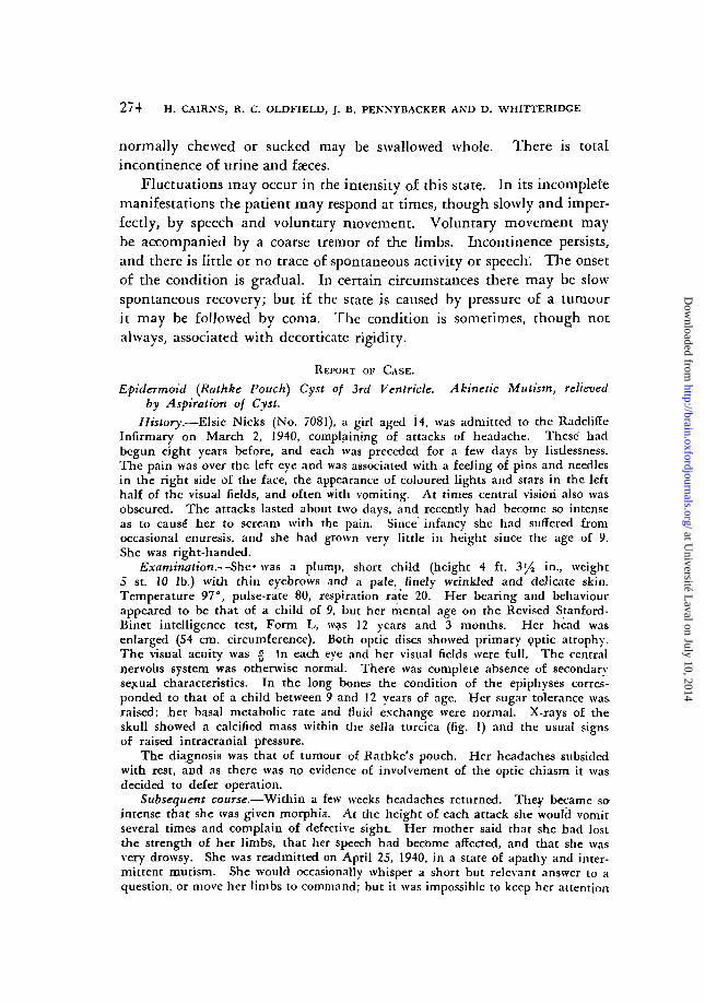
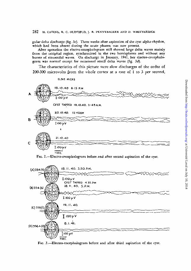
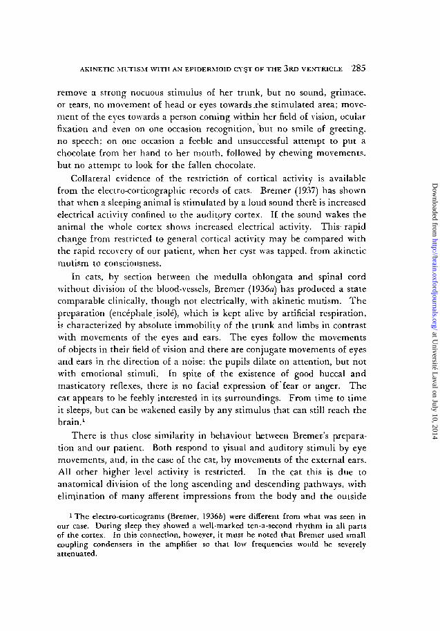
Examination.—She* was a plump, short child (height 4 ft. 3j/i in., weight.5 st. 10 lb.) with thin eyebrows and a pale, finely wrinkled and delicate skin.Temperature 97°, pulse-rate 80, respiration rate 20. Her bearing and behaviourappeared to be that of a child of 9, but her mental age on the Revised Stanford-Binet intelligence test, Form L, was 12 years and 3 months. Her head wasenlarged (54 cm. circumference). Both optic discs showed primary optic atrophy.The visual acuity was & In each eye and her visual fields were full. The centralnervous system was otherwise normal. There was complete absence of secondarysejcual characteristics. In the long bones the condition of the epiphyses corres-ponded to that of a child between 9 and 12 years of age. Her sugar tolerance wasraised; her basal metabolic rate and fluid exchange were normal. X-rays of theskull showed a calcified mass within the sella turcica (fig. 1) and the usual signsof raised intracranial pressure.
The diagnosis was that of tumour of Rathke's pouch. Her headaches subsidedwith rest, and as there was no evidence of involvement of the optic chiasm it wasdecided to defer operation.
Subsequent course.—Within a few weeks headaches returned. The^ became sointense that she was given morphia. At the height of each attack she would vomitseveral times and complain of defective sight Her mother said that she had lostthe strength of her limbs, that her speech had become affected, and that she wasvery drowsy. She was readmitted on April 25, 1940, in a state of apathy and inter-mittent mutism. She would occasionally whisper a short but relevant answer to aquestion, or move her limbs to command; but it was impossible to keep her attention
at Université L
aval on July 10, 2014http://brain.oxfordjournals.org/
Dow
nloaded from
Fie. la.
Fie.
Fie. 1 (a) and (fc).—Suprasellar cyst (cross hatched) and lateral ventricle outlinedby a series of X-ray pictures of an air bubble injected after aspiration of the cyst.The continuous line shows the actual outline, the dotted lines are an estimate ofthe outline not actually seen. In the lateral view patches of intrasellar calcificationare also represented.
at Université L
aval on July 10, 2014http://brain.oxfordjournals.org/
Dow
nloaded from
276 H., CAIRNS, R. C. OLDFIELD, J. B. PENNYBACKER AND D. WHITTERLDCE
thus employed for more than a few seconds at a time. She lapsednot into sleep but into a state of inertia and silence. All four limbswere moved weakly but without tremor. The abdominal reflexes wereno longer present and the plantar reflexes were now extensor in type. Sensi-bility appeared to be preserved, but with high threshold. Her peripheral circulationwas poor.
In the following days she became less accessible. She did not sleep excessively.When awake and undisturbed she made no movement, save that her eyes wouldfollow quite alertly any movement in the ward, and on a few occasions shewhispered, almost inaudibly, a monosyllabic response. For the most part she madeno reply to urgent appeals, though on one occasion she gave a feeble smite whentickled, and turned her head away. She had to be fed. When a chocolate had beenplaced in her hand she attempted to put it in her mouth, but she dropped it anddid not try to reclaim it. However, she then made some tentative chewing move-ments. When barley sugar was placed in her mouth it was immediately swallowedwhole, and without any display of interest or discomfort. Sugar and quinine wereswallowed with complete indifference. Sustained pin-prick on the trunk evoked aslow/ deliberate movement of both hands towards the stimulus, but no sound,grimace or tears, though once or twice her expression changed a little as thoughshe were on the point of crying. There was no trace of catatonia. She was totallyincontinent of urine' and faeces.
Ventricular tapping.—On April 28, three days after admission, she lAd fourattacks of extensor rigidity at short intervals, with limbs extended, head retracted,pulse and respiration slowed. Between the attacks extensor tonus remained exces-sive and she could not be rouse*!. The lateral Ventricles were tapped through theleft frontal and right parietal regions, and were found to be dilated, with an initialpressure of over 600 mm. H,O. After release of the ventricular pressure she couldbe roused and her general condition improved, but the state of akinetic mutismwas little affected. In the next few days she had slight and temporary right hemi-paresis. When she was awake, her eyes still followed every movement of anobserver approaching her bedside, and seemed always to give promise of speech.But repeated attempts usually failed to elicit any utterance. During sleep hereyes were normally closed. GJn May 6 she had a further attack of extensor rigiditywith opisthotonos lasting two to three minutes. On May 9 she responded tostrenuous appeals for a short period, recognizing one of us by name in a whisper,naming the hospital, and moving her limbs to command, _albeit slowly and incom-pletely, but it was impossible to keep her attention for more than a few momentsat a time. At this stage the right hemiparesis was less; m the face, jt was seen todiminish as she wakened.
First aspiration on 3rd ventricle cyst.—On May 9 the right lateral ventricle wastapped tfirough its frontal horn. The initial pressure was 190 mm. H,Oi Theneedle was advanced towards the anterior part of the 3rd ventricle, and after slightresistance a cyst was entered and 18 c.c. of brownish fluid were aspirated, containing2-8 per cent, of protein.and alsdV'chplesterol crystals. Five c.c. of air were injectedinto the cyst and subsequent X-rays showed that the cyst occupied the greater partof the 3rd ventricle (fig. 1).
Immediately the cyst was"tapped the child roused and made a noise, the firstloud sound she had made since admission to hospital two weeks before, but shewould riot speak. Within ten minutes, however, she gave her name, age, andaddress correctly without any trace of dysarthria, and then asked where she*was.She co-operated perfectly in an examination. Within the next week the right hemi-pariesis disappeared, and she got up. She was still at times incontinent. She had
at Université L
aval on July 10, 2014http://brain.oxfordjournals.org/
Dow
nloaded from
AKINETIC MUTISM WITH AN EP1DERMO1D CYST OF THE 3RD VENTRICLE 277
no memory of the two weeks in hospital before the cyst was emptied. She wasdischarged on May 22, at which time, apart from a defect of sustained attention,she was alert and intelligent and showed no neurological signs other than opticatrophy and occasional incontinence of urine.
In June, 1940, she began once more to have attacks of headache, vomiting, andimpaired sight. In August her visual fields showed for the first time an incom-plete upper quadrantic bitemporal hemianopia, but her visual acuity remainednormal. She continued to be incontinent of urine at nights.
On October 17, after a severe attack of headache and vomiting lasting severaldays, she became once more mute and was readmitted to hospital in a dehydratedstate. Her condition was similar to the state observed in April. She was moredrowsy than on previous occasions, but could be awakened, when she would watchthe movements of people in the ward. Addressed in a loud voice she would moveher eyes towards the sound. There was a sluggish menace reflex. She swallowedwithout difficulty and would withdraw from painful stimuli. She seemed to under-stand at least something of what was said to her: for example, she would at timesmake feeble attempts ' to put <?ut her tongue on command ; but she wascompletely devoid of speech, and her only emotional expression was afeeble grimace in response to strong painful stimuli. She was incontinent ofurine and faeces. Her pupils were irregularly small. Her extremities were coldand cyanosed. Her upper limbs always assumed a position of flexion, her lowerlimbs were extended and showed strong extensor tonus. The ankle-jerks were in-creased, there was bilateral ankle clonus, and both plantar reflexes were extensor.
She became stuporous and had to be fed by nasal tube. At 4.30 a.m. on October19 she had an'attack of extensor rigidity of the limbs and trunk with head retrac-tion and upward deviation of the eyes. This lasted two minutes and was followedby coma. The pulse-rate slowed from 80 to 64. Respirations slowed from 20 to 10,and were deep and stertorous.
Second aspiration of cyst.—On October 19, seven hours after the attack, thecyst was aspirated again through the right frontal horn, and 15 c.c of brownish fluidwere removed (initial ventricular pressure 80 .mm. H,O). Immediately after theaspiration, even before»the needle was withdrawn, she became more alert and startedmoving about in bed. She then sat up, as though waking from deep sleep.She took a bite from a biscuit but would not take it into her owri hand. Withinhalf an hour, during electro-encephalography, she exclaimed, " Oh',' dear " a numberof times in a stereotyped fashion, but made no other verbal response. Two hoursafter the cyst had been emptied she began to talk, and an hour later she rang fora bed-pan. For two days she was slow in answering questions and mildly confused,but she helped herself to food with obvious enjoyment. She named objects slowlybut without error, and could cut with scissors, though somewhat clumsily. Heramnesia extended from some days before to several hours after aspiration of thecyst.
She soon- became bright and cheerful. With the improvement in mental stateall sjgns of extensor rigidity disappeared. Her tendon-jerks, abdominal and plantarreflexes were normally present. She was discharged from hospital on October 23.
Third aspiration of cyst.—She was readmitted on November 17 afttr a par-ticularly severe attack of headache, with which wa3- associated incontinence. Shewns drowsy, but conld be easily roused and would talk in response to questions,though she did not volunteer any remarks. Her abdominal reflexes were absent,but there were no other neurological signs in the limbs or trunk. On November 18the.cyst was tapped again, and 12 c.c. of brownish fluid were removed (initialventricular pressure of cerebrospinal fluid, 220 mm. HjO). Improvement was slow,
BRAIN—VOL. LXIV. 19
at Université L
aval on July 10, 2014http://brain.oxfordjournals.org/
Dow
nloaded from
2 7 8 H. CAIRNS, R. C. OLDFIELD, J. B. PENNYBACKER ANT3 D. WH1T*TER1DGE
but within forty-eight hours she was once more moderately alert! Further surgicaltreatment was deferred on account of a left otitis media.
Operation.—On December 17, 1940, the 3rd ventricle was explored by resectionof part of the right frontal lobe and a transventricular approach. A thin-walledyellowish cyst presented at the foramen of Monro. It occupied the 3rd ventricleand was in places adherent to the walls by vascular adhesions, especially at thelamina terminalis and the anterior part of the medial-surface of each thalamus.In the floor of the 3rd ventricle there was a solid portion of tumour; this was notremoved, but the cystic part was dissected away completely.
Pathological report (Dr. Dorothy Russell).—-The specimens received for histo-logical examination included two larger portions, measuring 3 by 1 by 0-3 cm. and3-5 by 1 by 0-5 cm. respectively, and one smaller portion, measuring 07 by 05 by0-4 cm., of soft yellowish grey tissue. The larger bits bore in places a .smoothglistening surface to which some cholesterol crystals were attached. The remainingsurfaces were firm and jiodular.
In addition there was a portion of cerebral cortex and white matter measuring4 by 2-5 up to 3-8 cm. deep. This was macroscopioally normal apart fromoperative trauma, and was not examined microscopically.
Microscopically.—The tissue from the 3rd ventricle is composed, ior the.mostpart, of neuroglial cells which are mostly of pilocytic form. Hyaline degenerativechanges are present Ln many of them. The surface bears, Ln many -places, a well-differentiated epithelium of basal type composed of polygonal cells and, in rela-tionship with the neuroglia, a regular layer of columnar cells. Papillae composedof these cells penetrate the neuroglia for a considerable depth in some places. Super-ficially the epithelium frequently contains squamous and horny nests. The latterare occasionally present in the neuroglial tissue and may then be associated with aforeign body giant-cell reaction.
Post-operative course.—After operation there was a recurrence in mild degree ofthe mental state seen at earlier stages. In the first few days her speech was mainlylimited to " Yes " or " No." She moved her limbs to hold a drinking cup, to rubher nose, or to withdraw from painful stimuli, but not in response to commands.Her right upper limb now moved about, exploring the bedclothes and the sideof the -bedr From this state she gradually recovered, and by December 26 she wasable to get up. After a period of physical exercise she always became more alert,smiling and spontaneous in actions and speech, but she was still slower thannormal. Except under very strong stimulus her emotional display was belowaverage. She was still on occasions incontinent. At this time hypersomnia becameevident. When left to herself she would often fall asleep, sometimes even whilestanding up. This tendency to sleep by day was only slightly relieved by benzedrine.On discharge from hospital on January 8, 1941, she was still pathologically drowsy,and she showed a hypopituitary type of habitus, primary optic atrophy with normalvisual fields and acuity, fanning of the toes on stimulation of each sole, .but noother neurological signs. Her basal metabolic rate was — 10 per cent,.
Throughout the whole nine months of observation her temperature fluctuatedbetween 95° and 98-4°, but was usually about 97°. Her pulse showed no grossirregulaxjties; at first its rate was 80, but subsequently it varied between 54 and110, being slowest when the decorticate state and loss of consciousness were mostprofound. The fluctuations in temperature and pulse-rate did not entirely disappearafter aspiration .of the cyst and improvement in the general clinical state. At theheight of coma and extensor rigidity the respirations were slowed to 10 and weredeep and stertorous. There were never any sudden changes of colour, and nosweating.
at Université L
aval on July 10, 2014http://brain.oxfordjournals.org/
Dow
nloaded from
AKINETIC MUTISM WITH AN EPIDERMOID CIST OF THE 3RD VENTRICLE 279
During the nine months of observation she did not grow or charige in weight.Her blood-pressure fell during the period from 108/70 to 75/45. She never men-struated or showed any secondary sexual characteristics, and she never had polyuriaor polydipsia. Her blood sugar three hours after her midday meal was 0'130 percent,, and intravenous glucose produced no alleviation of the akinetic mutism.
When she was last seen in July, 1941, eight months after operation, she waswell and free from headache, mutism, drowsiness and polyuria, and she was ableto do routine work. She was forgetful, and still at times incontinent of urine.She still had not menstruated.
DISCUSSION.
The feature of this case which we wish to emphasize is the recurringepisodes of mutism and immobility associated with, pressure of a cyst ofRathke's pouch upon structures surrounding the 3rd ventricle.
The state was described by the child's parents and by nurses as oneof drowsiness, but drowsiness was not a feature of it. Except duringperiods of severe decorticate rigidity, the girl was always-easy to rouseand she did not sleep excessively. The most profound manifestations weremutism, loss of feeling tone, loss of emotional expression, of spontaneousand of most other voluntary movements, and total incontinence of urineand faeces of a degree and persistence rarely met with in brain lesions notassociated with coma. She was incapable of originating active manifesta-tions of any kind, with the notable exception that ocular fixation andmovement occurred in response to the movement of external objects andto sounds. It was this which gave the child an appearance of alertnessso incongruous with her silence and general immobility.1
All feeling appeared to be in abeyance. To painful stimuli therewas reflex withdrawal and at times, after a prolonged latent period, a slowand ineffective voluntary movement of her hands towards the point ofstimulation, but never any sign or sound of displeasure. The sight offood evoked no sign of pleasure, though at times she appeared to under-stard what it was and would make a slow, feeble attempt to put it in hermouth. Pleasant and unpleasant substances, hard foodstuffs that shouldat irst have been chewed or sucked, were" all swallowed promptly withoutanr expression of pleasure, distaste, or discomfort. There was disturbanceof consciousness at the highest levels; as evidenced by total amnesia for
1 These ocular movements are comparable in some ways to the tendency to attendto ".very visual stimulus (" hypermetamorphosis ") observed in macaques by Kliiveram Bucy (1939) after bilateral removal of the temporal lobes, including the uncusanc the greater part of the hippocampus; but our patient differed from the mon-key;, in that her attention was attracted by movement of objects rather than byobjets themselves, and no motor reaction followed movement and fixation of hereyes.
at Université L
aval on July 10, 2014http://brain.oxfordjournals.org/
Dow
nloaded from
at Université L
aval on July 10, 2014http://brain.oxfordjournals.org/
Dow
nloaded from
AKINETIC MUTISM WITH AN EP1DERMOID CYST OF THE 3RD VENTRICLE 2 8 1
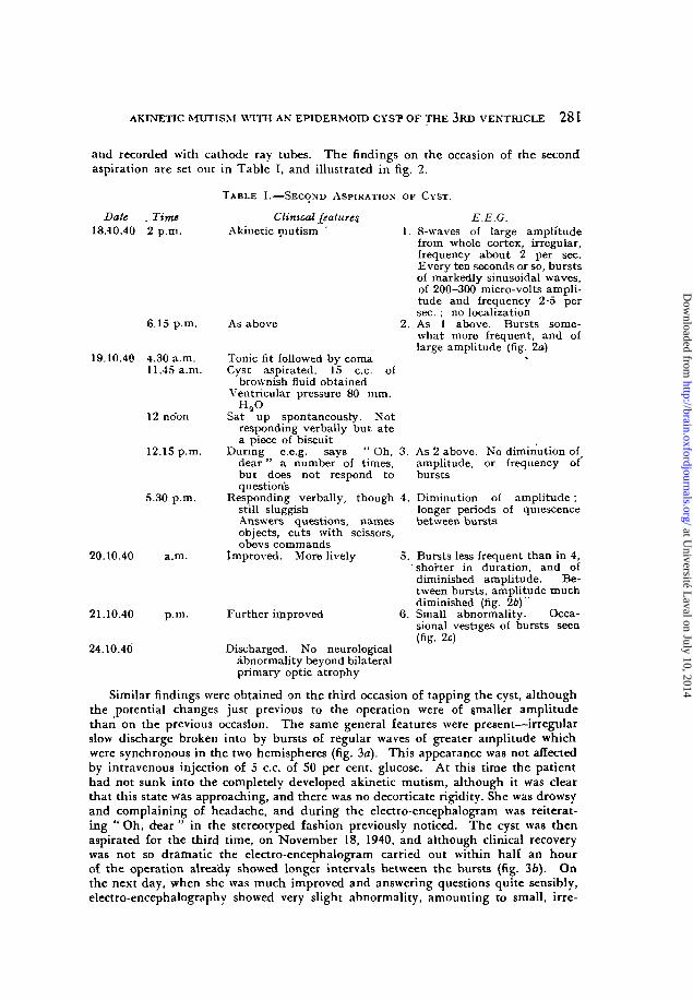
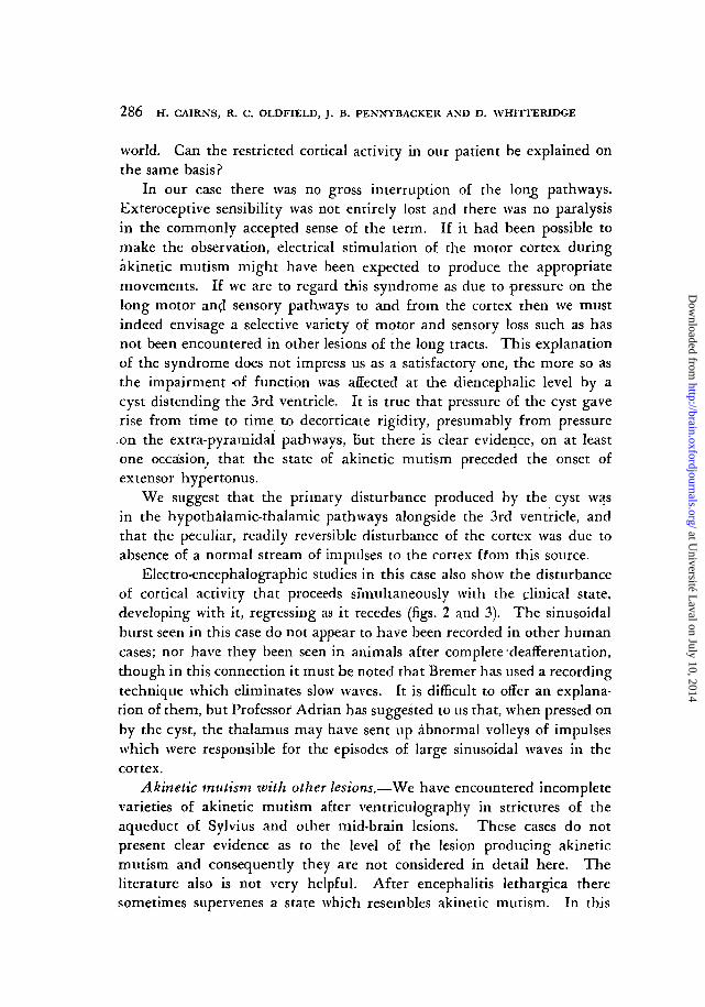
and recorded with cathode ray. tubes. The findings on the occasion of the secondaspiration are set out in Table I, and illustrated in fig. 2.
TABLE I.—SECOND ASPIRATION OF CYST.
Date . Time18.10.40 2 p.m.
Clinical featuresAkinetic mutism
6.15 p.m.
19.10.40 4.30 a.m.11.45 a.m.
12 noon
12.15 p.m.
5.30 p.m.
20.10.40 a.m.
21.10.40 p.m.
24.10.4(3
As above
Tonic fit followed by comaCyst aspirated. 15 c.c.
brownish fluid obtainedVentricular pressure 80 mm.
E.E.G.8-waves of large amplitudefrom whole cortex, irregular,frequency about 2 per sec.Every ten seconds or so, burstsof markedly sinusoidal waves,of 200-300 micro-volts ampli-tude and frequency 2-5 persec. ; no localizationAs 1 above. Bursts some-what more frequent, and oflarge amplitude (fig. 2a)
of
Sat up spontaneously. Notresponding verbally but atea piece of biscuit
During e.e.g. says " Oh, 3.dear" a number of times,but does not respond toquestions
Responding verbally, though 4.still sluggishAnswers questions, namesobjects, cuts with scissors,obevs commands
Improved. More lively 5.
Further improved 6.
Discharged. No neurologicalabnormality beyond bilateralprimary optic atrophy
As 2 above. No diminution ofamplitude, or frequency ofbursts
Diminution of amplitude ;longer periods of quiescencebetween bursts
Bursts less frequent than in 4,shorter in duration, and ofdiminished amplitude. Be-tween bursts, amplitude muchdiminished (fig. 26) ~Small abnormality. Occa-sional vestiges of bursts seen(fig. 2c)
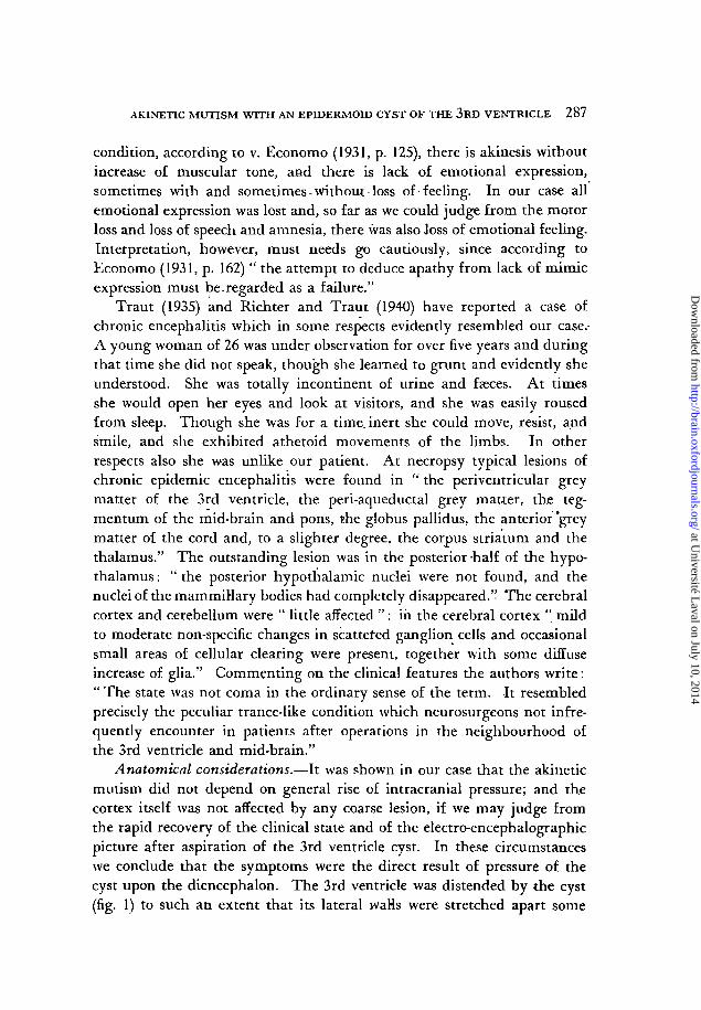
Similar findings were obtained on the third occasion of tapping the cyst, althoughthe potential changes just previous to the operation were of smaller amplitudethan on the previous occasion. The same general features were present—irregularslow discharge broken into by bursts of regular waves of greater amplitude whichwere synchronous in the two hemispheres (fig. 3a). This appearance was not affectedby intravenous injection of 5 c.c. of 50 per cent, glucose. At this time the patienthad not sunk into the completely developed akinetic mutism, although it was clearthat this state was approaching, and there was no decorticate rigidity. She was drowsyand complaining of headache, and during the electro-encephalogram was reiterat-ing " Oh, dear " in the stereotyped fashion previously noticed. The cyst was thenaspirated for the third time, on November 18, 1940, and although clinical recoverywas not so dramatic the electro-encephalogram carried out within half an hourof the operation already showed longer intervals between the bursts (fig. 3b). Onthe next day, when she was much improved and answering questions quite sensibly,electro-encephalography showed very slight abnormality, amounting to small, irre-
at Université L
aval on July 10, 2014http://brain.oxfordjournals.org/
Dow
nloaded from
282 H. CAIRNS, R. C. OLDFIELD, J. B. PENNYBACKER AiVD D. WH1TTERIDCE
gular delta discharge (fig. 3c). Three weeks after aspiration of the cyst alpha-rhythm,which had been absent during the acute phases-, was now present.
After operation the electro-encephalogram still showed large delta waves mainlyfrom the occipital region, synchronized in the two hemispheres and without anybursts of sinusoidal waves. On discharge in January, 1941, her electro-encephalo-gram was normal except for occasional small delta waves (fig. 3d).
The characteristics of this picture were slow discharges of the order of200-300 microvolts from the whole cortex at a rate of 1 to 3 per second,
ELSIE NICKS
18. IO.4O 6 1 5 P.M
B
CYST TAPPED I9.I0.4O. M-43A.M
ZOJO.4O. 12 MOON
ZI.IO.4O
IlOOpV
I SEC.'
Fic. 2.—Electro-encephalogram before and after second aspiration of the cyst.
I IOOJJVCYST TAPPED 4 50. PMId. I I . 4O. 5.P.M.
(D) 3 5 6 A ( i
JlOOpVI SEC.
Fic. 3.—Electro-encephalogram before and after third aspiration of. the cyst.
at Université L
aval on July 10, 2014http://brain.oxfordjournals.org/
Dow
nloaded from
AKINETIC MUTISM WITH AN EPIDERMOID CYST OF THE 3RD VENTRICLE 2 8 3
irregular in form and interspersed by periods of nearly sinusoidal dischargeof slightly greater ajnplitude at 2 to 5 per second (figs. 2 and 3). Thesesinusoidal bursts have never been seen by us in states of raised intracranialpressure, and indeed there was no significant rise of intracranial pressure inour patient at the times of electro-encephalographic observation. Followingaspiration of the cyst the abnormal wave form progressively disappearedwith concomitant improvement and disappearance of.mutism and akinesia.
Walter, Griffiths and Nevin (1939) have reported abnormalities of theelectro-encephalogram in one case of tumour involving the structuresaround the 3rd ventricle, and causing a state of pathological sleep. Theydescribe their patient's condition as " extremely drowsy," with deep, regularrespiration at 20 per minute. She resisted any attempt to open her eyes,but " with further stimulation she could generally be roused, so that shewould open her eyes, stir,.perhaps smile, and mutter a few meaninglesswords. At other times she was more awake, and then it was noticed thatshe moved all her limbs normally and also opened her eyes on command."The patient was doubly incontinent, had no cranial nerve signs so far asit was possible to determine, and no papillcedema. Tone was normal inall limbs, reflexes were normal in the arms, but the abdominal and deepleg reflexes were absent. The plantars were flexor. At lumbar puncturethe initial pressure of cerebrospinal fluid was 140- mm. After a few daysthe patient died in hyperthermia. At post-mortem examination a soft,greyish-red tumour, described as a perithelial sarcoma, was found., involv-ing the thalamus, hypothalamus, internal capsule and corpus striatumon the left side, sparing only the posterior third of these structures. Awedge-shaped mass of tumour extended across the middle line towards anarea of softening in the right globus pallidus. The infundibular stalk andpituitary were not involved.
The electro-encephalogram in this case showed continuous high-voltageslow discharge from all areas of the cortex. The potential changes reacheda level of 500 microvolts. They were irregular in form, and poorly syn-chronized in the two hemispheres. " The discharge persisted almostunchanged when the patient was partially aroused. There was no traceof the normal alpha and beta rhythm, but the potential of the slow dis-charge decreased by about 20 per cent, when the patient was awakenedas much as possible, the rhythm being the same."
The authors consider that such an electro-encephalographic picturemore closely resembles that found in normal sleep than in the comaassociated with raised intracranial pressure, in which, they state, " a veryregular rhythm of smaller amplitude may be found in all areas." They
at Université L
aval on July 10, 2014http://brain.oxfordjournals.org/
Dow
nloaded from
2 8 4 H. CAIRNS, R. C. OLDFlELDl J. B. PENNYBACKER AND D. WHITTERIDGE
suggest that this difference " may be useful in differentiating between thestupor of hydrocephalus and that of hypersomnia associated with a locallesion in the hypothalamic region." It is quite debatable whether hydro-cephalus disturbs consciousness except through the brain-stem. Certainlythere are many cases of hydrocephalus and .raised intracranial pressurein which there is no stupor or other disturbance of consciousness, and itmay be that this is an attempt to differentiate by electro-encephalographybetween two states that are not really distinguishable. The importantpoint is that in Walter, Griffiths and Nevin's case, as in ours, the electro-encephalographic picture was clearly associated with a lesion of thediencephalon rather than with a state of raised intracranial pressure. Inour case the slow, irregular discharge, such as Walter, Griffiths and Nevinsaw, is broken into by bursts of regular sinusoidal waves of 100-200 micro-volts. The state of coma in Walter's case differed from the conditionobtaining in our case, being more definitely like sleep, and the e.e.g. intheir case more closely resembled that of normal sleep as described byDavis et al. (1938).
The impairment of cortical function.—In this clinical state of akineticmutism the functions disturbed for the most part involve the cortex:voluntary movement, including speech, spontaneous activity of all kinds,emotional expression, perception and memory. There was as a rule absenceof all forms of voluntary movement, except that of movement and fixationof the eyes in response to sound or a movement in the outside world,functions which in man, according to Holmes (1938), can be evoked onlythrough the cortex.
There was no coarse lesion of the cortex or interruption of afferentcortical pathways from limbs or special sense organs in the ordinary way,no persisting motor or sensory palsy, beyond a fleeting left hemiparesison one occasion which only served to show the essential contrast betweenthis general somatic akinesia and a.specific defect of motor pathways.1
Furthermore, recovery from akinesia was quite unlike recovery from acortical injury. There was no stage of weak or clumsy movement, nodysphasia or dyspraxia. Ten minutes after 3rd ventricle cyst was emptiedthe child sat up in bed and said: " Where am I?" It was more like wakingfrom sleep.
During the height of the akinesia such cortical activity as was presentwas restricted, incomplete—a slow, deliberate movement of the hands to
1 This temporary left hemiparesis after simple tapping of the right lateral ventriclemay also indicate that the cortex in akinetic mutism, though healthy, is never-theless abnormally vulnerable.
at Université L
aval on July 10, 2014http://brain.oxfordjournals.org/
Dow
nloaded from
AKINETIC MUTISM WITH AN EPIDERMOLD CY§T OF THE 3RD VENTRICLE 2 8 5
remove a strong nocuous stimulus of her trunk, but no sound, grimace,or tears, no movement of head or eyes towards the stimulated area; move-ment of the eyes towards a person coming within her field of vision, ocularfixation and even on one occasion recognition, but no smile of greeting,no speech; on one occasion a feeble and unsuccessful attempt to put achocolate from her hand to her mouth, followed by chewing movements,but no attempt to look for the fallen chocolate.
Collateral evidence of the restriction of cortical activity is availablefrom the electro-corticographic records of cats. Bremer (1937) has shownthat when a sleeping animal is stimulated by a loud sound there is increasedelectrical activity confined to the auditory cortex. If the sound wakes theanimal the whole cortex shows increased electrical activity. This rapidchange from restricted to general cortical activity may be compared withthe rapid recovery of our patient, when her cyst was tapped, from akineticmutism to consciousness.
In cats, by section between the medulla oblongata and spinal cordwithout division of the blood-vessels, Bremer (1936a) has produced a statecomparable clinically, though not electrically, with akinetic mutism. Thepreparation (encephale isole), which is kept alive by artificial respiration,is characterized by absolute immobility, of the trunk and limbs in contrastwith movements of the eyes and ears. The eyes follow the movementsof objects in their field of vision and there are conjugate movements of eyesand ears in the direction of a noise; the pupils dilate on attention, but notwith emotional stimuli. In spite of the existence of good buccal andmasticatory reflexes, there is no facial expression of fear or anger. Thecat appears to be feebly interested in its surroundings. From time to timeit sleeps, but can be wakened easily by any stimulus that can still reach thebrain.1
There is thus close similarity in behaviour between Bremer-'s prepara-tion and our patient. Both respond to visual and auditory stimuli by eyemovements, and, in the case of the cat, by movements of the external ears.All other higher level activity is restricted. In the cat this is due toanatomical division of the long ascending and descending pathways, withelimination of many afferent impressions from the body and the outside
1The electro-corticograms (Bremer, 19366) were different from what was seen inour case. During sleep they showed a well-marked ten-a-second rhythm in all partsof the cortex. In this connection, however, it must be noted that Bremer used smallcoupling condensers in the amplifier so that low frequencies would be severelyattenuated.
at Université L
aval on July 10, 2014http://brain.oxfordjournals.org/
Dow
nloaded from
2 8 6 H. CAIRNS, R. C. OLDFIELD, J. B. PENNYBACKER AND D. WHITTERIDGE
world. Can the restricted cortical activity in our patient be explained onthe same basis?
In our case there was no gross interruption of the long pathways.Exteroceptive sensibility was not entirely lost and there was no paralysisin the commonly accepted sense of the term. If it had been possible tomalce the observation, electrical stimulation of the motor cortex duringakinetic mutism might have been expected to produce the appropriate,movements. If we are to regard this syndrome as due to pressure on thelong motor and sensory pathways to and from the cortex then we mustindeed envisage a selective variety of motor and sensory loss such as hasnot been encountered in other lesions of the long tracts. This explanationof the syndrome does not impress us as a satisfactory one, the more so asthe impairment of function was affected at the diencephalic level by acyst distending the 3rd ventricle. It is true that pressure of the cyst gaverise from time to time to decorticate rigidity, presumably from pressure.on the extra-pyramidal pathways, but there is clear evidence, on at leastone occasion, that the state of akinetic mutism preceded the onset ofextensor hypertonus.
We suggest that the primary disturbance produced by the cyst wasin the hypothalamic-thalamic pathways alongside the 3rd ventricle, andthat the peculiar, readily reversible disturbance of the cortex was due toabsence of a normal stream of impulses to the cortex ffom this source.
Electro-encephalographic studies in this case also show the disturbanceof cortical activity that proceeds simultaneously with the clinical state,developing with it, regressing as it recedes (figs. 2 and 3). The sinusoidalburst seen in this case do not appear to have been recorded in other humancases; nor have they been seen in animals after complete'deafferentation,though in this connection it must be noted that Bremer has used a recordingtechnique which eliminates slow waves. It is difficult to offer an explana-tion of them, tout Professor* Adrian has suggested to us that, when pressed onby the cyst, the thalamus may have sent up abnormal volleys of impulseswhich were responsible for the episodes of large sinusoidal waves in thecortex.
Akinetic mutism with other lesions.—We have encountered incompletevarieties of akinetic mutism after ventriculography in strictures of theaqueduct of Sylvius and other mid-brain lesions. These cases do notpresent clear evidence as to the level of the lesion producing akineticmutism and consequently they are not considered in detail here. Theliterature also is not very helpful. After encephalitis lethargica theresometimes supervenes a state which resembles akinetic mutism. In this
at Université L
aval on July 10, 2014http://brain.oxfordjournals.org/
Dow
nloaded from
AKINETIC MUTISM WITH AN EPIDERMOID CYST OF THE 3RD VENTRICLE 287
condition, according to v. Economo (1931, p. 125), there is akinesis withoutincrease of muscular tone, and there is lack of emotional expression,sometimes with and sometimes without loss of feeling. In our case allemotional expression was lost and, so far as we could judge from the motorloss and loss of speech and amnesia, there was also loss of emotional feeling.Interpretation, however, must needs go cautiously, since according toEconomo (1931, p. 162) " the attempt to deduce apathy from lack of mimicexpression must be.regarded as a failure."
Traut (1935) and Richter and Traut (1940) have reported a case ofchronic encephalitis which in some respects evidently resembled our case.-A young woman of 26 was under observation for over five years and duringthat time she did not speak, though she learned to grunt and evidently sheunderstood. She was totally incontinent of urine and fasces. At timesshe would open her eyes and look at visitors, and she was easily rousedfrom sleep. Though she was for a time, inert she could move, resist, andsmile, and she exhibited athetoid movements of the limbs. In otherrespects also she was unlike our patient. At necropsy typical lesions ofchronic epidemic encephalitis were found in " the periventricular greymatter of the 3rd ventricle, the peri-aqueductal grey matter, the teg-mentum of the mid-brain and pons, the globus pallidus, the anterior 'greymatter of the cord and, to a slighter degree, the corpus striatum and thethalamus." The outstanding lesion was in the posterior-half of the hypo-thalamus: " the posterior hypothalamic nuclei were not found, and thenuclei of the mammiHary bodies had completely disappeared." The cerebralcortex and cerebellum were " little affected ": in the cerebral cortex " mildto moderate non-specific changes in scatteted ganglion cells and occasionalsmall areas of cellular clearing were present, together with some diffuseincrease of glia." Commenting on the clinical features the authors write:" The state was not coma in the ordinary sense of the term. It resembledprecisely the peculiar trance-like condition which neurosurgeons not infre-quently encounter in patients after operations in the neighbourhood ofthe 3rd ventricle and mid-brain."
Anatomical considerations.—It was shown in our case that the akineticmutism did not depend on general rise of intracrahial pressure; and thecortex itself was not affected by any coarse lesion, if we may judge fromthe rapid recovery of the clinical state and of the electro-encephalographicpicture after aspiration of the 3rd ventricle cyst. In these circumstanceswe conclude that the symptoms, were the direct result of pressure of thecyst upon the diencephalon. The 3rd ventricle was distended by the cyst(fig. 1) to such an extent that its lateral waHs were stretched apart some
at Université L
aval on July 10, 2014http://brain.oxfordjournals.org/
Dow
nloaded from
2 8 8 H. CAIRNS, R. C. OLDFIELD, J. B. PENNYBACKER AND D. WHITTERIDGE
I'7 cm., as compared with the normal width, of 015 to 03 cm. This degreeof stretching of the 3rd ventricle is not exceptional in cases f̂- cystic orsolid epidermoid (Rathke pouch) tumours, in mospof which, it must benoted, the syndrome of akinetic mutism is not seen. We have so far failedto find in the literature any case of 3rd ventricle cyst which could bedefinitely labelled as akinetic mutism, notwithstanding the remark ofRichter and Traut quoted above. The symptoms most commonly seenwith epidermoid tumour in the 3rd ventricle are headache and vomiting,depression or absence of sexual function, diabetes insipidus, adiposity,disturbance of temperature regulation, failure of vision, hypersomnia, andcoma. The last two of these symptoms, and possibly also visual loss,might effectively mask akinetic mutism, but the capricious clinical effectsof 3rd ventricle tumours cannot often be explained by supposing thedominance of one symptom over another.
While it is not possible to determine which particular structures in thewall of the 3rd ventricle were so compressed as to produce this syndrome,the absence of chiasmal signs suggests that the cyst did not encroachupon the hypophysis and its stalk. The optic tracts are shieldedfrom pressure within the 3rd ventricle by the thalamus, and this doubtlesscontributes to the preservation of the eye movements that was such anoticeable feature of the clinical state. The presence of pupillary andexternal ocular movements excludes the pineal body, quadrigeminal plate,and the aqueduct of Sylvius. There remain as the main structures in theimmediate neighbourhood of the cyst the thalamus, hypothalamus, andfornix.
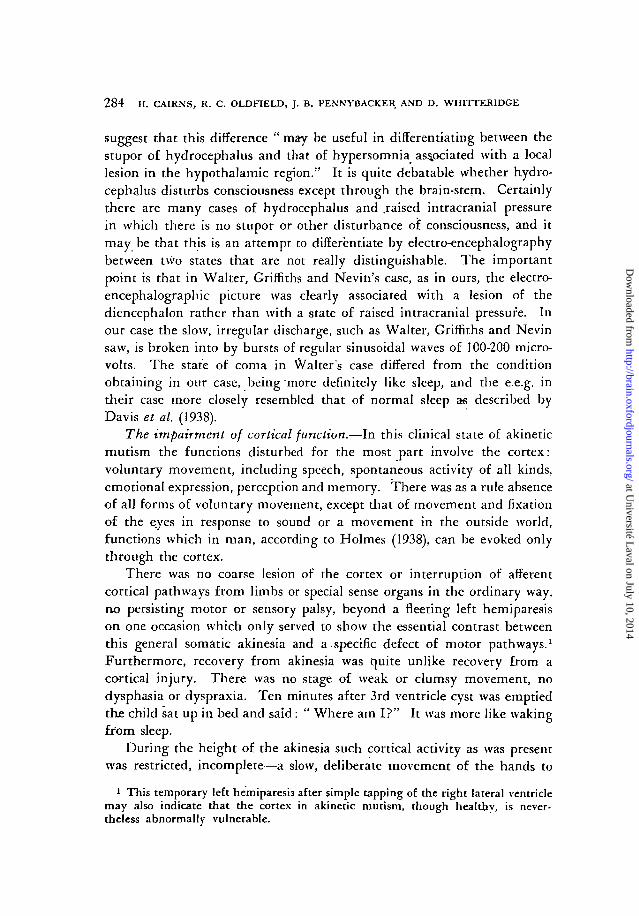
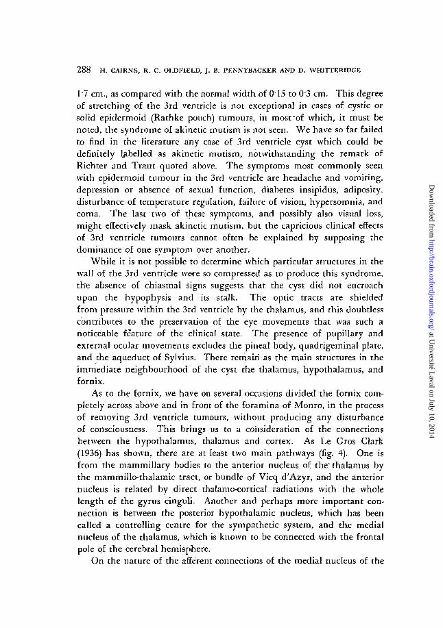
As to the fornix, we have on several occasions divided the fornix com-pletely across above and in front of the foramina of Monro, in the processof removing 3rd ventricle tumours, without producing any disturbanceof consciousness. This brings us to a consideration- of the connectionsbetween the hypothalamus, thalamus and cortex. As Le Gros Clark(1936) has shown, there axe at least two main pathways (fig. 4). One isfrom the mammillary bodies to the anterior nucleus of the" thalamus bythe rhammillo-thalamic tract, or bundle of Vicq d'Azyr, and the anteriornucleus 'is related by direct thalamo-cortical radiations with the wholelength of the gyms cinguli.. Another and perhaps more important con-nection is between the posterior hypothalamic nucleus, which has beencalled a controlling centre for the sympathetic .system, and the medialnucleus of the thalamus, which is known to be connected with the frontalpole of the cerebral hemisphere.
On the nature of the afferent connections of the medial nucleus of the
at Université L
aval on July 10, 2014http://brain.oxfordjournals.org/
Dow
nloaded from
AKINETIC MUTISM WITH AN EP1DERM01D CYST OF THE 3RD VENTRICLE 289
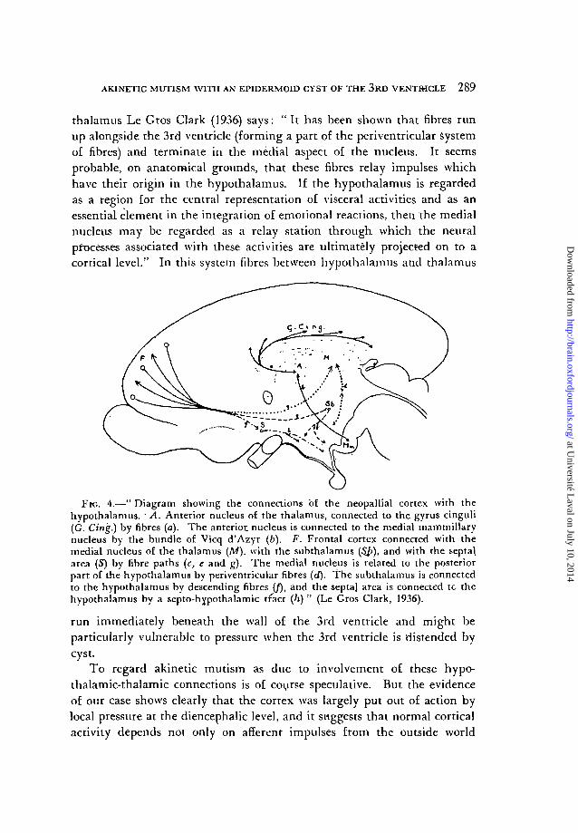
thalamus Le Gros Clark (1936) says: "I t has been shown that fibres runup alongside the 3rd ventricle (forming a part of the periventricular systemof fibres) and terminate in the medial aspect of the nucleus. It seemsprobable, on anatomical grounds, that these fibres relay impulses whichhave their origin in the hypothalamus. If the hypothalamus is regarded.as a region for the central representation of visceral activities and as anessential element in the integration of emotional reactions, then the medialnucleus may be regarded as a relay station through, which the neuralprocesses associated with these activities are ultimately projected on to acortical level." In this system fibres between hypothalamus and thalamus
Fie. 4.—" Diagram showing the connections of the neopallial cortex with thehypothalamus. • A. Anterior nucleus of the thalamus, connected to the gyrus cinguli(G. Cing.) by fibres (a).. The anterior, nucleus is connected to the medial mammillarynucleus by the bundle of Vicq d'Azyr (b). F. Frontal cortex connected with themedial nucleus of the thalamus (Af), with the subthalamus ($/>), and with the septa\area (S) by fibre paths (c, e and g). The medial nucleus is related to the posteriorpart of the hypothalamus by periventricular fibres (d). The subthalamus is connectedto the hypothalamus by descending fibres (/), and the septa) area is connected tc thehypothalamus by a septo-hypothalamic tract (/i) " (Le Gros Clark, 1936).
run immediately beneath the wall of the 3rd ventricle and might beparticularly vulnerable to pressure when the 3rd ventricle is distended bycyst.
To regard akinetic mutism as due to involvement of these hypo-thalamic-thalamic connections is of course speculative. But the evidenceof our case shows clearly that the cortex was largely put out of action bylocal pressure at the diencephalic level, and it suggests that normal corticalactivity depends nor only on afferent impulses from the outside world
at Université L
aval on July 10, 2014http://brain.oxfordjournals.org/
Dow
nloaded from
2 9 0 H. CAIRNS, R. C. OLDFIELD, J. B. PENNYBACKER AND D. WHITTERIDCE
and the somatic proprioceptors, but also on some other impulses mediatedby the- diencephalon. It may be, indeed, that consciousness depends onthe synthesis of impressions from the outside world with those from theinterior of the body, and that this is one of the main functions of thehypothalamic-thalamic system and its cortical connections.
SUMMARY.
A cyst distending the-3rd ventricle produced mutism, loss of voluntaryand emotional movement, with the exception of movement of the eyeballs,apparent loss of emotional feeling, and other symptoms which are atpresent conveniently described as akinetic mutism.- This syndrome wastreated on three occasions by aspiration of the 3rd ventricle cyst and oneach occasion there was rapid, almost immediate disappearance of thesymptoms.
In the state of akinetic mutism electro-encephalography showed highvoltage (200-300 microvolts) slow waves with bursts of sinusoidal waves.After the cyst was aspirated the e.e.g. returned to normal simultaneouslywith the disappearance of the symptoms.
The symptoms of akinetic mutism may be interpreted as due for themost part to disturbance of cortical function. They were shown not tobe due to a rise of intracranial pressure and were evidently due thereforeto disturbance at the diencephalic level. This disturbance was probablyan interruption of afferent impulses, not only of those related to extero-ceptive and somatic proprioceptive sensibility, but also of those of visceralsensibility.
We desire to thank Dr. Dorothy Russell for the pathological reportand the Medical Research Council for providing a technical assistant forelectro-encephalography.
BIBLIOGRAPHY.BREMER (1936a), Comples rendhs du SociHi de Biologic, 122, 460.
(I936i), ibid., 122, 464.(1937), ibid., 124, 842.
DAVIS, H., DAVIS, P. A., LOOMIS, A. L., HARVEY, E. N., and HOBART, G. (1938),}. Neurophysiol, 1, 24.
v. ECONOMO, C. (1931), " Encephalitis Lethargica " (Transl. by K. O. Newman),Oxford University Press.
HOLMES, G. (1938), B.M.J., 2, 107.KLUVER, H., and BUCY, P. C. (1939), Arch. Nenrol, and Psychiat., « , 979.L E GROS CLARK, W. E. (1936), Journal Mental Science, 82, 100.RICHTER, R. B., and TRAUT, E. F. (1940), Journ. Neurol. and Psychiat., 4«, 848.TRACT, E. F. (1935), J.A.M.A., 1M, 1210.WALTER, W. G., GRIFFITHS, G. M., 'and NEVIN, S. (1939), B.M./., 1, 107.
at Université L
aval on July 10, 2014http://brain.oxfordjournals.org/
Dow
nloaded from