air pollution, cognitive deficits and brain abnormalities...
TRANSCRIPT
Air pollution, cognitive deficits and brain abnormalities: A pilot studywith children and dogs
Lilian Calderón-Garcidueñas a,b,*, Antonieta Mora-Tiscareño a, Esperanza Ontiveros a,Gilberto Gómez-Garza a, Gerardo Barragán-Mejía a, James Broadway c, Susan Chapman d,Gildardo Valencia-Salazar a, Valerie Jewells e, Robert R. Maronpot f, Carlos Henríquez-Roldán g,Beatriz Pérez-Guillé a, Ricardo Torres-Jardón h, Lou Herrit b, Diane Brooks b, Norma Osnaya-Brizuela a,Maria E. Monroy a, Angelica González-Maciel a, Rafael Reynoso-Robles a, Rafael Villarreal-Calderon i,Anna C Solt j, Randall W. Engle c,1
a Instituto Nacional de Pediatría, Mexico City, MexicobDepartment of Biomedical and Pharmaceutical Sciences, The Center for Structural and Functional Neurosciences, College of Health Professions and Biomedical Sciences,The University of Montana, 32 Campus Drive, 287 Skaggs Building, Missoula, MT 59812, USAc School of Psychology, Georgia Institute of Technology, 654 Cherry Street, Atlanta, GA 30332-0170, USAdRockdale County Public Schools, 954 N. Main Street, Conyers, GA 30012, USAeRadiology Department, University of North Carolina, Chapel Hill, NC 27599, USAf Experimental Pathology Laboratories, Inc., P.O. Box 12766, Research Triangle Park, NC 27709, USAgDepartamento de Estadística, Universidad de Valparaíso, ChilehCentro de Ciencias de la Atmósfera, Universidad Nacional Autónoma de México, Mexico City 04510, MexicoiDavidson Honors College, The University of Montana, Missoula, MT 59812, USAj South Shore Psychiatric Program, Harvard University, Brockton, MA 02301, USA
a r t i c l e i n f o
Article history:Accepted 14 April 2008Available online xxxx
Keywords:Air pollutionBrain MRIChildrenDogsEndothelial pathologyFluid cognitionNeuroinflammationParticulate matter PMUltrafine PMWhite matter hyperintense lesions WML
a b s t r a c t
Exposure to air pollution is associated with neuroinflammation in healthy children and dogs in MexicoCity. Comparative studies were carried out in healthy children and young dogs similarly exposed to ambi-ent pollution in Mexico City. Children from Mexico City (n: 55) and a low polluted city (n:18) underwentpsychometric testing and brain magnetic resonance imaging MRI. Seven healthy young dogs with similarexposure to Mexico City air pollution had brain MRI, measurement of mRNA abundance of two inflam-matory genes cyclooxygenase-2, and interleukin 1 b in target brain areas, and histopathological evalua-tion of brain tissue. Children with no known risk factors for neurological or cognitive disorders residing ina polluted urban environment exhibited significant deficits in a combination of fluid and crystallized cog-nition tasks. Fifty-six percent of Mexico City children tested showed prefrontal white matter hyperin-tense lesions and similar lesions were observed in dogs (57%). Exposed dogs had frontal lesions withvascular subcortical pathology associated with neuroinflammation, enlarged Virchow–Robin spaces, gli-osis, and ultrafine particulate matter deposition. Based on the MRI findings, the prefrontal cortex was atarget anatomical region in Mexico City children and its damage could have contributed to their cognitivedysfunction. The present work presents a groundbreaking, interdisciplinary methodology for addressingrelationships between environmental pollution, structural brain alterations by MRI, and cognitive defi-cits/delays in healthy children.
! 2008 Elsevier Inc. All rights reserved.
1. Introduction
There is mounting evidence that exposure to air pollution cancause stroke-related sickness and death (Hong et al., 2002;Maheswaran et al., 2006), as well as brain damage and neurode-generation (Calderón-Garcidueñas et al., 2002, 2003b, 2004,2007a; Dorado-Martinez, Paredes-Carbajal, Mascher, Borgonio-Perez, & Rivas-Arancibia, 2001; Peters et al., 2006). Children are apopulation at risk for these latter effects since childhood and ado-
0278-2626/$ - see front matter ! 2008 Elsevier Inc. All rights reserved.doi:10.1016/j.bandc.2008.04.008
* Corresponding author. Address: Department of Biomedical and PharmaceuticalSciences, The Center for Structural and Functional Neurosciences, College of HealthProfessions and Biomedical Sciences, The University of Montana, 32 Campus Drive,287 Skaggs Building, Missoula, MT 59812, USA. Fax: +1 406 243 5228.
E-mail addresses: [email protected] (L. Calderón-Garcidueñas), [email protected] (R.W. Engle).
1 Fax: +1 404 894 8905.
Brain and Cognition xxx (2008) xxx–xxx
Contents lists available at ScienceDirect
Brain and Cognition
journal homepage: www.elsevier .com/ locate /b&c
ARTICLE IN PRESS
Please cite this article in press as: Calderón-Garcidueñas, L. et al., Air pollution, cognitive deficits and brain abnormalities: A pilot study ...,Brain and Cognition (2008), doi:10.1016/j.bandc.2008.04.008
lescence are crucial periods of brain development associated withdynamic behavioral, cognitive and emotional changes. There isan important knowledge gap regarding the impact that chronicexposure to air pollution has on cognitive performance, neuroin-flammation, and neurodegeneration in healthy children with noother risk factors for neurological diseases.
Urban dwelling children and their companion canine pets aresimilarly exposed to ambient environmental factors that may havepathophysiological consequences. We have previously demon-strated that health effects from air pollution in dogs mimic similareffects in humans (Calderón-Garcidueñas et al., 2002, 2003a,2003b, 2007a, 2007b) underscoring the utility of comparative stud-ies. Studies from our laboratory have shown that long-term expo-sure to severe air pollution causes neuroinflammation, and anaccumulation of the 42 amino acid-isoform of beta amyloid inadult residents of megacities (average age 54.7 ± 4.8 year) (Calde-rón-Garcidueñas et al., 2004), and that healthy dogs younger than1 year exhibit neuroinflammation along with disruption of theblood–brain-barrier (BBB), and accumulation of beta amyloid 42(Calderón-Garcidueñas et al., 2002). More recently, we have shownin highly exposed children and young adults average age 25.1 ± 1.5years, a significant upregulation of cyclooxygenase 2 (COX2), inter-leukin 1b (IL1b) and CD14 in olfactory bulb, frontal cortex, substan-tia nigrae and vagus nerves, disruption of the BBB, endothelialactivation, oxidative stress, and inflammatory cell trafficking (Cal-derón-Garcidueñas et al., 2008).
Children living in Mexico City (MC) exhibit evidence of chronicinflammation of the upper and lower respiratory tracts, alterationsin circulating inflammatory mediators, and breakdown of the nasalrespiratory epithelial barrier (Calderón-Garcidueñas et al., 2001,2003a, 2007a). These children also have elevated levels of plasmaendothelin-1 (Calderón-Garcidueñas et al., 2003a, 2007b), a potentvasoconstrictor peptide involved in the homeostatic regulation ofthe cerebral microcirculation and upregulated after exposure toair pollutants including particulate matter (PM; Chauhan, Breznan,Thomson, Karthikeyan, & Vincent, 2005; McCarron et al., 2006;Thomson, Kumarathasan, & Vincent, 2006). Dogs exposed to thepolluted environment in Mexico City exhibit respiratory tractchronic inflammation, early expression of neuronal nuclear NF jBand endothelial/glial inducible nitric oxide synthase, disruptionof the nasal and olfactory barriers and the blood–brain-barrier,accumulation of Ab42 in neurons, and increased olfactory bulband hippocampal apurinic/apyrimidinic sites as indicators of oxi-dative DNA damage (Calderón-Garcidueñas et al., 2002, 2003b,2004). T2 weighted magnetic resonance imaging (MRI) sequencesare especially sensitive to structural alterations of CNS tissue(Boretius et al., 2006; T’Hart et al., 1998). White matter inflamma-tory lesions are visualized as high signal intensity regions by MRI(Boretius et al., 2006; T’Hart et al., 1998).
The primary purpose of the present work was to evaluate theneuropsychological functioning and the structural brain alterationsas detected by MRI of clinically healthy children with a life timeresidency in two significantly different urban environments, onewith high concentrations of air pollutants (Mexico City), and theother (Polotitlán, Mexico State) with levels within the currentUSA National Ambient Air Quality Standards (NAAQS). We selecteda standardized neuropsychological instrument to analyze the de-gree of impairment of cognitive processes such as attention, work-ing memory and executive functions: the Wechsler IntelligenceScale for Children–Revised (WISC-R 1974). We compared perfor-mance on the WISC-R for the children living in the two cites, con-trolling for age differences either statistically or by age-normativescaling methods made available by the test publisher (El ManualModerno S. A. Mexico, 1981). Given that neuroinflammation isseen in both Mexico City healthy children and dogs (Calderón-Gar-cidueñas et al., 2002, 2003b, 2004, 2007a, 2008), we investigated
the gene expression profiling and the histopathology of the MRI-detectable lesions in healthy young dogs resident in the same areaas the MC children and thus exposed to the same levels of air pol-lution. Two key inflammatory genes: cyclooxygenase-2 (COX2),and interleukin-1beta (IL-1b), and the LPS receptor CD14, weremeasured by real time polymerase chain reaction. The gene selec-tion was based on the increasing evidence that neuroinflammatoryprocesses contribute to the cascade of events that lead to neurode-generation (Griffin & Mrak, 2002; Hoozemans, Veerhuis, Rozemul-ler, & Eikelenboom, 2006; Minghetti, 2005; Qin et al., 2007;Rothwell & Luheshi, 2000; Whitton, 2007). Given that Apolipopro-tein E (APOE) plays a crucial role in the maintenance and repair ofneurons, and since APOE 4 is associated with a wide variety ofneuropathological/neurological processes and it is a major knowngenetic risk factor for Alzheimer’s disease (Gozal, Capdevila, Khei-randish-Gozai, & Crabtree, 2007; Mahley, Weisgraber, & Huang,2006; Packard et al., 2007) the cohorts were genotyped for theApolipoprotein E alleles to determine if subjects had a known riskfactor for Alzheimer’s disease (ie., APOE e 4 allele carriers). We alsodetermined the allelic frequencies of the Asp299Gly TLR4 polymor-phism to determine if subjects were capable of responding to lipo-polysaccharides (one of the major organic components in MexicoCity particulate matter).
In the present study we have documented MRI prefrontal le-sions and cognitive deficits in children exposed to ambient air pol-lution in Mexico City. Comparative frontal MRI lesions werepresent in similarly exposed laboratory housed young Mexico Citydogs and subsequent histological and molecular analysis of the dogbrains identified neuroinflammatory changes in the frontal cortexand white matter tracts. This study identifies a possible link be-tween cognitive dysfunction/structural alterations to children’sbrains and chronic exposure to significant concentrations of airpollutants, including particulate matter. This study was done inclinically healthy cohorts with no known risk factors for cognitivedysfunction.
2. Procedure
2.1. Study areas
We selected a large polluted city and a control city. Mexico Citywas the selected megacity with high pollution and Polotitlán wasthe selected control city with lower pollution levels. Mexico Cityrepresents an extreme of urban growth and environmental pollu-tion (Bravo & Torres, 2002) covering an area of 2000 km2 sur-rounded by a series of volcanic and discontinuous mountainranges that restrict the natural ventilation of the topographic ba-sin. The basin has more than 30,000 industrial facilities and 4 mil-lion vehicles with an estimated annual emission of 2.6 million tonsof particulate and gaseous air pollutants (Bravo & Torres, 2002).The critical air pollutants are particulate matter (PM) and ozone(O3; Bravo & Torres, 2002). The climatic conditions in Mexico Cityare stable throughout the seasons, thus air pollutant concentra-tions are relatively consistent.
Residents in Mexico City have been chronically exposed to sig-nificant concentrations of O3, PM, and lipopolyssacharides-associ-ated with PM (LPS-PM) for the last two decades (Calderón-Garcidueñas et al., 2007b). The higher 8-h O3 and PM2.5 concentra-tions coincide with the times children are outdoors during theschool recess and physical education periods and when they playoutdoors at home (Villarreal-Calderón et al., 2002). Children inSouthwest Mexico City are exposed to a yearly average concentra-tion of PM2.5 of 25 lg/m3, a value well above the annual standard of15 lg/m3. Lipopolyssacharides detected in PM10 samples show arange of 15.3–20.6 ng/mg of PM10, and South Mexico City PM sam-ples show the highest endotoxin concentrations at 59 EU/mg PM10
2 L. Calderón-Garcidueñas et al. / Brain and Cognition xxx (2008) xxx–xxx
ARTICLE IN PRESS
Please cite this article in press as: Calderón-Garcidueñas, L. et al., Air pollution, cognitive deficits and brain abnormalities: A pilot study ...,Brain and Cognition (2008), doi:10.1016/j.bandc.2008.04.008
(Bonner et al., 1998; Osornio-Vargas et al., 2003). Significantsources of environmental endotoxins in Mexico City include openfield waste areas, wastewater treatment plants, and daily outdoordeposits of more than 500 metric tons of animal and human fecalmaterial (Estrada-García, Cerna, Thompson, & Lopez-Saucedo,2002).
In contrast, in the control city Polotitlán all criteria pollutants(O3, PM10, SO2, NO2, CO and Pb) levels are below the current USstandards, due to the fortunate combination of relatively few con-tributing emission sources from industry and cars and good venti-lation conditions due to regional winds. The selection of Polotitlánas the control city was based on the low air pollution levels plusfour additional factors. Polotitlán is at an altitude above sea levelsimilar to Mexico City. The choice of Polotitlán as a control city alsoprovided access to middle class children, and its relative proximityto Mexico City (114 km) facilitated the follow-up of these controlcohorts. Previous clinical studies with the Polotitlán control cohortindicated no air pollution-related health issues (Calderón-Gar-cidueñas et al., 2006; Calderón-Garcidueñas et al., 2007b).
2.2. Study population
The Institutional Review Boards for Human Studies at the Insti-tuto Nacional de Pediatria in Mexico City approved the study pro-tocol. We recruited 55 children from Mexico City and 18 childrenfrom Polotitlán. Children were selected from middle class families,determined by parental occupation and income criteria. Parentsand children gave written consent and oral assent to participation.Children underwent a physical by a pediatrician, drawing of bloodsamples, brain MRI scanning, and psychometric testing.
2.3. Psychometric testing
The Wechsler Intelligence Scale for Children–Revised WISC-R;(Wechsler, 1974) consists of 12 subtests (Information, Similarities,Arithmetic, Vocabulary, Comprehension, Digit Span, Picture Com-pletion, Picture Arrangement, Block Design, Object Assembly, Cod-ing, and Mazes). Composite IQ scores (Full Scale, Verbal, andPerformance) are derived from performance on different combina-tions of the subtests, reflecting different content/process domains.Standard administration procedures were followed.
2.4. Genotyping for the Apolipoprotein E (APOE) alleles, and theAsp299Gly Toll-like receptor 4 (TLR4) polymorphism
DNA was isolated from peripheral blood as described and gen-otyped for the HhaI restriction site polymorphism in the APOEgene (Hixson & Vernier, 1990). Since missense mutations such asAsp299Gly are associated with a blunted response to inhaledlipopolyssacharides, we determined the allelic frequencies of theAsp299Gly TLR4 polymorphism (Garantziotis, Hollingsworth, Zaas,& Schwartz, 2008) using an allelic discrimination assay protocolaccording to Applied Biosystems.
2.5. Children’s magnetic resonance imaging (MRI)
Thirty-six children had brain MRI, 23 from Mexico City (12 fe-males; Mean age = 10.73 years, SD = 2.734) and 13 from Polotitlán(7 females; Mean age = 10.69 years, SD = 2.097). The 3D MRI for allsubjects was acquired on a 1.5 Tesla 5T Signa Excite HD MR (Gen-eral Electric, Milwaukee WI, USA) with an 8 Channel Brain Array.White matter lesions (WML) were defined as hyperintense focalimages observed in two different sequences: T2 and T2 weightedwith fluid-attenuated inversion recovery (FLAIR). White matter le-sions were scored by lobe location, and number, and we obtained aquantitative measure of load by multiplying each lesion by a size-
dependent constant: 0.0042 ml for 1 (<3 mm), 0.114 ml for 2 (4–10 mm), and 0.90 ml for 3(>10 mm) according to the method usedby Kruit et al. (2004). The studies were coded and a pediatric radi-ologist and two neuroradiologists reviewed the studies indepen-dently, having access only to age and gender information in eachcase. The final index of number and extent of white matter lesionsfor each participant (Kruit score) was the result of the evaluation ofthe three readings.
2.6. Dogs’ studies
The Institutional Animal Care and Use Committee at the INP ap-proved the study protocol. We selected seven healthy 12-, 15-, and19-month-oldmongrel dogs, bred and reared at the INP animal facil-ity located in Southwest Mexico City. To examine the mRNA in thegenes of interest, COX2, IL1b and GFAP, we used fresh-frozen sam-ples from five age-matched control dogs from our previous studies(Calderón-Garcidueñas et al., 2003b) for comparison to the samplesfrom the Mexico City dogs. The pathology of Mexico City dogs wascompared with the materials from 14 control dogs from Tlaxcala, acity located 114 km east of Mexico City at 2254 m above sea level,with low levels of air pollutants (Calderón-Garcidueñas et al.,2003b). Mexico City dogs were whelped and housed in an outdoor-indoor kennel; husbandry was in compliance with the AmericanAssociation of LaboratoryAnimal Certification Standards. Dogswereunder daily veterinary observation during their entire life, and at notime was there any evidence of overt respiratory, cardiovascular, orneurological diseases. Dogs had all applicable vaccines, and weretreated with antihelmintics regularly. Mexico City dogs had a brainMRI 2–4 days prior to their euthanasia, conducted in accordancewith established American Veterinary Medical Association Guide-lines (Panel on Euthanasia, 2001).
We used the same scanning protocol for the dogs as for the chil-dren but only acquired axial and coronal images. The total scan-ning time was approximately 35 min, and dogs wereanesthetized with Zoletil-Virbac 10 mg/kg IM. The resultingimages were coded, a neuroradiologist reviewed the images, andthe final Kruit score for each dog was the result of the evaluationof his reading.
2.7. Dogs’ necropsy and tissue preparation
Pathology procedures were as in our earlier work (Calderón-Garcidueñas et al.,2002, 2003b, 2004). Necropsies were performedwithin 2–4 days after the MRI study to allow for the review of thestudies conjointly by the neuroradiologists and the neuropatholo-gist. Immunohistochemistry (IHC) antibodies included: cyclooxy-genase-2 COX2 (Santa Cruz Biotechnology 1:100), glial fibrillaryacidic protein GFAP (Abcam, Inc 1:500), and Zonula occludents-1ZO-1-FITC (In Vitrogen, 1:200). A veterinarian pathologist and ahuman neuropathologist read sections with no access to the codesregarding the dogs’ identification data. Electron microscopy wasperformed in selected frontal gray and white matter samples, aswell as lung and heart samples, examined with a Carl ZeissEM109T (Germany) or a JEM-1011 (Japan) microscope. For the con-focal microscopy vessel diameters and tight junctions (TJ) abnor-malities were assessed by two independent observers and vesselswere scored as normal or abnormal on the basis of the ZO-1 stain-ing of their TJs. Fluorescence was examined using a BioRad Radi-ance 2000 laser scanning confocal on an inverted Nikon TE 300microscope with an Argon laser line, 488/514 emission.
2.8. Estimation of mRNA abundance by real-time RT-PCR
RT-PCR protocols were performed as described previously (Cal-derón-Garcidueñas et al. 2004). Relative abundances of mRNAs
L. Calderón-Garcidueñas et al. / Brain and Cognition xxx (2008) xxx–xxx 3
ARTICLE IN PRESS
Please cite this article in press as: Calderón-Garcidueñas, L. et al., Air pollution, cognitive deficits and brain abnormalities: A pilot study ...,Brain and Cognition (2008), doi:10.1016/j.bandc.2008.04.008
encoding COX2, IL1b and CD14 were estimated by quantitativefluorogenic 50 nuclease (TaqMan) assay of the first strand cDNAsas described in Calderón-Garcidueñas et al. (2004).
2.9. Statistics
Statistics were performed using Stata (StataCorp, 2005). Weused several strategies to analyze the WISC-R data. Hierarchicalregressions on the independent variables Age, Residency, and Gen-der were done first to estimate the total variance accounted for inWISC-R raw scores and to locate sub-tests showing possible effectsof Residency after controlling for Age and Gender. For the sevensub-tests that did suggest a unique effect of Residency, we esti-mated the variance, uniquely accounted for by Residency. WISC-R raw scores were converted to age-normed ‘‘performance age”scores (the corresponding age for the raw score obtained by thesubject in each scale, according to the procedure described inappendix D and using Table 21 of The Wechsler Intelligence Scalefor Children–Revised. El Manual Moderno S. A. Mexico, 1981).Briefly raw scores obtained by each subject in each scale were usedto find the correspondent scalar age in Table 21. Data from eachchild were used to compare actual chronological age vs. scalarage within each cohort. We expected these measures to show theMC children performing substantially ‘‘behind” their control co-horts (Table 2). To test the dog data, we applied a parametric Stu-dent ‘‘t” test or a nonparametric Wilcoxon or Mann–Whitney testprocedure to compare two independent samples for the mRNAdog results in the target genes, and univariate descriptive measure-ments were summarized as mean values ± SEM (Table 5). Signifi-cance was assumed at p < .05.
3. Results
3.1. Demographic data
A total of 73 children participated, all from middle class fami-lies. Years of formal education were not different (p > .05) betweenmothers of the children from the two cities, Mean = 10.53 years,SD = 3.70 for the Mexico City mothers, and Mean = 9.31 years,SD = 3.66 for the Polotitlán mothers, respectively. Children sleptin bedrooms with no carpeting/draperies and had open windowsfor ventilation. All households had kitchens separated from the liv-ing and sleeping areas and used gas for cooking. There were 18children from Polotitlán (10 females; Mean age = 10.5 years,SD = 2.0) and 55 children from Mexico City (28 females, Meanage = 9.2 years, SD = 2.3), p = .036 Children had physical examsand their weight and height were within normal limits for theirage and gender. No overweight or obese children were included.
3.2. Psychometric data
Age has a large effect on psychometric intelligence scores forchildren. Therefore, the following analyses were used to controlfor mean age differences between our two cohorts. RegressingWISC-R raw scores on Age, Gender, and Residency accounted forsignificant variance and suggested a unique Residency effect in se-ven of the twelve sub-tests (Object Assembly, Picture Arrange-ment, Digit Span, Information, Arithmetic, Mazes, andVocabulary; the latter two are trends; Table 1). Variance partition-ing (Pedhazur, 1997) indicated that Residency accounted for be-tween 2% and 6% of the variance in these seven subtestsuniquely (Table 1, column 5), after controlling for variance sharedwith Age and Gender. With respect to the composite IQ scores andthe other five subtests for which unique effects of Residency didnot approach significance, mean differences were at least in the ex-
pected direction, with control children scoring on average one totwo points higher than MC children in each case.
We used WISC-R raw scores converted to age-normed ‘‘perfor-mance age” scores as a method for dealing with the confounding ofage differences and city of residence. Raw scores obtained by eachsubject in each scale were used to find the correspondent scalarage. Data from each child was used to compare actual chronologi-cal age vs. scalar age within each cohort.
The scalar age served to locate the actual children ‘‘age of per-formance” and could be taken as an indicator of whether a child’scognitive development is ‘‘on track, behind, or ahead” of his or hersown chronological age. Comparing performance age to chronolog-ical age within residency groups showed significant differences inthe Mexico City group for a number of WISC-R variables but nonein the Polotitlán group (Table 2). These results suggest that MexicoCity children, but not Polotitlán children, performed significantly‘‘behind” their normative level of cognitive development.
3.3. Children’s MRI data
The results of the brain MRI in 36 children are shown in Table 3.These children included 13 children from Polotitlán (7 females;Mean age = 10.69 years, SD = 2.097), and 23 children from MexicoCity (12 females; Mean age = 10.73 years, SD = 2.734). Age differ-ences were not significant between residence groups that werescanned, p > .05. The APOE, the TLR4 Asp299Gly polymorphism,the total number of white matter lesions (WML) and the Kruit loadscores are also shown (Table 3). Hyperintense areas localized pre-dominantly in subcortical prefrontal white matter characterizedthe white matter lesions. One Polotitlán child out of 13 tested(7.6%) exhibited a single white matter lesion, with APOE 3/4. Incontrast, 13 of the 23 Mexico City children tested (56.5%) hadwhite matter lesions, an outcome significantly different fromchance, X2
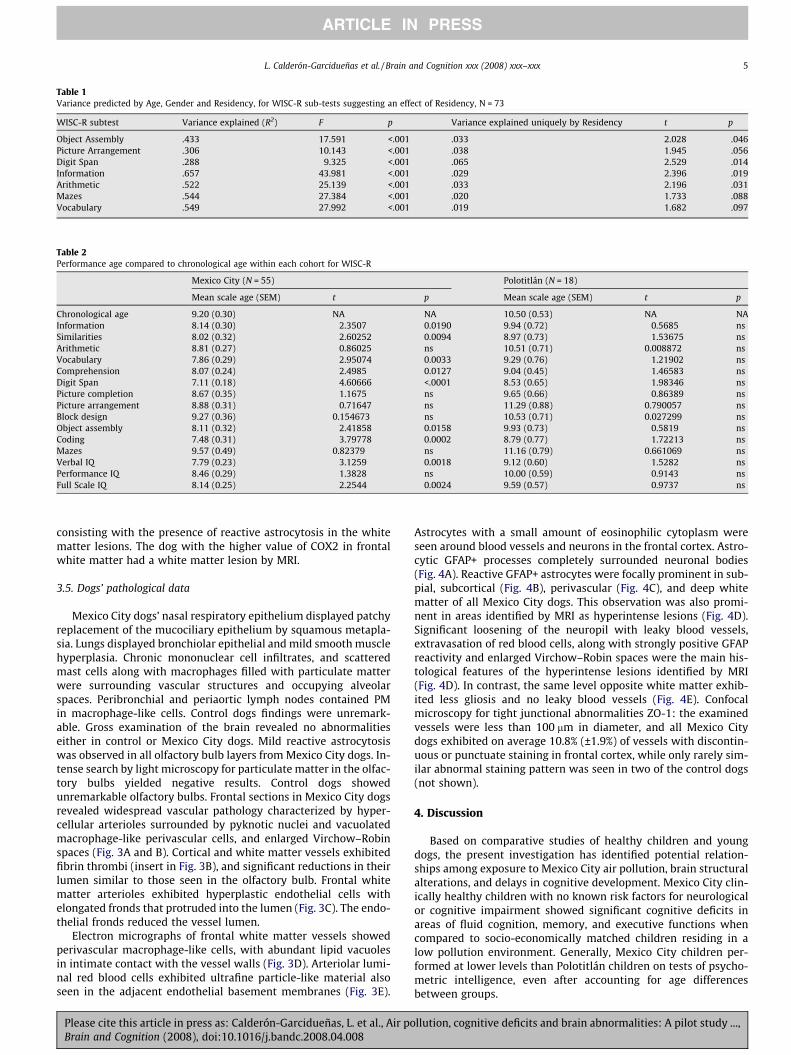
(1) = 8.33, p = .003 (test performed included 13 of 23MC and 1 of 13 Control children with WML). There were no APOE4 children in the Mexico City cohort, or any TLR 4 Asp299Gly car-riers in the Polotitlán group. Two Mexico City children ages 14 and17 had the TLR 4 Asp299Gly polymorphism associated with ablunted response to inhaled lipopolysaccharides. Fig. 1A–C illus-trate an 8-year-old girl with persistent white matter hyperintenseT2 and FLAIR lesions through 11 months of follow-up with sequen-tial MRI. Table 4 illustrates the results of the quantification withthe Kruit load scores of persistent white matter lesions in threeMexico City children that had 3–4 MRI in an 11-month period.
The results in this pilot study have suggested relationships be-tween city of residency and cognitive deficits/delays in children,and a relationship between city of residency and white matter le-sions in children.
3.4. Dog’s MRI and gene expression data
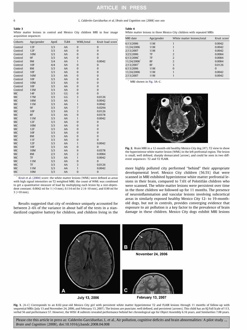
Four of the seven Mexico City dogs (57%) exhibited white mat-ter lesions (WML) in the frontal cortex (Fig. 2), these lesions werevery similar in their characteristics, size and location to the lesionsfound in the children. Table 5 shows the results of the mRNA forCOX2, IL1b and GFAP in samples of frontal cortex white matter inage-matched control versus Mexico City dogs. There was a signifi-cant upregulation of the inflammatory genes: COX2 and IL1bmRNA in Mexico City dogs, while GFAP showed no differences. Ta-ble 6 shows the mRNA data for the frontal white matter contrast-ing the values of COX2, IL1b and GFAP mRNA between the areasidentified as WML by MRI, and the opposite or adjacent anatomicalarea with no apparent lesion by MRI. The four dogs with whitematter lesions exhibited higher values of IL1b and/or COX2 in thesample corresponding to the MRI lesion. In addition, three of fourdogs also showed higher mRNA for the glial acidic fibrillary protein
4 L. Calderón-Garcidueñas et al. / Brain and Cognition xxx (2008) xxx–xxx
ARTICLE IN PRESS
Please cite this article in press as: Calderón-Garcidueñas, L. et al., Air pollution, cognitive deficits and brain abnormalities: A pilot study ...,Brain and Cognition (2008), doi:10.1016/j.bandc.2008.04.008
consisting with the presence of reactive astrocytosis in the whitematter lesions. The dog with the higher value of COX2 in frontalwhite matter had a white matter lesion by MRI.
3.5. Dogs’ pathological data
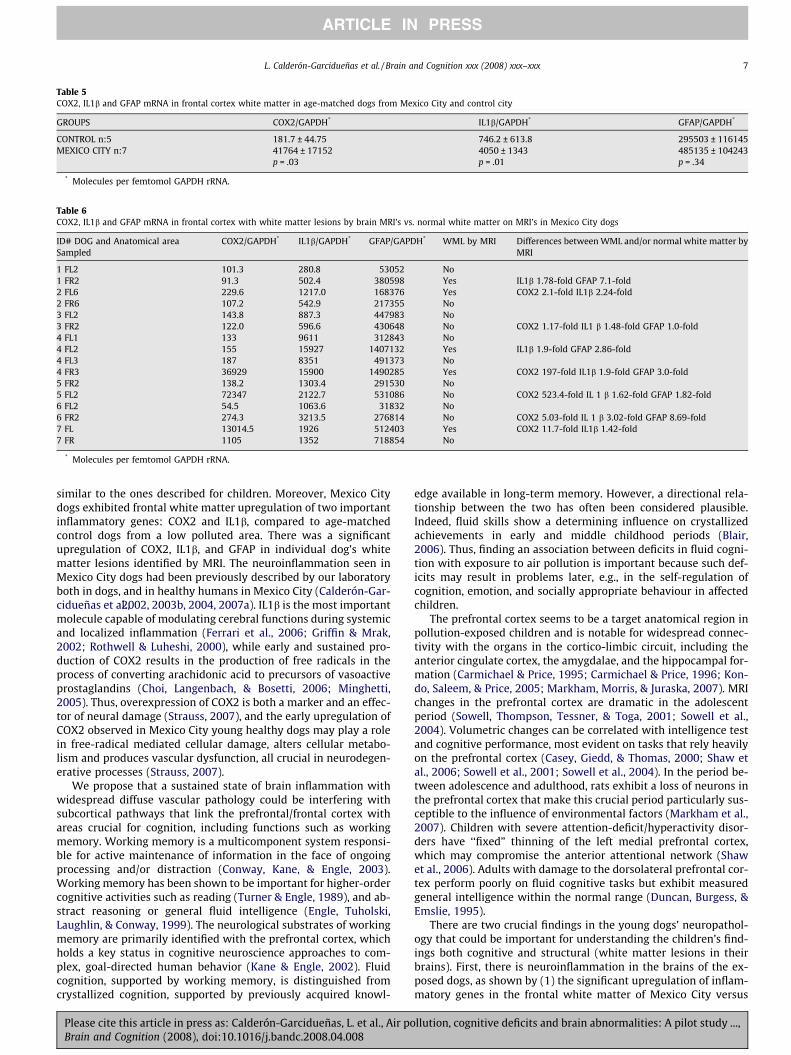
Mexico City dogs’ nasal respiratory epithelium displayed patchyreplacement of the mucociliary epithelium by squamous metapla-sia. Lungs displayed bronchiolar epithelial and mild smooth musclehyperplasia. Chronic mononuclear cell infiltrates, and scatteredmast cells along with macrophages filled with particulate matterwere surrounding vascular structures and occupying alveolarspaces. Peribronchial and periaortic lymph nodes contained PMin macrophage-like cells. Control dogs findings were unremark-able. Gross examination of the brain revealed no abnormalitieseither in control or Mexico City dogs. Mild reactive astrocytosiswas observed in all olfactory bulb layers fromMexico City dogs. In-tense search by light microscopy for particulate matter in the olfac-tory bulbs yielded negative results. Control dogs showedunremarkable olfactory bulbs. Frontal sections in Mexico City dogsrevealed widespread vascular pathology characterized by hyper-cellular arterioles surrounded by pyknotic nuclei and vacuolatedmacrophage-like perivascular cells, and enlarged Virchow–Robinspaces (Fig. 3A and B). Cortical and white matter vessels exhibitedfibrin thrombi (insert in Fig. 3B), and significant reductions in theirlumen similar to those seen in the olfactory bulb. Frontal whitematter arterioles exhibited hyperplastic endothelial cells withelongated fronds that protruded into the lumen (Fig. 3C). The endo-thelial fronds reduced the vessel lumen.
Electron micrographs of frontal white matter vessels showedperivascular macrophage-like cells, with abundant lipid vacuolesin intimate contact with the vessel walls (Fig. 3D). Arteriolar lumi-nal red blood cells exhibited ultrafine particle-like material alsoseen in the adjacent endothelial basement membranes (Fig. 3E).
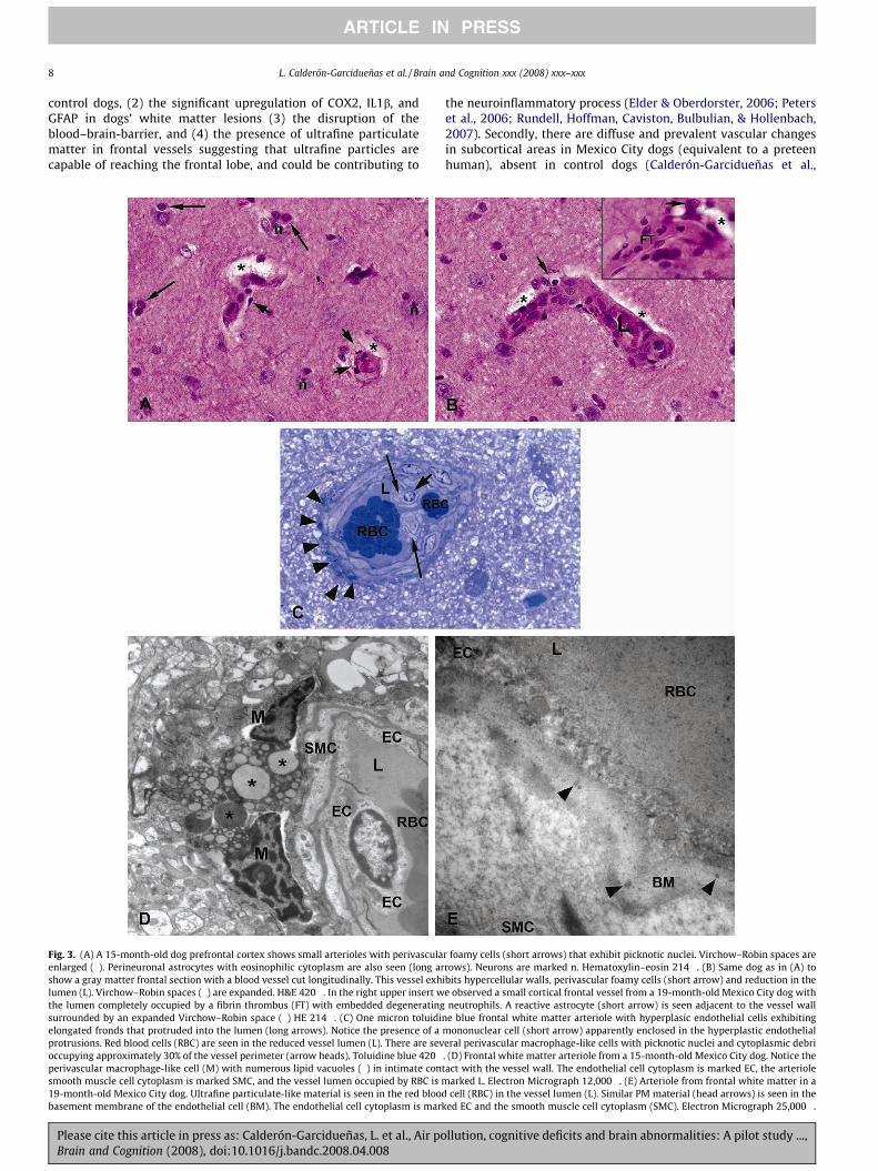
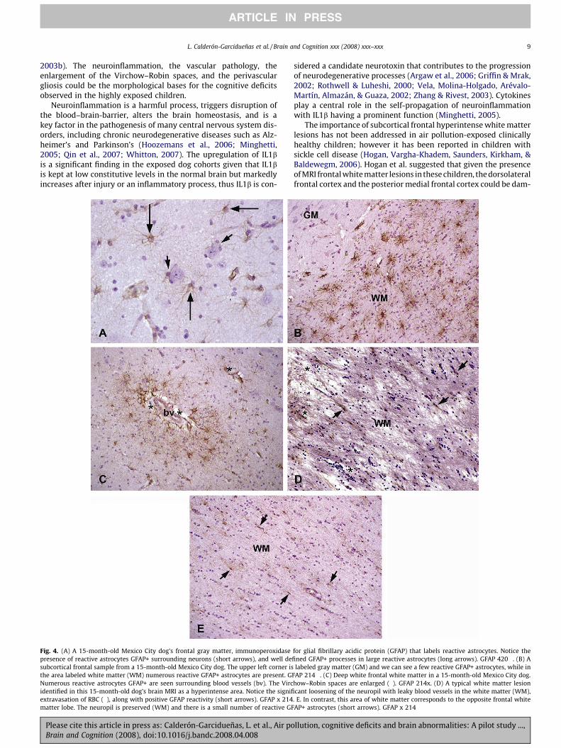
Astrocytes with a small amount of eosinophilic cytoplasm wereseen around blood vessels and neurons in the frontal cortex. Astro-cytic GFAP+ processes completely surrounded neuronal bodies(Fig. 4A). Reactive GFAP+ astrocytes were focally prominent in sub-pial, subcortical (Fig. 4B), perivascular (Fig. 4C), and deep whitematter of all Mexico City dogs. This observation was also promi-nent in areas identified by MRI as hyperintense lesions (Fig. 4D).Significant loosening of the neuropil with leaky blood vessels,extravasation of red blood cells, along with strongly positive GFAPreactivity and enlarged Virchow–Robin spaces were the main his-tological features of the hyperintense lesions identified by MRI(Fig. 4D). In contrast, the same level opposite white matter exhib-ited less gliosis and no leaky blood vessels (Fig. 4E). Confocalmicroscopy for tight junctional abnormalities ZO-1: the examinedvessels were less than 100 lm in diameter, and all Mexico Citydogs exhibited on average 10.8% (±1.9%) of vessels with discontin-uous or punctuate staining in frontal cortex, while only rarely sim-ilar abnormal staining pattern was seen in two of the control dogs(not shown).
4. Discussion
Based on comparative studies of healthy children and youngdogs, the present investigation has identified potential relation-ships among exposure to Mexico City air pollution, brain structuralalterations, and delays in cognitive development. Mexico City clin-ically healthy children with no known risk factors for neurologicalor cognitive impairment showed significant cognitive deficits inareas of fluid cognition, memory, and executive functions whencompared to socio-economically matched children residing in alow pollution environment. Generally, Mexico City children per-formed at lower levels than Polotitlán children on tests of psycho-metric intelligence, even after accounting for age differencesbetween groups.
Table 1Variance predicted by Age, Gender and Residency, for WISC-R sub-tests suggesting an effect of Residency, N = 73
WISC-R subtest Variance explained (R2) F p Variance explained uniquely by Residency t p
Object Assembly .433 17.591 <.001 .033 2.028 .046Picture Arrangement .306 10.143 <.001 .038 1.945 .056Digit Span .288 9.325 <.001 .065 2.529 .014Information .657 43.981 <.001 .029 2.396 .019Arithmetic .522 25.139 <.001 .033 2.196 .031Mazes .544 27.384 <.001 .020 1.733 .088Vocabulary .549 27.992 <.001 .019 1.682 .097
Table 2Performance age compared to chronological age within each cohort for WISC-R
Mexico City (N = 55) Polotitlán (N = 18)
Mean scale age (SEM) t p Mean scale age (SEM) t p
Chronological age 9.20 (0.30) NA NA 10.50 (0.53) NA NAInformation 8.14 (0.30) !2.3507 0.0190 9.94 (0.72) !0.5685 nsSimilarities 8.02 (0.32) !2.60252 0.0094 8.97 (0.73) !1.53675 nsArithmetic 8.81 (0.27) !0.86025 ns 10.51 (0.71) 0.008872 nsVocabulary 7.86 (0.29) !2.95074 0.0033 9.29 (0.76) !1.21902 nsComprehension 8.07 (0.24) !2.4985 0.0127 9.04 (0.45) !1.46583 nsDigit Span 7.11 (0.18) !4.60666 <.0001 8.53 (0.65) !1.98346 nsPicture completion 8.67 (0.35) !1.1675 ns 9.65 (0.66) !0.86389 nsPicture arrangement 8.88 (0.31) !0.71647 ns 11.29 (0.88) 0.790057 nsBlock design 9.27 (0.36) 0.154673 ns 10.53 (0.71) 0.027299 nsObject assembly 8.11 (0.32) !2.41858 0.0158 9.93 (0.73) !0.5819 nsCoding 7.48 (0.31) !3.79778 0.0002 8.79 (0.77) !1.72213 nsMazes 9.57 (0.49) 0.82379 ns 11.16 (0.79) 0.661069 nsVerbal IQ 7.79 (0.23) !3.1259 0.0018 9.12 (0.60) !1.5282 nsPerformance IQ 8.46 (0.29) !1.3828 ns 10.00 (0.59) !0.9143 nsFull Scale IQ 8.14 (0.25) !2.2544 0.0024 9.59 (0.57) !0.9737 ns
L. Calderón-Garcidueñas et al. / Brain and Cognition xxx (2008) xxx–xxx 5
ARTICLE IN PRESS
Please cite this article in press as: Calderón-Garcidueñas, L. et al., Air pollution, cognitive deficits and brain abnormalities: A pilot study ...,Brain and Cognition (2008), doi:10.1016/j.bandc.2008.04.008
Results suggested that city of residence uniquely accounted forbetween 2–6% of the variance in about half of the tests in a stan-dardized cognitive battery for children, and children living in the
more highly polluted city performed ‘‘behind” their appropriatedevelopmental level. Mexico City children (56.5%) that werescanned in MRI exhibited hyperintense white matter prefrontal le-sions in their brain, compared to 7.6% of Polotitlán children whowere scanned. The white matter lesions were persistent over timein the three children we followed up for 11 months. The presenceof neuroinflammation and vascular lesions involving subcorticalareas in similarly exposed healthy Mexico City 12- to 19-month-old dogs, but not in controls, provides converging evidence thatexposure to air pollution is a key factor to the prevalence of braindamage in these children. Mexico City dogs exhibit MRI lesions
Table 3White matter lesions in control and Mexico City children MRI in four imageacquisition sequences
Cohorts Age/gender ApoE TLR4 WML/total Kruit load score*
Control 12F 3/3 AA 0 0Control 12F 3/3 AA 0 0Control 10M 3/3 AA 0 0Control 9F 3/3 AA 0 0Control 9M 3/4 AA 1 0.0042Control 10F 4/4 AA 0 0Control 8M 3/3 AA 0 0Control 10F 3/3 AA 0 0Control 16M 3/3 AA 0 0Control 10F 3/3 AA 0 0Control 10M 3/3 AA 0 0Control 10F 3/3 AA 0 0Control 13M 3/3 AA 0 0MC 14F 3/3 GG 0 0MC 17M 3/3 GG 3 0.0126MC 18M 3/3 AA 1 0.0042MC 11M 3/3 AA 1 0.0042MC 9F 3/3 AA 7 0.0294MC 10F 3/3 AA 3 0.0126MC 8F 3/3 AA 9 0.0378MC 11M 3/3 AA 1 0.0042MC 12F 3/3 AA 0 0MC 10M 3/3 AA 0 0MC 12F 3/3 AA 0 0MC 10F 3/3 AA 0 0MC 8M 3/3 AA 0 0MC 11F 3/3 AA 0 0MC 12F 3/3 AA 1 0.0042MC 10F 3/3 AA 0 0MC 10M 3/3 AA 9 0.0378MC 8M 2/3 AA 2 0.0084MC 7F 3/3 AA 1 0.0042MC 11M 3/3 AA 0 0MC 7F 3/3 AA 3 0.0126MC 11M 3/3 AA 1 0.0042MC 10M 3/3 AA 0 0
* Kruit et al. (2004) score: the white matter lesions (WML) were defined as areaswith high signal intensities on T2 weighted MRI; the count of WML was combinedto get a quantitative measure of load by multiplying each lesion by a size-depen-dent constant: 0.0042 ml for 1 (<3 mm), 0.114 ml for 2 (4–10 mm), and 0.90 ml for3 (>10 mm).
Fig. 1. (A–C) Corresponds to an 8.02-year-old Mexico City girl with persistent white matter hyperintense T2 and FLAIR lesions through 11 months of follow-up withsequential MRIs (July 13 and November 24, 2006, and February 13, 2007). The lesions are punctate, well defined, and persistent (arrows). This child has an IQ Full Scale of 113,verbal 56 and performance 57. However, the WISC-R subtests revealed performance behind her chronological age for Object Assembly 6.16 years, and Similarities 7.08 years.
Table 4White matter lesions in three Mexico City children with repeated MRIs
MRI date Age/gender White matter lesions/total Kruit score*
6/13/2006 11M 1 0.004211/24/2006 11M 1 0.00422/13/2007 11M 1 0.00423/23/2006 7F 2 0.00846/13/2006* 7F 2 0.008411/24/2006* 8F 2 0.00842/13/2007* 8F 3 0.01266/13/2006 11M 0 011/24/2006 11M 1 0.00422/13/2007 11M 1 0.0042
* MRI shown in Fig. 3A–C.
Fig. 2. Brain MRI in a 12-month-old healthy Mexico City dog (#7). T2 view to showthe hyperintense white matter lesion (WML) in the left prefrontal region. The lesionis small, well defined, sharply demarcated (arrow), and could be seen in two diff-erent sequences: T2 and T2 FLAIR.
6 L. Calderón-Garcidueñas et al. / Brain and Cognition xxx (2008) xxx–xxx
ARTICLE IN PRESS
Please cite this article in press as: Calderón-Garcidueñas, L. et al., Air pollution, cognitive deficits and brain abnormalities: A pilot study ...,Brain and Cognition (2008), doi:10.1016/j.bandc.2008.04.008
similar to the ones described for children. Moreover, Mexico Citydogs exhibited frontal white matter upregulation of two importantinflammatory genes: COX2 and IL1b, compared to age-matchedcontrol dogs from a low polluted area. There was a significantupregulation of COX2, IL1b, and GFAP in individual dog’s whitematter lesions identified by MRI. The neuroinflammation seen inMexico City dogs had been previously described by our laboratoryboth in dogs, and in healthy humans in Mexico City (Calderón-Gar-cidueñas et al.,2002, 2003b, 2004, 2007a). IL1b is the most importantmolecule capable of modulating cerebral functions during systemicand localized inflammation (Ferrari et al., 2006; Griffin & Mrak,2002; Rothwell & Luheshi, 2000), while early and sustained pro-duction of COX2 results in the production of free radicals in theprocess of converting arachidonic acid to precursors of vasoactiveprostaglandins (Choi, Langenbach, & Bosetti, 2006; Minghetti,2005). Thus, overexpression of COX2 is both a marker and an effec-tor of neural damage (Strauss, 2007), and the early upregulation ofCOX2 observed in Mexico City young healthy dogs may play a rolein free-radical mediated cellular damage, alters cellular metabo-lism and produces vascular dysfunction, all crucial in neurodegen-erative processes (Strauss, 2007).
We propose that a sustained state of brain inflammation withwidespread diffuse vascular pathology could be interfering withsubcortical pathways that link the prefrontal/frontal cortex withareas crucial for cognition, including functions such as workingmemory. Working memory is a multicomponent system responsi-ble for active maintenance of information in the face of ongoingprocessing and/or distraction (Conway, Kane, & Engle, 2003).Working memory has been shown to be important for higher-ordercognitive activities such as reading (Turner & Engle, 1989), and ab-stract reasoning or general fluid intelligence (Engle, Tuholski,Laughlin, & Conway, 1999). The neurological substrates of workingmemory are primarily identified with the prefrontal cortex, whichholds a key status in cognitive neuroscience approaches to com-plex, goal-directed human behavior (Kane & Engle, 2002). Fluidcognition, supported by working memory, is distinguished fromcrystallized cognition, supported by previously acquired knowl-
edge available in long-term memory. However, a directional rela-tionship between the two has often been considered plausible.Indeed, fluid skills show a determining influence on crystallizedachievements in early and middle childhood periods (Blair,2006). Thus, finding an association between deficits in fluid cogni-tion with exposure to air pollution is important because such def-icits may result in problems later, e.g., in the self-regulation ofcognition, emotion, and socially appropriate behaviour in affectedchildren.
The prefrontal cortex seems to be a target anatomical region inpollution-exposed children and is notable for widespread connec-tivity with the organs in the cortico-limbic circuit, including theanterior cingulate cortex, the amygdalae, and the hippocampal for-mation (Carmichael & Price, 1995; Carmichael & Price, 1996; Kon-do, Saleem, & Price, 2005; Markham, Morris, & Juraska, 2007). MRIchanges in the prefrontal cortex are dramatic in the adolescentperiod (Sowell, Thompson, Tessner, & Toga, 2001; Sowell et al.,2004). Volumetric changes can be correlated with intelligence testand cognitive performance, most evident on tasks that rely heavilyon the prefrontal cortex (Casey, Giedd, & Thomas, 2000; Shaw etal., 2006; Sowell et al., 2001; Sowell et al., 2004). In the period be-tween adolescence and adulthood, rats exhibit a loss of neurons inthe prefrontal cortex that make this crucial period particularly sus-ceptible to the influence of environmental factors (Markham et al.,2007). Children with severe attention-deficit/hyperactivity disor-ders have ‘‘fixed” thinning of the left medial prefrontal cortex,which may compromise the anterior attentional network (Shawet al., 2006). Adults with damage to the dorsolateral prefrontal cor-tex perform poorly on fluid cognitive tasks but exhibit measuredgeneral intelligence within the normal range (Duncan, Burgess, &Emslie, 1995).
There are two crucial findings in the young dogs’ neuropathol-ogy that could be important for understanding the children’s find-ings both cognitive and structural (white matter lesions in theirbrains). First, there is neuroinflammation in the brains of the ex-posed dogs, as shown by (1) the significant upregulation of inflam-matory genes in the frontal white matter of Mexico City versus
Table 5COX2, IL1b and GFAP mRNA in frontal cortex white matter in age-matched dogs from Mexico City and control city
GROUPS COX2/GAPDH* IL1b/GAPDH* GFAP/GAPDH*
CONTROL n:5 181.7 ± 44.75 746.2 ± 613.8 295503 ± 116145MEXICO CITY n:7 41764 ± 17152
p = .034050 ± 1343p = .01
485135 ± 104243p = .34
* Molecules per femtomol GAPDH rRNA.
Table 6COX2, IL1b and GFAP mRNA in frontal cortex with white matter lesions by brain MRI’s vs. normal white matter on MRI’s in Mexico City dogs
ID# DOG and Anatomical areaSampled
COX2/GAPDH* IL1b/GAPDH* GFAP/GAPDH* WML by MRI Differences betweenWML and/or normal white matter byMRI
1 FL2 101.3 280.8 53052 No1 FR2 91.3 502.4 380598 Yes IL1b 1.78-fold GFAP 7.1-fold2 FL6 229.6 1217.0 168376 Yes COX2 2.1-fold IL1b 2.24-fold2 FR6 107.2 542.9 217355 No3 FL2 143.8 887.3 447983 No3 FR2 122.0 596.6 430648 No COX2 1.17-fold IL1 b 1.48-fold GFAP 1.0-fold4 FL1 133 9611 312843 No4 FL2 155 15927 1407132 Yes IL1b 1.9-fold GFAP 2.86-fold4 FL3 187 8351 491373 No4 FR3 36929 15900 1490285 Yes COX2 197-fold IL1b 1.9-fold GFAP 3.0-fold5 FR2 138.2 1303.4 291530 No5 FL2 72347 2122.7 531086 No COX2 523.4-fold IL 1 b 1.62-fold GFAP 1.82-fold6 FL2 54.5 1063.6 31832 No6 FR2 274.3 3213.5 276814 No COX2 5.03-fold IL 1 b 3.02-fold GFAP 8.69-fold7 FL 13014.5 1926 512403 Yes COX2 11.7-fold IL1b 1.42-fold7 FR 1105 1352 718854 No
* Molecules per femtomol GAPDH rRNA.
L. Calderón-Garcidueñas et al. / Brain and Cognition xxx (2008) xxx–xxx 7
ARTICLE IN PRESS
Please cite this article in press as: Calderón-Garcidueñas, L. et al., Air pollution, cognitive deficits and brain abnormalities: A pilot study ...,Brain and Cognition (2008), doi:10.1016/j.bandc.2008.04.008
control dogs, (2) the significant upregulation of COX2, IL1b, andGFAP in dogs’ white matter lesions (3) the disruption of theblood–brain-barrier, and (4) the presence of ultrafine particulatematter in frontal vessels suggesting that ultrafine particles arecapable of reaching the frontal lobe, and could be contributing to
the neuroinflammatory process (Elder & Oberdorster, 2006; Peterset al., 2006; Rundell, Hoffman, Caviston, Bulbulian, & Hollenbach,2007). Secondly, there are diffuse and prevalent vascular changesin subcortical areas in Mexico City dogs (equivalent to a preteenhuman), absent in control dogs (Calderón-Garcidueñas et al.,
Fig. 3. (A) A 15-month-old dog prefrontal cortex shows small arterioles with perivascular foamy cells (short arrows) that exhibit picknotic nuclei. Virchow–Robin spaces areenlarged ("). Perineuronal astrocytes with eosinophilic cytoplasm are also seen (long arrows). Neurons are marked n. Hematoxylin–eosin 214#. (B) Same dog as in (A) toshow a gray matter frontal section with a blood vessel cut longitudinally. This vessel exhibits hypercellular walls, perivascular foamy cells (short arrow) and reduction in thelumen (L). Virchow–Robin spaces (") are expanded. H&E 420#. In the right upper insert we observed a small cortical frontal vessel from a 19-month-old Mexico City dog withthe lumen completely occupied by a fibrin thrombus (FT) with embedded degenerating neutrophils. A reactive astrocyte (short arrow) is seen adjacent to the vessel wallsurrounded by an expanded Virchow–Robin space (") HE 214#. (C) One micron toluidine blue frontal white matter arteriole with hyperplasic endothelial cells exhibitingelongated fronds that protruded into the lumen (long arrows). Notice the presence of a mononuclear cell (short arrow) apparently enclosed in the hyperplastic endothelialprotrusions. Red blood cells (RBC) are seen in the reduced vessel lumen (L). There are several perivascular macrophage-like cells with picknotic nuclei and cytoplasmic debrioccupying approximately 30% of the vessel perimeter (arrow heads). Toluidine blue 420#. (D) Frontal white matter arteriole from a 15-month-old Mexico City dog. Notice theperivascular macrophage-like cell (M) with numerous lipid vacuoles (") in intimate contact with the vessel wall. The endothelial cell cytoplasm is marked EC, the arteriolesmooth muscle cell cytoplasm is marked SMC, and the vessel lumen occupied by RBC is marked L. Electron Micrograph 12,000#. (E) Arteriole from frontal white matter in a19-month-old Mexico City dog. Ultrafine particulate-like material is seen in the red blood cell (RBC) in the vessel lumen (L). Similar PM material (head arrows) is seen in thebasement membrane of the endothelial cell (BM). The endothelial cell cytoplasm is marked EC and the smooth muscle cell cytoplasm (SMC). Electron Micrograph 25,000#.
8 L. Calderón-Garcidueñas et al. / Brain and Cognition xxx (2008) xxx–xxx
ARTICLE IN PRESS
Please cite this article in press as: Calderón-Garcidueñas, L. et al., Air pollution, cognitive deficits and brain abnormalities: A pilot study ...,Brain and Cognition (2008), doi:10.1016/j.bandc.2008.04.008
2003b). The neuroinflammation, the vascular pathology, theenlargement of the Virchow–Robin spaces, and the perivasculargliosis could be the morphological bases for the cognitive deficitsobserved in the highly exposed children.
Neuroinflammation is a harmful process, triggers disruption ofthe blood–brain-barrier, alters the brain homeostasis, and is akey factor in the pathogenesis of many central nervous system dis-orders, including chronic neurodegenerative diseases such as Alz-heimer’s and Parkinson’s (Hoozemans et al., 2006; Minghetti,2005; Qin et al., 2007; Whitton, 2007). The upregulation of IL1bis a significant finding in the exposed dog cohorts given that IL1bis kept at low constitutive levels in the normal brain but markedlyincreases after injury or an inflammatory process, thus IL1b is con-
sidered a candidate neurotoxin that contributes to the progressionof neurodegenerative processes (Argaw et al., 2006; Griffin & Mrak,2002; Rothwell & Luheshi, 2000; Vela, Molina-Holgado, Arévalo-Martín, Almazán, & Guaza, 2002; Zhang & Rivest, 2003). Cytokinesplay a central role in the self-propagation of neuroinflammationwith IL1b having a prominent function (Minghetti, 2005).
The importance of subcortical frontal hyperintensewhitematterlesions has not been addressed in air pollution-exposed clinicallyhealthy children; however it has been reported in children withsickle cell disease (Hogan, Vargha-Khadem, Saunders, Kirkham, &Baldewegm, 2006). Hogan et al. suggested that given the presenceofMRI frontalwhitematter lesions in thesechildren, thedorsolateralfrontal cortex and the posteriormedial frontal cortex could be dam-
Fig. 4. (A) A 15-month-old Mexico City dog’s frontal gray matter, immunoperoxidase for glial fibrillary acidic protein (GFAP) that labels reactive astrocytes. Notice thepresence of reactive astrocytes GFAP+ surrounding neurons (short arrows), and well defined GFAP+ processes in large reactive astrocytes (long arrows). GFAP 420#. (B) Asubcortical frontal sample from a 15-month-old Mexico City dog. The upper left corner is labeled gray matter (GM) and we can see a few reactive GFAP+ astrocytes, while inthe area labeled white matter (WM) numerous reactive GFAP+ astrocytes are present. GFAP 214#. (C) Deep white frontal white matter in a 15-month-old Mexico City dog.Numerous reactive astrocytes GFAP+ are seen surrounding blood vessels (bv). The Virchow–Robin spaces are enlarged ("). GFAP 214x. (D) A typical white matter lesionidentified in this 15-month-old dog’s brain MRI as a hyperintense area. Notice the significant loosening of the neuropil with leaky blood vessels in the white matter (WM),extravasation of RBC ("), along with positive GFAP reactivity (short arrows). GFAP x 214. E. In contrast, this area of white matter corresponds to the opposite frontal whitematter lobe. The neuropil is preserved (WM) and there is a small number of reactive GFAP+ astrocytes (short arrows). GFAP x 214
L. Calderón-Garcidueñas et al. / Brain and Cognition xxx (2008) xxx–xxx 9
ARTICLE IN PRESS
Please cite this article in press as: Calderón-Garcidueñas, L. et al., Air pollution, cognitive deficits and brain abnormalities: A pilot study ...,Brain and Cognition (2008), doi:10.1016/j.bandc.2008.04.008
aged, thus a disruption to event-related potential markers couldindicate cortical disconnection in frontal areas. Children with sicklecell disease have a brain vasculopathy related to their abnormalhemoglobin, and thusavascular component is thekey for theirwhitematter lesions.Whitematter lesions in adults and elderly people areassociated with cognitive dysfunction, gait abnormalities, falls, anddepression (Breteleret al., 1994; Longstrethet al., 1996;vanStraatenet al., 2006).Whitematter lesions impair frontal lobe function (Tull-berg et al., 2004), and indicate abnormalities of the subcortical fibersystem (Gootjes et al., 2004). Histopathology correlates in the el-derly include enlarged Virchow–Robin spaces, degeneration of ax-ons and myelin, and gliosis (Scarpelli et al., 1994; Wahlund et al.,2001). Progressive white matter hyperintensities in older adultsare related to changes in regional cerebral blood flow (Kraut, Bea-son-Held, Elkins, & Resnick, 2008). Vascular changes predominantlyinvolving subcortical areas (e.g., white matter, basal ganglia, thala-mus, and hippocampus) are seen in both Alzheimer’s and vasculardementia patients, and inmixed dementia cases (Jellinger & Attems,2007). The commondenominator for the hyperintensewhitematterlesions detected by brain MRI appears to be a vascular lesion withperivascular gliosis and enlarged Virchow–Robin spaces. Based onour pilot results we suggest that the white matter lesions detectedin 56.5% of Mexico City children tested represent the extreme ofthe vascular lesions with a breakdown of the blood–brain-barrier,and that the vascular pathology is likely of a diffuse nature as seenin the young dogs.
In summary, clinically healthy children with no known risk fac-tors for neurological or cognitive disorders residing in a highly pol-luted urban environment exhibited deficits in fluid cognition,memory, and executive functions, relative to children living in aless polluted urban environment. In parallel, a number of the chil-dren living in the highly polluted city exhibited white matterhyperintense lesions in MRI, while only one child living in the lesspolluted city exhibited a single lesion. These white matter lesionsalong with the presence of more diffuse vascular pathology arelikely to interrupt subcortical prefrontal cortex connections andother white matter tracts, potentially contributing to the cognitivedysfunction. Ultrafine particulate matter reaching the frontal cor-tex in the highly exposed dogs and in young Mexico City adults(Calderón-Garcidueñas et al., 2008) is likely contributing to theneuroinflammation.
In the USA 158 million people live in areas where ozone exceedsthe 8 h standard, 29 million are exposed to PM10 and 88 million toPM2.5 (U.S.Environmental Protection Agency. Green Book: Particu-late Matter Nonattainment Area, E2004). Urban sites with high PMcontributions from vehicles and industry (Seagrave et al., 2006) arehighly visible polluted sources, although indoor pollution is alsovery important particularly for disadvantage populations living inhigh-density multiunit dwellings in large urban areas (Baxter,Clougherty, Laden, & Levy, 2007). The issue of air pollution causingcognitive deficits and brain structural changes in healthy childrenwith all their potential consequences ought to be of major publicimportance. Alterations in measures of fluid intelligence and cogni-tive control predict school performance, complex learning, abilityto control attention and avoid distraction, reading and listeningcomprehension, reasoning, and of key importance from the socialpoint of view: the ability to block impulsive anti-social behavior.
MRI techniques have made it possible to study structural andmetabolic brain development across age groups, including children(Evans, 2006), and regional white matter alterations are related todecreases in performance on neuropsychological tests (Brickmanet al., 2006). Thus, as suggested by the interdisciplinary methodol-ogy of the present investigation, it is possible to comprehensivelyexamine brain structural changes and to determine the interrela-tionships between age, white matter changes, hippocampal mea-surements, cognitive function, and the cumulative doses of air
pollutants. The work here presents a pioneering attempt to answersuch urgent questions, and initial findings give encouragementthat future, large-scale work in this area has great potential to givemuch-needed answers.
References
Argaw, A. T., Zhang, Y., Snyder, B. J., Zhao, M. L., Kopp, N., Lee, S. C., et al. (2006). IL-1beta regulates blood–brain-barrier permeability via reactivation of thehypoxia-angiogenesis program. Journal of Immunology, 177, 5574–5584.
Baxter, L. K., Clougherty, J. E., Laden, F., & Levy, J. I. (2007). Predictors ofconcentrations of nitrogen dioxide, fine particulate matter, and particleconstituents inside of lower socioeconomic status urban homes. Journal ofExposure Science and Environmental Epidemiology, 17, 433–444.
Blair, C. (2006). How similar are fluid cognition and general intelligence? Adevelopmental neuroscience perspective on fluid cognition as an aspect ofhuman cognitive ability. The Behavioral and Brain Sciences, 29, 109–125.
Bonner, J. C., Rice, A. B., Lindroos, P. M., O’Brien, P. O., Dreher, K. L., Rosas, I., et al.(1998). Induction of the lung myofibroblast PDGF receptor system by urbanambient particles from Mexico City. American Journal of Respiratory Cell andMolecular Biology, 19, 672–680.
Boretius, S., Schmelting, B., Watanabe, T., Merkler, D., Tammer, R., Czéh, B., et al.(2006). Monitoring of EAE onset and progression in the common marmosetmonkey by sequential high-resolution 3D MRI. NMR in Biomedicine, 19, 41–49.
Bravo, H. A., & Torres, R. J. (2002). Air Pollution levels and trends in the Mexico Citymetropolitan area. In M. Fenn, L. Bauer, & T. Hernández (Eds.), Urban airpollution and forests: Resources at risk in the Mexico City Air Basin (pp. 121–159).New York: Springer-Verlag.
Breteler, M. M., van Swieten, J. C., Bots, M. L., Grobbee, D. E., Claus, J. J., van den Hout,J. H., et al. (1994). Cerebral white matter lesions, vascular risk factors, andcognitive function in a population-based study: The Rotterdam Study.Neurology, 44, 1246–1252.
Brickman, A. M., Zimmerman, M. E., Paul, R. H., Grieve, S. M., Tate, D. F., Cohen, R. A.,et al. (2006). Regional white matter and neuropsychological functioning acrossthe adult lifespan. Biological Psychiatry, 60, 444–453.
Calderón-Garcidueñas, L., Rodriguez-Alcaraz, A., Valencia-Salazar, G., Mora-Tiscareño, A., Garcia, R., Osnaya, N., et al. (2001). Nasal biopsies of childrenexposed to air pollutants. Toxicologic Pathology, 29, 558–564.
Calderón-Garcidueñas, L., Azzarelli, B., Acuña, H., Garcia, R., Gambling, T. M., Osnaya,N., et al. (2002). Air pollution and brain damage. Toxicologic Pathology, 30,373–389.
Calderón-Garcidueñas, L., Mora-Tiscareño, A., Fordham, L. A., Valencia-Salazar, G.,Chung, C. J., Rodriguez-Alcaraz, A., et al. (2003a). Respiratory damage in childrenexposed to urban pollution. Pediatric Pulmonology, 36, 148–161.
Calderón-Garcidueñas, L., Maronpot, R. R., Torres-Jardón, R., Henríquez-Roldán, C.,Schoonhoven, R., Acuña-Ayala, H., et al. (2003b). DNA damage in nasal and braintissues of canines exposed to air pollutants is associated with evidence ofchronic brain inflammation and neurodegeneration. Toxicologic Pathology, 31,524–538.
Calderón-Garcidueñas, L., Reed, W., Maronpot, R. R., Henriquez-Roldan, C., Delgado-Chavez, R., Calderón-Garcidueñas, A., et al. (2004). Brain inflammation andAlzheimer’s-like pathology in individuals exposed to severe air pollution.Toxicologic Pathology, 32, 650–658.
Calderón-Garcidueñas, L., Mora-Tiscareño, A., Fordham, L. A., Chung, C. J., Valencia-Salazar, G., Flores-Gomez, S., et al. (2006). Lung radiology and pulmonaryfunction of children chronically exposed to air pollution. Environmental HealthPerspectives, 114, 1432–1437.
Calderón-Garcidueñas, L., Franco-Lira, M., Torres-Jardón, R., Henríquez-Roldán, C.,Barragan-Mejia, G., Valencia-Salazar, G., et al. (2007a). Pediatric respiratory andsystemic effects of chronic air pollution exposure: Nose, lung, heart, and brainpathology. Toxicologic Pathology, 15, 1–9.
Calderón-Garcidueñas, L., Vincent, R., Mora-Tiscareño, A., Franco-Lira, M.,Henríquez-Roldán, C., Barragán-Mejía, G., et al. (2007b). Elevated plasmaendothelin-1 and pulmonary arterial pressure in children exposed to airpollution. Environmental Health Perspectives, 115, 1248–1253.
Calderón-Garcidueñas, L., Solt, A. C., Henríquez-Roldán C Torres-Jardón, R., Nuse, B.,Herritt, L., et al. (2008). Long-term air pollution exposure is associated withneuroinflammation, an altered innate immune response, disruption of theblood–brain-barrier, ultrafine particulate deposition, and accumulation ofamyloid b42 and a synuclein in children and young adults. ToxicologicPathology, 36, 289–310.
Carmichael, S. T., & Price, J. L. (1995). Sensory and premotor connections of theorbital and medial prefrontal cortex of macaque monkeys. Journal ofComparative Neurology, 363, 642–664.
Carmichael, S. T., & Price, J. L. (1996). Connectional networks within the orbital andmedial prefrontal cortex of macaque monkeys. Journal of ComparativeNeurology, 371, 179–207.
Casey, B. J., Giedd, J. N., & Thomas, K. M. (2000). Structural and functional braindevelopment and its relation to cognitive development. Biological Psychology,54, 241–257.
Chauhan, V., Breznan, D., Thomson, E., Karthikeyan, S., & Vincent, R. (2005). Effectsof ambient air particles on the endothelin system in human pulmonaryepithelial cells (A549). Cell Biology and Toxicology, 21, 191–205.
10 L. Calderón-Garcidueñas et al. / Brain and Cognition xxx (2008) xxx–xxx
ARTICLE IN PRESS
Please cite this article in press as: Calderón-Garcidueñas, L. et al., Air pollution, cognitive deficits and brain abnormalities: A pilot study ...,Brain and Cognition (2008), doi:10.1016/j.bandc.2008.04.008
Conway, A. R. A., Kane, M. J., & Engle, R. W. (2003). Working memory capacity andits relation to general intelligence. Trends in Cognitive Sciences, 7, 547–552.
Choi, S. H., Langenbach, R., & Bosetti, F. (2006). Cyclooxygenase-1 and 2 enzymesdifferentially regulate the brain upstream NF-jB pathway and downstreamenzymes involved in prostaglandin biosynthesis. Journal of Neurochemistry, 98,801–811.
Dorado-Martinez, C., Paredes-Carbajal, C., Mascher, D., Borgonio-Perez, G., & Rivas-Arancibia, S. (2001). Effects of different ozone doses on memory, motor activityand lipid peroxidation levels, in rats. International Journal of Neurosciences, 108,149–161.
Duncan, J., Burgess, P., & Emslie, H. (1995). Fluid intelligence after frontal lobelesions. Neuropsychologia, 33, 261–268.
Elder, A., & Oberdorster, G. (2006). Translocation and effects of ultrafine particlesoutside the lung. Clinics in Occupational and Environmental Medicine, 5, 785–796.
Engle, R. W., Tuholski, S. W., Laughlin, J. E., & Conway, A. R. (1999). Workingmemory, short-term memory, and general fluid intelligence: A latent-variableapproach. Journal of Experimental Psychology: General, 128, 309–331.
Estrada-García, T., Cerna, J. F., Thompson, M. R., & Lopez-Saucedo, C. (2002). Faecalcontamination and enterotoxigenic Escherichia coli in street-vended chili saucesin Mexico and its public health relevance. Epidemiology and Infection, 129,223–226.
Evans, A. C. (2006). The NIH MRI study of normal brain development. Neuroimage,30, 184–202.
Ferrari, C. C., Pott-Godoy, M. C., Tarelli, R., Chertoff, M., Depino, A. M., & Pitossi, F. J.(2006). Progressive neurodegeneration and motor disabilities induced bychronic expression of IL-1 beta in the substantia nigrae. Neurobiology ofDisease, 24, 183–193.
Garantziotis, S., Hollingsworth, J. W., Zaas, A. K., & Schwartz, D. A. (2008). The effectof Toll-like receptors and Toll-like receptor genetics in human disease. AnnualReviews of Medicine, 59, 343–359.
Gootjes, L., Teipel, S. J., Zebuhr, Y., Schwarz, R., Leinsinger, G., Scheltens, P., et al.(2004). Regional distribution of white matter hyperintensities in vasculardementia, Alzheimer’s disease and healthy aging. Dementia and GeriatricCognitive Disorders, 18, 180–188.
Gozal, D., Capdevila, O. S., Kheirandish-Gozai, L., & Crabtree, V. M. (2007). APOEepsilon 4 allele, cognitive dysfunction, and obstructive sleep apnea in children.Neurology, 69, 243–249.
Griffin, W. S., & Mrak, R. E. (2002). Interleukin-1 in the genesis and progression ofand risk for development of neuronal degeneration in Alzheimer’s disease.Journal of Leukocyte Biology, 72, 233–238.
Hixson, J. E., & Vernier, D. T. (1990). Restriction isotyping of human apolipoprotein Eby gene amplification and cleavage with HhaI. Journal of Lipid Research, 31,545–548.
Hogan, A. M., Vargha-Khadem, F., Saunders, D. E., Kirkham, F. J., & Baldewegm, T.(2006). Impact of frontal white matter lesions on performance monitoring: ERPevidence for cortical disconnection. Brain, 129, 2177–2188.
Hong, Y. C., Lee, J. T., Kim, H., Ha, E. H., Schwartz, J., & Christiani, D. C. (2002). Effectsof air pollutants on acute stroke mortality. Environmental Health Perspectives,110, 187–191.
Hoozemans, J. J., Veerhuis, R., Rozemuller, J. M., & Eikelenboom, P. (2006).Neuroinflammation and regeneration in the early stages of Alzheimer’sdisease pathology. International Journal of Developmental Neurosciences, 24,157–165.
Jellinger, K. A., & Attems, J. (2007). Neuropathological evaluation of mixeddementia. Journal of the Neurological Sciences, 257, 80–87.
Kane, M. J., & Engle, R. W. (2002). The role of prefrontal cortex in working-memorycapacity, executive attention, and general fluid intelligence: An individual-differences perspective. Psychonomic Bulletin & Review, 9, 637–671.
Kraut, M. A., Beason-Held, L. L., Elkins, W. D., & Resnick, S. M. (2008). The impact ofmagnetic resonance imaging-detected white matter hyperintensities onlongitudinal changes in regional cerebral blood flow. Journal of Cerebral BloodFlow Metabolism, 28, 190–197.
Kruit, M. C., van Buchem, M. A., Hofman, P. A. M., Bakkers, J. T. N., Terwindt, G. M.,Ferrari, M. D., et al. (2004). Migraine as a risk factor for subclinical brain lesions.Journal of the American Medical Association, 291, 427–434.
Kondo, H., Saleem, K. S., & Price, J. L. (2005). Differential connections of theperirhinal and parahippocampal cortex with the orbital and medial prefrontalnetworks in macaque monkeys. Journal of Comparative Neurology, 493, 479–509.
Longstreth, W. T., Jr., Manolio, T. A., Arnoldm, A., Burke, G. L., Bryan, N., Jungreis, C.A., et al. (1996). Clinical correlates of white matter findings on cranial magneticresonance imaging of 3301 elderly people. The Cardiovascular Health Study.Stroke, 27, 1274–1282.
Maheswaran, R., Pearson, T., Campbell, M. J., Haining, R. P., McLeod, C. W., Smeeton,N., et al. (2006). A protocol for investigation of the effects of outdoor airpollution on stroke incidence, phenotypes and survival using the South LondonStroke Register. International Journal of Health Geographics, 5, 10.
Mahley, R. W., Weisgraber, K. H., & Huang, Y. (2006). Apolipoprotein E4: A causativefactor and therapeutic target in neuropathology, including Alzheimer’s disease.Proceedings of the National Academy of Sciences, 103, 5644–5651.
McCarron, R. M., Chen, Y., Tomori, T., Strasser, A., Mechoulam, R., Shohami, E., et al.(2006). Endothelial-mediated regulation of cerebral microcirculation. Journal ofPhysiology and Pharmacology, 57(Suppl 11), 133–144.
Markham, J. A., Morris, J. R., & Juraska, J. M. (2007). Neuron number decreases in therat ventral, but not dorsal medial prefrontal cortex between adolescence andadulthood. Neurosciences, 144, 961–968.
Minghetti, L. (2005). Role of inflammation in neurodegenerative disease. CurrentOpinions in Neurology, 18, 315–321.
Osornio-Vargas, A. R., Bonner, J. C., Alfaro-Moreno, E., Martinez, L., Garcia-Cuellar, C.,Ponce-de-Leon-Rosales, S., et al. (2003). Proinflammatory and cytotoxic effectsof Mexico City air pollution particulate matter in vitro are dependent on particlesize and composition. Environmental Health Perspectives, 111, 1289–1293.
Packard, C. J., Westendorp, R. G., Stott, D. J., Caslake, M. J., Murray, H. M., Shepherd, J.,et al.Prospective study of Pravastatin in the elderly risk group. (2007).Association between apolipoprotein E4 and cognitive decline in elderlyadults. Journal of the American Geriatrics Society, 55, 1777–1785.
Panel on Euthanasia (2001). Journal of the American Veterinary Medical Association,218, 669–696.
Pedhazur, E. J. (1997). Multiple regression in behavioral research: Explanation andprediction. New York: Holt, Rinehart, & Wilson.
Peters, A., Veronesi, B., Calderón-Garcidueñas, L., Gehr, P., Chen, L. C., Geiser, M.,et al. (2006). Translocation and potential neurological effects of fine andultrafine particles a critical update. Particle and Fibre Toxicology, 3, 13.
Qin, L., Wu, X., Block, M. L., Liu, Y., Breese, G. R., Hong, J. S., et al. (2007). Systemic LPScauses chronic neuroinflammation and progressive neurodegeneration. Glia, 55,453–462.
Rothwell, N. J., & Luheshi, G. N. (2000). Interleukin-1 in the brain: Biology, pathologyand therapeutic target. Trends in Neurosciences, 23, 618–625.
Rundell, K. W., Hoffman, J. R., Caviston, R., Bulbulian, R., & Hollenbach, A. M. (2007).Inhalation of ultrafine and fine particulate matter disrupts systemic vascularfunction. Inhalation Toxicology, 19, 133–140.
Seagrave, J., McDonald, J. D., Bedrick, E., Edgerton, E. S., Gigliotti, A. P., Jansen, J. J.,et al. (2006). Lung toxicity of ambient particulate matter from southeastern U.S.sites with different contributing sources: Relationships between compositionand effects. Environmental Health Perspectives, 114, 1387–1393.
Scarpelli, M., Salvolini, U., Diamanti, L., Montironi, R., Chiaromoni, L., & Maricotti, M.(1994). MRI and pathological examination of post-mortem brains: The problemof white matter high signal areas. Neuroradiology, 36, 393–398.
Shaw, P., Lerch, J., Greenstein, D., Sharp, W., Clasen, L., Evans, A., et al. (2006).Longitudinal mapping of cortical thickness and clinical outcome in children andadolescents with attention-deficit/hyperactivity disorder. Archives of GeneralPsychiatry, 63, 540–549.
Strauss, K. I. (2007). Antiinflammatory and neuroprotective actions of COX2inhibitors in the injured brain. Brain Behavior and Immunity (November 7 Epub).
Sowell, E. R., Thompson, P. M., Tessner, K. D., & Toga, A. W. (2001). Mappingcontinued brain growth and gray matter density reduction in dorsal frontalcortex: Inverse relationship during postadolescence brain maturation. Journal ofNeurosciences, 21, 8819–8820.
Sowell, E. R., Thompson, P. M., Leonard, C. M., Welcome, S. E., Kan, E., & Toga, A. W.(2004). Longitudinal mapping of cortical thickness and brain growth in normalchildren. Journal of Neurosciences, 24, 8223–8231.
Thomson, E., Kumarathasan, P., & Vincent, R. (2006). Pulmonary expression ofpreproET-1 and preproET-3 mRNAs is altered reciprocally in rats afterinhalation of air pollutants. Experimental Biology Medicine (Maywood), 231,979–984.
T’Hart, B. A., Bauer, J., Muller, H. J., Melchers, B., Nicolay, K., Brok, H., et al. (1998).Histopathological characterization of magnetic resonance imaging-detectablebrain white matter lesions in a primate model of multiple sclerosis. AmericanJournal of Pathology, 153, 649–663.
Tullberg, M., Fletcher, E., DeCarli, C., Mungas, D., Reed, B. R., Harvey, D. J., et al.(2004). White matter lesions impair frontal lobe function regardless of theirlocation. Neurology, 63, 246–253.
Turner, M. J., & Engle, R. W. (1989). Is working memory capacity task dependent?Journal of Memory and Language, 28, 127–154.
U.S. Environmental Protection Agency (2004). Green Book: Particulate matternonattainment area summary. Available from: <http://www.epa.gov/oar/oaqps/greenbk/pnsum.html> (accessed on 9-27-2004).
van Straaten, E. C., Fazekas, F., Rostrup, E., Scheltens, P., Schmidt, R., Pantoni, L., et al.(2006). Impact of white matter hyperintensities scoring method on correlationswith clinical data: The LADIS study. Stroke, 37, 836–840.
Vela, J. M., Molina-Holgado, E., Arévalo-Martín, A., Almazán, G., & Guaza, C.(2002). Interleukin-1 regulates proliferation and differentiation ofoligodendrocyte progenitor cells. Molecular and Cellular Neurosciences, 20,489–502.
Villarreal-Calderón, A., Acuña, H., Villarreal-Calderón, J., Garduño, M., Henríquez-Roldán, C. F., Calderón-Garcidueñas, L., et al. (2002). Assessment of physicaleducation time and after-school outdoor time in elementary and middle schoolstudents in south Mexico City: The dilemma between physical fitness and theadverse health effects of outdoor pollutant exposure. Archives of EnvironmentalHealth, 57, 450–460.
Wahlund, L. O., Barkhof, F., Fazekas, F., Bronge, L., Augustin, M., Sjogren, M., et al.(2001). A new rating scale for age-related white matter changes applicable toMRI and CT. Stroke, 32, 1318–1322.
Wechsler, D. (1974). Wechsler intelligence scale for children—revised. New York, NewYork 10017, USA: The Psychological Corporation.
Wisc-R en Español (1981). El Manual. S.A. Mexico: Moderno.Whitton, P. S. (2007). Inflammation as a causative factor in the etiology of
Parkinson’s disease. British Journal of Pharmacology, 150, 963–976.Zhang, J., & Rivest, S. (2003). Is survival possible without arachidonate metabolites
in the brain during systemic infection? News in Physiological Sciences, 18,137–142.
L. Calderón-Garcidueñas et al. / Brain and Cognition xxx (2008) xxx–xxx 11
ARTICLE IN PRESS
Please cite this article in press as: Calderón-Garcidueñas, L. et al., Air pollution, cognitive deficits and brain abnormalities: A pilot study ...,Brain and Cognition (2008), doi:10.1016/j.bandc.2008.04.008