advantageous sensitivity in the dna homolog of the rna dopamine aptamer
TRANSCRIPT

This article was downloaded by: [Purdue University]On: 01 October 2013, At: 08:21Publisher: Taylor & FrancisInforma Ltd Registered in England and Wales Registered Number: 1072954 Registeredoffice: Mortimer House, 37-41 Mortimer Street, London W1T 3JH, UK
Journal of Immunoassay andImmunochemistryPublication details, including instructions for authors andsubscription information:http://www.tandfonline.com/loi/ljii20
ADVANTAGEOUS SENSITIVITY IN THEDNA HOMOLOG OF THE RNA DOPAMINEAPTAMEREunhye Kim a & Insook Rhee Paeng aa Department of Chemistry , Seoul Women's University , Seoul ,Republic of KoreaAccepted author version posted online: 15 Apr 2013.Publishedonline: 24 Sep 2013.
To cite this article: Eunhye Kim & Insook Rhee Paeng (2014) ADVANTAGEOUS SENSITIVITY IN THE DNAHOMOLOG OF THE RNA DOPAMINE APTAMER, Journal of Immunoassay and Immunochemistry, 35:1,83-100, DOI: 10.1080/15321819.2013.792833
To link to this article: http://dx.doi.org/10.1080/15321819.2013.792833
PLEASE SCROLL DOWN FOR ARTICLE
Taylor & Francis makes every effort to ensure the accuracy of all the information (the“Content”) contained in the publications on our platform. However, Taylor & Francis,our agents, and our licensors make no representations or warranties whatsoever as tothe accuracy, completeness, or suitability for any purpose of the Content. Any opinionsand views expressed in this publication are the opinions and views of the authors,and are not the views of or endorsed by Taylor & Francis. The accuracy of the Contentshould not be relied upon and should be independently verified with primary sourcesof information. Taylor and Francis shall not be liable for any losses, actions, claims,proceedings, demands, costs, expenses, damages, and other liabilities whatsoever orhowsoever caused arising directly or indirectly in connection with, in relation to or arisingout of the use of the Content.
This article may be used for research, teaching, and private study purposes. Anysubstantial or systematic reproduction, redistribution, reselling, loan, sub-licensing,systematic supply, or distribution in any form to anyone is expressly forbidden. Terms &Conditions of access and use can be found at http://www.tandfonline.com/page/terms-and-conditions

ADVANTAGEOUS SENSITIVITY IN THE DNA HOMOLOGOF THE RNA DOPAMINE APTAMER
Eunhye Kim and Insook Rhee Paeng
Department of Chemistry, Seoul Women’s University, Seoul, Republic of Korea
& A competitive enzyme-linked aptamer assay for DA was performed by using two aptamers,individually; one is a 57 mer-RNA aptamer and the other is its homolog DNA aptamer. Thedifference between the RNA aptamer and the DNA aptamer are based on their particular nucleo-tides. It is known that the lack of a hydroxyl group in the 2’ position of DNA is related with itschemical and biological stability. Thus, the use of the DNA homolog of the RNA aptamer couldimprove the affinity toward DA due to stability and finally, lower the detection limit. In this paper,we report advantageous sensitivity and specificity of its homolog DNA aptamer assay as comparedto the RNA aptamer assay. Both aptamer assays were performed with 0.01 mgmL�1 of eachaptamer and 1.205� 10�8M DA-HRP conjugate using the optimized method. A dose-responsecurve was constructed, and the limit of detection for the DA was determined as 6.3� 10�8Mfor RNA aptamer assay, and 3.2� 10�12M for the homolog DNA aptamer assay, respectively.These results demonstrated that the assay sensitivity was more than 104 times improved with theDNA homolog of the RNA aptamer compared to its original RNA aptamer obtained throughSELEX process. Also these results confirmed that the DNA homolog of the RNA aptamer canmaintained the binding site and retained a function in both structure. Thus, the switching tothe DNA version of RNA aptamer is possible to bind more stably and still able to bind to dopamine.
Keywords aptamer, deoxyribonucleic acid, dopamine, enzyme assay, homolog aptamer,ribonucleic acid
INTRODUCTION
Aptamers are artificial nucleic acids such as functional single-strandedoligonucleotides (ssDNA or RNA), which are selected in vitro by the system-atic evolution of ligands by exponential enrichment (SELEX) process.[1–3]
An aptamer can be generated against various targets such as chemicals,drugs, proteins, and other molecules, with specificity and selectivity.This opens up the door for aptamers to be feasible for a wide range ofapplications in multiple-target detection and sensing.[4,5] Due to their high
Address correspondence to Insook Rhee Paeng, Department of Chemistry, Seoul Women’sUniversity, Seoul 139-774, Republic of Korea. E-mail: [email protected]
Journal of Immunoassay and Immunochemistry, 35:83–100, 2014Copyright # Taylor & Francis Group, LLCISSN: 1532-1819 print/1532-4230 onlineDOI: 10.1080/15321819.2013.792833
Dow
nloa
ded
by [
Purd
ue U
nive
rsity
] at
08:
21 0
1 O
ctob
er 2
013

specificity, adaptability, higher temperature stability, and ease of modifi-cation, aptamers appeared as alternative candidates to antibodies foranalytical devices and several assays have been reported.[6] Thus, novelaptamer-based binding assay have been emerged as a powerful tool.
It has been known that the functional tertiary structures formed byRNA cannot be replicated by a deoxy version of the same sequence(by changing U; RNA base to T; DNA base). DNA is more valuable andattractive than RNA in terms of stability as it can be replaced. Recently,Walsh and DeRosa[7] demonstrated that a DNA homolog of the RNA DAaptamer maintained the binding site in both structures and retained afunction according to the fluorescence anisotropy.
Dopamine (DA) is an organic chemical with smallmolecular weight of 189Da and functions as a neurotransmitter in the brain.[8] As a member ofthe catecholamine family, DA is a precursor to the norepinephrine andepinephrine neurotransmitters in the biosynthetic pathways. It plays majorroles in evaluating the activity of the hormonal, cardiovascular, and centralnervous systems.[9,10] In normal biological systems, the concentration of DAis usually 10�8M to 10�6M.[11] Deficient DA neurotransmission is implicatedin attention-deficit hyperactivity disorder (ADHD) and Parkinson’s disease.[12,13]
Thus, it is essential to development of a sensitive and selective method fordetermination of DA in biological fluids such as serum and urine.
There are several methods to detect DA using a variety of techniques,including high-performance liquid chromatography,[14–16] electroche-mistry,[17–19] colorimetry using unmodified gold nanoparticles,[20] andchemiluminescence.[21–23] Some methods require sample treatment,expensive equipment, professional technique, and can be time consuming.Additionally, DA is very sensitive to oxygen and light, so that DA is easily dete-riorated due to the oxidative conversion of the phenolic group to a quinoidform.[24] Recently, we reported enzyme-linked immunosorbent assay(ELISA)-based methods[25] as rapid screening methods and enzyme-linkedaptamer assay (ELAA) based on using RNA DA aptamer (67-mer)[26] fordetermination of DA and its metabolite in serum.
Here, we present a competitive ELAA based on both the RNA DAaptamer (57-mer) and its homolog DNAs aptamer (57-mer) as bio-recognizer. These two aptamers are thoroughly examined and comparedfor determination of DA and its metabolites, respectively, and the resultswere evaluated. From those, this article come to a conclusion that thehomolog DNA aptamer could lead to a better detection limit, and moreresistant to matrix effect. To the author’s knowledge, it is the first timeexamining both an RNA aptamer and its homolog by using ELAA methodin human serum. The biotinylated aptamer of the 50-end was immobilizedonto neutravidin of a microtiter plate through avidin-biotin interaction. Theoptimizations in both aptamers assay were conducted about concentration
84 E. Kim and I. R. Paeng
Dow
nloa
ded
by [
Purd
ue U
nive
rsity
] at
08:
21 0
1 O
ctob
er 2
013
of reagents, incubation temperature, thermal treatment of the aptamer,mono- and divalent ions addition to stabilize the conformation of aptamers.This article describes optimized sensitivity (LOD), assay performance inserum matrix (% recovery), and assay specificity (cross-reactivity), of bothaptamers.
MATERIALS AND METHODS
Reagents
An RNA DA aptamer was designed using the sequences by Mannironiet al.[27] This aptamer was modified on the 50-end biotin modificationwith a DMT(dimethoxytrityl)-biotin amidite. The biotin-50-aptamer [Mw.(18894.6), mp (86.2�C)] was purchased from ST Pharm. (Seoul, Korea).The sequence of the 57-mer RNA aptamer is: biotin-50-GUC UCU GUGUGC GCC AGA GAC ACU GGG GCA GAU AUG GGC CAG CAC AGAAUG AGG CCC- 3’.
A DNA homolog aptamer of the 57-mer RNA aptamer was alsomodified through 50-end biotin. The following 57-mer DNA aptamer[Mw. (18074.8), mp (83.1�C)] was chemically synthesized from Geno Tech.(Daejon, Korea): biotin-C6-50-GTC TCT GTG TGC GCC AGA GAC ACTGGG GCA GAT ATG GGC CAG CAC AGA ATG AGG CCC- 30.
Hydrochlorides of DA, norepinephrine, epinephrine, 3-methoxytyramine,3,4-dihydroxyphenylacetic acid and homovanillic acid were purchased fromSigma (St. Louis, MO, USA). Neutravidin was obtained fromPierce (Rockford,IL, USA). Horseradish peroxidase (HRP), Ovalbumin (OVA), 3,3’,5,50-tetramethylbenzidine dihydrochloride (TMB) and phosphate-citrate bufferwith sodium perborate were obtained from Sigma (St. Louis, MO, USA).Horseradish peroxidase conjugated with DA (HRP-DA conjugate) was pre-pared through membrane tube (MWCO: 12,000) dialysis after the proceduredescribed by EDC method.[28] Sodium bicarbonate, sodium carbonate,sodium phosphate monobasic, sodium phosphate dibasic, sodium chloride,and magnesium chloride were purchased from Duksan Pure Chemical(Ansan, Kyonggido, Korea). All chemicals were of analytical-reagent gradeor better.
Buffers
All solutions were prepared using triply-deionized distilled water(Milli-Q water purification system, Millipore, Billerica, MA, USA) followedby a 0.20 mmmembrane filter system. The coating buffer was a 50mM bicar-bonate buffer (pH 9.6). The assay buffer optimized was a 10mM phosphatebuffer (pH 7.2) without mono- and divalent ions. Especially in the RNA
Advantageous Sensitivity in the DNA Homolog Aptamer 85
Dow
nloa
ded
by [
Purd
ue U
nive
rsity
] at
08:
21 0
1 O
ctob
er 2
013
aptamer assay, the assay buffer was filtered again by a 0.20mm membranefilter system. The wash buffer was a 10mM Phosphate Buffered Saline(PBS) containing 0.05% tween 20 (PBST). The blocking agent was 1% (w=v)OVA in assay buffer. The substrate buffer was a 50mMphosphate–citrate buffer(pH 5.0). The substrate solution was prepared by adding TMB tablet in 10mLphosphate–citrate buffer. The stopping solution was 2M H2SO4.
Instrumentation
Enzyme activity was measured using a Emax1 precision microplate
reader (Molecular Device Co., Sunnyvale, CA, USA) at 450nm. ELAA wasperformed in flat-bottomed polystyrene immuno plates (Nunc, Roskilde,Denmark). Microplates were washed using Multiwasher III (Tricontinent,Grass Valley, CA, USA) to eliminate unbounded species. Centrifugationwas performed in a Supra22K from Hanil sci. (Incheon, Korea), anda VS-30000i from Vision sci. (Bucheon, Kyonggido, Korea).
Optimization of Assay Conditions
In a competitive assay, the assay sensitivity is dependent on the con-centrations of both binder and competitor. Thus, the optimization wasperformed by varying concentrations of aptamer and DA-HRP conjugate,in the RNA aptamer assay and its homolog DNA aptamer assay, respectively.
On the RNA aptamer assay, the RNA aptamer was diluted to0.05 mgmL�1, 0.03 mgmL�1, and 0.01 mgmL�1. The DA-HRP conjugateswere also diluted to concentrations of 1.205� 10�8M and 6.025� 10�9M.There are many factors optimized on the aptamer assay:[29,30] incubationtemperature, denaturation of the aptamer, mono-, or divalent metal ionaddition in the assay buffer. The incubation temperature was examinedat 4�C or RT, respectively. The effect of denaturation on the aptamer wasconducted with the RNA aptamer with or without thermal treatment.The thermal denaturation was performed by heating the RNA aptamer at95�C for 10min and then cooling it rapidly in an ice bath. The influenceof mono-(Naþ) or divalent ions (Mg2þ) was checked by measuring theperformance with or without those in the assay buffer: 10mM phosphatebuffer, phosphate buffer containing 2.5mM or 5mM Mg2þ ion,phosphate buffer containing 10mM or 50mM or 150mM Naþ ion, andphosphate buffer containing both 150mM Naþ and 5mM Mg2þ ion.
The optimization for the DNA homolog aptamer assay was alsoperformed in the same manner as the RNA aptamer optimization. TheDNA aptamer was diluted to 0.05 mgmL�1, 0.03 mgmL�1, 0.01 mgmL�1,0.008mgmL�1, and 0.002mgmL�1. The DA-HRP conjugates of 2.41� 10�8M,1.205� 10�8M and 6.025� 10�9M concentration were also diluted.
86 E. Kim and I. R. Paeng
Dow
nloa
ded
by [
Purd
ue U
nive
rsity
] at
08:
21 0
1 O
ctob
er 2
013
Enzyme-Linked Aptamer Assay (ELAA) for DA Standards
After optimization, ELAA for DA standards was performed. Theselected concentrations of 57-mer aptamer (RNA or DNA) and DA-HRPconjugate were 0.01 mgmL�1 and 1.205� 10�8M, respectively.
100mL of neutravidin (1mgmL�1) in coating buffer was immobilizedon the plate at 4�C for 16 hr. After washing, the plates were blockedwith 300 mL of blocking agent in 10mM phosphate buffer for 30minand unbound species were washed with wash buffer. Then, 100 mL of thebiotinylated DNA aptamer (0.01mgmL�1) or RNA aptamer (0.01mgmL�1)was added to each well respectively. After 1hr incubation, the wells werewashed with wash buffer and a stock solution of DA standard (1.0� 10�2M)in assay buffer was prepared. Each 50mL of various concentrations of standardDA solution (from 2.0� 10�4M to 2.0� 10�14M) was added to the wells intriplicate and the plate was incubated for 1hr. Then 50mL DA-HRP conjugate(1.205� 10�8M) was added to each well followed by incubation for 30minand the plates were washed with wash buffer. 100mL of TMB in substratebuffer was added in the wells, and incubated for 10min. The color develop-ment was stopped by adding 50mL of 2M H2SO4. Then, absorbance wasdetected by E-max at 450nm. All experiments except the coating stepwere performed at RT. All of the washing steps were washed three times withwash buffer to eliminate unbound species.
Cross-Reactivity Study
The cross-reactivity study of ELAA to DA was performed in the samemanner as the studies conducted for DA assay. The cross-reactants suchas epinephrine, norepinephrine, 3-methoxytyramine, 3,4-dihydroxypheny-lacetic acid and homovanillic acid are structurally similar to DA asoxidation products and metabolites of DA. The percent cross-reactivitywas calculated using these cross-reactants.
Aptamer Assay for DA in Serum
A frozen liquid human serum (S7023) was purchased from Sigma andkept at �20�C until used. Diluted serum solution was filtered by a 3 kDadialysis membrane to extract the proteins in serum. A stock solution ofDA standard (2.0� 10�2M) was prepared in serum and serially diluted withserum, and prepared as standard DA solution.
Recovery Study
Free DA (9.482mgmL�1, 0.9482mgmL�1, 0.09482mgmL�1 for RNA apta-mer, and 3.783ngmL�1, 0.3783ngmL�1, 0.03783ngmL�1, 0.003783ngmL�1
Advantageous Sensitivity in the DNA Homolog Aptamer 87
Dow
nloa
ded
by [
Purd
ue U
nive
rsity
] at
08:
21 0
1 O
ctob
er 2
013

for DNA aptamer) were spiked in non treated serum solution, and then eachserum solution was diluted with assay buffer, followed by filtering througha 3kDa dialysis membrane. Moreover, the percent recoveries of each concen-tration were analyzed in ten replicate samples.
RESULTS AND DISCUSSION
A competitive enzyme-linked aptamer assay for DA was performed byusing two aptamers, individually; one is a 57 mer-RNA aptamer and theother is its homolog DNA aptamer. The difference between the RNAaptamer and the DNA aptamer is based on their particular nucleotides.It is known that the lack of a hydroxyl group in the 2’ position of DNAis related with its chemical and biological stability. Thus, the use of theDNA homolog of the RNA aptamer could improve the affinity toward DAdue to stability and finally, lower the detection limit.
RNA Aptamer Assay
The sensitivity of competitive assay design depends on two major var-iants: both the concentrations of binder (DA aptamers) and competitor(DA-HRP conjugate). Thus, the optimization was performed by varyingconcentrations of aptamer and DA-HRP in this assay, respectively.Cross-combination of both reagents was examined to optimize the assayperformance based on the dose-response curve obtained. Concentra-tions of the RNA aptamer (0.05 gmL�1, 0.03 gmL�1, and 0.01 gmL�1)and competitor, DA-HRP conjugate (1.205� 10�8M and 6.025� 10�9M)were examined, and dose-response curves were performed. Figure 1a showsthe binder dilution curve obtained by varying amounts of aptamer. In thiscase, there was no difference from the point of view on assay sensitivity.0.01 mgmL�1 of the aptamer was selected based on non excess conditionsand conservation of reagent. Also, it could be observed that a sigmoid curvewhen 1.205� 10�8M of the competitor, DA-HRP conjugate was used(Figure 1b). Incubation temperature which could affect the stability ofthe aptamer was investigated. To evaluate the effect of incubation tempera-ture in the aptamer assay, the assay temperature was examined at 4�C andat RT (Figure 1c). These results showed better sensitivity with a largesignal difference at RT compared to at 4�C. Hereafter, experiments wereperformed at RT. It had led to conflicting results with the DA assay using67-mer RNA aptamer, because it met with better sensitivity at 4�C.[26]
Additionally, the assay was performed with the aptamer with andwithout a thermal treatment (90�C for 10min, rapid cooling in ice bath)before its immobilization, and the results were compared in terms of
88 E. Kim and I. R. Paeng
Dow
nloa
ded
by [
Purd
ue U
nive
rsity
] at
08:
21 0
1 O
ctob
er 2
013

sensitivity. The thermal treatment unfolds the aptamer strand and makes thebiotin label at 50-end available for the interaction with neutravidin immobi-lized on the plate. Also, this can be used to unfold existing 3D structures thatcan hinder the target binding site. As shown in Figure 1d, the aptamer dena-turation did not improve the efficiency of the assay. This states that the DAaptamer holds the same folding structure even after the denaturation.
FIGURE 1 Optimization of DARNA aptamer assay conditions. (a) (RNA aptamer): 0.05 mgmL�1(!), 0.03mgmL�1 (�) and 0.01 mgmL�1 (.). (b) (DA-HRP conjugate): 1.205� 10�8M (.) and 6.025� 10�9M (�).(c) Incubation temperature: RT (.) and 4�C (�). (d) Aptamer denaturation: no (.) and yes (�).
Advantageous Sensitivity in the DNA Homolog Aptamer 89
Dow
nloa
ded
by [
Purd
ue U
nive
rsity
] at
08:
21 0
1 O
ctob
er 2
013
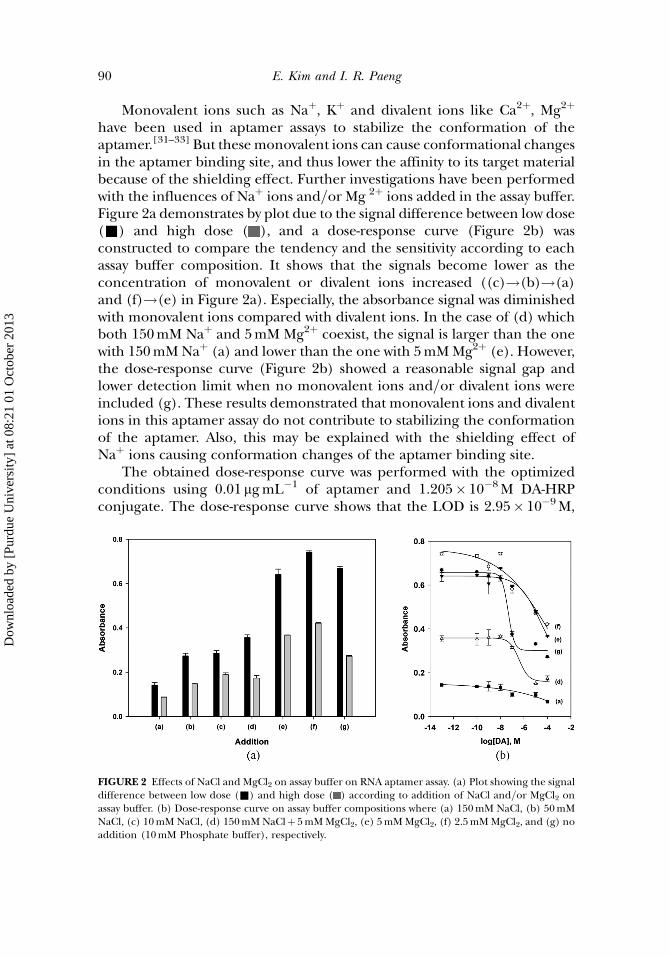
Monovalent ions such as Naþ, Kþ and divalent ions like Ca2þ, Mg2þ
have been used in aptamer assays to stabilize the conformation of theaptamer.[31–33] But thesemonovalent ions can cause conformational changesin the aptamer binding site, and thus lower the affinity to its target materialbecause of the shielding effect. Further investigations have been performedwith the influences of Naþ ions and=or Mg 2þ ions added in the assay buffer.Figure 2a demonstrates by plot due to the signal difference between low dose(&) and high dose ( ), and a dose-response curve (Figure 2b) wasconstructed to compare the tendency and the sensitivity according to eachassay buffer composition. It shows that the signals become lower as theconcentration of monovalent or divalent ions increased ((c)!(b)!(a)and (f)!(e) in Figure 2a). Especially, the absorbance signal was diminishedwith monovalent ions compared with divalent ions. In the case of (d) whichboth 150mM Naþ and 5mM Mg2þ coexist, the signal is larger than the onewith 150mMNaþ (a) and lower than the one with 5mMMg2þ (e). However,the dose-response curve (Figure 2b) showed a reasonable signal gap andlower detection limit when no monovalent ions and=or divalent ions wereincluded (g). These results demonstrated that monovalent ions and divalentions in this aptamer assay do not contribute to stabilizing the conformationof the aptamer. Also, this may be explained with the shielding effect ofNaþ ions causing conformation changes of the aptamer binding site.
The obtained dose-response curve was performed with the optimizedconditions using 0.01 mgmL�1 of aptamer and 1.205� 10�8M DA-HRPconjugate. The dose-response curve shows that the LOD is 2.95� 10�9M,
FIGURE 2 Effects of NaCl and MgCl2 on assay buffer on RNA aptamer assay. (a) Plot showing the signaldifference between low dose (&) and high dose ( ) according to addition of NaCl and=or MgCl2 onassay buffer. (b) Dose-response curve on assay buffer compositions where (a) 150mM NaCl, (b) 50mMNaCl, (c) 10mM NaCl, (d) 150mM NaClþ 5mMMgCl2, (e) 5mMMgCl2, (f) 2.5mMMgCl2, and (g) noaddition (10mM Phosphate buffer), respectively.
90 E. Kim and I. R. Paeng
Dow
nloa
ded
by [
Purd
ue U
nive
rsity
] at
08:
21 0
1 O
ctob
er 2
013
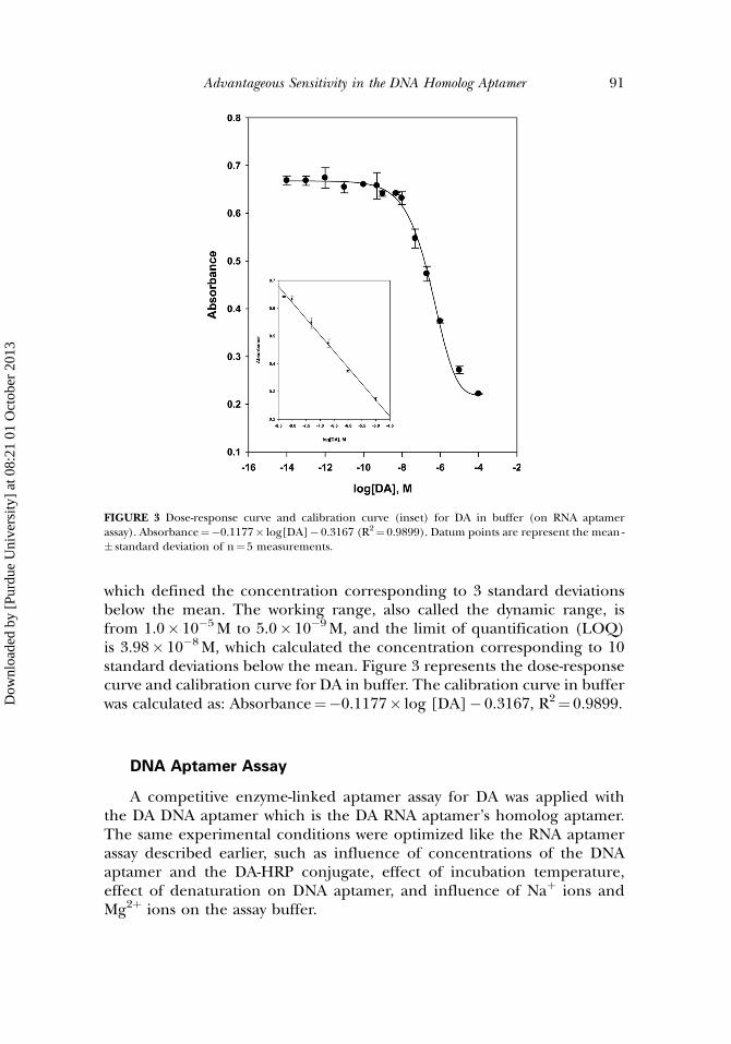
which defined the concentration corresponding to 3 standard deviationsbelow the mean. The working range, also called the dynamic range, isfrom 1.0� 10�5M to 5.0� 10�9M, and the limit of quantification (LOQ)is 3.98� 10�8M, which calculated the concentration corresponding to 10standard deviations below the mean. Figure 3 represents the dose-responsecurve and calibration curve for DA in buffer. The calibration curve in bufferwas calculated as: Absorbance¼�0.1177� log [DA]� 0.3167, R2¼ 0.9899.
DNA Aptamer Assay
A competitive enzyme-linked aptamer assay for DA was applied withthe DA DNA aptamer which is the DA RNA aptamer’s homolog aptamer.The same experimental conditions were optimized like the RNA aptamerassay described earlier, such as influence of concentrations of the DNAaptamer and the DA-HRP conjugate, effect of incubation temperature,effect of denaturation on DNA aptamer, and influence of Naþ ions andMg2þ ions on the assay buffer.
FIGURE 3 Dose-response curve and calibration curve (inset) for DA in buffer (on RNA aptamerassay). Absorbance¼�0.1177� log[DA]� 0.3167 (R2¼ 0.9899). Datum points are represent the mean -� standard deviation of n¼ 5 measurements.
Advantageous Sensitivity in the DNA Homolog Aptamer 91
Dow
nloa
ded
by [
Purd
ue U
nive
rsity
] at
08:
21 0
1 O
ctob
er 2
013
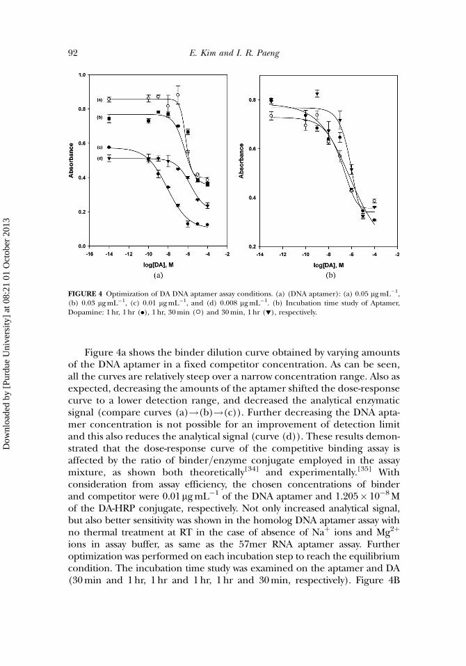
Figure 4a shows the binder dilution curve obtained by varying amountsof the DNA aptamer in a fixed competitor concentration. As can be seen,all the curves are relatively steep over a narrow concentration range. Also asexpected, decreasing the amounts of the aptamer shifted the dose-responsecurve to a lower detection range, and decreased the analytical enzymaticsignal (compare curves (a)!(b)!(c)). Further decreasing the DNA apta-mer concentration is not possible for an improvement of detection limitand this also reduces the analytical signal (curve (d)). These results demon-strated that the dose-response curve of the competitive binding assay isaffected by the ratio of binder=enzyme conjugate employed in the assaymixture, as shown both theoretically[34] and experimentally.[35] Withconsideration from assay efficiency, the chosen concentrations of binderand competitor were 0.01mgmL�1 of the DNA aptamer and 1.205� 10�8Mof the DA-HRP conjugate, respectively. Not only increased analytical signal,but also better sensitivity was shown in the homolog DNA aptamer assay withno thermal treatment at RT in the case of absence of Naþ ions and Mg2þ
ions in assay buffer, as same as the 57mer RNA aptamer assay. Furtheroptimization was performed on each incubation step to reach the equilibriumcondition. The incubation time study was examined on the aptamer and DA(30min and 1hr, 1hr and 1hr, 1hr and 30min, respectively). Figure 4B
FIGURE 4 Optimization of DA DNA aptamer assay conditions. (a) (DNA aptamer): (a) 0.05 mgmL�1,(b) 0.03 mgmL�1, (c) 0.01 mgmL�1, and (d) 0.008 mgmL�1. (b) Incubation time study of Aptamer,Dopamine: 1 hr, 1 hr (.), 1 hr, 30min (�) and 30min, 1 hr (!), respectively.
92 E. Kim and I. R. Paeng
Dow
nloa
ded
by [
Purd
ue U
nive
rsity
] at
08:
21 0
1 O
ctob
er 2
013
illustrates a dose-response curve obtained with these conditions. The incu-bation time of the aptamer for 1hr andDA for 1hr presents the best sensitivity.
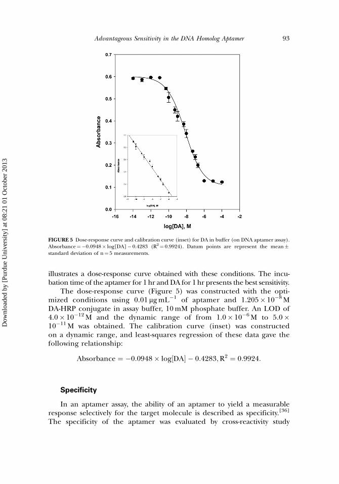
The dose-response curve (Figure 5) was constructed with the opti-mized conditions using 0.01 mgmL�1 of aptamer and 1.205� 10�8MDA-HRP conjugate in assay buffer, 10mM phosphate buffer. An LOD of4.0� 10�12M and the dynamic range of from 1.0� 10�6M to 5.0�10�11M was obtained. The calibration curve (inset) was constructedon a dynamic range, and least-squares regression of these data gave thefollowing relationship:
Absorbance ¼ �0:0948� log½DA� � 0:4283;R2 ¼ 0:9924:
Specificity
In an aptamer assay, the ability of an aptamer to yield a measurableresponse selectively for the target molecule is described as specificity.[36]
The specificity of the aptamer was evaluated by cross-reactivity study
FIGURE 5 Dose-response curve and calibration curve (inset) for DA in buffer (on DNA aptamer assay).Absorbance¼�0.0948� log[DA]� 0.4283 (R2¼ 0.9924). Datum points are represent the mean�standard deviation of n¼ 5 measurements.
Advantageous Sensitivity in the DNA Homolog Aptamer 93
Dow
nloa
ded
by [
Purd
ue U
nive
rsity
] at
08:
21 0
1 O
ctob
er 2
013
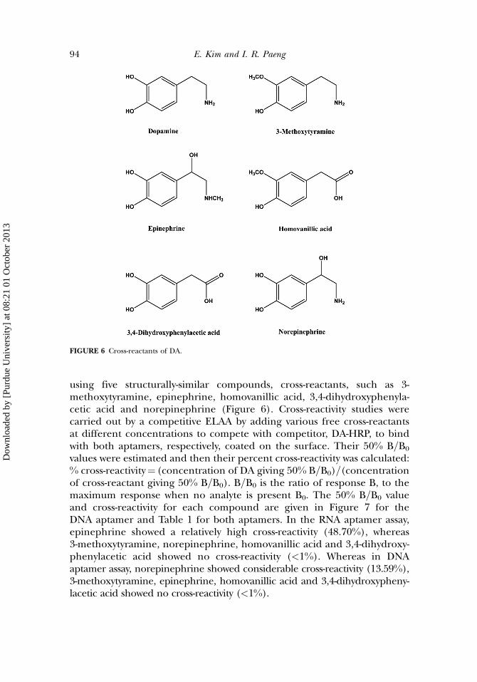
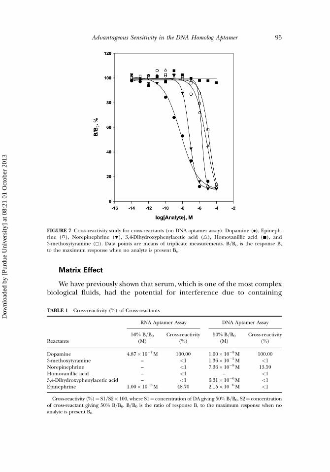
using five structurally-similar compounds, cross-reactants, such as 3-methoxytyramine, epinephrine, homovanillic acid, 3,4-dihydroxyphenyla-cetic acid and norepinephrine (Figure 6). Cross-reactivity studies werecarried out by a competitive ELAA by adding various free cross-reactantsat different concentrations to compete with competitor, DA-HRP, to bindwith both aptamers, respectively, coated on the surface. Their 50% B=B0
values were estimated and then their percent cross-reactivity was calculated:% cross-reactivity¼ (concentration of DA giving 50% B=B0)=(concentrationof cross-reactant giving 50% B=B0). B=B0 is the ratio of response B, to themaximum response when no analyte is present B0. The 50% B=B0 valueand cross-reactivity for each compound are given in Figure 7 for theDNA aptamer and Table 1 for both aptamers. In the RNA aptamer assay,epinephrine showed a relatively high cross-reactivity (48.70%), whereas3-methoxytyramine, norepinephrine, homovanillic acid and 3,4-dihydroxy-phenylacetic acid showed no cross-reactivity (<1%). Whereas in DNAaptamer assay, norepinephrine showed considerable cross-reactivity (13.59%),3-methoxytyramine, epinephrine, homovanillic acid and 3,4-dihydroxypheny-lacetic acid showed no cross-reactivity (<1%).
FIGURE 6 Cross-reactants of DA.
94 E. Kim and I. R. Paeng
Dow
nloa
ded
by [
Purd
ue U
nive
rsity
] at
08:
21 0
1 O
ctob
er 2
013
Matrix Effect
We have previously shown that serum, which is one of the most complexbiological fluids, had the potential for interference due to containing
FIGURE 7 Cross-reactivity study for cross-reactants (on DNA aptamer assay): Dopamine (.), Epineph-rine (�), Norepinephrine (!), 3,4-Dihydroxyphenylacetic acid (4), Homovanillic acid (&), and3-methoxytyramine (&). Data points are means of triplicate measurements. B=Bo is the response B,to the maximum response when no analyte is present Bo.
TABLE 1 Cross-reactivity (%) of Cross-reactants
RNA Aptamer Assay DNA Aptamer Assay
Reactants50% B=B0
(M)Cross-reactivity
(%)50% B=B0
(M)Cross-reactivity
(%)
Dopamine 4.87� 10�7M 100.00 1.00� 10�8M 100.003-methoxytyramine – <1 1.36� 10�5M <1Norepinephrine – <1 7.36� 10�8M 13.59Homovanillic acid – <1 – <13,4-Dihydroxyphenylacetic acid – <1 6.31� 10�6M <1Epinephrine 1.00� 10�6M 48.70 2.15� 10�6M <1
Cross-reactivity (%)¼ S1=S2� 100, where S1¼ concentration of DA giving 50% B=B0, S2¼ concentrationof cross-reactant giving 50% B=B0. B=B0 is the ratio of response B, to the maximum response when noanalyte is present B0.
Advantageous Sensitivity in the DNA Homolog Aptamer 95
Dow
nloa
ded
by [
Purd
ue U
nive
rsity
] at
08:
21 0
1 O
ctob
er 2
013
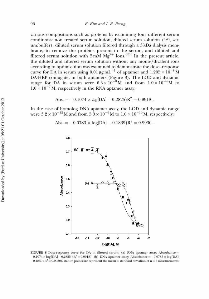
various compositions such as proteins by examining four different serumconditions: non treated serum solution, diluted serum solution (1:9, ser-um:buffer), diluted serum solution filtered through a 3 kDa dialysis mem-brane, to remove the proteins present in the serum, and diluted andfiltered serum solution with 5mM Mg2þ ions.[26] In the present article,the diluted and filtered serum solution without any mono-=divalent ionsaccording to optimization was examined to demonstrate the dose–responsecurve for DA in serum using 0.01 mgmL�1 of aptamer and 1.205� 10�8MDA-HRP conjugate, in both aptamers (Figure 8). The LOD and dynamicrange for DA in serum were 6.3� 10�8M and from 1.0� 10�4M to1.0� 10�7M, respectively in the RNA aptamer assay:
Abs: ¼ �0:1074� log DA½ � � 0:2825 R2 ¼ 0:9918� �
:
In the case of homolog DNA aptamer assay, the LOD and dynamic rangewere 3.2� 10�12M and from 5.0� 10�6M to 1.0� 10�10M, respectively:
Abs: ¼ �0:0783� log DA½ � � 0:1839 R2 ¼ 0:9930� �
:
FIGURE 8 Dose-response curve for DA in filtered serum: (a) RNA aptamer assay, Absorbance¼�0.1074� log[DA]�0.2825 (R2¼ 0.9918). (b) DNA aptamer assay, Absorbance¼�0.0783� log[DA]�0.1839 (R2¼ 0.9930). Datum points are represent the mean� standard deviation of n¼ 5measurements.
96 E. Kim and I. R. Paeng
Dow
nloa
ded
by [
Purd
ue U
nive
rsity
] at
08:
21 0
1 O
ctob
er 2
013
The correlation diagram of the absorbance obtained both in buffer and inserum was constructed:
Abs: in serumð Þ ¼ 1:0114� Abs: in bufferð Þþ 0:0182 onRNA aptamer R2 ¼ 0:9959
� �
Abs: in serumð Þ ¼ 0:8492� Abs: in bufferð Þþ 0:1548 onhomologDNA aptamerðR2 ¼ 0:9813Þ:
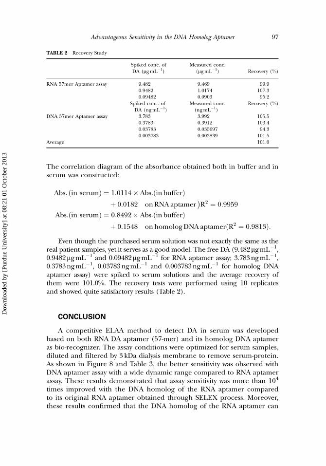
Even though the purchased serum solution was not exactly the same as thereal patient samples, yet it serves as a goodmodel. The free DA (9.482mgmL�1,0.9482mgmL�1 and 0.09482mgmL�1 for RNA aptamer assay; 3.783ngmL�1,0.3783ngmL�1, 0.03783ngmL�1 and 0.003783ngmL�1 for homolog DNAaptamer assay) were spiked to serum solutions and the average recovery ofthem were 101.0%. The recovery tests were performed using 10 replicatesand showed quite satisfactory results (Table 2).
CONCLUSION
A competitive ELAA method to detect DA in serum was developedbased on both RNA DA aptamer (57-mer) and its homolog DNA aptameras bio-recognizer. The assay conditions were optimized for serum samples,diluted and filtered by 3 kDa dialysis membrane to remove serum-protein.As shown in Figure 8 and Table 3, the better sensitivity was observed withDNA aptamer assay with a wide dynamic range compared to RNA aptamerassay. These results demonstrated that assay sensitivity was more than 104
times improved with the DNA homolog of the RNA aptamer comparedto its original RNA aptamer obtained through SELEX process. Moreover,these results confirmed that the DNA homolog of the RNA aptamer can
TABLE 2 Recovery Study
Spiked conc. ofDA (mgmL�1)
Measured conc.(mgmL�1) Recovery (%)
RNA 57mer Aptamer assay 9.482 9.469 99.90.9482 1.0174 107.30.09482 0.0903 95.2
Spiked conc. ofDA (ngmL�1)
Measured conc.(ngmL�1)
Recovery (%)
DNA 57mer Aptamer assay 3.783 3.992 105.50.3783 0.3912 103.40.03783 0.035697 94.30.003783 0.003839 101.5
Average 101.0
Advantageous Sensitivity in the DNA Homolog Aptamer 97
Dow
nloa
ded
by [
Purd
ue U
nive
rsity
] at
08:
21 0
1 O
ctob
er 2
013
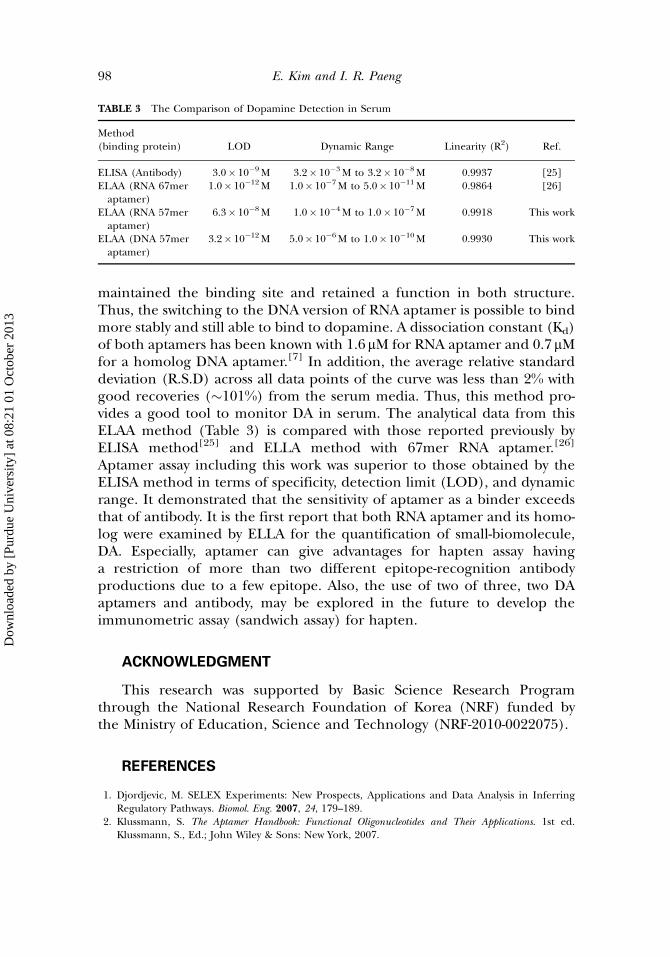
maintained the binding site and retained a function in both structure.Thus, the switching to the DNA version of RNA aptamer is possible to bindmore stably and still able to bind to dopamine. A dissociation constant (Kd)of both aptamers has been known with 1.6mM for RNA aptamer and 0.7mMfor a homolog DNA aptamer.[7] In addition, the average relative standarddeviation (R.S.D) across all data points of the curve was less than 2% withgood recoveries (�101%) from the serum media. Thus, this method pro-vides a good tool to monitor DA in serum. The analytical data from thisELAA method (Table 3) is compared with those reported previously byELISA method[25] and ELLA method with 67mer RNA aptamer.[26]
Aptamer assay including this work was superior to those obtained by theELISA method in terms of specificity, detection limit (LOD), and dynamicrange. It demonstrated that the sensitivity of aptamer as a binder exceedsthat of antibody. It is the first report that both RNA aptamer and its homo-log were examined by ELLA for the quantification of small-biomolecule,DA. Especially, aptamer can give advantages for hapten assay havinga restriction of more than two different epitope-recognition antibodyproductions due to a few epitope. Also, the use of two of three, two DAaptamers and antibody, may be explored in the future to develop theimmunometric assay (sandwich assay) for hapten.
ACKNOWLEDGMENT
This research was supported by Basic Science Research Programthrough the National Research Foundation of Korea (NRF) funded bythe Ministry of Education, Science and Technology (NRF-2010-0022075).
REFERENCES
1. Djordjevic, M. SELEX Experiments: New Prospects, Applications and Data Analysis in InferringRegulatory Pathways. Biomol. Eng. 2007, 24, 179–189.
2. Klussmann, S. The Aptamer Handbook: Functional Oligonucleotides and Their Applications. 1st ed.Klussmann, S., Ed.; John Wiley & Sons: New York, 2007.
TABLE 3 The Comparison of Dopamine Detection in Serum
Method(binding protein) LOD Dynamic Range Linearity (R2) Ref.
ELISA (Antibody) 3.0� 10�9M 3.2� 10�3M to 3.2� 10�8M 0.9937 [25]ELAA (RNA 67meraptamer)
1.0� 10�12M 1.0� 10�7M to 5.0� 10�11M 0.9864 [26]
ELAA (RNA 57meraptamer)
6.3� 10�8M 1.0� 10�4M to 1.0� 10�7M 0.9918 This work
ELAA (DNA 57meraptamer)
3.2� 10�12M 5.0� 10�6M to 1.0� 10�10M 0.9930 This work
98 E. Kim and I. R. Paeng
Dow
nloa
ded
by [
Purd
ue U
nive
rsity
] at
08:
21 0
1 O
ctob
er 2
013

3. Yang, Y.; Yang, D.; Schluesener, H. J.; Zhang, Z. Advances in SELEX and Application of Aptamers inthe Central Nervous System. Biomol. Eng. 2007, 24, 583–592.
4. Tombelli, S.; Minunni, M.; Mascini, M. Analytical Applications of Aptamers. Biosens. Bioelectron.2005, 20, 2424–2434.
5. Iliuk, A. B.; Hu, L.; Tao, W. A. Aptamer in Bioanalytical Applications. Anal. Chem. 2011, 83, 4440–4452.6. O’Sullivan, C. K. Aptasensors – The Future of Biosensing? Anal. Bioanal. Chem. 2002, 372, 44–48.7. Walsh, R.; DeRosa, M. C. Retention of Function in the DNA Homolog of the RNA Dopamine
Aptamer. Biochem. Biophys. Res. Commun. 2009, 388, 732–735.8. Cooper, J. R.; Bloom, F. E.; Roth, R. H. The Biochemical Basis of Neuropharmacology, 7th ed.; New York:
Oxford University Press, 1996.9. Perry, M.; Li, Q.; Kennedy, R. T. Review of Recent Advances in Analytical Techniques for
the Determination of Neurotransmitters. Anal. Chim. Acta 2009, 653, 1–22.10. Damier, P.; Hirsch, E. C.; Agid, Y.; Graybiel, A. M. Review of Recent Advances in Analytical
Techniques for the Determination of Neurotransmitters. Brain 1999, 122(8), 1437–1448.11. Zheng, J.; Zhou, X. Sodium Dodecyl Sulfate-Modified Carbon Paste Electrodes for Selective
Determination of Dopamine in the Presence of Ascorbic Acid. Bioelectrochemistry 2007, 70, 408–415.12. Kienast, T.; Heinz, A. Dopamine and theDiseased Brain.CNSNeurol. Disord. Drug Targets 2006, 5(1), 109–131.13. Swanson, C. J.; Perry, K. W.; Koch-Krueger, S.; Katner, J.; Svensson, K. A.; Bymaster, F. P. Effect of the
Attention Deficit=Hyperactivity Disorder Drug Atomoxetine on Extracellular Concentrations of Nore-pinephrine and Dopamine in Several Brain Regions of the Rat. Neuropharmacology 2006, 50, 755–760.
14. Carrera, V.; Sabater, E.; Vilanova, E.; Sogorb, M. A. A Simple and RapidHPLC–MSMethod for the Sim-ultaneous Determination of Epinephrine, Norepinephrine, Dopamine and 5-Hydroxytryptamine:Application to the Secretion of Bovine Chromaffin Cell Cultures. J. Chromatogr. B 2007, 847(2), 88–94.
15. Li, N.; Guo, J.; Liu, B.; Yu, Y.; Cui, H.; Mao, L.; Lin, Y. Determination of Monoamine Neuro-transmitters and Their Metabolites in a Mouse Brain Microdialysate by Coupling High-PerformanceLiquid Chromatography with Gold Nanoparticle-Initiated Chemiluminescence. Anal. Chim. Acta2009, 645, 48–55.
16. Syslova, K.; Rambousek, L.; Kuzma, M.; Najmanova, V.; Bubenikova-Valesova, V.; Slamberova, R.;Kacer, P. Monitoring of Dopamine and its Metabolites in Brain Microdialysates: Method CombiningFreeze-Drying with Liquid Chromatography–Tandem Mass Spectrometry. J. Chromatogr. A 2011,1218, 3382–3391.
17. Wang, Y.; Li, Y.; Tang, L.; Lu, J.; Li, J. Application of Graphene-Modified Electrode for SelectiveDetection of Dopamine. Electrochem. Commun. 2009, 11, 889–892.
18. Pournaghi-Azar, M. H.; Dastangoo, H.; Fadakar bajeh baj, R. Simultaneous Determination ofDopamine and its Oxidized Product (Aminochrom), by Hydrodynamic Amperometry and AnodicStripping Voltammetry, using the Metallic Palladium and Uranalyl Hexacyanoferrate CoatedAluminum Electrodes. Biosens. Bioelectron. 2010, 25, 1481–1486.
19. Baldrich, E.; Gomez, R.; Gabriel, G.; Munoz, F. X. Magnetic Entrapment for Fast, Simple andReversible Electrode Modification with Carbon Nanotubes: Application to Dopamine Detection.Biosens. Bioelectron. 2011, 26, 1876–1882.
20. Zheng, Y.; Wang, Y.; Yang, X. Aptamer-based Colorimetric Biosensing of Dopamine using UnmodifiedGold Nanoparticles. Sens. Actuators B 2011, 156, 95–99.
21. Zhao, S.; Huang, Y.; Shi, M.; Liu, R.; Liu, Y. M. Chemiluminescence Resonance EnergyTransfer-Based Detection for Microchip Electrophoresis. Anal. Chem. 2010, 82, 2036–2041.
22. Nalewajko, E.; Wiszowata, A.; Kojło, A. Determination of catecholamines by flow-Injection Analysisand High-Performance Liquid Chromatography with Chemiluminescence Detection. J. Pharm.Biomed. Anal. 2007, 43(5), 1673–1681.
23. Chen, Y. C.; Lin, W. Y. Enhancement of Chemiluminescence of the KIO4–Luminol System by GallicAcid, Acetaldehyde and Mn2þ: Application for the Determination of Catecholamines. Luminescence2010, 25, 43–49.
24. Sanchez-Rivera, A. E.; Corona-Avendano, S.; Alarcon-Angeles, G.; Rojas-Hernandez, A.; Ramirez-Silva, M. T.; Romero-Romo, M. A. Spectrophotometric Study on the Stability of Dopamineand the Determination of its Acidity Constants. Spectrochim. Acta Part A 2003, 59, 3193–3203.
25. Kim, J.; Jeon, M.; Paeng, K.-J.; Paeng, I. R. Competitive Enzyme-Linked Immunosorbent Assayfor the Determination of Catecholamine, Dopamine in Serum. Anal. Chim. Acta 2008, 619, 87–93.
Advantageous Sensitivity in the DNA Homolog Aptamer 99
Dow
nloa
ded
by [
Purd
ue U
nive
rsity
] at
08:
21 0
1 O
ctob
er 2
013
26. Park, H.; Paeng, I. R. Development of Direct Competitive Enzyme-Linked Aptamer Assay forDetermination of Dopamine in Serum. Anal. Chim. Acta 2011, 685, 65–73.
27. Mannironi, C.; Nardo, A. D.; Fruscoloni, P.; Tocchini-Valentini, G. P. In Vitro Selection of DopamineRNA Ligands. Biochemistry 1997, 36, 9726–9734.
28. Hermanson, G. T. Bioconjugate Techniques, 2nd ed.; Academic Press: Burlington, MA, 2008.29. Baldrich, E.; Restrepo, A.; O¢Sullivan, C. K. Aptasensor Development: Elucidation of Critical
Parameters for Optimal Aptamer Performance. Anal. Chem. 2004, 76, 7053–7063.30. Cho, E. J.; Collett, J. R.; Szafranska, A. E.; Ellington, A. D. Optimization of Aptamer Microarray
Technology for Multiple Protein Targets. Anal. Chim. Acta 2006, 564, 82–90.31. Hianik, T.; Ostatna, V.; Sonlajtnerova, M.; Grman, I. Influence of Ionic Strength, pH and Aptamer
Configuration for Binding Affinity to Thrombin. Bioelectrochemistry 2007, 70, 127–133.32. Zhu, Z.; Schmidt, T.; Mahrous, M.; Guieu, V.; Perrier, S.; Ravelet, C.; Peyrin, E. Optimization of the
Structure-Switching Aptamer-Based Fluorescence Polarization Assay for the Sensitive TyrosinamideSensing. Anal. Chim. Acta 2011, 707, 191–196.
33. Xu, W.; Lu, Y. Label-Free Fluorescent Aptamer Sensor Based on Regulation of Malachite GreenFluorescence. Anal. Chem. 2010, 82, 574–578.
34. Bachas, L. G.; Meyerhoff, M. E. Theoretical Models for Predicting the Effect of Bridging GroupRecognition and Conjugate Substitution on Hapten Enzyme Immunoassay Dose-Response Curves.Anal. Biochem. 1986, 156, 223–238.
35. Cha, G. S.; Meyerhoff, M. E. Solid Phase Enzyme-Linked Competitive Binding Assay for Riboflavin.Anal. Biochem. 1988, 168, 216–227.
36. Datta, P.; Xu, L.; Malik, S.; Landicho, D.; Ferreri, L.; Halverson, K.; Roby, P. V.; Zebelman, A. M.;Kenny, M. A. Effect of Antibody Specificity on Results of Selected Digoxin Immunoassays amongVarious Clinical Groups. Clin. Chem. 1996, 42(3), 373–379.
100 E. Kim and I. R. Paeng
Dow
nloa
ded
by [
Purd
ue U
nive
rsity
] at
08:
21 0
1 O
ctob
er 2
013