advanced cardiac life support - emcmedicaltraining.com · advanced cardiac life support participant...
TRANSCRIPT

1
EMERGENCY MEDICAL CONSULTANTS INC. Florida’s Premier Provider Of Quality Medical Training Programs
Nationally Accredited and OSHA Programs CEU Provider
Since 1988
ADVANCED CARDIAC LIFE SUPPORT
PARTICIPANT PREPARATION PACKET 2016 - 2020
This information is derived from the 2015 ECC Guidelines
This packet contains prep information for the ACLS Course as well as EKG and BLS reviews. We strongly recommend completing these exams prior to the course.
-MANDATORY REQUIREMENTS- You must bring the AHA ACLS textbook to class with your completed online AHA Self Assessment.
Instructions can be found on page 2 of your red ACLS textbook. Passing Score = 70% (If a score of 70% is not achieved in each section, please review the text and retest the section.)
★!If you are attending the BLS section following ACLS, refer to page 76 for additional instructions. (There is a mandatory pretest if you are choosing to do BLS)
COURSE DATE / TIME: LOCATION:
NAME:
ã2019 Emergency Medical Consultants This material is protected by Copyright and may not be reproduced without written consent
(772) 878-3085 * Fax: (772) 878-7909 * Email: [email protected] 597 SE Port Saint Lucie Blvd * Port Saint Lucie, Florida 34984
2019A Visit Our Website- www EMCmedicaltraining.com

2

3
EMERGENCY MEDICAL CONSULTANTS INC. Florida’s Premier Provider Of Quality Medical Training Programs
Nationally Accredited and OSHA Programs CEU Provider
Since 1988
This copyrighted prep packet is a supplement for those students taking an ACLS program with EMC. Welcome to Emergency Medical Consultants’ Stress Free ACLS Course. We are pleased that you have chosen us to provide you with this outstanding course and are sure that this will be a worthwhile learning experience for you as a medical professional. Please remember you will need to be able to perform the AHA CPR skills and must be familiar with basic dysrhythmia recognition and pharmacology before the course.
In order to keep our program “stress free” and to assure that all participants meet the AHA requirements for ACLS proficiency, preparation is required prior to the actual class. We will be using the latest Emergency Cardiac Care Standards for BLS and ACLS.
The American Heart Association mandates that each participant have a textbook to review prior to the course. Currently there is a Textbook and a Resource Text available. The resource text provides a more in depth and detailed prospective of the AHA guidelines. The Text can be purchased through an AHA vendor or borrowed from your hospital or departments’ Education Center if your facility provides a library.
Enclosed you will find information to help you prepare for the required skills stations and ACLS didactic evaluation. Please take the time to look through this information, begin to learn drug uses and doses, review the algorithm and EKG sections, and take the pretests (answer keys are included). This will ensure a stress free day! It is important to prepare for the day by reviewing information prior to class for optimal success. For more EKG practice, log onto www.Skillstat.com Also refer to pg. ii of your ACLS text to access the AHA online pretest located at www.heart.org/eccstudent. This contains information regarding pharmacology, EKG and relevant to the exam is available for review. A score will be provided upon completion, 70% must be obtained prior to the class
We strive to make our program realistic and relevant, thus, the scenarios that you will be required to manage will relate to the work that you do.
All information is based on the American Heart Association ACLS standards at the time of printing and thought to be correct. Providers are encouraged to review the ACLS textbook and their specific policies prior to implementing any procedures or administering any medication based on this study packet.
We look forward to meeting you at the course and will be happy to answer any questions you may have - just call our office at 772-878-3085.
Sincerely, Shaun Fix and the ACLS Staff Emergency Medical Consultants, Inc

4
Guidelines - 2015 Changes
The latest ACLS guidelines from the American Heart Association 2015 ECC Committee were published in late 2015 and implementation began in early 2016. These guidelines will be utilized from 2016 through 2020.
This section contains a brief synopsis of the guidelines that were new in 2015 as well as associated rationale.
CPR Changes-Adult
New change: Immediate recognition and activation of the emergency response system. Latest guidelines indicate the following sequence:
1) Call for nearby help upon finding the victim unresponsive. 2) Check for pulse and respirations simultaneously. No longer than 10 seconds. 3) Activate the emergency response system/ get a defibrillator. 4) Start with 30 compressions 5) Open the airway and give 2 breaths. 6) Resume compression/respiration cycle
Rationale: Calling for help as soon as you find an unresponsive individual is intended to minimize response delay time. Checking the pulse and respirations simultaneously also fosters rapid assessment times, instead of wasting time with a slow, methodical, step by step approach. New Change: Chest Compression Rate-In adult victims of cardiac arrest, perform chest compressions at a rate of 100-120/min. Rationale: Studies indicate that a faster compression rate results in better end tidal CO2 results and BP intra arrest. Additionally individuals who had a return of spontaneous circulation (ROSC) had a more favorable outcome. However rates above 120 were associated with a decrease in compression depth. New Change: Chest Compression Depth-Perform chest compressions to a depth of at least 2 inches/5cm for an average adult. Avoid depths of greater than 2.4 inches. Rationale: A compression depth of approximately 2 inches is associated with greater likelihood of favorable outcomes compared with shallower compressions Potential injuries may result in compression depth greater than 2.4 inches. It is clinically difficult to judge compression depth without the use of a feedback device. Keep in mind that compressions are often found to be too shallow rather than too deep. New Change: Shock first vs CPR first- CPR should be started immediately upon finding an individual in cardiac arrest. The defibrillator should be used as soon as possible. CPR should be continued while the AED or defibrillator pads are applied. Shock should immediately follow when indicated. Rationale: Studies have gone round and round on the topic of whether it is better to provide 1.5-3 minutes of CPR before defibrillation, as compared to delivering a shock as soon as the AED is available. The most recent studies have shown that there is no difference in outcome when CPR is performed for a specified amount of time prior to defibrillation. So the bottom line is now-- CPR until the defibrillator is available. Minimize interruptions in CPR while pads are applied, then if indicated, shock as soon as you can.

5
New Change: Chest Recoil--Avoid leaning on the chest between chest compressions to allow full chest recoil for adults in cardiac arrest. Rationale: Leaning on the chest wall returns compressions prohibits full chest wall recoil. Full chest wall recoil occurs when the sternum return to its natural position during the decompression phase of CPR. Full recoil promotes venous return and increased cardiopulmonary blood flow. New Change: Minimizing Interruptions in Chest Compressions—Minimize the frequency and duration of interruptions in compressions to maximize the number of chest compressions delivered per minute. Rationale: Minimizing interruptions, whether intentional (rhythm analysis, ventilation) or unintended (rescuer distraction) maximizes coronary perfusion and blood flow during CPR. New Change: CPR with an Advanced Airway in Place—With an advanced airway in place, deliver 1 breath every 6 seconds (10 breaths per minute) while continuous chest compressions are being performed. Rationale: This simple single rate for adults, children and infants-rather than a range of breaths per minute-should be easier to learn, remember and perform.
ACLS Changes
All previous BLS changes apply to ACLS, in addition to the following:
New Change: Targeted Temperature Management—All comatose (lacking meaningful response to verbal commands) adult patients with return of spontaneous circulation (ROSC) following cardiac arrest should have targeted temperature management (TTM) with a target temperature between 32-36°C. The selected temperature should be achieved and maintained constantly for at least 24 hours. Rationale: Improved neurological outcome has been documented in individuals in whom hypothermia was induced. The current recommendation is to select a single target temperature and maintain it for at least 24 hours. The selected temperature (between 32-36°C) may be determined by clinician preference or clinical factors. New Change: Out of Hospital Cooling—Routine use of prehospital cooling of patients with rapid infusion of cold IV fluids after ROSC is no longer recommended. Rationale: Previously, it was believed that early initiation of cooling might provide added benefit and facilitate continued in-hospital cooling. Recently published high quality studies demonstrate no benefit to prehospital cooling and also identified potential complications when using cold IV fluids in the prehospital environment. New Change: Vasopressin in Resuscitation—Vasopressin in combination with epinephrine offers no advantage as a substitute for standard dose epinephrine in cardiac arrest. Rationale: Both epinephrine and vasopressin have been proven to be helpful in the cardiac arrest situation to improve ROSC. Review of the available evidence shows the efficacy of the two drugs is similar and there is no demonstrated benefit from administering both epinephrine and vasopressin compared with epinephrine alone. Therefore, in the interest of simplicity, vasopressin has been removed from the Adult Cardiac Arrest Algorithm.

6
New Change: The Role of Epinephrine in Resuscitation—It may be reasonable to administer epinephrine as soon as feasible after the onset of cardiac arrest due to an initial non-shockable (PEA/Asystole) rhythm. Rationale: A very large observational study of cardiac arrest with a non-shockable rhythm (PEA/Asystole) compared epinephrine given at 1-3 minutes with epinephrine given at three later time intervals (4 to 6, 7 to 9, and greater than 9 minutes). The study found an association between early administration of epinephrine and increased ROSC, survival to hospital discharge, and neurologically intact survival. New Change: Defibrillation-Medication Sequence- Defibrillate as soon as possible in the arrest patient (v,fib or puseless v.tach). Immediately resume CPR for 2 minutes and then defibrillate again. Following the second defibrillation, resume CPR and administer Epinepherine. Rationale: The interval from collapse to defibrillation is one of the most important indicators of survival from cardiac arrest. Previous studies have shown that vasopressors are not proven to increase survival rates from v.fib or pulseless v.tach, although they may still be considered useful to increase cerebral and coronary blood flow during CPR. Therefor the initial focus has shifted to administering epinephrine after the second defibrillation. New Change: Oxygen Administration in the Acute Coronary Syndrome (ACS) Patient-administer oxygen to the ACS patient if their oxygen saturation is less than 90%, or is unknown. Rationale: Oxygen should be administered to the ACS patient if the patient is obviously dyspneic, hypoxemic, has obvious signs of heart failure or the oxygen saturation falls below 90%. If applied, the oxygen levels should be titrated to achieve 90% or greater (up to 99%) saturation. The usefulness of O2 has not been determined in normoxic patients with suspected or confirmed ACS. Unless the above criteria is met, providers may consider withholding supplementary oxygen. 2018 Focused Updates— (November 2018)
1. Lidocaine has returned to the VF/pVT algorithim. You may use either Amiodarone or Lidocaine as the IVP or IV infusion antiarrhythmic of choice.
2. Routine use of Magnesium for cardiac arrest is not recommended in adult patients. Magnesium may be considered for torsades de points. (ie polymorphic VT with long QT interval)
3. There is insufficient evidence to support or refute the routine use of a beta blocker early (within the 1st hour after ROSC)
4. Insufficient evidence to support the routine use of Lidocaine within the 1st hour after ROSC. The
exception would be considered in specific circumstances, such as during EMS transport, when recurrent VF/pVT might prove to be challenging.

7
Table of Contents
Topic
Course Agendas 8
Basic Tips for Patient Management 9
Basic Life Support- A Critical Component of ACLS 10
Pharmacology Overview 12
ACLS Rhythms and Algorithms
Ventricular Fibrillation or Pulseless Ventricular Fibrillation 28 Asystole / Pulseless Electrical Activity 29 Post Arrest Care 30 Symptomatic Bradycardia 31 Bradycardias- EKG samples 32 Heart Blocks EKG samples 33 For Stabilization of Rhythm After VF or VT Conversion 34 Hypotension 35 Supraventricular Tachycardia - Stable 36 Atrial Fibrillation- Stable 37 Supraventricular Tachycardia - Unstable 38 Tachycardias- EKG samples 39 Ventricular Tachycardia – Stable 40 Ventricular Tachycardia - Unstable 41 Acute Coronary Syndromes 42 The 12 Lead ECG 43 Acute Stroke 44 Inclusion Criteria for Fibrinolytic Therapy 45 Pulmonary Edema 46 Tips for Drips 47 Special Arrest Situations 48
Practice Exams
EKG 56 ACLS Pre-Course Exam 59
Glossary 69 EKG Answer Key 73 ACLS Pretest Answer Key 74
BLS Completion Information 75

8
ACLS COURSE AGENDA
2-DAY TRAINING PROGRAM (You will be advised of class start times when you enroll))
DAY ONE Introduction to ACLS, Overview of the ACLS Program
AHA Videos: Science and Prep Break Pharmacology I and II Acute Coronary Syndromes and Stroke
Lunch Small group interactive teaching stations:
§ Airway Management, AED, and BLS § Perfusing Patient Algorithms § Non-perfusing Patient Algorithms
DAY TWO AHA Megacode and Team Videos
Overview of rhythms and algorithms / Code team concept Break Small group Patient Management Scenario Practice Lunch ACLS evaluation stations
§ Multiple choice exam § Patient Management Simulation
ACLS COURSE AGENDA 1-DAY REFRESHER PROGRAM
New Science Review / Key Points Overview of Rhythms and Algorithm Resuscitation Team Concept / Putting it all together
Break
Airway Management, AED, and BLS/ Begin Patient Management Scenarios
Lunch
Patient Management Evaluation Stations continued as needed Written Exam
9
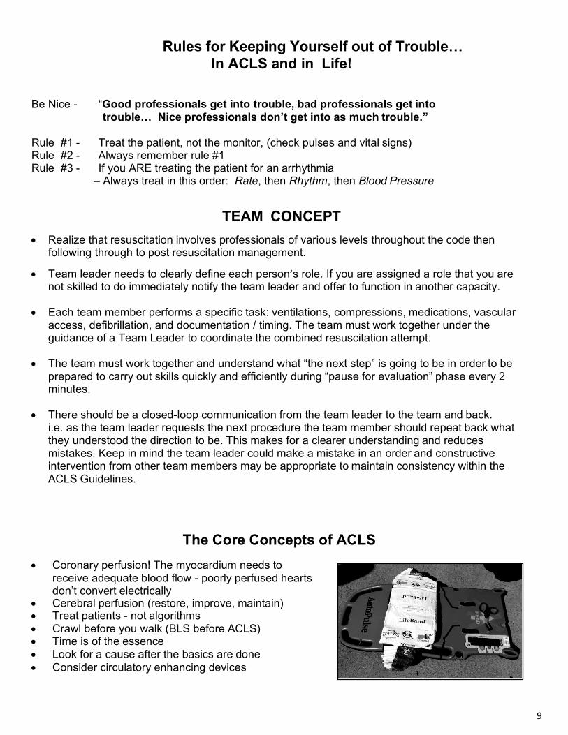
Rules for Keeping Yourself out of Trouble… In ACLS and in Life!
Be Nice - “Good professionals get into trouble, bad professionals get into
trouble… Nice professionals don’t get into as much trouble.” Rule #1 - Treat the patient, not the monitor, (check pulses and vital signs) Rule #2 - Always remember rule #1 Rule #3 - If you ARE treating the patient for an arrhythmia
– Always treat in this order: Rate, then Rhythm, then Blood Pressure
TEAM CONCEPT • Realize that resuscitation involves professionals of various levels throughout the code then
following through to post resuscitation management.
• Team leader needs to clearly define each person’s role. If you are assigned a role that you are not skilled to do immediately notify the team leader and offer to function in another capacity.
• Each team member performs a specific task: ventilations, compressions, medications, vascular
access, defibrillation, and documentation / timing. The team must work together under the guidance of a Team Leader to coordinate the combined resuscitation attempt.
• The team must work together and understand what “the next step” is going to be in order to be
prepared to carry out skills quickly and efficiently during “pause for evaluation” phase every 2 minutes.
• There should be a closed-loop communication from the team leader to the team and back.
i.e. as the team leader requests the next procedure the team member should repeat back what they understood the direction to be. This makes for a clearer understanding and reduces mistakes. Keep in mind the team leader could make a mistake in an order and constructive intervention from other team members may be appropriate to maintain consistency within the ACLS Guidelines.
The Core Concepts of ACLS
• Coronary perfusion! The myocardium needs to receive adequate blood flow - poorly perfused hearts don’t convert electrically
• Cerebral perfusion (restore, improve, maintain) • Treat patients - not algorithms • Crawl before you walk (BLS before ACLS) • Time is of the essence • Look for a cause after the basics are done • Consider circulatory enhancing devices
10
Basic Life Support (CPR) - A critical component of ACLS The most current research available suggests that quality CPR is a crucial variable in survival from
resuscitation attempts - even more important than previously thought. Every participant in an ACLS
class must correctly demonstrate adult CPR skills and use of an AED.
Follow these guidelines when performing Basic Life Support skills:
PUSH HARD: Compress the chest of an adult at least 2 inches, allowing for complete recoil of the
chest between compressions. 2.4 inches is the maximum depth. Do not lean on the
chest in between compressions.
PUSH FAST: Compress the chest at a rate of at 100-120 compressions per minute.
(30 compressions delivered between 15- 18 seconds)
USE CORRECT RATIOS: Deliver 30 compressions and 2 ventilations, in 2-minute blocks. Change
compressors every two minutes during pauses in compressions. At that time, also check rhythm and
pulses if indicated if the viewed rhythm is one that could produce a pulse.
MINIMIZE INTERRUPTIONS IN COMPRESSIONS: Stop CPR only for essential procedures, such as
rotating compressors, rhythm checks and pulse check if indicated, no more frequently than every 2
minutes. Make the pause in compressions as brief as possible.
VENTILATE CAUTIOUSLY: Deliver breaths over 1 second, using just enough volume to produce
visible chest rise. With an advanced airway in place, deliver 1 breath every 6 seconds (10 breaths
per minute) while continuous compressions are being performed. For rescue breathing in a perfusing
patient, deliver 10 -12 breaths per minute.
This equates to one breath approximately every 5-6 seconds. DEFIBRILLATE APPROPRIATELY: Deliver one shock, as soon as possible, and then immediately
resume chest compressions. Check the rhythm and pulse if indicated after 2 minutes of CPR.
11
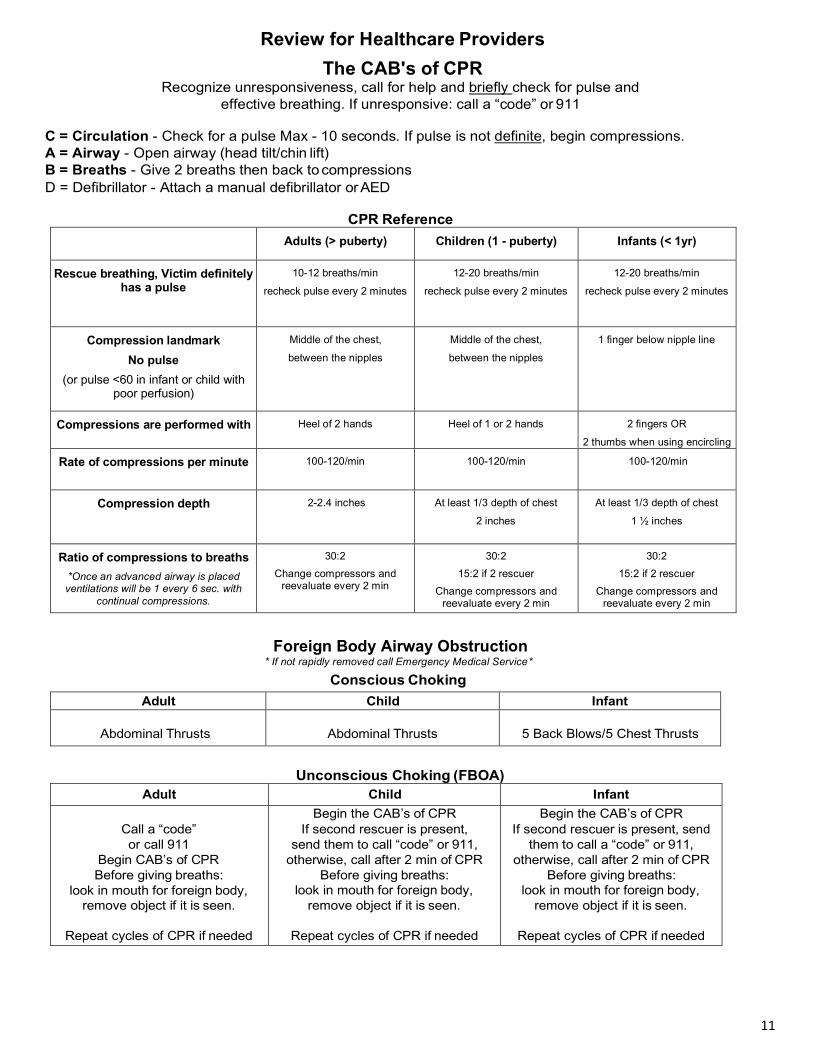
Review for Healthcare Providers The CAB's of CPR
Recognize unresponsiveness, call for help and briefly check for pulse and effective breathing. If unresponsive: call a “code” or 911
C = Circulation - Check for a pulse Max - 10 seconds. If pulse is not definite, begin compressions. A = Airway - Open airway (head tilt/chin lift) B = Breaths - Give 2 breaths then back to compressions D = Defibrillator - Attach a manual defibrillator or AED
CPR Reference
Adults (> puberty) Children (1 - puberty) Infants (< 1yr)
Rescue breathing, Victim definitely has a pulse
10-12 breaths/min
recheck pulse every 2 minutes
12-20 breaths/min
recheck pulse every 2 minutes
12-20 breaths/min
recheck pulse every 2 minutes
Compression landmark No pulse
(or pulse <60 in infant or child with poor perfusion)
Middle of the chest,
between the nipples
Middle of the chest,
between the nipples
1 finger below nipple line
Compressions are performed with Heel of 2 hands Heel of 1 or 2 hands
2 fingers OR
2 thumbs when using encircling hands technique Rate of compressions per minute 100-120/min 100-120/min 100-120/min
Compression depth 2-2.4 inches At least 1/3 depth of chest
2 inches
At least 1/3 depth of chest
1 ½ inches
Ratio of compressions to breaths *Once an advanced airway is placed ventilations will be 1 every 6 sec. with
continual compressions.
30:2 Change compressors and
reevaluate every 2 min
30:2 15:2 if 2 rescuer
Change compressors and reevaluate every 2 min
30:2 15:2 if 2 rescuer
Change compressors and reevaluate every 2 min
Foreign Body Airway Obstruction
* If not rapidly removed call Emergency Medical Service *
Conscious Choking
Adult Child Infant
Abdominal Thrusts
Abdominal Thrusts
5 Back Blows/5 Chest Thrusts
Unconscious Choking (FBOA) Adult Child Infant
Call a “code” or call 911
Begin CAB’s of CPR Before giving breaths:
look in mouth for foreign body, remove object if it is seen.
Repeat cycles of CPR if needed
Begin the CAB’s of CPR If second rescuer is present,
send them to call “code” or 911, otherwise, call after 2 min of CPR
Before giving breaths: look in mouth for foreign body,
remove object if it is seen.
Repeat cycles of CPR if needed
Begin the CAB’s of CPR If second rescuer is present, send
them to call a “code” or 911, otherwise, call after 2 min of CPR
Before giving breaths: look in mouth for foreign body,
remove object if it is seen.
Repeat cycles of CPR if needed

12
PHARMACOLOGY
Ideally, all medications are given through a large bore IV of NS or LR in the antecubital or external jugular. If an IV site is unavailable the next choice is Intraosseous (IO). In reality use “what ya got.”
For Patients in Cardiac Arrest: • Give meds rapidly early in sequence during compressions • Flush all meds with 20mL’s of fluid • Circulate meds with 2 minutes of CPR • Elevate the extremity to flush meds
ENDOTRACHEAL ADMINISTRATION The LEAST effective route
(Lidocaine, Epinephrine Atropine, Narcan)
• 2 - 2.5 times the IV dose • Stop compressions • Use at least 10mL’s total (dilute in NS or sterile water) • Ventilate several times • Resume CPR
For Patients in cardiac arrest:
BLS ASSESSMENT (C.A.B.(D))
Determine unresponsiveness and briefly check for effective breathing. If unresponsive, call a “code” or 911.
C(1) = Circulation: evaluate for signs of a pulse (10 seconds max). If pulse is absent: begin and maintain compressions, 30 rapid, deep compressions at 100-120 per min
A(2) = Airway: open it with a head tilt / chin lift or jaw thrust if neck trauma is suspected
B(3) = Breathing: administer 2 breaths after the 30 compressions. If an advanced airway device is placed, ventilate once each 6 seconds while compressions continue.
D(4) = Defibrillation: bring and attach a monitor/defibrillator or an AED to the patient shock as soon as appropriate

13
PRIMARY ASSESSMENT (A.B.C.D) A (1) - Advanced airway procedures: Reserved for those skilled at these procedures: Tracheal (intubation), or Perilaryngeal tube (LMA, King, Combitube, etc)
B (2) - Breathing: assessed, assured, and secured. Be sure whichever tube is placed is causing chest rise, apply supplemental O2, then secure the device. Monitor CO2 once intubated. Exhaled CO2 is an effective means for measuring ET placement and the quality of CPR.
C (3) - Circulatory interventions: Establish or confirm vascular access and begin cardiac pharmacology. What is the cardiac rhythm? Hint: in cardiac arrest the first medication is always a vasopressor such as Epinephrine. Then Epi may be administered every 3-5 minutes.
D (4) - Disability: check for neurologic function: Assess for responsiveness, level of consciousness and pupil dialation.
-- AVPU: Alert, Voice, Painful, Unresponsive
E (5) – Exposure: remove necessary clothing to perform a physical exam, look for obvious signs of injury, unusual marking, or medic alert bracelets.
SECONDARY ASSESSMENT Evaluates differential diagnosis
Sign and symptoms Allergies Medications Past medical history (especially relating to the current i llness) Last meal consumed Events
Potential reversible causes of cardiac arrest: 5 H’s & 5 T’s: Hypoxia Toxins (overdose) Hypovolemia Thromboemboli - Pulmonary Hyper/Hypokalemia Thromboemboli - Coronary Hypothermia Tension Pneumothorax Hydrogen ion (acidosis) Tamponade (cardiac)
Post Cardiac Arrest Induced Hypothermia: Targeted Temperature Mgmt. Numerous studies show improved neurological recovery for post arrest patients who are cooled to the low 90’s F° (32-36°C) following a successful resuscitation if the patient has no appropriate neurological response. The hypothermic state is maintained for 24+ hours.
For Perfusing Patients (people with pulses)
Begin with the basics of all patient care: • Assess and maintain Circulation, Airway, and Breathing • Evaluate the patient’s symptoms and related (targeted) history, begin a physical exam
Things to do: 1. Administer oxygen - only if needed (Dyspnea, Hypoxia - O2 sat < 94%, CHF) 2. Assess and monitor vital and diagnostic signs: 3. Respirations, Pulse, B/P, Pulse Oximetry, Monitor EKG Rhythm 4. Establish vascular access 5. Obtain 12 lead ECG and Chest X-ray 6. Obtain Labs-bleeding times, cardiac enzymes, etc.
14
OXYGEN
Indications: • Cardiac patients with signs of hypoxia (dyspnea, rales, O2 sat < 94% generally. BUT< 90% specifically in ACS) • Suspected hypoxia of any cause • Cardiac arrest Actions:
• Reverses hypoxia Dosage: • Nasal cannula @ 2 - 6 Lpm = 24 - 44% FiO2 • Simple plastic face mask @ 6 - 10 Lpm = 40 - 60% FiO2 • Non-rebreather mask @ 10 - 15 Lpm = 90 - 100% FiO2
• Patients with inadequate rate or depth of respirations:
Bag-valve mask @ 10 - 15 Lpm with an oxygen reservoir = 90 - 100% FiO2
Side effects: • Rare: Possible respiratory depression in a hypoxic drive patient • NEVER withhold O2 in patients who need it Quick tip:
Let the patient’s need be your guide. In general: • Ideally maintain oxygen levels at 94 - 99% SaO2 • Monitor closely; high O2 may cause oxygen toxicity and impede cellular healing • Administer low flow oxygen for patients with chest pain or stroke. • Ventilate 10 -12 breaths per minute for the apneic patient with a pulse,
10 breaths per minute for the pulseless patient with an advanced airway in place • Deliver just enough volume to see the chest rise, around 500 - 600mL
EPINEPHRINE
Classification: Adrenergic (sympathetic) stimulator Indications: • Cardiac arrest • Symptomatic bradycardia refractory to Atropine & transcutaneous pacing (drip only) Actions: • Positive b effects, including increased heart rate, contractility, and automaticity • Positive a effects, including peripheral vasoconstriction. Dosage: • Bolus: 1mg IV repeat at 3 - 5 minute intervals
Infusion: • 4mg/250mL’s (16 mcg/mL) D5W or NS. • For Bradycardia: Infuse 2 -10mcg/min (14-70 mL/hr) titrate • For Hypotension 0.1-0.5 mcg/kg/min (8-40 mcg/min)(30-150 mL/hr) titrate to SBP above 90
Route: • IV/IO, ET, IV infusion
Side effects: • Tachycardia, hypertension, increased O2 demand, PVC’s, tachyarrhythmias

15
ATROPINE Classification: Parasympatholytic (blocks acetylcholine from the parasympathetic nervous system)
Indications: • Symptomatic bradycardia
Actions: • Increases heart rate and conduction through the AV node.
Dosage: • Bolus 0.5mg IV. Repeat at 3 - 5 minute intervals, not to exceed approximately 3mg
Route: • IV/IO, ET
Side effects: • Tachycardia, dilated pupils, angina. Smaller doses may cause bradycardia
• Note: Give 0.5 if alive. To minimize the possibility of tachycardia in perfusing patients
AMIODARONE Cordarone
Classification: Antidysrhythmic
Indications: • VT or VF • Rapid atrial arrhythmias (Usually not as an initial agent)
Actions: • Prolongs the recovery period of cardiac cells after they have carried an impulse • Effects sodium, potassium, and calcium channels and a and b channels
Dosage: • VF/VT-Cardiac arrest: 300mg in 20 - 30mL’s, may repeat 150mg in 3 - 5min X 1 • Perfusing patients (VT some SVT’s): 150mg IV/IO over 10 minutes
• May repeat in 10 minutes IF NEEDED • Use infusion (below) for continued stabilization of a converted rhythm
Infusion: • 900mg/500mL (1.8mg/mL) / Infuse @ 1mg/min (33mL/hr) x 6hrs then 0.5mg/min (17mL/hr)
Max combined daily dose 2.2grams in any 24 hour period Side effects: • Hypotension, bradycardia (can be minimized by slowing drug infusion) • Sinus bradycardia, atrioventricular block • Congestive heart failure • Ventricular proarrhythmias (especially if used in conjunction with Procainamide)
Contraindications: • Marked sinus bradycardia due to severe sinus node dysfunction • Second- or third-degree AV block • Cardiogenic shock

16
AMIODARONE (Continued) • Note: Early Amiodarone was diluted by some manufacturers in a carrier solution that foams
when agitated. Draw up slowly and avoid shaking the drug vial. • Note: Don’t give antidysrhythmic drugs to bradycardic patients. Premature beats still deliver blood.
Remember to stabilize rate, then rhythm, then blood pressure. Classification: Antidysrhythmic Indications: • VF, VT, PVC’s
Actions: • Sodium channel blocker
LIDOCAINE Xylocaine
• Depresses ventricular irritability and automaticity • Increases fibrillation threshold Dosage: • VF & Pulseless VT = 1.0 - 1.5mg/kg. Repeat at half dose if necessary. Max: 3mg/kg • VT or PVC’s = 0.5 - 0.75 mg/kg up to 1 - 1.5mg/kg
• then 0.5 - 0.75mg/kg every 5 - 10 minutes IF NEEDED, not to exceed 3mg/kg Infusion: • Maintenance Infusion: Mix 2gm/500mL D5W (4mg/mL)
• Infuse @ 1 - 4mg/min (15 - 60 mL/hr) Route: • IV/IO, ET Side effects:
• Muscle tremors, paresthesias, CNS symptoms – seizures Classification: Antidysrhythmic Indications:
PROCAINAMIDE Pronestyl
• Supraventricular arrhythmias especially A-fib and A-flutter • Control of rapid ventricular rate due to accessory pathway in pre-excited atrial rhythms • Stable monomorphic VT with normal QT interval • PSVT not controlled by Adenosine Actions: • Depresses atrial and ventricular automaticity • Slows down conduction through all the pacemakers Dosage: • 20 - 50mg/min bolus (1gm/50mL @ 60 - 90mL/hr) not to exceed 17mg/kg Infusion: • Maint. Infusion: Mix 2gm/500mL D5W (4mg/mL). Infuse @ 1 - 4mg/min (15 - 60 mL/hr) Side effects:
• Hypotension (especially with rapid injection), widening of QRS complex. Avoid use in patients with preexisting prolonged QT interval and Torsades de Points
End points of administration: Arrhythmia suppressed, Hypotension develops, QRS widens by 50%, Max dose is (17mg/kg)

17
Classification: Antidysrhythmic
Indications:
ADENOCARD Adenosine
• Supraventricular Tachycardia (specifically Atrial Tachycardia) • may try in regular wide tach (aberrant SVT)
Actions: • Abolishes reentry, slows AV conduction
Dosage: • 6mg IV/IO rapidly, followed by saline flush. May be repeated at 12mg rapid IV if needed.
Decrease dose to half for patients taking Persantine (Dipyridamole) or Tegretol (Carbamazepine)
Route: • IV/IO push-rapid (Adenosine has less than 10 second half life)
Side effects: • Transient reentry dysrhythmias, chest pain, palpitations, flushing, headache • Warn the patient that he may not feel well and push the monitor’s record button before
pushing the drug. Push…flush…fast!! Classification:
CARDIZEM Diltiazem HCl
Antidysrhythmic (Calcium channel antagonists)
Indications: • Supraventricular tachydysrhythmias (Especially A-fib and A-flutter)
Actions: • Calcium channel antagonist • Slows conduction • Smooth muscle dilation
Dosage: • 15 - 20mg (0.25 mg/kg) over 2 minutes, may repeat with 25mg (0.35mg/kg) IVP in 15
minutes if needed • Infusion: Mix 1:1 (eg: 125mg/100mL) (1 mg/mL) infuse at 5 - 15mg/hr
Route: • IV push slowly and IV infusion
Side effects: • Bradycardia, hypotension (Do not use in patient with WPW history)
• Note: Reverse calcium channel blocker adverse effects with calcium administration

18
VERAPAMIL Isoptin, Calan
Classification: Antidysrhythmic (calcium channel antagonist)
Indications: • Supraventricular tachydysrhythmias (Especially Afib and Aflutter)
Actions: • Calcium channel antagonist • Slows conduction • Smooth muscle dilation
Dosage: • 2.5 - 5mg IVP over 1- 2 minutes • May repeat at 5 - 10mg after 15 - 30 minutes
Route: • IV push slowly
Side effects: • Bradycardia, hypotension (do not use in patient with WPW history)
DIGITALIS Lanoxin, Digoxin
Classification: Cardiac glycoside
Indications: • CHF (Better for chronic management than acute) • Chronic Atrial fibrillation
Actions: • Increases stroke volume by increasing force of contraction • Slows conduction through the AV node
Dosage: • Loading Dosage: 10 - 15mcg/kg lean body weight (usually 0.5 - 1mg) • Maintenance Dosage: is determined by patient’s size, renal function, and blood levels

19
Classification: Antidysrhythmic (electrolyte)
MAGNESIUM SULFATE
(Electrolyte, which has antidysrhythmic properties if ectopy is due to hypomagnesemia)
Indications: • Refractory ventricular dysrhythmias, Torsades de Pointes, hypomagnesemia
Actions: • Stabilizes tissue membranes (including myocardial cells), elevates Magnesium levels
Dosage: • VF or pulseless VT: 1 - 2gm IV push • VT with a pulse: 1 - 2gm diluted in 10mL over 1 - 2 minutes • Hypomagnesemia without ectopy: 0.5 - 1gm/hr infusion Route: • IV Push or IV infusion
Side effects: • Mild bradycardia, hypotension
Caution: • Overdosage: diarrhea, circulatory collapse, paralysis
Classification: Alkalinizer, buffer
Indications:
SODIUM BICARBONATE NaHCO3
• Metabolic acidosis from any cause (arrest, shock, renal failure, ketoacidosis) • Tricyclic antidepressant overdose • Hyperkalemia Actions: • Increases pH, reverses acidosis
Dosage: • 1mEq/kg IV push, followed by 0.5mEq/kg every 10 minutes based on ABG’s
• (may be given as a slow infusion in overdoses where bicarb is indicated) Route: • IV push or IV infusion
Side effects: • Hypernatremia, hyperosmolality, metabolic alkalosis • Note: The “Give one amp of bicarb” routine only works on TV. Unless the patient weighs 50kg,
one amp is under-dosing. Pay attention to weight based dosing.
20
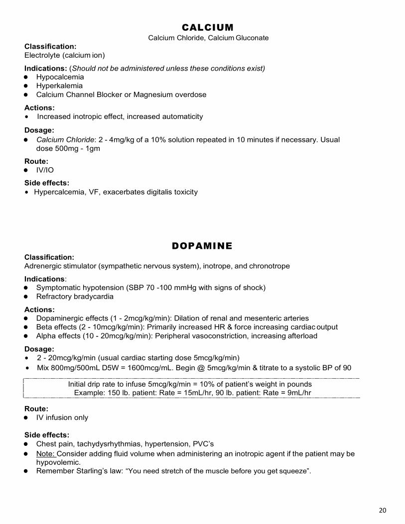
Classification: Electrolyte (calcium ion)
CALCIUM Calcium Chloride, Calcium Gluconate
Indications: (Should not be administered unless these conditions exist) • Hypocalcemia • Hyperkalemia • Calcium Channel Blocker or Magnesium overdose
Actions: • Increased inotropic effect, increased automaticity
Dosage: • Calcium Chloride: 2 - 4mg/kg of a 10% solution repeated in 10 minutes if necessary. Usual
dose 500mg - 1gm
Route: • IV/IO
Side effects: • Hypercalcemia, VF, exacerbates digitalis toxicity
Classification:
DOPAMINE
Adrenergic stimulator (sympathetic nervous system), inotrope, and chronotrope
Indications: • Symptomatic hypotension (SBP 70 -100 mmHg with signs of shock) • Refractory bradycardia
Actions: • Dopaminergic effects (1 - 2mcg/kg/min): Dilation of renal and mesenteric arteries • Beta effects (2 - 10mcg/kg/min): Primarily increased HR & force increasing cardiac output • Alpha effects (10 - 20mcg/kg/min): Peripheral vasoconstriction, increasing afterload
Dosage: • 2 - 20mcg/kg/min (usual cardiac starting dose 5mcg/kg/min) • Mix 800mg/500mL D5W = 1600mcg/mL. Begin @ 5mcg/kg/min & titrate to a systolic BP of 90
Route: • IV infusion only
Side effects: • Chest pain, tachydysrhythmias, hypertension, PVC’s • Note: Consider adding fluid volume when administering an inotropic agent if the patient may be
hypovolemic. • Remember Starling’s law: “You need stretch of the muscle before you get squeeze”.

21
Classification:
NOREPINEPHRINE Levophed
Adrenergic stimulator (sympathetic nervous system). Vasopressor Indications: • Hypotension refractory to Dopamine • SBP < 70 mmHg and low peripheral resistance
Actions: • Primarily alpha effects causing an increase in systemic vascular resistance through
vasoconstriction Dosage: • Mix 4mg/250ml D5W or NS = 16mcg/mL • Begin infusion at 0.1-0.5mcg/kg/min (8-40mg=30-150mL/hr)
Route: • IV infusion only
Side Effects: • Increased myocardial work and oxygen consumption. May cause tachycardia and myocardial ischemia. Severe tissue necrosis if infiltrated
NITROPRUSSIDE Nipride
Classification: Antianginal, antihypertensive
Indications: • Hypertension • CHF with PE
Actions: • Smooth muscle dilator causing a decrease in preload, afterload, and a resulting increase in venous pooling. Works more on the arterial side than nitroglycerine
Dosage: • Infusion: Mix 50mg/250mL D5W (200mcg/mL) and start at 0.5 – 8 mcg/kg/min (start at: 15mL/hr)
Route: • IV infusion only
Side effects: • Hypotension, headache, thiocyanate toxicity possible when metabolized
22
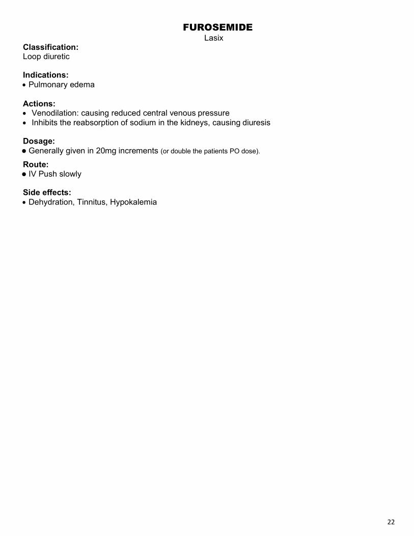
Classification: Loop diuretic
Indications: • Pulmonary edema
FUROSEMIDE Lasix
Actions: • Venodilation: causing reduced central venous pressure • Inhibits the reabsorption of sodium in the kidneys, causing diuresis
Dosage: • Generally given in 20mg increments (or double the patients PO dose).
Route: • IV Push slowly
Side effects: • Dehydration, Tinnitus, Hypokalemia

23
Classification: Antianginal, Antihypertensive
Indications:
NITROGLYCERIN Nitrostat, Tridil
• Angina, MI, CHF (provided patient has SBP > 90 mmHg) Actions: • Smooth muscle dilator causing a decrease in preload, afterload, and a resulting increase in venous pooling, thus reducing the workload of the myocardium • May also reduce coronary artery vasospasm Dosage: • Tablet or metered spray: 1 SL (0.3 - 0.4mg dose) every 5 minutes • Infusion: 10 mcg/min to start (Mix 50mg/250mL = 200mcg/mL. Start at approx. 3mL/hr & titrate) Route: • SL, IV infusion
Side effects: • Hypotension, headache, tachycardia following hypotension
Caution / Avoid: • Erectile Dysfunction Drugs ex: Viagra & Levitra (24hrs), Cialis (48hrs), Right Ventricular Infarct,
Brady or Tachy (without CHF)
Classification: Narcotic analgesic
Indications:
MORPHINE
• Chest pain during STEMI not relieved by 3 doses of NTG • Pulmonary edema
Actions: • Potent analgesic • Promotes venous pooling causing a decrease in preload • Reduces anxiety
Dosage: • 2 - 4mg increments
Route: • IV push slowly
Side effects: • Respiratory depression, Hypotension, Nausea • Use with caution in unstable angina / Non ST elevated patients (mortality increase noted)

24
Classifications: Anticoagulant, antipyretic, analgesic
Indications: • Chest discomfort of cardiac nature • Unstable angina
ASPIRIN
Actions: • Blocks formation of thromboxin A2 which is responsible for platelet aggregation and vasoconstriction,
thus keeping platelets from becoming lodged in partially occluded coronary vessels. Route: • Oral
Dosage: • 160 - 325mg chewable tablets
BETA BLOCKERS Metoprolol (Lopressor), Sotolol (Betapace), Esmolol (Breviblock)
Classification: Beta Adrenergic Blocker
Indications: • Secondary management ACS after patient is stable; usually 6-8 hours • Supraventricular tachydysrhythmias, refractory to other therapies
Actions: • Decreases heart rate, stroke volume, automaticity, and conductivity
Dosage: • Metoprolol (Lopressor): 5mg; may repeat in 5 minutes to max of 15mg • Sotolol (Betapace): 100mg over 5 minutes (for VT) • Esmolol (Brevibloc): Load with 500mcg/kg over 1 min, then maintenance =
50mcg/kg over 4 min, (may repeat loading and increase maintenance if unsuccessful) Route: • Depends on the drug
*Oral doses are generally used unless acutely hypertensive Contraindications: • CHF, Hypotension, Asthma, Bradycardia, Heart Blocks
25
Classification: Anticoagulant Indications:
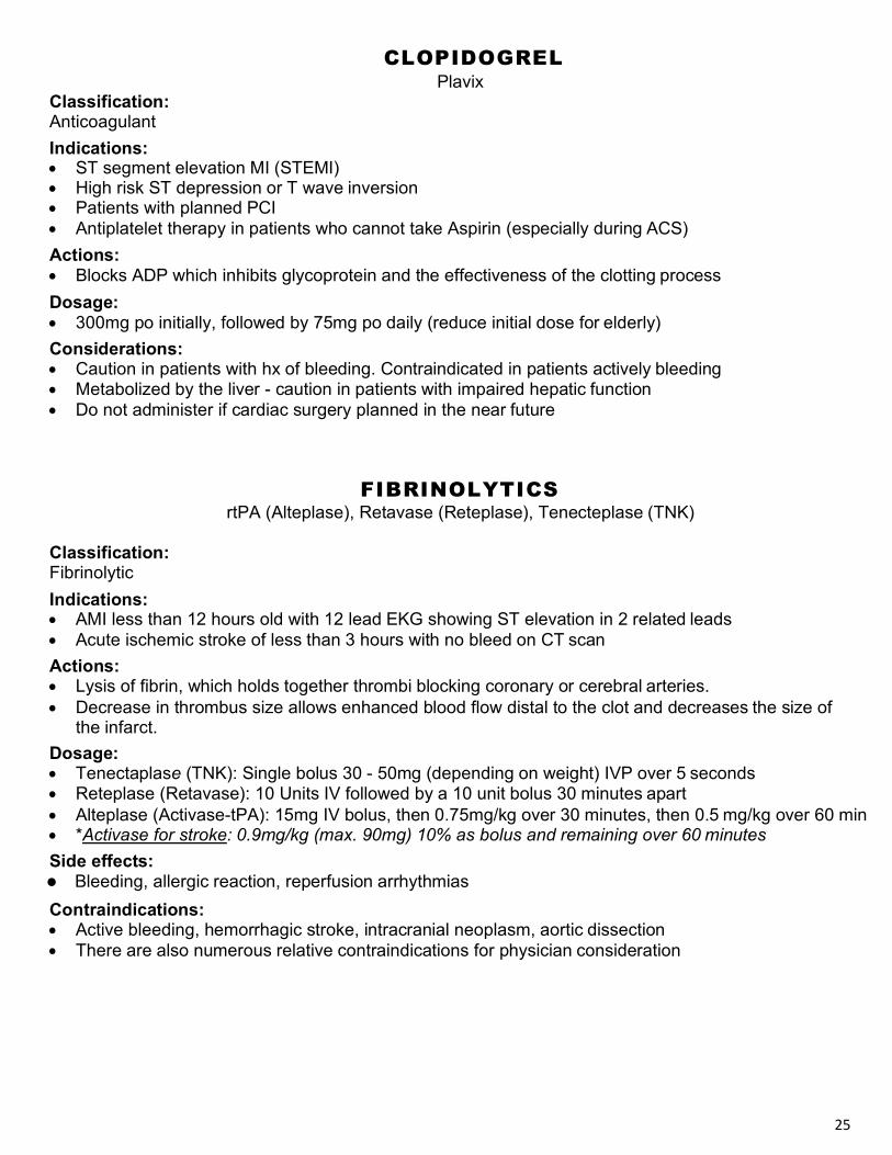
CLOPIDOGREL Plavix
• ST segment elevation MI (STEMI) • High risk ST depression or T wave inversion • Patients with planned PCI • Antiplatelet therapy in patients who cannot take Aspirin (especially during ACS) Actions: • Blocks ADP which inhibits glycoprotein and the effectiveness of the clotting process Dosage: • 300mg po initially, followed by 75mg po daily (reduce initial dose for elderly) Considerations: • Caution in patients with hx of bleeding. Contraindicated in patients actively bleeding • Metabolized by the liver - caution in patients with impaired hepatic function • Do not administer if cardiac surgery planned in the near future
Classification: Fibrinolytic Indications:
FIBRINOLYTICS
rtPA (Alteplase), Retavase (Reteplase), Tenecteplase (TNK)
• AMI less than 12 hours old with 12 lead EKG showing ST elevation in 2 related leads • Acute ischemic stroke of less than 3 hours with no bleed on CT scan Actions: • Lysis of fibrin, which holds together thrombi blocking coronary or cerebral arteries. • Decrease in thrombus size allows enhanced blood flow distal to the clot and decreases the size of
the infarct. Dosage: • Tenectaplase (TNK): Single bolus 30 - 50mg (depending on weight) IVP over 5 seconds • Reteplase (Retavase): 10 Units IV followed by a 10 unit bolus 30 minutes apart • Alteplase (Activase-tPA): 15mg IV bolus, then 0.75mg/kg over 30 minutes, then 0.5 mg/kg over 60 min • *Activase for stroke: 0.9mg/kg (max. 90mg) 10% as bolus and remaining over 60 minutes Side effects: • Bleeding, allergic reaction, reperfusion arrhythmias Contraindications: • Active bleeding, hemorrhagic stroke, intracranial neoplasm, aortic dissection • There are also numerous relative contraindications for physician consideration
26
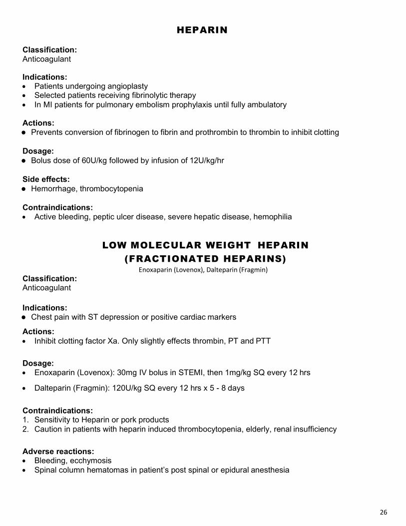
HEPARIN
Classification: Anticoagulant
Indications: • Patients undergoing angioplasty • Selected patients receiving fibrinolytic therapy • In MI patients for pulmonary embolism prophylaxis until fully ambulatory
Actions: • Prevents conversion of fibrinogen to fibrin and prothrombin to thrombin to inhibit clotting
Dosage: • Bolus dose of 60U/kg followed by infusion of 12U/kg/hr
Side effects: • Hemorrhage, thrombocytopenia
Contraindications: • Active bleeding, peptic ulcer disease, severe hepatic disease, hemophilia
LOW MOLECULAR WEIGHT HEPARIN (FRACTIONATED HEPARINS)
Enoxaparin (Lovenox), Dalteparin (Fragmin) Classification: Anticoagulant
Indications: • Chest pain with ST depression or positive cardiac markers
Actions: • Inhibit clotting factor Xa. Only slightly effects thrombin, PT and PTT
Dosage: • Enoxaparin (Lovenox): 30mg IV bolus in STEMI, then 1mg/kg SQ every 12 hrs
• Dalteparin (Fragmin): 120U/kg SQ every 12 hrs x 5 - 8 days
Contraindications: 1. Sensitivity to Heparin or pork products 2. Caution in patients with heparin induced thrombocytopenia, elderly, renal insufficiency
Adverse reactions: • Bleeding, ecchymosis • Spinal column hematomas in patient’s post spinal or epidural anesthesia

27
GLYCOPROTEIN IIb/IIIa INHIBITORS ReoPro (Abciximab), Aggrastat (Tirofiban), Integrilin (Eptifabide)
Indications:
• Chest pain with ST segment depression • Non Q wave MI • Unstable Angina
Action: • Blocks enzyme glycoprotein IIb/IIIa, which is essential for platelet aggregation
Dosage: • Eptifabide (Integrelin): 180mcg/kg IV over 1 - 2 min followed by infusion of 2mcg/kg/min
(decrease to 0.5mcg/min pre cardiac cath). Drug available in 100mL bolus vials and 100mL infusion vials, which can be spiked directly for administration.
• Tirofiban (Aggrastat): Infuse 0.4mcg/kg/min x 30 min and then 0.1mcg/kg/min for 18 - 24 hrs • Abciximab (ReoPro): 0.25mg/kg IV followed by infusion of 1mcg/min for 18 - 24 hrs
Side effects: • Bleeding (more likely in females, pt < 75 lbs, > 65yr, hx of GI disease, or receiving fibrinolytics) • Nausea, vomiting, hypotension, bradycardia • Further risk of bleeding when used in combination with Aspirin and Heparin
Contraindications: • Active internal bleeding / bleeding in past 30 days. Platelets < 100,000 • B/P Systolic >180, Diastolic >100
Classification: Antihypertensive
Action:
ACE INHIBITORS Enalapril (Vasotec), Captopril (Capoten), Lisinopril (Prinivil)
• Selectively suppresses the renin-angiotensin-aldosterone system • Inhibits conversion of angiotensin I to angiotensin II, resulting in dilation of arterial & venous vessels • Attenuates cardiac remodeling post MI
Indications: • Hypertension, CHF • Post MI (first 24 hours then long term)
Dosage: • Vasotec: 5 - 40mg po Q day, 0.625 - 1.25mg IV over 5 min every 6hr • Capoten: 12.5 - 50mg po BID/TID • Prinivil: 10 - 40mg po Q day
Route: • IV, PO
Side effects: • Hypotension, chest pain, tachycardia, dysrhythmias

28
Continual CO2 Monitoring Ø Good indicator of CPR qual ity
Ø CO2 waveforms provide a more sensit ive and rapid evaluation of respiratory
funct ion than pulse oximetry
Ø Specif ica lly evaluates perfusion at the alveol i level
Ø Normal CO2 is 35-40 mmhg
Ø High CO2 denotes respiratory acidosis =venti late more effectively and more frequently
Ø Low CO2 dur ing cardiac arrest indicates low perfusion =may be common dur ing arrest due to CPR being the only perfusion
Ø In a code, attempt to mainta in CO2 above 10 mmhg ( ideally 20’s)
Use End Tidal CO2 (after intubation) to evaluate:
Ø ET tube placement ( is there any CO2?)
Ø Effect iveness of Compressions ( is the CO2 level above 10 mmhg?) If not , evaluate compressions.
Ø Terminat ion Cri teria: Cont inual CO2 <10 for 20 min= “0” survival

29
VENTRICULAR FIBRILLATION
or
PULSELESS VENTRICULAR TACHYCARDIA
“Circle of Life” Core concepts of Resuscitation
Assess CAB's and Begin CPR Attach monitor / defibrillator
Defibrillate (*device specific dose)
Administer Oxygen Continue CPR in 2 minute cycles
â [Secondary procedures]
Secure Airway and Establish IV or IO with NS or LR during CPR
â ������������ @ device specific
dose Continue CPR 2 minutes
â Given during CPR
Epinephrine 1mg (Continue Epinephrine Q 3-5 min.)
â *Defibrillate @ device specific dose
Continue CPR 2 minutes â
Antidysrhythmic of choice Given during CPR
Amiodarone 300mg OR Lidocaine 1-1.5 mg/kg
â *Defibrillate @ device specific dose
Continue CPR 2 minutes
Repeat Sequence of CPR 2 min- Defibrillate-1 Medication Repeat Epinephrine Q 3-5 minutes
Repeat Amiodarone 150mg 1x OR
Lidocaine 0.5 - 0.75mg/kg up to 3mg/kg max
Evaluate for & treat reversible causes anytime during the
sequence
E Quick tip
The sequence should be: Hypoxia Toxins (overdose) Hypovolemia Thrombosis - Pulmonary Hydrogen ion (Acidosis) Thrombosis - Coronary Hyper/Hypokalemia Tamponade - (Cardiac) Hypothermia Tension Pneumothorax
CPR à Drug à Shock àCPR
E Tips for successfully managing this case:
< Don’t forget:
§ Continue CPR § Throughout and
for 2min between shocks
§ Monitor for effective CPR - Use ETCo2
§ 2” compression § Full recoil § No rush to
intubate § Start/upgrade IV
or IO § Gather focused
history
Primary goal: continue effective CPR followed by rotating medications.
*Verbalize appropriate drug, dose, route, flush, and reevaluate patient every 2 minutes.
Once a rhythm is restored, maintain ventilations as appropriate then stabilize in order: 1. rate 2. rhythm 3. blood pressure
*Device specific dose relates to the type and brand of defibrillator used and may range from 120 joules to 360 joules depending on your specific machine.
*Low energy biphasic 120 – 150J *Standard biphasic 200 – 360J *Monophasic 360 J *If unknown, use max dose
*Subsequent shocks may be at the same or higher dose.
*Become familiar with the recommendations of your specific defibrillator
30
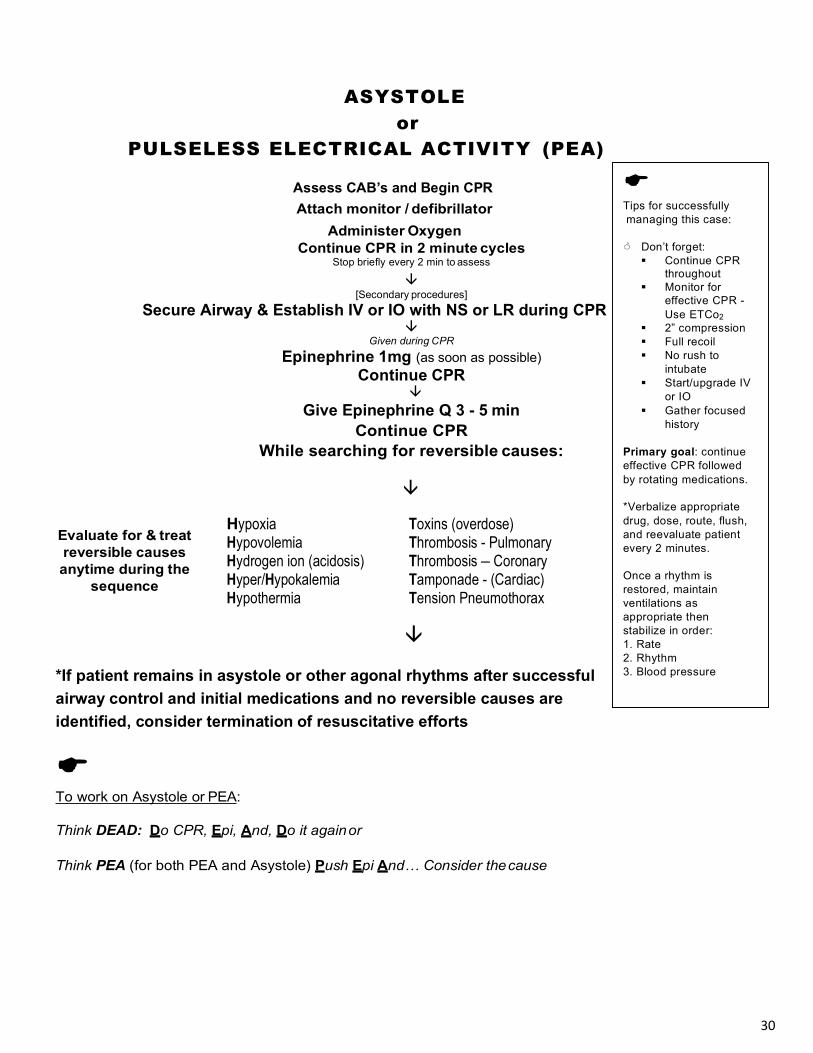
ASYSTOLE
or PULSELESS ELECTRICAL ACTIVITY (PEA)
Assess CAB’s and Begin CPR
Attach monitor / defibrillator Administer Oxygen
Continue CPR in 2 minute cycles Stop briefly every 2 min to assess
â [Secondary procedures]
Secure Airway & Establish IV or IO with NS or LR during CPR â
Given during CPR Epinephrine 1mg (as soon as possible)
Continue CPR â
Give Epinephrine Q 3 - 5 min Continue CPR
While searching for reversible causes:
â
Evaluate for & treat reversible causes anytime during the
sequence
Hypoxia Toxins (overdose) Hypovolemia Thrombosis - Pulmonary Hydrogen ion (acidosis) Thrombosis – Coronary Hyper/Hypokalemia Tamponade - (Cardiac) Hypothermia Tension Pneumothorax
â
*If patient remains in asystole or other agonal rhythms after successful airway control and initial medications and no reversible causes are identified, consider termination of resuscitative efforts
E To work on Asystole or PEA:
Think DEAD: Do CPR, Epi, And, Do it again or
Think PEA (for both PEA and Asystole) Push Epi And… Consider the cause
E Tips for successfully managing this case:
< Don’t forget:
§ Continue CPR throughout
§ Monitor for effective CPR - Use ETCo2
§ 2” compression § Full recoil § No rush to
intubate § Start/upgrade IV
or IO § Gather focused
history
Primary goal: continue effective CPR followed by rotating medications.
*Verbalize appropriate drug, dose, route, flush, and reevaluate patient every 2 minutes.
Once a rhythm is restored, maintain ventilations as appropriate then stabilize in order: 1. Rate 2. Rhythm 3. Blood pressure
31
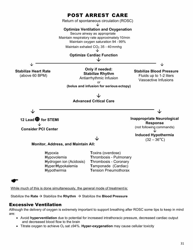
POST ARREST CARE
Return of spontaneous circulation (ROSC) â
Optimize Ventilation and Oxygenation Secure airway as appropriate
Maintain respiratory rate approximately 10/min Maintain oxygen saturation 94 - 99%
Maintain exhaled CO2 35 - 40 mmhg â
Optimize Cardiac Function â
â â
Stabilize Heart Rate (above 60 BPM)
Only if needed: Stabilize Rhythm
Antiarrhythmic Infusion or
(bolus and infusion for serious ectopy)
Stabilize Blood Pressure Fluids up to 1-2 liters Vasoactive Infusions
â Advanced Critical Care
â â
12 Lead Å for STEMI â
Consider PCI Center
Inappropriate Neurological Response
(not following commands) â
Induced Hypothermia â (32 – 36°C)
Monitor, Address, and Maintain All:
Hypoxia Toxins (overdose) Hypovolemia Thrombosis - Pulmonary Hydrogen ion (Acidosis) Thrombosis - Coronary Hyper/Hypokalemia Tamponade (Cardiac) Hypothermia Tension Pneumothorax
E While much of this is done simultaneously, the general mode of treatment is:
Stabilize the Rate à Stabilize the Rhythm à Stabilize the Blood Pressure
Excessive Ventilation Although the delivery of oxygen is extremely important to support breathing after ROSC some tips to keep in mind are:
• Avoid hyperventilation due to potential for increased intrathoracic pressure, decreased cardiac output and decreased blood flow to the brain
• Titrate oxygen to achieve O2 sat ≥94%. Hyper-oxygenation may cause cellular toxicity
32
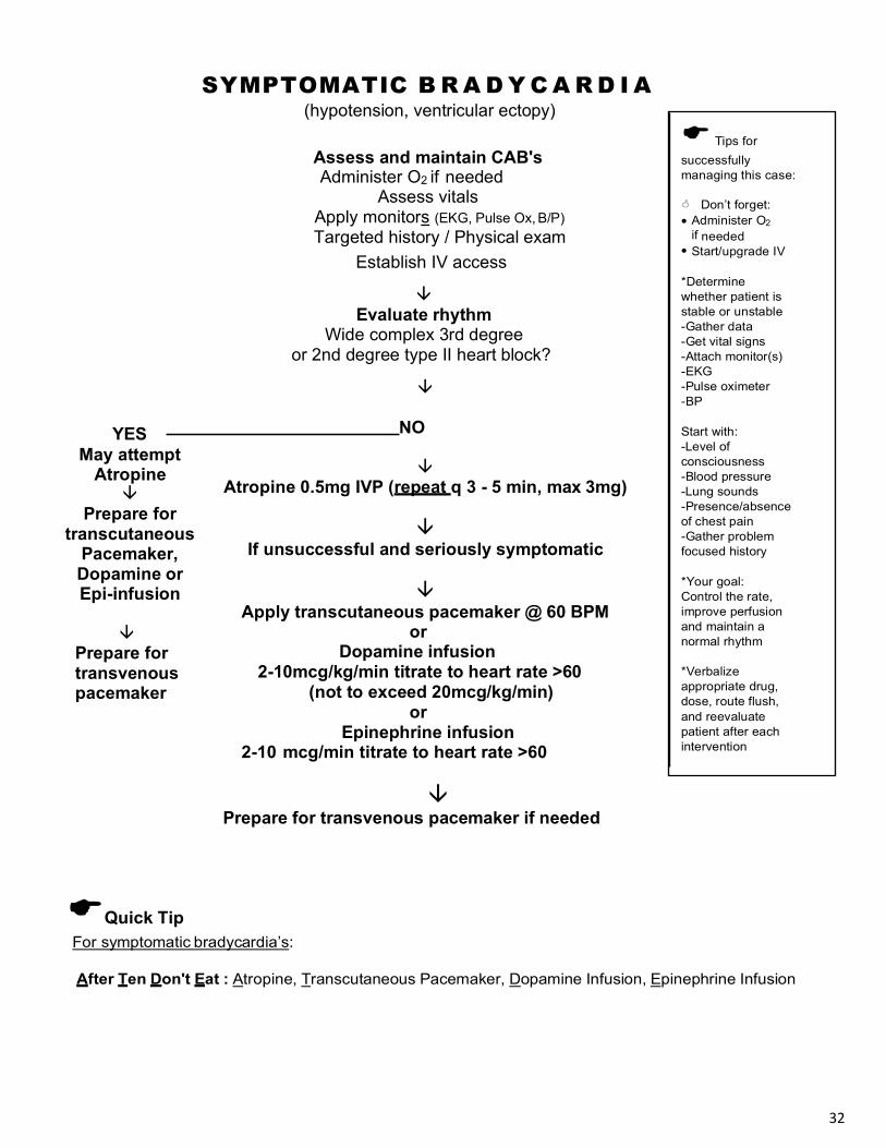
SYMPTOMATIC B R A D Y C A R D I A
(hypotension, ventricular ectopy)
Assess and maintain CAB's Administer O2 if needed
Assess vitals Apply monitors (EKG, Pulse Ox, B/P) Targeted history / Physical exam
Establish IV access
â Evaluate rhythm
Wide complex 3rd degree or 2nd degree type II heart block?
â NO
â Atropine 0.5mg IVP (repeat q 3 - 5 min, max 3mg)
â
If unsuccessful and seriously symptomatic
â Apply transcutaneous pacemaker @ 60 BPM
or Dopamine infusion
2-10mcg/kg/min titrate to heart rate >60 (not to exceed 20mcg/kg/min)
or Epinephrine infusion
2-10 mcg/min titrate to heart rate >60
â Prepare for transvenous pacemaker if needed
EQuick Tip For symptomatic bradycardia’s:
After Ten Don't Eat : Atropine, Transcutaneous Pacemaker, Dopamine Infusion, Epinephrine Infusion
2
YES May attempt
Atropine â
Prepare for transcutaneous
Pacemaker, Dopamine or Epi-infusion
â
Prepare for transvenous pacemaker

33
Bradycardias
Bradycardias are treated if the patient is symptomatic. ie,- has signs of poor perfusion or PVC’s

34
Heart Blocks
In the acute setting, heart blocks are treated as bradycardias. However, there is some controversy over whether to use Atropine in the MI setting. Also, for wide 3o blocks and 2o
type II blocks, many experts choose to avoid Atropine and apply the pacemaker or chronotripic infusions.

35
For Stabilization of Rhythm after VF or VT Conversion Evidence recommends t reating the under lying cause rather than t reating the PVC’s unless the
PVC’s occur f requently o r in groups (i.e. Salvos or VT). “Routine use not recommended” .
Assess and maintain CAB’s
Administer O2 if needed Assess vitals
Apply monitors (EKG, B/P, Resp Pulse Ox)
Targeted history/ Physical exam Establish IV access
Look for underlying causes and consider whether pharmacologic intervention is appropriate. If indicated:
â Antidysrhythmic of choice May bolus if not already done
Otherwise, move to infusion section below Amiodarone 150mg over 10 min
OR Lidocaine 0.5-1.5mg/kg
â
Repeat antidysrhythmic if needed
â If effective, consider an antidysrhythmic infusion of the agent used in the bolus
Infusions: Amiodarone 1mg/min for 6 hours (900mg in 500mLs @ 33mLs/hr)
Then 0.5mg/min for 18 hours (17mLs/hr) OR
Lidocaine or Procainamide 1 - 4mg/min (2grams in 500mLs @ 15mLs/hr)
E Quick tip Generally choose only 1 antidysrhythmic until expert consult:
• Amiodarone bolus can be given 1x then repeated every 10 minutes (max 2.2 grams in 24 hrs)
• Lidocaine bolus 1 - 1.5mg/kg then repeated @ half doses to max of 3mg/kg
• Procainamide 20 - 50mg/min to max of 17mg/kg
• Magnesium 1 - 2 grams over several minutes (ok to mix with others if needed)

36
HYPOTENSION (Symptomatic with systolic < 90 mmHg)
Assess and maintain CAB’s
Administer O2 if needed Assess vitals
Apply monitors (EKG, B/P, Resp Pulse Ox) Review history/ Physical exam
Establish IV access
â Administer fluid bolus’ (1-2 liters)
(If lung sounds are clear) â
If needed and lung sounds are still clear Repeat fluid bolus
â
ß Reassess BP à If still low
â
Dopamine drip 2-10mcg/kg/min (generally start at 5mcg/kg/min)
(not to exceed 20mcg/kg/min)
*Reminder: Treat the rate, then the rhythm, then the blood pressure
E Quick tip: If hypotension is caused by a dysrhythmia, FIX THE RHYTHM:
• Try to identify cause of hypotension (hypovolemia, pump failure, profound vasodilation) to help identify the most effective treatment
• Watch for unwanted cardiac symptoms such as tachycardia or ectopy when using Norepinephrine,
Dopamine, or Dobutamine
May Consider: Norepinephrine:
If SBP <70 and patient has signs of shock 0.1-0.5 mcg/kg/min
May Consider: Epinephrine infusion
0.1-0.5 mcg/kg/min
37
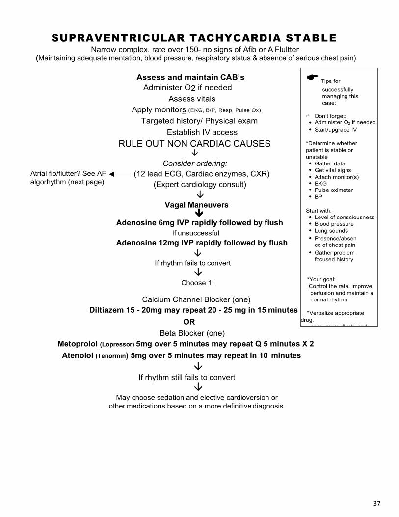
SUPRAVENTRICULAR TACHYCARDIA STABLE Narrow complex, rate over 150- no signs of Afib or A Flultter
(Maintaining adequate mentation, blood pressure, respiratory status & absence of serious chest pain)
Assess and maintain CAB’s Administer O2 if needed
Assess vitals Apply monitors (EKG, B/P, Resp, Pulse Ox)
Targeted history/ Physical exam Establish IV access
RULE OUT NON CARDIAC CAUSES â
Consider ordering: Atrial fib/flutter? See AF algorhythm (next page)
(12 lead ECG, Cardiac enzymes, CXR) (Expert cardiology consult) â
Vagal Maneuvers â
Adenosine 6mg IVP rapidly followed by flush If unsuccessful
Adenosine 12mg IVP rapidly followed by flush â
If rhythm fails to convert â
Choose 1:
Calcium Channel Blocker (one) Diltiazem 15 - 20mg may repeat 20 - 25 mg in 15 minutes
OR Beta Blocker (one)
Metoprolol (Lopressor) 5mg over 5 minutes may repeat Q 5 minutes X 2 Atenolol (Tenormin) 5mg over 5 minutes may repeat in 10 minutes â If rhythm still fails to convert â
May choose sedation and elective cardioversion or other medications based on a more definitive diagnosis
2

38
ATRIAL FIBRILLATION STABLE WITH RAPID VENTRICULAR RESPONSE
Sustained rate over 150 (maintaining adequate mentation, blood pressure, respiratory status, & absence of chest pain)
Assess and maintain CAB’s
Administer O2 if needed Assess vitals
Apply monitors (EKG, B/P, Resp Pulse Ox)
Targated history/ Physical exam Establish IV access
â Consider ordering:
(12 lead ECG, Cardiac enzymes, CXR) (Expert cardiology consult) â
Control rate with: Choose 1: Calcium Channel Blocker
Diltiazem 15 - 20mg may repeat 20 - 25mg in 15 minutes (consider infusion)
OR Beta Blocker
Metoprolol (Lopressor) 5mg over 5 minutes may repeat Q 5 minutes X 2 May choose other Beta blockers: Atenolol, Esmolol
Convert rhythm after expert cardiology consult? Duration of fib?
â â
<48 hrs >48 hrs â â
Convert rhythm by the Delay rhythm conversion unless unstable: same means as the patient R/O emboli or Anticoagulation up to 4 weeks who had emboli ruled out *Once emboli R/O, May consider any of the
following: 1. Elective cardioversion.
~ Start: 120 - 200J Biphasic / 200J Monophasic 2. Amiodarone 150mg over 10min then infusion 3. Digitalis 10 - 15mcg/kg (0.5 - 1.0mg)
*Be cautious with medications that may convert A-fib prior to cardiac consult (Amiodarone)
2
39
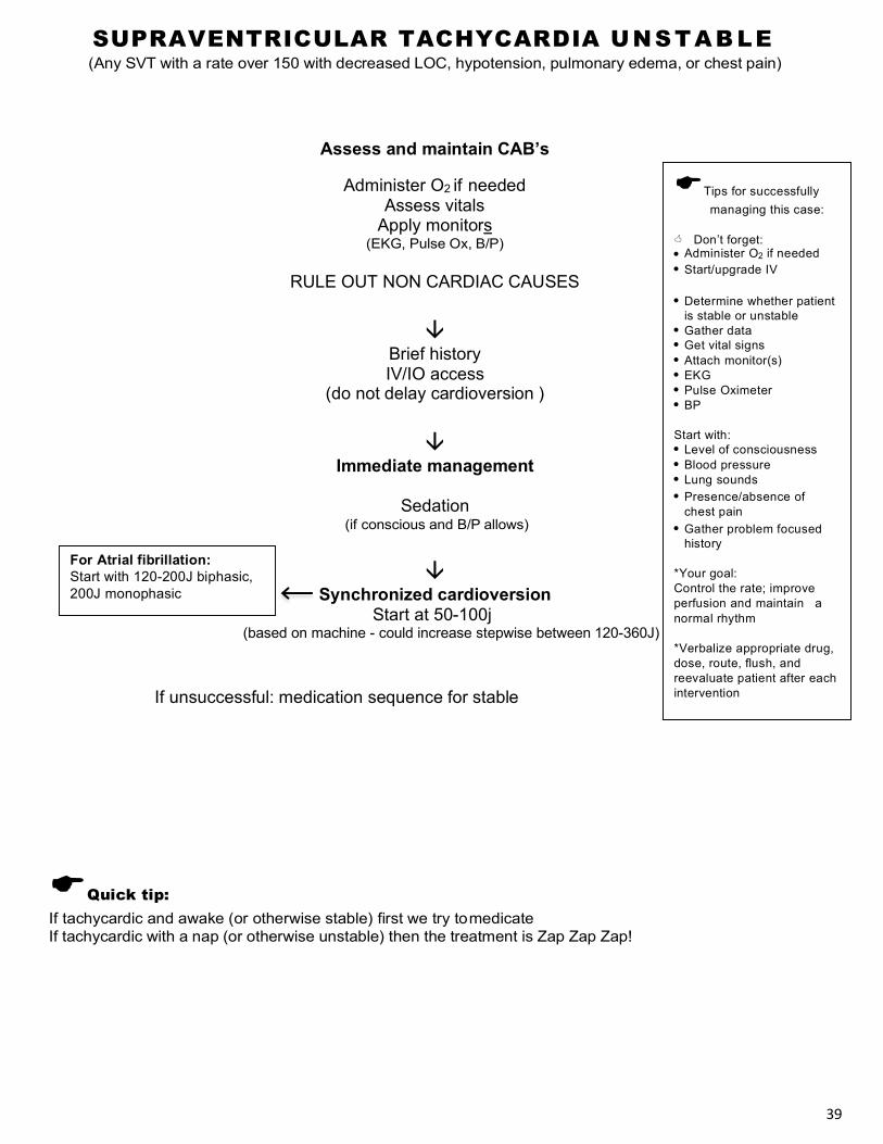
SUPRAVENTRICULAR TACHYCARDIA UNSTAB LE (Any SVT with a rate over 150 with decreased LOC, hypotension, pulmonary edema, or chest pain)
Assess and maintain CAB’s
Administer O2 if needed Assess vitals
Apply monitors (EKG, Pulse Ox, B/P)
RULE OUT NON CARDIAC CAUSES
â
Brief history IV/IO access
(do not delay cardioversion )
â Immediate management
Sedation
(if conscious and B/P allows)
â Synchronized cardioversion
Start at 50-100j (based on machine - could increase stepwise between 120-360J)
If unsuccessful: medication sequence for stable
EQuick tip: If tachycardic and awake (or otherwise stable) first we try to medicate If tachycardic with a nap (or otherwise unstable) then the treatment is Zap Zap Zap!
ETips for successfully managing this case:
< Don’t forget: • Administer O2 if needed • Start/upgrade IV
• Determine whether patient
is stable or unstable • Gather data • Get vital signs • Attach monitor(s) • EKG • Pulse Oximeter • BP
Start with: • Level of consciousness • Blood pressure • Lung sounds • Presence/absence of
chest pain • Gather problem focused
history
*Your goal: Control the rate; improve perfusion and maintain a normal rhythm
*Verbalize appropriate drug, dose, route, flush, and reevaluate patient after each intervention
For Atrial fibrillation: Start with 120-200J biphasic, 200J monophasic
40
ycardia and awake, first we must medicate. If tachycardia with a nap...then the treatment's zap zap zap
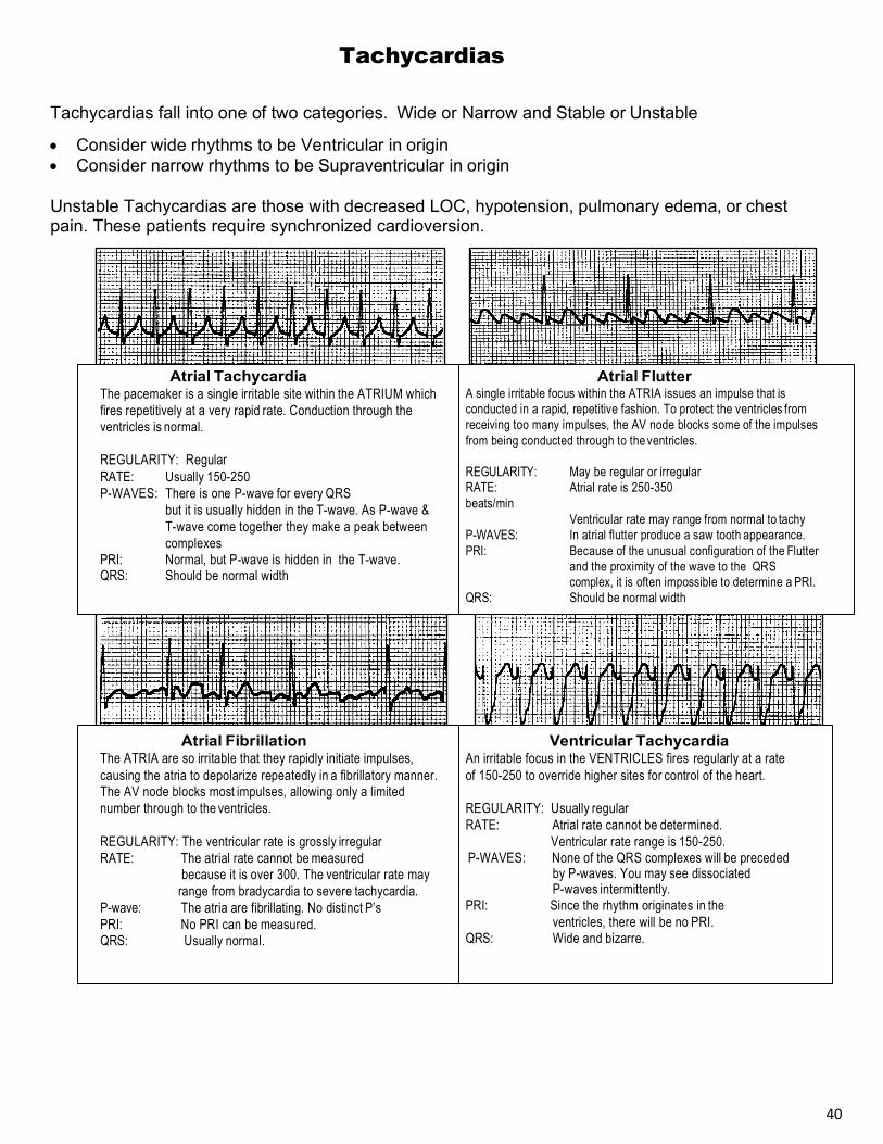
Tachycardias
Tachycardias fall into one of two categories. Wide or Narrow and Stable or Unstable
• Consider wide rhythms to be Ventricular in origin • Consider narrow rhythms to be Supraventricular in origin
Unstable Tachycardias are those with decreased LOC, hypotension, pulmonary edema, or chest pain. These patients require synchronized cardioversion.
Atrial Tachycardia The pacemaker is a single irritable site within the ATRIUM which fires repetitively at a very rapid rate. Conduction through the ventricles is normal.
REGULARITY: Regular RATE: Usually 150-250
P-WAVES: There is one P-wave for every QRS but it is usually hidden in the T-wave. As P-wave & T-wave come together they make a peak between complexes
PRI: Normal, but P-wave is hidden in the T-wave. QRS: Should be normal width
Atrial Flutter
A single irritable focus within the ATRIA issues an impulse that is conducted in a rapid, repetitive fashion. To protect the ventricles from receiving too many impulses, the AV node blocks some of the impulses from being conducted through to the ventricles.
REGULARITY: May be regular or irregular RATE: Atrial rate is 250-350 beats/min
Ventricular rate may range from normal to tachy P-WAVES: In atrial flutter produce a saw tooth appearance. PRI: Because of the unusual configuration of the Flutter
and the proximity of the wave to the QRS complex, it is often impossible to determine a PRI.
QRS: Should be normal width
Atrial Fibrillation The ATRIA are so irritable that they rapidly initiate impulses, causing the atria to depolarize repeatedly in a fibrillatory manner. The AV node blocks most impulses, allowing only a limited number through to the ventricles.
REGULARITY: The ventricular rate is grossly irregular RATE: The atrial rate cannot be measured
because it is over 300. The ventricular rate may range from bradycardia to severe tachycardia.
P-wave: The atria are fibrillating. No distinct P’s PRI: No PRI can be measured. QRS: Usually normal.
Ventricular Tachycardia An irritable focus in the VENTRICLES fires regularly at a rate of 150-250 to override higher sites for control of the heart.
REGULARITY: Usually regular RATE: Atrial rate cannot be determined. Ventricular rate range is 150-250. P-WAVES: None of the QRS complexes will be preceded
by P-waves. You may see dissociated P-waves intermittently.
PRI: Since the rhythm originates in the ventricles, there will be no PRI.
QRS: Wide and bizarre.

41
VENTRICULAR TACHYCARDIA STABLE (Maintaining adequate mentation, blood pressure, respiratory status, and absence of chest pain)
Wide complex, rate over 150, regular with no P waves or signs of A-fib or flutter
Assess and maintain CAB’s Administer O2 if needed
Assess vitals Apply monitors
(EKG, Pulse Ox, B/P) Targeted history/ Physical exam
Establish IV access â
(Consider ordering) (12 lead ECG, Cardiac enzymes, CXR) (Cardiology consult)
â
ß Preferred Antidyshythmic
â Consider the following at any time
â
Sedation and synchronized cardioversion Begin at 100j, and increase PRN.
(based on machine – could increase stepwise between 120-360J)
Prepare an infusion of the antidysrhythmic
medication used if conversion is successful
EQuick tip
Find the cause: Patients don’t have Ventricular Tach because they are low on Amiodarone (or any other antidysrhythmic). Medications are a temporary “Band-Aid” for ventricular irritability, but it is likely to recur if the cause is not diagnosed and treated.
E Tips for successfully managing this case:
< Don’t forget: • Administer O2 if needed • Start/upgrade IV
• Determine whether
patient is stable or unstable
• Gather data • Get vital signs • Attach monitor(s) • EKG • Pulse oximeter • BP
Start with: • Level of consciousness • Blood pressure • Lung sounds • Presence/absence of
chest pain • Gather problem focused
history
*Your goal: Control the rate, improve perfusion and maintain a normal rhythm
*Verbalize appropriate drug, dose, route, flush, and reevaluate patient after each intervention
May use: (generally only one) Procainamide 20-50 mg/min
~Or~ Amiodarone 150 mg IV drip over 10 min May repeat 150 mg IV
~Or~ Sotolol 100 mg over 5 min
~Or~ Lidocaine 0.5-1.5 mg/kg ½ initial dose for repeat dose May repeat to max total 3mg/kg
~Or~ Magnesium 1 - 2 gm IV for Torsades or suspected hypomagnesemia
42
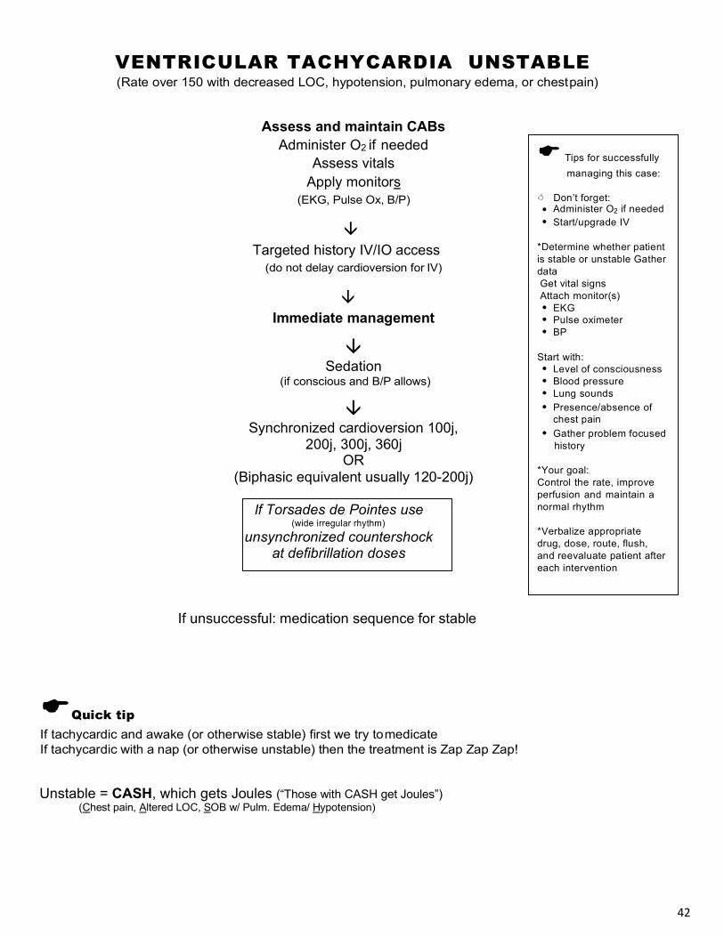
VENTRICULAR TACHYCARDIA UNSTABLE (Rate over 150 with decreased LOC, hypotension, pulmonary edema, or chest pain)
Assess and maintain CABs Administer O2 if needed
Assess vitals Apply monitors
(EKG, Pulse Ox, B/P)
â Targeted history IV/IO access
(do not delay cardioversion for IV)
â Immediate management
â Sedation
(if conscious and B/P allows)
â Synchronized cardioversion 100j,
200j, 300j, 360j OR
(Biphasic equivalent usually 120-200j)
If unsuccessful: medication sequence for stable
EQuick tip If tachycardic and awake (or otherwise stable) first we try to medicate If tachycardic with a nap (or otherwise unstable) then the treatment is Zap Zap Zap!
Unstable = CASH, which gets Joules (“Those with CASH get Joules”)
(Chest pain, Altered LOC, SOB w/ Pulm. Edema/ Hypotension)
If Torsades de Pointes use (wide irregular rhythm)
unsynchronized countershock at defibrillation doses
E Tips for successfully managing this case:
< Don’t forget:
• Administer O2 if needed • Start/upgrade IV
*Determine whether patient is stable or unstable Gather data Get vital signs Attach monitor(s) • EKG • Pulse oximeter • BP
Start with:
• Level of consciousness • Blood pressure • Lung sounds • Presence/absence of
chest pain • Gather problem focused
history
*Your goal: Control the rate, improve perfusion and maintain a normal rhythm
*Verbalize appropriate drug, dose, route, flush, and reevaluate patient after each intervention

43
Nondiagnostic ECG or enzymes, Admit to ED/ chest pain unit
Serial ECGs, Serial cardiac markers
ACUTE CORONARY SYNDROMES Assess and maintain CAB’s Administer O2 only if needed
Assess vitals Apply monitors
(EKG, Pulse Ox, B/P) Targeted history /Physical exam
Establish IV access Perform 12 LEAD ECG
(electrolytes, enzymes-troponin, coags)
â
â â
ECG + for AMI <12 hrs High risk Acute Coronary Syndromes
(ST elevation in 2 or more related leads) -ST depression/T wave inversion -High risk unstable angina
â (female, rales Hx MI, diabetes, -IV Nitroglycerine (continuing ischemia, HTN, PE) hypotension, tachycardia, atrial fib) -Heparin or LMWH -AMI >12 hrs -Ace inhibitors (after 6 hrs) â -B Blockers (after stable) -IV Nitroglycerine
â -Heparin or LMWH Immediate: Prepare patient for: -Antiplatelets (GPIIb/IIIa inhibitors) PCI (Percutaneous Coronary intervention) -Ace inhibitors (after 6 hours) #1 choice for pt, <75 yrs old; -B Blockers (after stable) Cath, Stent, CABG â Ideal first contact to cath time 90 min As Available: Cardiac cath to evaluate OR
â â
Fibrinolytics If suitable for revascularization Ideal door or EMS to drug time 30 min PCI
CABG
Perform simultaneously with initial assessment
Oxygen (to maintain saturation *90 - 99%) Nitroglycerine SL or spray Aspirin Morphine IV if pain not relieved by 3 NTG. Only recommended if STEMI
ETips for successfully managing this case:
< Don’t forget to: • Use a pain scale to help your
patient rate the pain
• Perform PQRST assessment to determine if the cause of pain is likely myocardial ischemia or injury
• Determine time of onset
early
• History/physical should include screening for Fibrinolytic contraindications
• Assess vital signs before and
after administering Nitrates
• Obtain 12 lead EKG early
• Administer Morphine only if Nitro fails to relieve the pain
• Reassess vital signs and
pain frequently

44
Related leads on the ECG: S – Septal: V1, V2 A – Anterior: V3, V4 L – Lateral: V5, V6, I, AVL I – Inferior: II, III, AVF
THE 12 LEAD ECG
“The Imposters”
non AMI causes of ST and QRS changes
Left Bundle branch block QRS > 0.12 sec, QRS inverted in V1, upright in V6, S-T elevation, depression, and T
wave inversion seen throughout. Cannot accurately diagnose MI
Right bundle branch block
QRS 0.12 sec or wider, rSR pattern in V1, (QRS upright in V1), S-T elevation, depression, and T wave inversion may be seen throughout. May be able to detect MI, especially if comparison ECG available
Other causes of Wide QRS Ventricular rhythms, (PVCs, VT), electronic pacemakers, medications, (Quinidine,
Pronestyl), any depolarization abnormality can cause repolarization abnormalities
Left Ventricular hypertrophy Strain pattern of depressed S-T segments, large QRS complexes in chest leads
Digitalis ST segment “sags”. May also be seen with calcium ingestion
Pericarditis Widespread ST elevation, T waves upright, no pathological Q waves, Possible PR interval depression in V6. Clinical correlation is necessary. Look for viral syndrome: fever, malaise. Patient will prefer to lean forward, obtaining some relief
Angina Pectoris Flat (plane) depressions of S-T segment. Inverted T waves possible. ECG changes may
improve with pain relief.
Prinzmetal’s angina Slope elevation of S-T, especially in V4-V6. Changes may resolve with pain relief.
Early repolarization Normal variant. S-T slightly elevated with normal concave slope in most leads. J point is elevated, possibly with “fishhook” appearance.
In 2 or more related leads
Arouses suspicion for injury
Arouses suspicion for ischemia
(may be Angina or early MI)
45
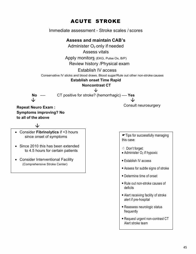
ACUTE STROKE
Immediate assessment - Stroke scales / scores
Assess and maintain CAB’s Administer O2 only if needed
Assess vitals Apply monitors (EKG, Pulse Ox, B/P)
Review history /Physical exam Establish IV access
Conservative IV sticks and blood draws. Blood sugar/Rule out other non-stroke causes Establish onset Time Rapid
Noncontrast CT â
No ---- CT positive for stroke? (hemorrhagic) ---- Yes â â
Repeat Neuro Exam : Symptoms improving? No to all of the above
â
Consult neurosurgery
• Consider Fibrinolytics if <3 hours since onset of symptoms
• Since 2010 this has been extended
to 4.5 hours for certain patients
• Consider Interventional Facility (Comprehensive Stroke Center)
ETips for successfully managing this case:
< Don’t forget: • Administer O2 if hypoxic
• Establish IV access
• Assess for subtle signs of stroke
• Determine time of onset
• Rule out non-stroke causes of deficits
• Alert receiving facility of stroke alert if pre-hospital
• Reassess neurologic status frequently
• Request urgent non-contrast CT Alert stroke team

46
INCLUSION CRITERIA FOR FIBRINOLYTIC THERAPY
CARDIAC
Inclusion criteria:
Inclusion criteria:
STROKE
q Chest pain &/or symptoms of acute MI q QRS duration <120 ms (.12 sec) q ST segment elevation >1mV (1mm) in 2 or
more related leads q II, II, aVF q V1, V2, V3, V4, V5, V6 q I, aVL
Exclusion criteria: q Active internal bleeding q History of CVA/TIA
Recent (< 2 months)
q Intracranial/intraspinal surgery, trauma q Brain tumor, aneurism q Arteriovenous malformation q Bleeding disorder/anticoagulant
Recent (<2 weeks)
q Major surgery q Trauma q Organ biopsy q GI or GU bleeding q Severe uncontrolled HTN (200/120) q Pregnancy/ Menses q Diabetic eye problems &/or other hemorrhagic opthalmic condition q Disoriented, uncooperative q Prolonged/traumatic CPR q Aortic dissection q Allergy to steptokinase
q Diagnosis of ischemic stroke causing measurable neurologic deficit q Onset of symptoms <3 hours before beginning treatment q Age ≥ 18 years Exclusion criteria: q Head trauma or prior stroke in previous 3 months q Symptoms suggest subarachnoid hemorrhage q Arterial puncture at noncompressible site in previous 7 days q History of previous intracranial hemorrhage q Elevated blood pressure (systolic >185 mm Hg or diastolic >110 mm Hg) q Evidence of active bleeding on examination q Acute bleeding diathesis, including but not limited to
-Platelet count <100 000/mm3 -Heparin received within 48 hours, resulting in an aPTT greater than the upper limit of normal -Current use of anticoagulant with INR >1.7 or PT >15 seconds
q Blood glucose concentration <50 mg/dl (2.7 mmol/L) q Ct demonstrates multilobar infarction (hypodensity >1/3 cerebral hemisphere) Relative Exclusion Criteria Patients may receive rtPA but risk/benefit must be carefully weighed if presented with the following: q Only minor or rapidly improving stoke symptoms (clearing spontaneously q Seizure at onset with postictal residual neurologic impairments q Major surgery or serious trauma within previous 14 days q Recent gastrointestinal or urinary tract hemorrhage (within previous 21 days) q Recent acute myocardial infarction (within previous 3 months)
47
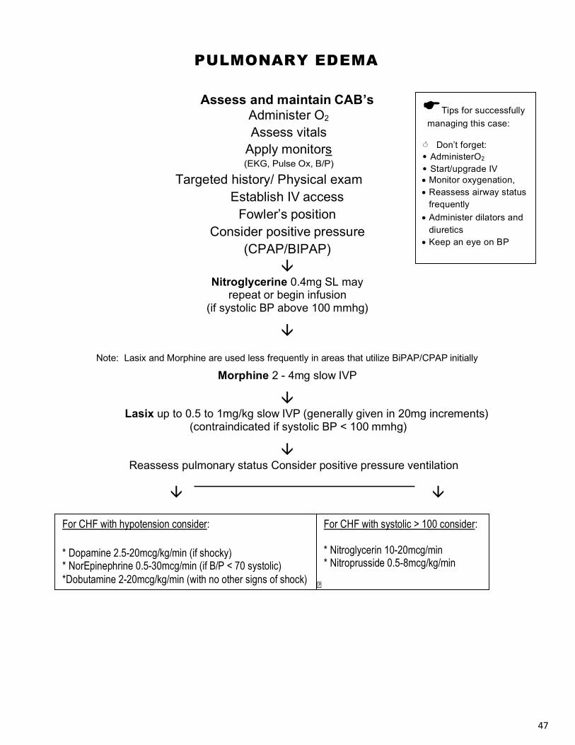
PULMONARY EDEMA
Assess and maintain CAB’s Administer O2
Assess vitals Apply monitors (EKG, Pulse Ox, B/P)
Targeted history/ Physical exam Establish IV access
Fowler’s position Consider positive pressure
(CPAP/BIPAP) â
Nitroglycerine 0.4mg SL may repeat or begin infusion
(if systolic BP above 100 mmhg)
â
Note: Lasix and Morphine are used less frequently in areas that utilize BiPAP/CPAP initially
Morphine 2 - 4mg slow IVP
â Lasix up to 0.5 to 1mg/kg slow IVP (generally given in 20mg increments)
(contraindicated if systolic BP < 100 mmhg)
â Reassess pulmonary status Consider positive pressure ventilation
â â
�
For CHF with hypotension consider:
* Dopamine 2.5-20mcg/kg/min (if shocky) * NorEpinephrine 0.5-30mcg/min (if B/P < 70 systolic) *Dobutamine 2-20mcg/kg/min (with no other signs of shock)
For CHF with systolic > 100 consider:
* Nitroglycerin 10-20mcg/min * Nitroprusside 0.5-8mcg/kg/min
ETips for successfully managing this case:
< Don’t forget: • AdministerO2
• Start/upgrade IV • Monitor oxygenation, • Reassess airway status
frequently • Administer dilators and
diuretics • Keep an eye on BP

48
TIPS FOR DRIPS
The following is merely one of the methods for calculating drip medications. It should not be considered “the only way” or “the ACLS way” to mix and administer infusions. If you are familiar with another method, use what works for you.
1mL
¾ mL
¼ mL
½ mL
FOR EXAMPLE: Antidysrhythmic: mix 2gm in 500mL Cardiac stimulants: Mix 4mg in 250mL
2000mg/500mL=4mg/mL 4000mcg/250mL=16mcg/mL
To use the clock method to calculate your drip rate you must figure the mixed concentration (the amount of drug per mL). This is done by dividing the amount of the fluid volume in the IV bag (500mL, etc) into the supplied drug amount. This number gives you the amount of medication administered per mL. Then, insert this number in the “60” slot on your clock, ½ of it at the 30. ¼ of it at the 15 and ¾ of it at the 45. Remember, when drugs are diluted for infusions, the concentration becomes the next lowest unit (for example, add a gram of drug to a bag and the concentration becomes mg/mL).
Clock Method:
Think of a mini drip chamber or IV pump as a clock. A clock
has 60 seconds in 1 minute.
A drip chamber has 60 drops in 1mL.
4mg/min 60mL/hr
16mcg/min 60mL/hr
3 mg/min 45mL/hr
1mg/min15mL/hr
12mcg/min 45mL/hr 4mcg/min
15mL/hr
2mg/min 30mL/hr
8mcg/min 30mL/hr

49
Special Arrest Situations Other Considerations in ACLS Management
In general, Critical Care Personnel are quite good at performing ACLS skills-intubations, defibrillation, vascular access, and appropriate medical treatment according to ACLS type protocols. However, in our haste to rapidly treat patients, we may miss clues as to why this person arrested and why he is not responding to your best ACLS treatment.
Occasionally, the provider may need to pull some “tricks” out of their drug box based on history labs, bystander information, the scene, and the fact that the patient is not responding to the standard ACLS treatment.
PLEASE NOTE: These are merely recommendations from the AHA guidelines and should not be construed as the only standard. All treatment should be approved by the supervising physician.
Some interesting facts to keep in mind: These things will cause PEA: Pulmonary emboli, Acidosis, AMI, Tension Pneumothorax, Cardiac Tamponade, Hypoxia, Hypovolemia, Hyperkalemia, Drug overdose.
These things will case Asystole: Hypoxia, hypothermia, hypokalemia, hyperkalemia, acidosis, drug overdose, and death. V-Fib or pulseless VT can be caused by anything.
Electrolyte Imbalances Certainly, if labs have been “drawn” or if values are rapidly available this information can be utilized to guide treatment.
Since providers do not all have the ability to rapidly obtain and evaluate ABG’s or blood work, here are a few clues for patients not responding to your routine regime; consider these early:
Renal dialysis patients • May have Pre dialysis Acidosis, Hyperkalemia, Hypoglycemia, or Post dialysis
Hypokalemia & Hypovolemia. Diabetics • May have Acidosis, Hypoglycemia, Hypovolemia, Hyperkalemia, Hypokalemia. Alcoholics • May have Hypokalemia, Hypoglycemia, Hypomagnesemia. Prolonged Vomiting • May have Dehydration, Metabolic, Alkaolsis, Hypokalemia.
Prolonged Diarrhea • May have Dehydration, Acidosis, Hypokalemia, Hypomagnesemia.

50
Management of Electrolyte Related Arrests, In Addition To Standard ACLS, could Include:
Hyperkalemia (>6.5 mmol/L) (One of the most potentially life threatening):
• Most Commonly occurs in renal failure patients, though other conditions can cause “release” from the cells.
While Performing Standard ACLS: 1. Administer Calcium Chloride 500mg-1gram to stabilize myocardial cells
2. Administer Sodium Bicarbonate 50 mEq to shift potassium into the cells
3. A mix of Glucose (25g) and Insulin 10u may be infused over 15 minutes
Hypomagnesemia (< 1.3 mEq/L)
• Most commonly occurs in the malnourished, chronic alcoholic, or chronic diarrhea
• May cause VT, Polymorphic VT (Torsades de point) • Administer 1-2g magnesium IV bolus
Metabolic Acidosis (Ph < 7.35) • Occurs during an extended period of arrest or in a patient who is without CPR for an
extended period of time initially • May occur with medical conditions (Ketoacidosis) or overdoses
• Initially provide adequate CPR and ventilations
• Ideally use ABG’s to guide treatment, but may be given based on history
• Administer Sodium Bicarbonate 1mEq/kg, repeat at half dose
Unique Respiratory Conditions • COPD and asthma have caused arrests in the old and the young from respiratory failure
and acidosis. There have also been numerous cases of Tension Pneumothoraxes.
• The Pneumothorax patient may require pleural decompression if they fail to respond to standard ACLS, have poor BVM compliance, absent lung sounds, and other TPT signs.
• Lateral pressure applied to the chest during exhalation may help expel trapped air and reduce intrathoracic pressure and the incidence of barotrauma during resuscitation.
• Intubated asthma patients in arrest should be ventilated slower and with less tidal volume.

51
Unique Respiratory Conditions (Continued) • Any Intubated patient who deteriorates after stabilization, along with managing cardiac
dysfunction, should be evaluated for the following:
§ Dislodged ET Tube
§ Obstructed ET Tube
§ Pneumothorax
§ Equipment Failure (ventricular)
Drowning
• Hypoxia and Acidosis are the initial causes.
• Consider C-Spine management (if indicated), sodium bicarbonate, and hypothermia. • Hypothermia BLS management
o Protect from heat loss; cover victim. o Begin CPR without delay (even though pulse may be difficult to detect). o Check rectal or tympanic temperature. <86°F(30°C)=Severe, >86°F(30°C)=mild to
moderate.
• Hypothermia ALS management o Perform ALS skills (ET) gently especially in severe hypothermia. o Move towards aggressive core warming (warm lavages, bypass, etc.) as primary. o Withhold antiarrhythmics until core temp is >86°F(30°C). o Perform all other ACLS procedures.
• Do not terminate efforts until temperature is near normal. Trauma Arrests
• Though survivability rates are low, scene management should include spinal immobilization, airway control, bleeding control, and a rapid search for reversible conditions such as Tension Pneumothorax, and V-Fib. IV fluids, and medication should take place enroute to a trauma facility
• Utilize the H’s and T’s to rapidly evaluate reversible causes.
• Some areas may choose not to resuscitate trauma arrests due to poor prognosis.

52
Pregnancy and Obesity
• Be prepared for a difficult airway and a smaller glottic opening • If Pulmonary Embolism is suspected, consider Emergent Fibrinolytics • Do not change the standard ACLS protocols
• Manually, shift fetus to the left. (as effective as tilting and easier to perform CPR).
• Consider C-Section if there is no response to 5 minutes of ACLS
• If the pregnant female was receiving Magnesium, administer 1 gram Calcium Chloride
• Compressions may need to be performed slightly higher on the chest due to the larger abdomen
Anaphylaxis
• Allergic reactions, while usually easily reversible, may progress to cardiovascular collapse due to profound vasodilation and hypoperfusion
• For patients in near arrest or arrest states, administer Epinephrine IV .05-0.1mg
~ rather than SQ or IM, then follow standard ACLS to manage arrhythmias
• Emphasis should be placed on securing an appropriate airway early on • In severe shock, administer large fluid boluses – 1 liter at a time (4-8 liters)
• Vasopressin can be considered in cardiac arrest if Epinephrine is unsuccessful Arrests Related to Overdose
• The initial management for drug induced arrests is basically unchanged. Follow the standard BLS and ACLS regime while searching for reversible causes.
The following will outline consideration in specific overdoses:
Cocaine - Tachyarrhythmias, vasoconstriction, pulmonary edema, seizures, HTN, & hypothermia • SVT: Often short-lived, not requiring therapy
o ~However, for sustained SVT consider administering benzodiazepines (Valium, Ativan, etc.)
• Hemodynamically stable VT: Consider benzodiazepines. If persistent, administer standard antidysrhythmics. Follow with Sodium Bicarbonate 1 mEq/kg IV

53
Arrests Related to Overdoes (Continued)
• Hypertension- Treat initially with benzodiazepine. Follow with vasodilator such as
Nitroglycerine or Nitroprusside. (Nitroglycerine preferred if concurrent chest pain)
• **Do not use b blockers – blocking b stimulus may allow the a blocking properties of
cocaine to function unopposed, potentially increasing blood pressure
~A pure -blocker such as Phentolamine (1mg q 2 - 3 min up to 10mg) may be used
• Pulmonary edema - Standard medical management including + pressure ventilation • Acute Coronary Syndromes - With cocaine use, more often due to spasm rather than
thrombus. Use O2, ASA, NTG, titrated doses of benzodiazepine Tricyclic Antidepressants (Elavil, Tofranil, Amitryptyline, etc.) - Cardiotoxic when overdosed. Expect mental status changes, Tachycardias, Prolonged QT intervals, and anticholinergic effects. Interventions include:
• Symptoms = 3 C’s – Convulsions, Coma, Cardiac dysrhythmia then Acidosis and
Hypotension • Consider activated charcoal in non cardiac arrest within 1 hour of ingestion • Terminate seizures with benzodiazepines • During arrest consider sodium bicarbonate 1mcg/kg • Pre arrest or post arrest cardiovascular collapse with widened RS
o Administer sodium barcarbonate, consider fluid bolus NaCL as needed Digitalis - Overdose may cause bradyarrhythmias and heart failure, ventricular arrhythmias, and hyperkalemia. There is no evidence to support antidotes during cardiac arrest. Pre or post arrest cardio-toxicity treatment should include:
• Activated charcoal within 1 hour of ingestion
• Use standard ACLS treatment for critical bradycardias • Cautious use of transvenous pacemakers due to pacemaker induced dysrhythmias.
• K + >5mEq/L patients have a poor prognosis • Fab fragment therapy (digibind):
o 2 vials per mg of drug ingested o 10-20 vials for unknown amount ingestion
54
Calcium Channel Blocker and b Blocker toxicity - May cause hypotension, decreased contractility,
bradycardias, decreased LOC, seizures, hypoglycemia & hyperkalemia. With b blockers,
hyperglycemia with Ca+ Channel Blockers, rapid progression to shock.
There is no evidence to support antidotes during cardiac arrest. Cardiovascular Toxicity (collapse) pre
or post cardiac arrest could include:
• O2, ECG, monitor BP, establish vascular access • Volume for hypotension • Check blood glucose
• Activated charcoal within 1 hour of ingestion with mild hemodynamic effects
• A mix of high dose Insulin 1u/kg + 0.5g/kg dextrose may improve hemodynamic ability by improving myocardial energy utilization
For Calcium Channel Blocker overdose, to treat myocardial dysfunction
~ (not cardiac arrest): 1. NS boluses 500 - 1000mL
2. Epinephrine infusion 2 - 100 mcg/min
3. Calcium chloride 0.2mL/kg if shock refractory to fluids and Epinephrine
4. May use calcium gluconate 10% (0.3mEq/kg)
5. Pacing for bradycardia For b blocker overdose, to treat myocardial dysfunction (not cardiac arrest):
• NS boluses • Epinephrine infusion 2-100 mcg/min • Calcium chloride 0.2 mL/kg
o May use calcium gluconate 10% (0.3mEq/kg)
• Glucagon 3 - 10mg IV

55
Narcotics or Benzodiazepines - Generally cause CNS and respiratory depression. There is no
indication for treating these conditions during cardiac arrest.
Narcotic (Opioid) Overdose: • No indication to treat during cardiac arrest
• May treat respiratory depression secondary to overdose
• Naloxone (narcan) 0.4mg increments Benzodiazepines: • No indication to treat during cardiac arrest
• May treat respiratory depression secondary to overdose only in known nonhabitual users (i.e.: overdoes given during a procedure)
• Flumazenil 0.2mg increments up to 1mg Cyanide Poisoning • Found in industry and jewelry cleaners • Very common in smoke inhalation from fires
• Causes CNS depression, metabolic acidosis, and cardiovascular collapse • Along with standard BLS and ACLS resuscitation regimes:
§ Administer Cyanide poison kit (carried by EMS, hospital ED and pharmacies) o IV
Sodium Nitrate, IV Sodium Thiosulfate (for cyanide not from toxic smoke) o Cyano kit-
Hydroxocobalamin IV (for any cyanide including toxic smoke)
Local Anesthetic Toxicity
• Accidental IV administration of anesthetics such as Lidocaine and Mepivacaine may cause toxicity, seizures and cardiovascular collapse
• A rapid IV of 20% long chain fatty acid emulsion (LipidRescue™) may redistribute the toxin or stabilize the myocardial cells
• Studies document 1.5 mL/kg repeated Q5 may be more effective than epinephrine in these cases

56
Glossary (Guide to ACLS abbreviations and terms) ACLS - Advanced Cardiac Life Support ACS (Acute coronary syndrome) - A range of cardiac conditions involving decreased oxygen delivery to myocardial tissue. Includes; myocardial ischemia, angina, unstable angina, and acute myocardial infarction. Bolus - IV push, or administration of moderate or high volume of fluid over a relatively short period of time. Cardiac output - The amount of blood ejected from the left ventricle in 1 minute. Expressed by the equation: Cardiac output = Heart rate x stroke volume. Chronotropic effect - Pharmacologic effect causing increase in heart rate. Code - A semi slang term denoting cardiac arrest or the management of cardiac arrest. (i.e., The patient “coded” or we’re “coding” the patient) Combitube or King Airway - Two Peri-laryngeal type airway devices used as an alternative to intubation. These require less time and skill than placing an ET tube. Ejection fraction - The percentage of blood in the ventricle that is ejected each time the ventricle contracts. Hypothermia - Low body temperature. For ACLS, defined as core body temperature <94° F. Inotropic effect - Pharmacologic effect causing increase in force of contraction of the ventricle. Infarction - Death of tissue,in the case of myocardial infarction; due to lack of oxygen, most often occlusion of coronary artery. IO (Intraosseous) - an alternative route for administration of medications and fluids when intravenous access is not available. Utilizes a marrow aspirating type needle placed in the marrow cavity of the lower leg, upper arm or sternum. Ischemia - A state of low oxygenation in myocardial cells. LMA (Laryngeal mask airway) - a noninvasive airway, used as a BLS airway device, when intubation is delayed or not possible. Occludes the supraglottic area and facilitates oxygen flow to the trachea. PCI (Percutaneous cardiac intervention) - Invasive cardiac procedures, such as angioplasty or stenting. Perfusion - Delivery of oxygenated blood to tissues. Perfusing - A state where oxygenated blood is delivered to tissues. (i.e., a patient with a pulse) Stroke volume - The amount of blood ejected from the ventricle in 1 contraction. Tidal volume - The amount of air taken into the lungs in one breath or ventilation. Vasopressor- Medication which causes vascular constriction.
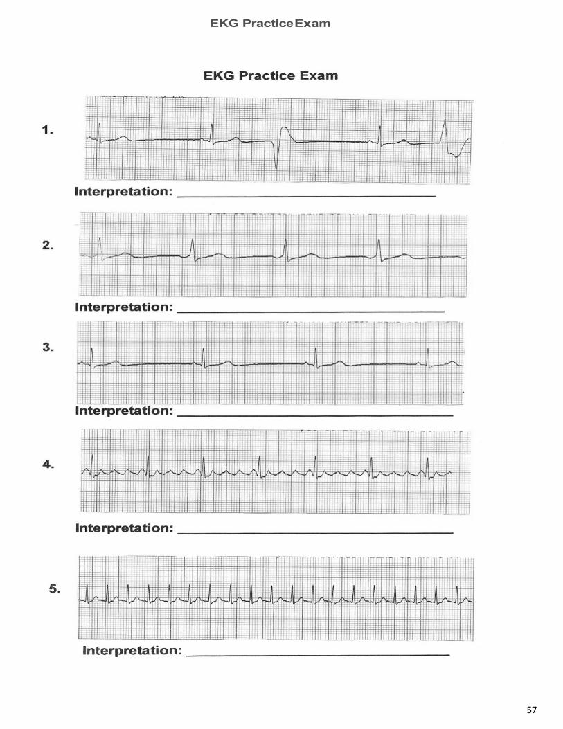
57
EKG Practice Exam

58
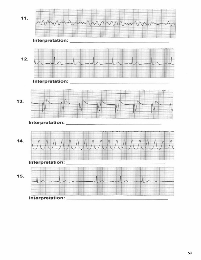
59
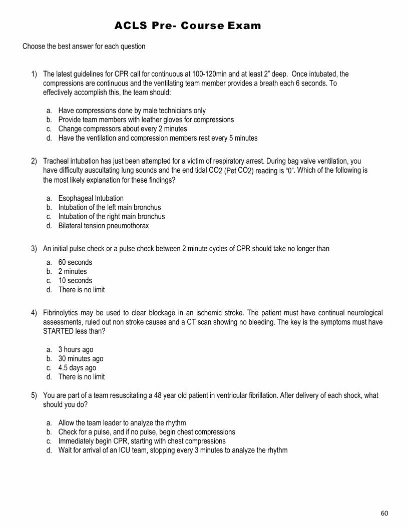
60
ACLS Pre- Course Exam
Choose the best answer for each question
1) The latest guidelines for CPR call for continuous at 100-120min and at least 2” deep. Once intubated, the compressions are continuous and the ventilating team member provides a breath each 6 seconds. To effectively accomplish this, the team should:
a. Have compressions done by male technicians only b. Provide team members with leather gloves for compressions c. Change compressors about every 2 minutes d. Have the ventilation and compression members rest every 5 minutes
2) Tracheal intubation has just been attempted for a victim of respiratory arrest. During bag valve ventilation, you
have difficulty auscultating lung sounds and the end tidal CO2 (Pet CO2) reading is “0”. Which of the following is the most likely explanation for these findings?
a. Esophageal Intubation b. Intubation of the left main bronchus c. Intubation of the right main bronchus d. Bilateral tension pneumothorax
3) An initial pulse check or a pulse check between 2 minute cycles of CPR should take no longer than
a. 60 seconds b. 2 minutes c. 10 seconds d. There is no limit
4) Fibrinolytics may be used to clear blockage in an ischemic stroke. The patient must have continual neurological
assessments, ruled out non stroke causes and a CT scan showing no bleeding. The key is the symptoms must have STARTED less than?
a. 3 hours ago b. 30 minutes ago c. 4.5 days ago d. There is no limit
5) You are part of a team resuscitating a 48 year old patient in ventricular fibrillation. After delivery of each shock, what
should you do?
a. Allow the team leader to analyze the rhythm b. Check for a pulse, and if no pulse, begin chest compressions c. Immediately begin CPR, starting with chest compressions d. Wait for arrival of an ICU team, stopping every 3 minutes to analyze the rhythm

61
6) A patient remains in VF after a defibrillation shock, tracheal intubation, CPR, Epinephrine 1mg IV, and another shock. Which of the following drug-dose combinations should this patient receive next?
a. Amiodarone 300mg b. Lidocaine 3mg/kg c. Procainamide 20-50mg/min, up to a total dose of 17mg/kg d. Magnesium 10g
7) Which of the following poses the greatest risk during defibrillation?
a. Defibrillating a patient found on a metal floor b. Defibrillating a patient with a nitro patch on the left shoulder c. O2 blowing over the chest during defibrillation d. Using defibrillation pads
8) EMS arrives at the side of a 55 year old man in cardiac arrest. They begin CPR and apply defibrillation pads to his chest. Later they have intubated the patient and started a large bore IV. What is the best way for them to confirm effective CPR is being performed?
a. Continued CPR should produce a normal blood pressure b. Watch the EKG for signs for tachycardia c. Monitor waveform capnography via exhaled CO2 (PETCO2) d. Evaluate pulse oximetry
9) A patient arrives in the ED. CPR continues with ventilations provided through a tracheal tube inserted in the field. Chest compressions produce a femoral pulse that disappears during a “stop compressions” pause. During the pause the cardiac monitor shows narrow QRS complexes at a rate of 40 bpm. 1mg Epinephrine was just given. At this point what is the next action you should take?
a. Begin reviewing the H’s & T’s for causes b. Administer Atropine 1mg IV push c. Send a stat blood sample for tox screen d. Withhold compressions to allow the heart to “prime”
10) During a code you observe a rhythm on the monitor. The rate is 160. No pulse is present. This best describes which rhythm?
a. PEA b. SVT c. V-Tach d. Malfunctioning cardiac monitor
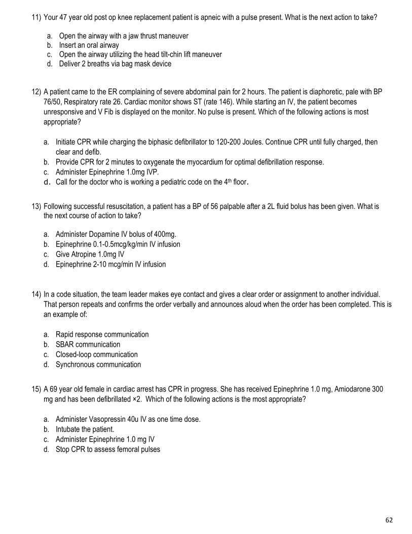
62
11) Your 47 year old post op knee replacement patient is apneic with a pulse present. What is the next action to take?
a. Open the airway with a jaw thrust maneuver b. Insert an oral airway c. Open the airway utilizing the head tilt-chin lift maneuver d. Deliver 2 breaths via bag mask device
12) A patient came to the ER complaining of severe abdominal pain for 2 hours. The patient is diaphoretic, pale with BP 76/50, Respiratory rate 26. Cardiac monitor shows ST (rate 146). While starting an IV, the patient becomes unresponsive and V Fib is displayed on the monitor. No pulse is present. Which of the following actions is most appropriate? a. Initiate CPR while charging the biphasic defibrillator to 120-200 Joules. Continue CPR until fully charged, then
clear and defib. b. Provide CPR for 2 minutes to oxygenate the myocardium for optimal defibrillation response. c. Administer Epinephrine 1.0mg IVP. d. Call for the doctor who is working a pediatric code on the 4th floor.
13) Following successful resuscitation, a patient has a BP of 56 palpable after a 2L fluid bolus has been given. What is
the next course of action to take? a. Administer Dopamine IV bolus of 400mg. b. Epinephrine 0.1-0.5mcg/kg/min IV infusion c. Give Atropine 1.0mg IV d. Epinephrine 2-10 mcg/min IV infusion
14) In a code situation, the team leader makes eye contact and gives a clear order or assignment to another individual. That person repeats and confirms the order verbally and announces aloud when the order has been completed. This is an example of: a. Rapid response communication b. SBAR communication c. Closed-loop communication d. Synchronous communication
15) A 69 year old female in cardiac arrest has CPR in progress. She has received Epinephrine 1.0 mg, Amiodarone 300
mg and has been defibrillated ×2. Which of the following actions is the most appropriate? a. Administer Vasopressin 40u IV as one time dose. b. Intubate the patient. c. Administer Epinephrine 1.0 mg IV d. Stop CPR to assess femoral pulses

63
16) An 88 year old man in normothermic cardiac arrest is being managed by Paramedics, he has had 20 minutes of asystole. Paramedics intubated him, confirmed proper tube placement, gained IV access, and have given Epinephrine 1mg IV X 4 and 100 MEQ Bicarcb. Which is an appropriate action for Paramedics to take?
a. Consider terminating in the field following physician consult b. Transport but “fake” CPR c. Never stop resuscitation efforts without an EEG d. Stop efforts only if there is no response to CPR and 4 defibrillatory shocks to confirm asystole.
17) A 50 year old man has a 3 mm ST elevation in leads V2 to V6. Severe chest pain continues despite Aspirin 160– 325mg, Nitroglycerine SL x 4, and Morphine 10 mg IV. BP 150/90 mm Hg; HR 100 bpm. Which of the following treatment combinations is most appropriate for this patient at this time (assume no complications to any medication)?
a. Calcium channel blocker IV + heparin bolus IV b. ACE inhibitor IV + Lidocaine infusion c. Magnesium sulfate IV + Lovenox SQ d. Fibrinolytic agents + heparin IV
18) Within 45 minutes of ED arrival, which of the following evaluation sequences should be performed for a 70 year old woman with rapid onset of headache, garbled speech, and right arm weakness?
a. History, physical, and neurologic exams, noncontrast head CT with radiologist interpretation b. History, physical, and neurologic exams, noncontrast head CT, start of fibrinolytic treatment if scan is positive
for hemorrhagic stroke c. History, physical, and neurologic exams, lumbar puncture, head CT if LP negative for blood d. History, physical and neurologic exams, contrast head CT, start of fibrinolytic therapy when
improvement in neurologic signs is noted
19) Which of the following conditions most closely mimics the signs and symptoms of an acute stroke? a. Acute insulin induced hypoglycemia b. Acute hypoxia c. Isotonic dehydration and hypovolemia d. Acute vasovagal or orthostatic hypotension
20) Which of the following rhythms is an appropriate indication for transcutaneous pacing? a. Sinus bradycardia with no symptoms b. Normal sinus rhythm with hypotension and shock c. Complete heart block with pulmonary edema d. Acute vasovagal or orthostatic hypotension

64
21) A patient receiving CPR is intubated. His PETCO2 (end tidal CO2) shows 8mmhg. What clinical adjustment may
help to correct this?
a. Compress deeper or faster b. Increase the ventilatory rate c. Decrease the ventilatory rate d. Escalating energy defibrillation
22) Which of the following patients’ needs immediate synchronized cardioversion?
a. A 78 year old woman with fever, pneumonia, chronic congestive heart failure, and sinus tachycardia at 125 bpm
b. A 55 year old man with multifocal atrial tachycardia at 125 bpm, respiratory rate of 12 breaths/min, and a BP of 134/86 mm Hg
c. A 69 year old woman with a history of coronary artery disease, chest pain, a 2mm ST elevation, and sinus tachycardia at 130 bpm
d. A 62 year old man with a history of rheumatic mitral valve disease, obvious shortness of breath, HR 160 bpm, and a BP of 88/70 mm Hg
23) Which of the following patients is most likely presenting with stable tachycardia that you should not cardiovert?
a. A 25 year old wheezing asthmatic woman who has pneumonia on chest X ray, who is taking albuterol, and who has the following vital signs: temp-101.2 F, HR-150 bpm, resp-20/min
b. A 55 year old man with diaphoresis, bilateral rales, and the following vital signs: HR- 160/min, BP- 90/55, resp 18/min, rhythm- rapid atrial flutter
c. A 62 year old man with wide complex tachycardia at a rate of 150, chest pain, shortness of breath, and palpitations
d. A 55 year old woman with chest pain, shortness of breath, extreme weakness and dizziness, BP 88/54, and a narrow complex tachycardia at a rate of 150 bpm.
24) You prepare to cardiovert an unstable 48 year old tachycardic woman with the monitor/defibrillator on “synchronization” mode. She suddenly becomes unresponsive and pulseless and the rhythm changes to an irregular, chaotic, VF-like pattern. You charge to 200 Joules and press the SHOCK button, but the defibrillator fails to deliver a shock, Why?
a. The defibrillator/monitor battery failed b. The “synch” switch failed c. You cannot shock VF in “synch” mode d. A monitor lead has lost contact, producing the “pseudo VF” rhythm
25) Your team is administering CPR to an 80 year old woman. Which of the following actions best demonstrates recommended CPR technique?
a. Changing out compressors when they get tired b. Allowing full chest recoil c. Giving 15 compressions: 2 breaths, when you
have 2 rescuers present d. Checking the pulse frequently.

65
26) A previously healthy 50 year old man complains of chest tightness, palpitations, and dizziness. HR is 170 bpm, BP is 86/50, and the ECG shows a narrow complex tachycardia. The patient failed to respond to initial vagal maneuvers and 2 rounds of Adenosine. As your next action, which of the following treatments should be attempted?
a. IV Amiodarone b. Unsynchronized countershock c. IV Diltiazem d. Synchronized countershock
27) A 75 year old man presents to the ED with a 1 day complaint of indigestion, palpitations, and general weakness. His HR is 180, RR 16, B/P 140/80 and O2 saturation at 97%. Which of the following therapies is most appropriate next?
a. Sedation, analgesia, then immediate cardioversion b. Oxygen via nasal cannula, labs for cardiac enzymes c. Vagal maneuver d. Administer oxygen via mask
28) A 66 year old malnourished, chronic alcoholic presents with polymorphic VT that resembles Torsades de Pointes. His HR is irregular at 120 to 160 bpm and B/P is 95/65. He has no related symptoms and no signs of impaired heart function. Which of the following treatments is most appropriate at this time?
a. IV Amiodarone b. IV Magnesium c. IV Lidocaine d. IV Procainamide
29) You have just resuscitated a 46 year old from V-Fib using CPR, 1 defibrillation at 200 joules, 1mg of epinephrine, 1
defibrillation at 300 joules, 300mg of Amiodarone then 1 defibrillation at 360 joules. You continued CPR throughout and now see NSR on the monitor. What should your next step be?
a. Check for a pulse, assure ventilation & oxygen, maintain B/P b. Check B/P, hang a Lidocaine drip, mildly hyperventilate patient c. Check pulse, increase oxygen percentage, increase B/P to at least 130 systolic d. Check B/P, infuse dopamine, continue CPR for 2 minutes
30) Which of the following patients is most likely to present with vague or atypical signs and unusual symptoms of an AMI?
a. A 50 year old woman with moderate coronary artery disease recently confirmed by angiography b. A 56 year old man who smokes 3 packs per day but has no history of heart disease c. A 45 year old woman diagnosed with type I diabetes 22 years ago d. A 48 year old man in the ICU after coronary artery bypass surgery
31) You found your 60 year old diabetic female patient unresponsive. She does not appear to be breathing. Your next
action is?
a. Open airway b. Administer rescue breaths c. Hyperventilate d. Check carotid pulse

66
32) You are managing a 64 year old with sub-sternal pressure. BP-100/60, HR 70, RR 16, Pulse oximetery 98%. What assessment step is important now?
a. Oxygen and PETCO2 b. Chest x-ray c. Laboratory testing d. Obtaining a 12-lead ECG
33) What is the preferred method of initial access for medication administration during cardiac arrest in most
patients?
a. Instraosseous b. Endotracheal c. Central Venous d. Peripheral IV site
34) As you apply your area’s AED during CPR, the unit does not analyze and continually states: “check pads”. Your
next action should be?
a. Continue chest compressions b. Call the biomed department c. Turn the unit off then on, unplug the pads d. Evaluate the pads expiration date
35) A common problem that causes the most negative impact on resuscitation efforts is?
a. Failure to obtain vascular access b. Failure to intubate c. Failure to perform endotracheal intubation d. Interruptions and delay in chest compressions
36) A 50 y.o. drowning victim has been pulled from the water. He is unresponsive without respiratory effort or pulses present. CPR is started. He is immediately placed on a cardiac monitor which shows sinus tachycardia with a heart rate of 122. Pulses are not palpable. What is the next action to take? a. Administer Epinephrine 1.0mg IVP. b. Defibrillate at 200 Joules c. Examine the H & T’s for possible reversible causes. d. Roll victim to the side, administer back blows to expel water from lungs.
37) Above all else, which action increases cardiac arrest survival potential the most?
a. Pausing chest compressions briefly after a defibrillation to evaluate rhythm b. Administering Epinephrine every 3-5 minutes c. Using manual defibrillator paddles in the synchronized mode d. Providing quality compressions in 2 minute uninterrupted cycles
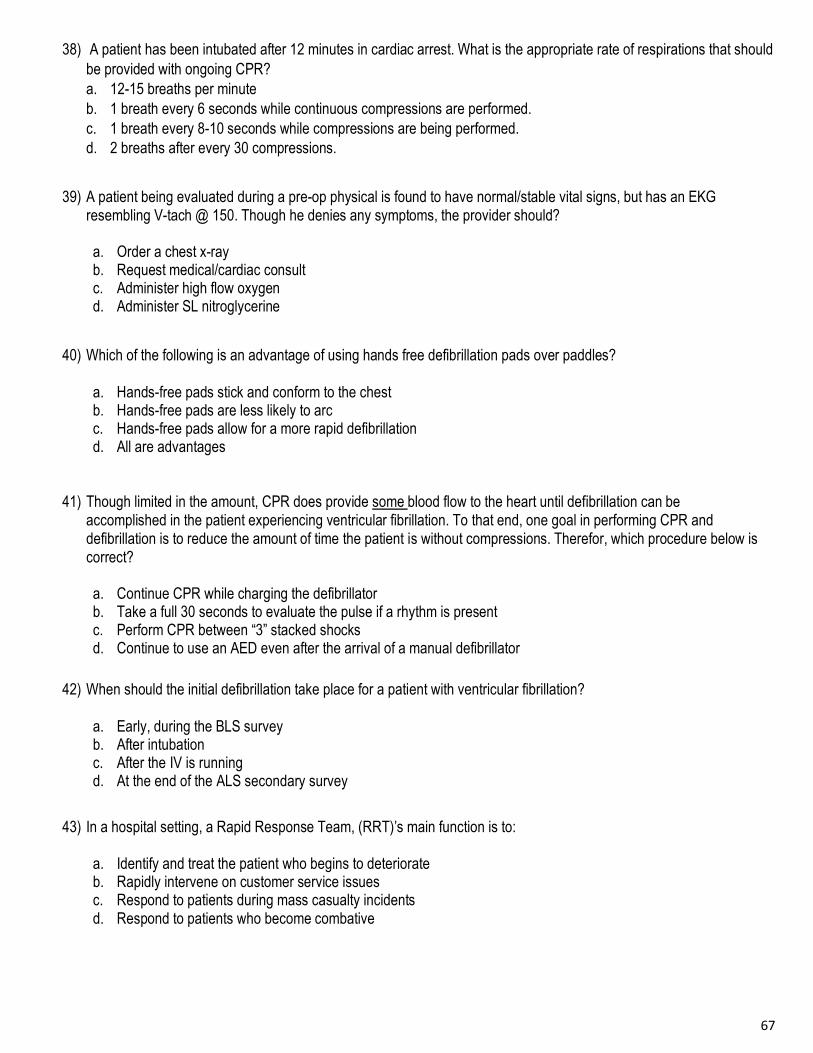
67
38) A patient has been intubated after 12 minutes in cardiac arrest. What is the appropriate rate of respirations that should
be provided with ongoing CPR? a. 12-15 breaths per minute b. 1 breath every 6 seconds while continuous compressions are performed. c. 1 breath every 8-10 seconds while compressions are being performed. d. 2 breaths after every 30 compressions.
39) A patient being evaluated during a pre-op physical is found to have normal/stable vital signs, but has an EKG
resembling V-tach @ 150. Though he denies any symptoms, the provider should?
a. Order a chest x-ray b. Request medical/cardiac consult c. Administer high flow oxygen d. Administer SL nitroglycerine
40) Which of the following is an advantage of using hands free defibrillation pads over paddles?
a. Hands-free pads stick and conform to the chest b. Hands-free pads are less likely to arc c. Hands-free pads allow for a more rapid defibrillation d. All are advantages
41) Though limited in the amount, CPR does provide some blood flow to the heart until defibrillation can be accomplished in the patient experiencing ventricular fibrillation. To that end, one goal in performing CPR and defibrillation is to reduce the amount of time the patient is without compressions. Therefor, which procedure below is correct?
a. Continue CPR while charging the defibrillator b. Take a full 30 seconds to evaluate the pulse if a rhythm is present c. Perform CPR between “3” stacked shocks d. Continue to use an AED even after the arrival of a manual defibrillator
42) When should the initial defibrillation take place for a patient with ventricular fibrillation?
a. Early, during the BLS survey b. After intubation c. After the IV is running d. At the end of the ALS secondary survey
43) In a hospital setting, a Rapid Response Team, (RRT)’s main function is to:
a. Identify and treat the patient who begins to deteriorate b. Rapidly intervene on customer service issues c. Respond to patients during mass casualty incidents d. Respond to patients who become combative

68
44) Following a successful resuscitation and intubation, a patient with a heart rate and pulse of 80/minute should be
ventilated how often?
a. 1 breath every 3 to 4 seconds b. 1 breath every 5 to 6 seconds c. 2 breaths every 5 to 6 seconds d. 2 breaths every 6 to 8 seconds
45) You are treating a patient in a rural clinic. He has a HR of 40, B/P 86/60, RR 16 and skin is diaphoretic. You have
no pacemaker, so the physician chooses dopamine for the bradycardia. You know the appropriate dose is?
a. 2 - 10 mg/min b. 2 - 10 mcg/kg/min c. 1mg IV every 10 minutes d. 5 to 50 mcg/kg per minute
46) Guidelines suggest that adult compressions should be administered at a depth of 2-2.4 inches for maximum
effectiveness. Which of the following is not true regarding compression depth? a. Consistent compression depth of at least 2 inches is associated with better outcomes. b. It is clinically difficult to judge compression depth without the use of a feedback device. c. Potential complications can occur at depths of greater than 2.4 inches. d. Compressions are often delivered too hard rather than too shallow.
47) You are in a small hospital with no CT scan capability due to recent hospital damage. EMS Calls with a 40 year old
with left side weakness and facial droop for 50 minutes. An appropriate option might be?
a. Attempt to contact the patients’ private physician b. Have the EMS crew administer plavix c. Do not tell anyone about the CT scanner being down d. Divert the patient to a hospital 20 minutes away with CT capabilities
48) On which of the following should we withhold resuscitation?
a. Patient with a valid DNR b. Evidence of rigor mortis c. The area is not safe for the rescuer (medical team) d. All of the above
49) Your 50 year old patient presents with persistent epigastric pain. B/P 168/92, HR 94 regular, RR 12, pulse oximetry 96%. His EKG shows normal sinus rhythm without ST elevation or depression. Which is the most appropriate intervention to perform next?
a. Administer oxygen b. Obtain a problem-focused history and physical exam c. Give antacids d. Administer sublingual nitroglycerin

69
50) You receive a patient who was short of breath, but is now unresponsive with RR 8/min, HR 30/min, B/P –
100/50, pulse oximetry 91%. What intervention should be attempted initially?
a. Atropine IV push b. Epinephrine IV infusion c. Begin CPR d. Basic airway support and ventilations
51) An intubated post CHF patient begins to have secretions coming from the ET tube. The proper ET suctioning
procedure is?
a. Insert catheter into tube, suction during insertion for up to 30 seconds b. Do not insert catheter into tube, suction the mouth and nose for up to 30 seconds c. Insert catheter into tube, suction during withdrawal but for no longer than 10 seconds d. Initially withhold oxygen to stimulate the hypoxic drive.
52) A 65 year old presents with a complete heart block. HR 36, B/P 80/60, RR 16 and pulse oximetry 93%. After oxygen, which of these is the most appropriate next intervention?
a. Dopamine slow push (IV bolus) b. Atropine IV bolus c. Lidocaine IV bolus then infusion d. Fluid bolus
53) A 70 year old experienced a sudden onset of right arm weakness and called EMS. B/P 150/80, HR 76, RR 16 and pulse oximetry 96%. What is the most appropriate action for the EMS team to perform next?
a. 12-lead ECG assessment b. Administration of 100% supplementary oxygen c. Cincinnati Stroke Scale assessment d. Administer fractionated Heparin
54) You respond to the home of a 72 year old complaining of chest pain and shortness of breath. B/P 104/62, HR
100/min, RR 18min, pulse oximetry is 92% on room air. Distended neck veins are noted and the lungs are clear. Based on his 12 lead EKG, Right ventricular infarct is suspected. Which of the following interventions should be avoided?
a. Nitroglycerine b. Oxygen c. IV fluids to maintain systolic BP greater than 100. d. Transport to a facility with PCI capabilities, if available.
55) Adult cardiac arrest patients who have a return of spontaneous circulation (ROSC) following successful resuscitation
should have targeted temperature management (TTM) if they remain unresponsive to verbal commands or comatose. Which of the following regarding TTM is true? a. Aim for temperature range between 32-36°C and maintain constantly for at least 24 hours. b. The cooling process should be started in the prehospital setting with rapid infusion of cold intravenous (IV) fluids. c. The temperature range should be between 32-34°C and maintained for 12-24 hours. d. Current guidelines suggest targeted temperature has not been found to be beneficial to post arrest patients.
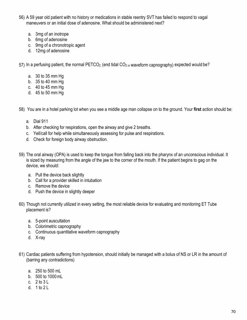
70
56) A 59 year old patient with no history or medications in stable reentry SVT has failed to respond to vagal
maneuvers or an initial dose of adenosine. What should be administered next?
a. 3mg of an inotrope b. 6mg of adenosine c. 9mg of a chronotropic agent d. 12mg of adenosine
57) In a perfusing patient, the normal PETCO2, (end tidal CO2 or waveform capnography) expected would be?
a. 30 to 35 mm Hg b. 35 to 40 mm Hg c. 40 to 45 mm Hg d. 45 to 50 mm Hg
58) You are in a hotel parking lot when you see a middle age man collapse on to the ground. Your first action should be: a. Dial 911 b. After checking for respirations, open the airway and give 2 breaths. c. Yell/call for help while simultaneously assessing for pulse and respirations. d. Check for foreign body airway obstruction.
59) The oral airway (OPA) is used to keep the tongue from falling back into the pharynx of an unconscious individual. It is sized by measuring from the angle of the jaw to the corner of the mouth. If the patient begins to gag on the device, we should:
a. Pull the device back slightly b. Call for a provider skilled in intubation c. Remove the device d. Push the device in slightly deeper
60) Though not currently utilized in every setting, the most reliable device for evaluating and monitoring ET Tube
placement is?
a. 5-point auscultation b. Colorimetric capnography c. Continuous quantitative waveform capnography d. X-ray
61) Cardiac patients suffering from hypotension, should initially be managed with a bolus of NS or LR in the amount of (barring any contradictions)
a. 250 to 500 mL b. 500 to 1000 mL c. 2 to 3 L d. 1 to 2 L
71
62) Defibrillation safety is important. Which location below would be the least safe area to utilize an AED or manual defibrillator?
a. On a metal floor of a ship b. In the snow c. In a hot tub d. In an area that is using anesthesia gasses
63) Post-cardiac arrest, once the respiratory and cardiac functions are optimized, the patient should be considered for
transfer/transport to an area capable of performing which procedures if indicated?
a. Cerebral coiling b. Targeted temperature management and PCI (Re-perfusion) c. CT scan and fibrinolytics d. Central lines and pacemakers
64) Post arrest 02 saturations should be maintained between 94 – 99% due to the fact that a 100% saturation could actually cause an FIO2 (blood gas) of 3 or more times normal, the main reason for avoiding the hyper-oxygenated state in adults is:
a. High oxygen levels can cause blindness b. There is no problem with hyper-oxygenation or hyperventilation c. Hyper-oxygenation reduces blood pressure d. Oxygen toxicity reduces the damaged cells ability to survive
65) Considering the previous question; Excessive ventilation (too many breaths per min.) is known to inhibit cardiac function by increasing intra thoracic pressure; It may also cause?
a. Cerebral vasoconstriction resulting in decreased blood flow to the brain. b. Increased intrathoracic pressure c. Decreased cardiac output d. All of the above

72
NAME: COURSE: # MISSED: GRADE:
1. A B C D 34. A B C D 2. A B C D 35. A B C D 3. A B C D 36. A B C D 4. A B C D 37. A B C D 5. A B C D 38. A B C D 6. A B C D 39. A B C D 7. A B C D 40. A B C D 8. A B C D 41. A B C D 9. A B C D 42. A B C D 10. A B C D 43. A B C D 11. A B C D 44. A B C D 12. A B C D 45. A B C D 13. A B C D 46. A B C D 14. A B C D 47. A B C D 15. A B C D 48. A B C D 16. A B C D 49. A B C D 17. A B C D 50. A B C D 18. A B C D 51. A B C D 19. A B C D 52. A B C D 20. A B C D 53. A B C D 21. A B C D 54. A B C D 22. A B C D 55. A B C D 23. A B C D 56. A B C D 24. A B C D 57. A B C D 25. A B C D 58. A B C D 26. A B C D 59. A B C D 27. A B C D 60. A B C D 28. A B C D 61. A B C D 29. A B C D 62. A B C D 30. A B C D 63. A B C D 31. A B C D 64. A B C D 32. A B C D 65. A B C D 33. A B C D

73
74
EKG Practice Exam - Key
1. Sinus Bradycardia with Multifocal PVC’s
2. Junctional Rhythm
3. Sinus Bradycardia
4. Atrial Flutter
5. Supraventricular Tachycardia, A-Tach
6. Sinus Tachycardia
7. Atrial Fibrillation
8. Idioventricular Rhythm
9. Complete Heart Block, 3rd Degree AVB
10. Mobitz II, 2nd Degree AVB type II 11. Ventricular Fibrillation
12. Normal Sinus Rhythm
13. Ventricular Pacer, 100% capture
14. Ventricular Tachycardia (monomorphic)
15. Mobitz 1, 2nd degree AVB type 1, Wenkebach
75
ACLS Pretest Answer K ey
1. C 34. A 2. A 35. D 3. C 36. A 4. A 37. D 5. C 38. B 6. A 39. B 7. C 40. D 8. C 41. A 9. A 42. A 10. A 43. A 11. C 44. B
12. A 45. B 13. B 46. D 14. C 47. D 15. C 48. D 16. A 49. B 17. D 50. D 18. A 51. C 19. A 52. B 20. C 53. C 21. A 54. A 22. D 55. A 23. A 56. D 24. C 57. B 25. B 58. C 26. D 59. C 27. C 60. C 28. B 61. D 29. A 62. C 30. C 63. B 31. D 64. D 32. D 65. D 33. D

76
EMERGENCY MEDICAL CONSULTANTS INC. Florida’s Premier Provider Of Quality Medical Training Programs
Nationally Accredited and OSHA Programs CEU Provider
Since 1988
The following pages contain a Pretest for anyone choosing to complete the BLS course
after ACLS or PALS .
This information in this packet comes from the 2015 BLS textbook; Please use the book to review.
You must score a 76% (-7)
In order to take this program
Completing these test questions prior to the course is mandatory if you plan on attending the CPR section
after the ACLS program.
(772) 878-3085 * Fax: (772) 878-7909 * Email: [email protected] 597 SE Port Saint Lucie Blvd * Port Saint Lucie, Florida 34984
Visit Our Website- www EMCmedicaltraining.com

77

78
Emergency Medical Consultants 772-878-3085 www.EMCmedicaltraining.com
Skills Review for Healthcare Providers
The CAB's of CPR
Simultaneously Determine unresponsiveness and check for effective breathing If unresponsive: call a “code” or 911
C = Circulation- Check for a pulse Max - 10 seconds. If pulse is not definite, begin compressions. A = Airway- Open airway (head tilt/chin lift) B = Breaths- Give 2 breaths then back to compressions D = Defibrillator- Attach a manual defibrillator or AED
CPR Reference Adults (> puberty) Children (1 - puberty) Infants (< 1yr)
Rescue breathing, Victim definitely has a pulse
10-12 breaths/min
recheck pulse every 2 minutes
12-20 breaths/min
recheck pulse every 2 minutes
12-20 breaths/min
recheck pulse every 2 minutes
Compression landmark No pulse
(or pulse <60 in infant or child with poor perfusion)
Middle of the chest,
between the nipples
Middle of the chest,
between the nipples
1 finger below nipple line
Compressions are performed with
Heel of 2 hands Heel of 1 or 2 hands
2 fingers OR
2 thumbs when using encircling hands technique
Rate of compressions per minute 100-120/min 100-120/min 100-120/min
Compression depth 2-2.4 inches At least 1/3 depth of chest
2 inches
At least 1/3 depth of chest
1 ½ inches
Ratio of compressions to breaths *Once an advanced airway is placed ventilations will be 1 every 6 sec. with
continual compressions.
30:2 Change compressors and
reevaluate every 2 min
30:2 15:2 if 2 rescuer
Change compressors and reevaluate every 2 min
30:2 15:2 if 2 rescuer
Change compressors and reevaluate every 2 min
Foreign Body Airway Obstruction * If not rapidly removed call Emergency Medical Service *
Conscious choking
Adult Child Infant
Abdominal Thrusts
Abdominal Thrusts
5 Back Blows/5 Chest Thrusts
Unconscious choking Adult Child Infant
Call a “code” or call 911
Begin CAB’s of CPR Before giving breaths:
look in mouth for foreign body, remove object if it is seen.
Repeat cycles of CPR if needed
Begin CPR
If second rescuer is present, send them to call a “code” or 911, otherwise, call after 2 min of CPR
Before giving breaths: look in mouth for foreign body,
remove object if it is seen.
Repeat cycles of CPR if needed
Begin CPR
If second rescuer is present, send them to call a “code” or 911, otherwise, call after 2 min of CPR
Before giving breaths: look in mouth for foreign body,
remove object if it is seen.
Repeat cycles of CPR if needed
79
INFORMATION TO KEEP IN MIND:
1. Know the maximum time that should be spent checking for the presence of a pulse.
2. Know the preferred techniques/devices for providing ventilations if you are a single rescuer
versus having multiple resources in the professional setting.
3. Know the concept of scene safety/awareness before providing care.
4. Know which patients require ventilations and which require ventilations plus compressions.
5. Know the best way to open the airway for an Adult, Child, Infant or spinally injured patient.
6. Know the location, depth and rate of compressions for an Adult, Child and Infant.
7. Know when to start compressions for an Adult, Child and Infant, be able to explain chest recoil (release) and high quality CPR.
8. Know the compression to ventilation ratio for both 1 and 2-rescuer for Adult, Child and Infant.
9. Know how to reduce the incidence of air being introduced into the patient’s stomach versus their lungs.
10. Understand how an AED affects the heart (shock to organize the rhythm), and know the steps for using an AED on an Adult, Child or Infant; pediatric use and placement.
11. Know how to incorporate CPR before, during and after AED use.
12. Know the changes in CPR, which are incorporated once a victim has an advanced airway “tube” placed by a medical professional.
13. Know the sequence, procedures and roles for 1 rescuer versus 2-rescuer CPR.
14. Know the procedures for conscious and unconscious choking for Adult, Child and Infant.
15. Know how to determine effectiveness of ventilations and compressions being provided during CPR
16. Know the elements of effective team dynamics and communicating during an emergency.

80
MANDATORY BLS Pre-Course Exam
1. An elderly woman collapses to the floor in a bingo hall. Your first action should be:
A. Open the airway and give 2 breaths. B. Go grab the defibrillator off the wall in the hallway. C. Yell out/ call for help while simultaneously assessing for pulse and respirations. D. Check for a carotid pulse.
2. You are performing 1 rescuer CPR on a 75-year-old female with a history of chest pain and diabetes. An
AED has just been made available to you. What is the first action that you should take at this time?
A. Finish the 5 cycles of chest compressions that you have started. B. Place the AED pads on the chest. C. Secure an electrical outlet to plug the AED into. D. Turn the AED on.
3. You are attending your nephew’s birthday party when a 5 year old child suddenly starts choking on a hotdog. What should you do?
A. Administer 2 rescue breaths. B. Perform a blind sweep of the victim’s mouth. C. Deliver 5 back-slaps. D. Position yourself behind the child and administer abdominal thrusts (Heimlich Maneuver).
4. Opioids are medications that are used to treat pain but have a high potential for abuse. Addiction rate to the medications is a growing problem and they can cause respiratory and or cardiac arrests. Currently, more adults die every year from opioid overdoses than car accidents. What is the name of the medication that is utilized to reverse the effects of respiratory depression?
A. Naloxone. B. Ativan. C. Lasix. D. Magnesium Sulfate.
5. Your middle age neighbor is mowing his grass when he clutches his chest and drops to the ground. He has no pulse or respirations. Your son calls 911 while you initiate chest CPR. How fast should the compression rate be?
A. 100 compressions per minute. B. 100-120 compressions per minute. C. 80-100 compressions per minute D. 120-150 compressions per minute.
6. Bystanders have pulled a young woman with a pulse but no respirations out of a lake. One of them is administering rescue breaths at a rate of one every 5-6 seconds while waiting for EMS to arrive. Which of the following is true about rescue breaths?
A. Each breath should result in visible chest rise. B. Give each breath over 1 second. C. The pulse should be checked every 2 minutes.. D. All of the above.

81
7. Which of the following situations will slightly delay AED usage while the situation is made safe for AED application?
A. A person found lying on a metal floor inside a meat cooler. B. A person found submerged in a bathtub. C. A person who collapsed in snow. D. A person who has a transdermal nitro patch on their arm.
8. When utilizing a bag valve mask device it is important to remember:
A. That this device requires training and is best suited for a 2-rescuer situation. B. The E-C clamp technique should be used while lifting the jaw to provide a good seal. C. To squeeze the bag for 1 second while watching the chest rise. D. All of the above.
9. What is the correct ratio for compressions to ventilations in infant CPR with 2 rescuers present?
A. 20 compressions to 4 breaths. B. 15 compressions to 2 breaths. C. The rate remains 30 compressions to 2 breaths. D. 15 compressions to 1 breath.
10. The maximum amount of time that should be taken to check for a pulse on an adult, infant or child is:
A. 15 seconds B. 10 seconds C. 30 seconds D. 5 seconds
11. You are the second rescuer providing ventilations to an adult victim in cardiac arrest. You observe the hand placement of the person who is providing compressions to be incorrect. You advise them to reposition their hands. This is an example of what type of team dynamic communication?
A. Knowledge Sharing. B. Closed Loop Communication. C. Constructive Intervention. D. Open Communication
12. While providing CPR to a victim, an AED becomes available and a shock is indicated and administered. What should you do next?
A. Administer 2 more shocks; to total 3. B. Immediately restart CPR, starting with compressions. C. Give 2 breaths first then resume CPR. D. Check the carotid pulse for no longer than 10 seconds.
13. What is the purpose of defibrillation?
A. To stop a chaotic rhythm and restore the heart’s normal rhythm. B. To increase the rate of complete heart block. C. To provide a blood pressure. D. To treat cardiac standstill.

82
14. Current guidelines suggest that adult compressions should be administered at a depth of 2-2.4 inches. Which of the following is not true regarding chest compression depth?
A. Compressions are often delivered too hard rather than too shallow. B. It may be difficult to accurately judge compression depth without the use of a feedback device. C. Consistent compression depth of at least 2 inches is associated with better outcomes. D. Potential complications can occur at depths of greater than 2.4 inches.
15. What is the correct rate of ventilations to provide when an advanced airway is in place?
A. 1breath every 3-5 seconds. B. 1 breath every 6-8 seconds. C. 1 breath every 10 seconds. D. 1 breath every 6 seconds.
16. You begin your shift on the med/surg floor. You begin your rounds and when you walk into a patient’s room, you notice the patient has agonal respirations. What should you do first?
A. Give the patient oxygen B. Check a pulse, and begin compressions if indicated C. Do nothing, the patient is asleep D. Begin the Heimlich maneuver
17. When is the two thumb encircling technique is used?
A. On an infant when two rescuers are available B. When the infant is choking C. When performing CPR on a pediatric victim D. When performing one rescuer CPR on an infant and you become tired
18. At the beginning of your work shift, you are assigned the role of compressor during a cardiac arrest. This is known as:
A. Mutual respect B. Closed loop communications C. Clear roles and responsibilities D. Constructive intervention
19. While assisting with a cardiac arrest, you are instructed to take over bag valve mask ventilations. You repeat back “ you would like for me to take over bag valve mask ventilations.” In team dynamics, what is this called?
A. Closed loop communications B. Knowing your limitations C. Knowledge sharing D. Mutual respect
20. In relation to the “Team concept” of resuscitation, if adequate medical staff is available, in order to reduce fatigue and increase effectiveness of compressions the team should?
A. Assign several people to switch off on compressions every 2 min B. Have each compressor work until they state they need relief C. Put a rescuer on each side of the victim and rotate each set of 30 compressions D. Always assign the largest, strongest person to compressions

83
21. You arrive to find a hospital maintenance worker lying on the ground, next to a ladder. He appears unconscious, your first action should be?
A. Check for breathing and a pulse B. Shake and shout, check unresponsiveness C. Begin compressions at 30:2 D. Assure the area is safe for you to be in
22. After performing the choking procedure for a conscious victim who becomes unconscious, the next procedure is to?
A. Perform a finger sweep B. Attempt ventilations C. Straddle the victim D. Begin CPR compressions
23. High quality CPR is the critical component to resuscitation, especially compressions; which concept is correct?
A. Compression depth has a higher priority than recoil or relaxation B. Compression and recoil (refill) are equally important C. Ventilation is the priority for all victims D. 30 ventilations per minute are optimum for the best outcome
24. When performing compressions on a child for CPR or unconscious foreign body airway procedures the proper depth is?
A. ½" or ½ the depth of the chest B. 1 ½ " to 2 1 ½ " inches C. Varies based on age and weight D. 2" or 1/3 the depth of the chest
25. While at a school event, a teacher chokes on gum. He runs towards the office before falling unconscious. Immediately after performing 30 compressions, the next step is?
A. Open the airway and look into the mouth before ventilating B. Perform a finger sweep and attempt breaths C. Readjust the airway with a jaw thrust maneuver D. Check pulse for no more than 10 seconds
26. The resuscitation team is made up of various professionals with different levels of license and skill sets. In order to function efficiently the team members must?
A. Wait for a physician to order CPR and defibrillation B. Always be prepared to perform any skills, even if not licensed to C. Realize their strengths, abilities and limitations D. Decide if they feel CPR is worth the physical effort
84
27. A victim begins to choke, and you find them grabbing their throat and coughing uncontrollably, you should?
A. Perform the Heimlich maneuver B. Perform the modified Heimlich maneuver C. Perform chest thrust if they are pregnant or obese D. Allow them to continue coughing
28. You are assisting with a cardiac arrest at a surgi- center. Someone brings in an AED that you are not familiar with, in relation to utilizing this:
A. Have an overhead page put out for someone familiar with the unit B. Perform 2 minutes of CPR, then apply the pads C. Wait for EMS or someone familiar with the AED before using D. Turn on the unit and follow the directions
29. The accepted ratio of compressions to ventilations for a 6 year old drowning victim when only 1 rescuer is available is?
A. 15:1 B. 30:1 C. 30:2 D. 15:2
30. AED Pads require firm contact to the skin to be most effective, which of the following will negatively effect that contact?
A. Water on the patients chest B. Suntan oil on the skin C. Hair on the chest D. All of the above

85

86
NAME: COURSE: Mandatory Pre Test Questions for those completing BLS after ACLS or PALS # MISSED: GRADE:
1. A B C D 2. A B C D 3. A B C D 4. A B C D 5. A B C D 6. A B C D 7. A B C D 8. A B C D 9. A B C D 10. A B C D 11. A B C D 12. A B C D 13. A B C D 14. A B C D 15. A B C D 16. A B C D 17. A B C D 18. A B C D 19. A B C D 20. A B C D 21. A B C D 22. A B C D 23. A B C D 24. A B C D 25. A B C D 26. A B C D 27. A B C D 28. A B C D 29. A B C D 30. A B C D
REMINDER:
You must score a 76% to be eligible for the BLS Completion section
after ACLS or PALS