adult human case of toxocariasis with pulmonary … · we reported toxocariasis (visceral larva...
TRANSCRIPT
Volumen 68, Broj 10 VOJNOSANITETSKI PREGLED Strana 881
Correspondence to: Djordje Považan, Institute for pulmonary diseases of Vojvodina, Sremska Kamenica, Institutski put 4,21 204 Sremska Kamenica, Serbia. Phone: +381 21 4805 120. E-mail: [email protected]
C A S E R E P O R T UDC: 616.993::[616.24-002:616.155.3DOI: 10.2298/VSP1110881P
Adult human case of toxocariasis with pulmonary migratory infiltrateand eosinophilia
Toksokarijaza kod odrasle osobe sa migratornim plućnim infiltratom ieozinofilijom
Djordje Považan*, Mirna Djurić*, Vera Uzurov-Dinić†,Dušan Lalošević‡,Vesna Lalošević§, Svetozar Sečen†, Anika Považan*
*Institute for Pulmonary Diseases of Vojvodina, Sremska Kamenica, Serbia; †ClinicalCenter of Vojvodina, Novi Sad, Serbia; ‡Pasteur Institute, Novi Sad, Serbia; §University
of Novi Sad, Faculty of Agriculture, Novi Sad, Serbia
Abstract
Introduction. Toxocariasis is a zoonosis which is in Serbiacharacterized with a very high infection rate of dogs and ex-cessive contamination of the soil with the eggs of Toxocaracanis, the agent of the disease. Toxocara-induced infectionshave in recent years been established in a few hundreds ofchildren, but toxocariasis has rather rarely been diagnosed inadults. Case report. We reported toxocariasis (visceral larvamigrans) in an adult, manifested by migratory pulmonary in-filtrates and positive serological test finding to Toxocara.Conclusion. Human toxocariasis is a rare disease in adults,therefore it should be considered in adult patients presentedwith eosinophilia and migratory pulmonary infiltrates.
Key words:toxocariasis; lung diseases; diagnosis, differential;eosinophilia; fluorescent antibody technique; adult.
Apstrakt
Uvod. Toksokarijaza je zoonoza koja se u Srbiji karakteriševrlo visokom stopom infekcije pasa i jako kontaminiranimzemljištem jajima paraliza Toxocara canis, uzročnika bolesti.Poslednjih godina dokazane su infekcije ovim uzročnikomkod više stotina dece, međutim, kod odraslih osoba, toksoka-rijaza se veoma retko dijagnostikuje. Prikaz bolesnika. Pri-kazali smo toksokarijazu (visceral larva migrans – VLM) kod od-rasle osobe koja se manifestovala migratornim plućnim infil-tratima i pozitivnim serološkim analizama prema Toxocara ca-nis. Zaključak. Humana toksokarijaza je retko oboljenje kododraslih osoba, pa na nju treba misliti kod bolesnika sa eozi-nofilijom i migratornim plućnim infiltratima.
Ključne reči:toksokarijaza; pluća, bolesti; dijagnoza, diferencijalna;eozinofilija; fluorescentna antitela, tehnika; odrasleosobe.
Introduction
Toxocariasis is a well-known parasitic zoonosiscaused by Toxocara canis, with the soil transmission cyclewhich includes dogs and other canis as natural hosts andmany mammals and birds as paratenic or transport hosts.Toxocariasis is present in nearly 100% of the dog popula-tion in Serbia, due to transplacental infection of puppiesand climate conditions suitable for embryonation of Toxo-cara ova in the soil, especially in urban places and childrenplaygrounds 1, 2. The major route of transmission to humansis through the ingestion of embryonated ova from the soil.As in other regions with a temperate climate, in Serbiatoxocariasis has been diagnosed mostly in children, as vis-ceral and rarely ocular involvement 3–5. However, in adults
toxocariasis has obscure symptoms and in Serbia it hasbeen extremely rarely confirmed, mostly in its ocular form5. Migratory eosinophilic pneumonia induced by the vis-ceral larva migrans in adults is not a common manifestationof toxocariasis, motivating us to report the case of a 20-year the female patient.
Although most human infections are asymptomatic, twowell-defined clinical syndromes are classically recognized:visceral larva migrans and ocular larva migrans. Additionaltwo less severe syndromes have also been described onemostly in children (covert toxocariasis), and the other pra-vailing in adults (common toxocariasis) 6–8. The diagnosis isestablished on the basis of laboratory findings of excessiveeosinophilia, particularly the finding of specific antibodies toToxocara in blood. Exceptianally polymorphic clinical pres-
Strana 882 VOJNOSANITETSKI PREGLED Volumen 68, Broj 10
Považan Dj, et al. Vojnosanit Pregl 2011; 68(10): 881–885.
entation makes the clinical diagnosis of toxocariasis difficult,so the diagnostic algorithm should be strictly applied 9.
We reported case of an adult toxocariasis with pulmo-nary involvement, the diagnosis difficult to establish.
Case report
In 2006, a 20-year old female patient was admitted tothe Institute for Pulmonary Diseases of Vojvodina (IPDV) inSremska Kamenica, Serbia, with a few months long historyof symptoms, including occasional subfebrile body tem-peratures, fatigue, hyperhydrosis and bilateral lung lesionsseen on chest computerized tomography (CT) finding, ac-companied with persisting eosinophilia in the blood. Due tothese symptoms, the patient visited the doctor several timesand had total blood count analyses. Having performed CTscreening of the chest to more accurately enlighten the mor-phology of lung lesions seen on the chest radiography, thepatient was referred to the Insitute for Pulmonary Diseases ofVojvodina (IPDV).
On admission, the patient was conscious, oriented, sub-febrile, withouth cardiac disease, with a slightly enlargedlymph node on the left neck, complaining about fatigue andexcessive sweating, free of cough or skin lesions. The anam-nestical data revealed the patient came from the country andhad puppy pets.
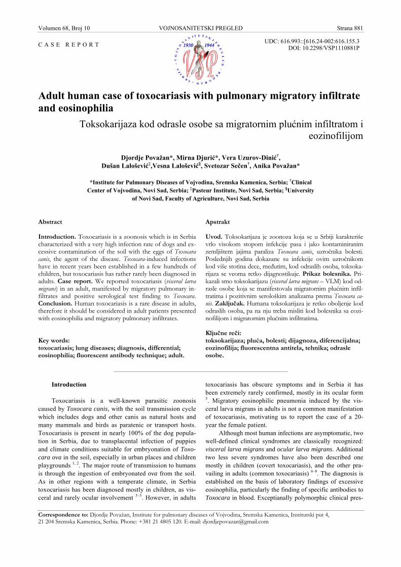
The chest radiography finding was presented with anexcessively marked bronchovascular contour and a tiny,roundish, inhomogeneous lesions partially in the intermedi-ary and lower lung portions bilaterally and one infiltrate onthe left basal field (Figure 1). The CT finding obtained im-mediately prior to admission was presented with roundish,uncleanly delineated to the periphery, inhomogeneous, hy-perdense lesions of 6–8 mm in the diameter, localized on theleft, in the projection of the basal and the Fowler's segment.The finding suggested micronodular infiltrative lesions wereinvolved in these localizations, and one infiltrate over 1 cmon the left (Figure 1).
The following blood test findings were obtained: eryth-rocyte sedimentation rate was 20/35, C-reactive protein(CRP) level < 6 mg/L, and white blood count (WBC) 5.3 109/L, with 18% of eosinophils, and immunoglobulin levelsof IgA 4.4 g/L, IgM 3.6 g/L, IgG 13.1 g/L. The tuberculin
and virological tests were negative. The sputum and catheterbiopsy samples examined for Mycobacterium tuberculosis bysmear and by culture were negative. Bacteriological findingsof the nose and throat swab samples, as well as the sputumones were negative, as well. The cytological sputum analysisrecurrently suggested the presence of eosinophil granulo-cytes. The stool assay for parasite eggs and larvae was nega-tive.
Lung function tests were normal, and the bronchialchallenge with carbachol was negative, as well. Blood gasanalyses at rest and on exertion of 80 W were normal. Al-lergy examination by a prick test revealed no hypersensitiv-ity to examined standard inhalant allergens.
Ultrasound (US) scanning of the upper abdomenshowed the liver approximated 151 mm in diameter, with ahomogeneous structure, and the pancreas, spleen and gallbladder were normal, as well.
The bronchoscopy finding was normal, and all the his-tological samples (catheter biopsy and transbronchial biopsy)taken from the left lung in the course of bronchoscopy hadnormal features, excluding the presence of few eosinophils.Bronchoalveolar lavage (BAL) of the middle lobe revealed theincreased presence of eosinophils (24%), with 64% macro-phages, 8% lymphocytes and 4% segmented neutrophils.
To better enlighten descrete lung lesions, a control CTscreening of the chest was performed three weeks later, re-vealing infiltrative migratory lesions of an altered localiza-tion and density as compared to those seen in the former CTfinding.
Due to enlarged cervical lymph nodes and as recom-mended by the hematologist, magnetic resonance (MR) im-aging of the neck and endocranium was performed, revealingcervical lymphadenomegaly (enlarged nuchal lymph nodesbilaterally with the largest diameter of 12.8 mm, as well asthe supraclavicular ones on the right, reaching 11.8 mm indiameter), without pathological lesions in the endocranium.
A high percentage of eosinophils (17%) persisted inblood. Taking into account the age of the patient, laboratory
and chest CT findings, the diagnosis of the hypereosinophilicsyndrome and eosinophilic pneumonia was established.
Corticosteroid therapy (prednisone 30 mg per day) wasinitiated resulting in a complete radiological regression andeosinophil count reduction in ten days. The patient's general
Fig. 1 – A) the patient's chest radiography; B) computerized tomography scan on ad-mission, the largest infiltrate (arrowhead)
Volumen 68, Broj 10 VOJNOSANITETSKI PREGLED Strana 883
Považan Dj, et al. Vojnosanit Pregl 2011; 68(10): 881–885.
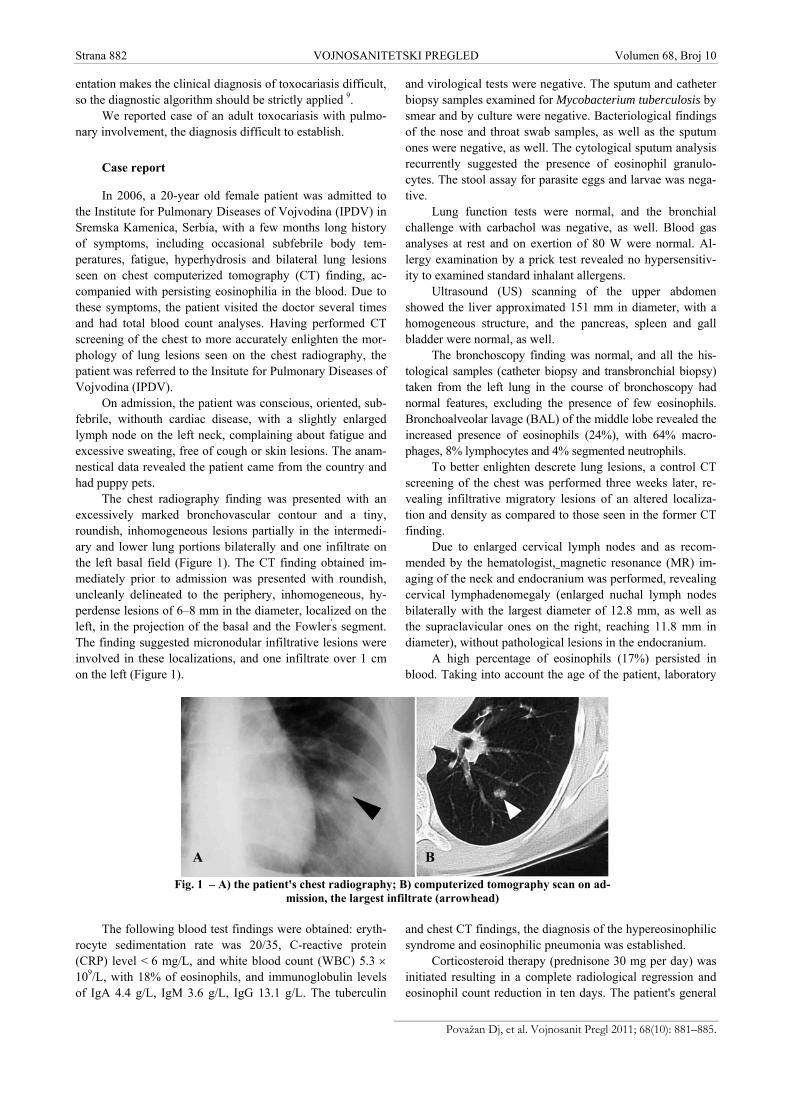
condition was satisfactory, but elevated eosinophil counts inthe peripheral blood persisted. After a 6-month corticosteroidtreatment, eosinophil count was normalized (5% of eosino-phils), so corticosteroids were discontinued. But, as the eo-sinophil count increased to 28% as soon as the corticosteroidtreatment had been discontinued (Figure 2), corticosteroidswere reintroduced into the treatment to include daily proni-sone doses of 30 mg per day.
Fig. 2 – Eosinophilia: course and levels with and withoutcorticosteroid therapy
To enlighten the etiology of persisting eosinophilia, ad-ditional analyses were performed: antistreptolysin titre, an-tiechinococcal antibodies, antinuclear, antimitochondrial,antithyroid, antiparietal, anti-smooth muscle and anticardiacantibodies, and they were negative. Complement 3 (C3) andcomplement 4 (C4) findings were normal, too. The ultra-sound (US) and hormone findings of the thyroid were nor-mal, as well. The cytological finding of the bone marrowsample was presented with an elevated percentage (15%) ofeosinophil cells. The repeate examinations of the stool andperianal print samples revealed no eggs of intestinal para-sites. The assay for eosinophils of the nose secretion sampleestablished 300 eosinophils per low magnification micro-scopic field.
Cytological analysis of the sputum and nose secretionsample, and the histological assay of the transbronchial bi-opsy sample established the presence of eosinophils, accom-panied with an increased percentage of eosinophils in theBAL and bone marrow sample and eosinophilia in bloodsuggest idiopathic hypereosinophilic syndrome with pulmo-nary involvement.
At the and, the indirect immunofluorescent test fortoxocariasis in the serum was significantly positive, in the ti-tre of 1 : 80 in the course of corticosteroid treatment.
A team of experts including a pulmologist, hematolo-gist, parasitologist and an immunologist, indicated that thetreatment with low-dose prednisolone accompanied with afurther follow-up of the patient, should also include an ant-helmintic drug (albendazole in the dose of 15 mg/kg/bodymass over 30 days), establishing the final diagnosis: Toxoca-riasis, Pneumonia eosinophilica, Eosinophilia persistens.
At subsequent controls the patient was free of symp-toms, with normal eosinophil count in blood, without patho-
logical lesions in radiograpy, and the negative finding of theimmunofluorescent test for toxocariasis was achieved aftertwo years.
Discussion
Migratory pulmonary infiltrates, eosinophilia in bloodand bronchial exudate and positive serology suggested thediagnosis of toxocariasis in our patient. After the hospitaltreatment and a long-term outpatient treatment and control, aregression of eosinophilia and negative serological assay twoyears later, strongly confirmed the diagnosis of the classicallarva migrans syndrome caused by Toxocara canis. A longdiagnostic procedure in our patient was due to a great rarityof toxocariasis in adult population in Serbia. So far, just afew cases of ocular involvement in adults have been regis-tered 5, in contrast to a great number of cases in children withdiverse clinical manifestations 2.
The infection of children is easy to explain. The geo-oral transmission, especially in children with geophagia, iswell-known. In adult patients without risky behavior, withgood hygienic standards, the Toxocara infection is possibleby ingestion of infected meat of other paratenic hosts. Meatas a source of infection has been mentioned in the literature.Serological investigations in Britain have shown that the pigserves as a paratenic host for Toxocara canis, with 4.5% ofpigs having antibodies against Toxocara canis 10, 11. Seroepi-demiological studies throughout the world have shown themost common prevalence of the general population is 2%–7% in moderate climate, but significantly higher in tropicalregions. In spite of numerous studies in this field performedin Serbia, we lack accurate data, but it has been well estab-lished that toxocariasis is a rare disease in adulthood 9.
Parasitic infections may induce pulmonary eosinophiliaand pulmonary lesions, so it is necessary to have a goodknowledge on inducing agents of pulmonary eosinophilia, itsmanifestations, diagnostic approach and treatment 12.
Toxocariasis is one of the inducing agents of eosinoph-ilic lung infiltrates. A study carried out in Korea reports 102patients with pulmonary infiltrates diagnosed by CT screen-ing of the chest who also had a positive serological finding intoxocariasis and blood eosinophilia. On the control chest CTfinding, 35% of the patients had migratory infiltrates, and48% of them were presented with regression of pulmonarylesions 13. Computerized tomography chest screening pro-vides a better morphological presentation of pulmonary le-sions, so we apply this imaging technique as other authors.Our reported patient also had migratory infiltrates seen onthe control chest CT finding, as described in this case report.Migratory nodular lesions were also seen on the control CTfinding in a 30-year old male patient with eosinophilia and apositive serological finding of Toxocara canis 14.
The relevance of an elevated percentage of eosinophilsin the BAL fluid has also been recognized by the authorsfrom Osaka, who reported a 38-year old female patient withthe symptoms of cough, blood eosinophilia, lung infiltratesand 94% of eosinophils in the BAL fluid, while we found24%. The diagnosis of visceral larva migrans (VLM) was
Strana 884 VOJNOSANITETSKI PREGLED Volumen 68, Broj 10
Považan Dj, et al. Vojnosanit Pregl 2011; 68(10): 881–885.
made on the basis of the positive results in the enzyme-linked immunosorbent assay for Toxocara canis, supportedby the clinical symptoms and laboratory findings 15.
The relevance of chest CT screening and BAL analysishas also been recognized by Polish authors who reported a32-year old patient with Toxocara infection. The diagnosiswas confirmed by serological tests with anti-Toxocara canisantibodies, bronchial lavage and chest CT scan with dis-seminated lung lesions 16.
Toxocariasis is one of the inducing agents of eosino-philia in the peripheral blood and eosinophilic infiltrates inan organism. The authors from Israel report that many casesmay be diagnosed just as hypereosinophilia syndrome in casethe serological test with Toxocara is not applied 17. It was thecase in our patient who, despite the applied corticosteroidtreatment developed an increase and persistence of eosino-phils, indicating a larger-scale investigations including animmunofluorescent assay which confirmed toxocariasis.There is no doubt it is crucial to establish an accurate diag-nosis, requiring a good knowledge of diagnostic algorithmfor eosinophilia. Due to an activated immunoinflammatoryreaction, our patient developed enlarged lymph nodes of theneck.
The diagnosis of larval migrans is usually done by im-munodiagnostic methods 18.
Serological tests with Toxocara are recommended inpatients with pulmonary infiltrates of unknown etiology 13,and the diagnosis of VLM is established by positive sero-logical findings for Toxocara canis, together with the clinicalsymptoms and laboratory findings 14–16.
Having obtained a significantly positive indirect immu-nofluorescent test finding of toxocariasis in serum in our pa-tient, we introduced albendazole in the treatment. Theauthors from Osaka reported an adult patient with VLM andpulmonary infiltrates, with histologically established eosi-nophilic pneumonia persisting for seven weeks prior to in-troducing the antihelminthic therapy with albendazole 15, thatwas also the case in our patient in whom all blood findingsdid not regain normal levels before albendazole had been ap-plied.
The duration of the treatment with albendozole, whichis diversely approached, is also analyzed by the authors whoreport a 42-year old patient with fever, productive cough,dyspnea, ground glass opacities mainly in the upper andmiddle lung fields on chest radiography. The symptoms dis-
appeared on antibiotic treatment, but eosinophilia persisted,so the investigations were, as in our case, extended andToxocara canis larva migrans was diagnosed. Four-week al-bendazole treatment was applied, but as eosinophilia reoccurafter one month discontinuation, the treatment was prolongedfor additional eight weeks 19.
Numerous studies investigated undesirable side effectsof albendazole. Some authors, however, recognize its effi-cacy without side effects for the tratment of toxocariasis inchildren 20. Jevtić et al. 21 analyze a long-term treatment withhigh albendazole doses in patients with cystic echinococco-sis, establishing significantly elevated transaminase levels inserum, which in most patients regain to normal in sixmonths. New efficient drugs of this group should thereforebe discovered 21, 22.
The clinical forms are non-specific but frequent andvaried (neurological, opthalmologic, pulmonary, cutaneousand sometimes rheumatological) 23. The following skinmanifestations have been described in patients with toxoca-riasis: chronic urticaria, chronic pruritis and eczema. In manycases, these skin manifestations appear as sole symptoms ofthe disease, developing after antihelmitic treatment 7. Oculartoxocariasis is usually a unilateral disease and the authors re-port a 71-year old female patient with ocular toxocariasis ofthe left eye and unilaterally deteriorated sight and strabism,pointing out the exposure to pets – dogs and cats, as riskfactors for Toxocara infection 8.
Our patient had cervical lymphadenopathy, which wasresolved after the applied treatment. It correlates with a casereport of a 7-year old boy with toxocariasis reporting that theinitial corticosteroid cotreatment succeeded by thiabendazoleresulted in a regression of lymphadenopathy and normaliza-tion of a total blood and eosinophil count 24. It is thereforesuggested that infection with Toxocara canis should be con-sidered in cases with generalized lymphadenopathy accom-panied with fever, hepatosplenomegaly and eosinophilia.
Conclusion
We reported a case of visceral larva migrans in an adultpatient with migratory pulmonary infiltrates and positive se-rology. Due to a prolonged diagnostic management of ourpatient, we emphasize this little known zoonosis in adult pa-tients which should be considered in case of eosinophilia andpulmonary infiltrates.
R E F E R E N C E S
1. Kulišić Z, Pavlović I, Milutinović MJ, Aleksić-Bakrac N. Intestinalparasites of dogs and role of dogs in epidemiology of larva mi-grans in the Belgrade area. Helminthologia 1998; 35(2): 79–82.
2. Lalosević D, Gebauer E, Malenković M. The role of toxocariasis inthe etiology of hypereosinophilic syndrome in children. MedPregl 1993; 46(11–12): 434–7. (Croatian)
3. Lalosević D, Oros A, Lalosević V, Knezević K, Knezević S, Bozić K, etal. Manifestations of visceral and ocular symptoms of toxoca-riasis in a 6-year-old boy. Med Pregl 2001; 54(Suppl 1): 51–3.Croatian.
4. Lalosević D, Radulović S, Mićović Z, Misita V. Identification ofToxocara canis larvae in ocular granuloma. Vojnosanit Pregl1994; 51(3): 242–4.
5. Stankovic-Babic G, Zlatanovic G, Djordjevic-Jocic J, Kostovska V, Ce-kic S. Ocular toxocariasis - A case report. Acta Fac Med Naiss2007; 24(3): 121–4.
6. Rubinsky-Elefant G, Hirata CE, Yamamoto JH, Ferreira MU. Hu-man toxocariasis: diagnosis, worldwide seroprevalences andclinical expression of the systemic and ocular forms. Ann TropMed Parasitol 2010; 104(1): 3–23.
Volumen 68, Broj 10 VOJNOSANITETSKI PREGLED Strana 885
Považan Dj, et al. Vojnosanit Pregl 2011; 68(10): 881–885.
7. Gavignet B, Piarroux R, Aubin F, Millon L, Humbert P. Cutaneousmanifestations of human toxocariasis. J Am Acad Dermatol2008; 59(6): 1031–42.
8. Stanković-Babić G, Zlatanović G, Đorđević-Jocić J, Kostovska V, CekićS. Ocular toxocariasis: a case report. Acta Fac Med Naiss 2007;24(3): 121–4.
9. Lalošević D, Lalošević V. Toxocariasa – larva migrans in humansand animals. Belgrade: Zadužbina Andrejević; 2008. p. 1–105.
10. Helwigh AB, Lind P, Nansen P. Visceral larva migrans: migratory pat-tern of Toxocara canis in pigs Int J Parasitol 1999; 29(4): 559–65.
11. Stevenson P. Toxocara and ascaris infection in British pigs: a se-rological survey. Vet Rec 1979; 104(23): 526–8.
12. Chitkara RK, Krishna G. Parasitic pulmonary eosinophilia.Semin Respir Crit Care Med 2006; 27(2): 171–84.
13. Yoon YS, Lee CH, Kang YA, Kwon SY, Yoon HI, Lee JH, et al. Im-pact of toxocariasis in patients with unexplained patchy pulmo-nary infiltrate in Korea. J Korean Med Sci 2009; 24(1): 40–5.
14. Hisamatsu Y, Ishii H, Kai N, Amemiya Y, Otani S, Morinaga R, et al.Case of toxocariasis showing migratory nodular shadows with ha-los. Nihon Kokyuki Gakkai Zasshi 2008; 46(5): 420–4. (Japanese)
15. Inoue K, Inoue Y, Arai T, Nawa Y, Kashiwa Y, Yamamoto S, et al.Chronic eosinophilic pneumonia due to visceral larva migrans.Intern Med 2002; 41(6): 478–82.
16. Kuziemski K, Jassem E, Mierzejewska E, Goljan J, Slominski JM.Lung manifestation of visceral larva migration syndrome dueto Toxocara canis infection. Pneumonol Alergol Pol 1999;67(11–12): 554–7.
17. Sayar D, Mazilis A, Kassem E, Klein A. Toxocariasis as a causeof hypereosinophilia. Harefuah 2009; 148(1): 14–6, 89. (He-brew)
18. Ishiyamna S, Ono K, Rai SK, Uga S. Method for detecting circu-lating Toxocara canis antigen and its application in human se-rum samples. Nepal Med Coll J 2009; 11(1): 9–13.
19. Takamatsu K, Sumitani M, Nanjyou S, Nishijima M, Syoji S, Taki-fuji N, et al. Case of Toxocara canis larva migrans cured by ad-ditional treatment with albendazole. Nihon Kokyuki GakkaiZasshi 2008; 46(10): 836–41.
20. Saporito L, Scarlata F, Colomba C, Infurnari L, Giordano S, TitoneL. Human toxocariasis: a report of nine cases. Acta Paediatr2008; 97(9): 1301–2.
21. Jevtić M, Mikić D, Arsić-Komljenović G, Stanković N, Ristanović E,Sjeničić G, et al. Adverse effects of long term, continual admini-stration of high doses of albendazole in the treatment of echi-nococcal disease. Vojnosanit Pregl 2008; 65(7): 539–44. (Ser-bian)
22. Mikić D, Jevtić M, Arsić-Komljenović G, Ristanović E, Stanković N,Sjenicić G, et al. Impossibility of the treatment of inoperableliver multicystic echinococcosis due to adverse reactions toantihelminitics. Vojnosanit Pregl 2009; 66(10): 833–9. (Ser-bian)
23. Degouy A, Menat C, Aubin F, Piarroux R, Woronoff-Lemsi MC,Humbert P. Toxocariasis. Presse Med 2001; 30(39–40 Pt 1):1933–8.
24. Szczepański T, Sońta-Jakimczyk D, Janik-Moszant A, Olejnik I.Generalized lymphadenopathy as initial presentation of toxo-cariasis in a seven-year-old boy. Pediatr Infect Dis J 1996;15(8): 717–8.
Received on January 27, 2010.Revised on May 4, 2010.
Accepted on May 10, 2010.