additional uses of trypan blue staining during cataract surgery
TRANSCRIPT
results, it cannot be excluded that these occurrences will continueto happen with future MemoryLens implantations.
GUY KLEINMANN, MDDAVID J. APPLE, MD
Salt Lake City, Utah, USAIRMINGARD M. NEUHANN, MD
Teubingen, GermanyTHOMAS F. NEUHANN, MD
Munich, Germany
REFERENCE
1. Neuhann IM, Werner L, Izak AM, et al. Late postoperative opacification
of a hydrophilic acrylic (hydrogel) intraocular lens; a clinicopathological
analysis of 106 explants. Ophthalmology 2004; 111:2094–2101
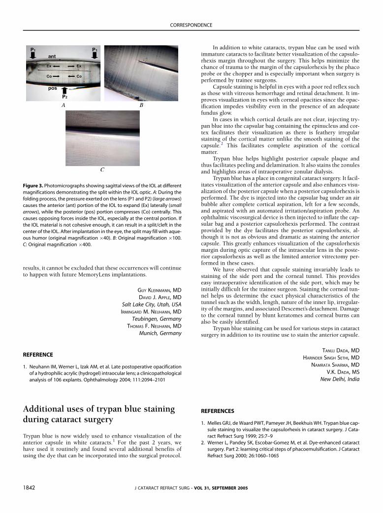
Figure 3. Photomicrographs showing sagittal views of the IOL at different
magnifications demonstrating the split within the IOL optic. A: During the
folding process, the pressure exerted on the lens (P1 and P2) (large arrows)
causes the anterior (ant) portion of the IOL to expand (Ex) laterally (small
arrows), while the posterior (pos) portion compresses (Co) centrally. This
causes opposing forces inside the IOL, especially at the central portion. If
the IOL material is not cohesive enough, it can result in a split/cleft in the
center of the IOL. After implantation in the eye, the split may fill with aque-
ous humor (original magnification �40). B: Original magnification �100.
C: Original magnification �400.
CORRESPONDENCE
Additional uses of trypan blue stainingduring cataract surgery
Trypan blue is now widely used to enhance visualization of theanterior capsule in white cataracts.1 For the past 2 years, wehave used it routinely and found several additional benefits ofusing the dye that can be incorporated into the surgical protocol.
J CATARACT REFRACT SURG -1842
In addition to white cataracts, trypan blue can be used withimmature cataracts to facilitate better visualization of the capsulo-rhexis margin throughout the surgery. This helps minimize thechance of trauma to the margin of the capsulorhexis by the phacoprobe or the chopper and is especially important when surgery isperformed by trainee surgeons.
Capsule staining is helpful in eyes with a poor red reflex suchas those with vitreous hemorrhage and retinal detachment. It im-proves visualization in eyes with corneal opacities since the opac-ification impedes visibility even in the presence of an adequatefundus glow.
In cases in which cortical details are not clear, injecting try-pan blue into the capsular bag containing the epinucleus and cor-tex facilitates their visualization as there is feathery irregularstaining of the cortical matter unlike the smooth staining of thecapsule.2 This facilitates complete aspiration of the corticalmatter.
Trypan blue helps highlight posterior capsule plaque andthus facilitates peeling and delamination. It also stains the zonulesand highlights areas of intraoperative zonular dialysis.
Trypan blue has a place in congenital cataract surgery. It facil-itates visualization of the anterior capsule and also enhances visu-alization of the posterior capsule when a posterior capsulorhexis isperformed. The dye is injected into the capsular bag under an airbubble after complete cortical aspiration, left for a few seconds,and aspirated with an automated irritation/aspiration probe. Anophthalmic viscosurgical device is then injected to inflate the cap-sular bag and a posterior capsulorhexis performed. The contrastprovided by the dye facilitates the posterior capsulorhexis, al-though it is not as obvious and dramatic as staining the anteriorcapsule. This greatly enhances visualization of the capsulorhexismargin during optic capture of the intraocular lens in the poste-rior capsulorhexis as well as the limited anterior vitrectomy per-formed in these cases.
We have observed that capsule staining invariably leads tostaining of the side port and the corneal tunnel. This provideseasy intraoperative identification of the side port, which may beinitially difficult for the trainee surgeon. Staining the corneal tun-nel helps us determine the exact physical characteristics of thetunnel such as the width, length, nature of the inner lip, irregular-ity of themargins, and associated Descemet’s detachment. Damageto the corneal tunnel by blunt keratomes and corneal burns canalso be easily identified.
Trypan blue staining can be used for various steps in cataractsurgery in addition to its routine use to stain the anterior capsule.
TANUJ DADA, MDHARINDER SINGH SETHI, MD
NAMRATA SHARMA, MDV.K. DADA, MS
New Delhi, India
REFERENCES
1. Melles GRJ, deWaard PWT, Pameyer JH, BeekhuisWH. Trypan blue cap-
sule staining to visualize the capsulorhexis in cataract surgery. J Cata-
ract Refract Surg 1999; 25:7–9
2. Werner L, Pandey SK, Escobar-Gomez M, et al. Dye-enhanced cataract
surgery. Part 2: learning critical steps of phacoemulsification. J Cataract
Refract Surg 2000; 26:1060–1065
VOL 31, SEPTEMBER 2005