acyanotic congenital heart disease · acyanotic congenital heart ... defects. iap ug teaching...
TRANSCRIPT
IAP UG Teaching slides 2015‐16 1
ACYANOTIC CONGENITAL HEART
DISEASE
IAP UG Teaching slides 2015‐16 2
ATRIAL SEPTAL DEFECT (ASD)
IAP UG Teaching slides 2015‐16 3
ATRIAL SEPTAL DEFECT (ASD)
• Isolated anomaly In 10%
• M:F ratio : 1:2
• 30‐50 % of children have ASD as part of cardiac defects.
IAP UG Teaching slides 2015‐16 4
ASD
• Increasingly referred as murmur and detected in
infancy
• Auscultatory Findings helpful in detection
• ECG quite useful
IAP UG Teaching slides 2015‐16 5
ASD TYPES
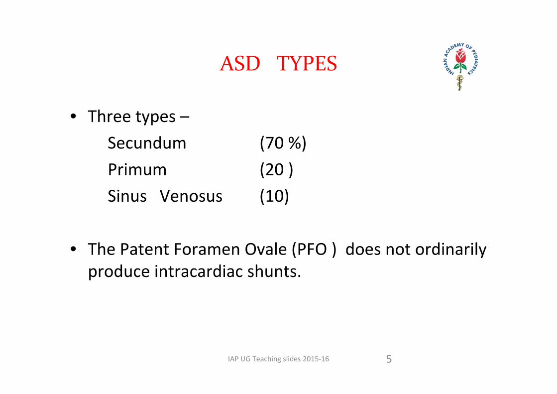
• Three types – Secundum (70 %) Primum (20 ) Sinus Venosus (10)
• The Patent Foramen Ovale (PFO ) does not ordinarily produce intracardiac shunts.
IAP UG Teaching slides 2015‐16 6
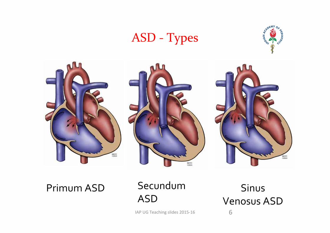
ASD ‐ Types
Primum ASD Secundum ASD
Sinus Venosus ASD

IAP UG Teaching slides 2015‐16 7
Pre tricuspid L R shunt
Asymptomatic
Detected in Late infancy & Childhood
Pulse NBP NJVP A=V
ASD
IAP UG Teaching slides 2015‐16 8
ASD
Auscultation
Wide & Fixed Split of S2 ‐ No change with Respiration /
Standing. P2 can be loud but no PAH
Ejection systolic murmur at Pulmonary Area
Mid Diastolic murmur at LLSB
IAP UG Teaching slides 2015‐16 9
•ECG : RAD / rSR : RSR : rR in V1 or V3 R ( 85%)
•CXR : Variable Cardiac size
Right atrial enlargement
PBF •Echocardiography : Location of ASD / Size
Dilatation of RV / RA / PAParadoxical motion of IVS
ASD
IAP UG Teaching slides 2015‐16 10
NATURAL HISTORY OF ASD
Spontaneous closure ?
• ASD <4mm by 18months > 90% closure.• ASD 4‐8mm by 18m 75% closure.• ASD > 8mm rarely close spontaneously.
• If untreated CHF and PAH develops in adults• SBE prophylaxis is not required unless associated defects present.
IAP UG Teaching slides 2015‐16
ATRIAL SEPTAL DEFECT (ASD), OTHER THANPRIMUM TYPE
• Mode of diagnosis: – Physical examination, ECG, X‐ray Chest, transthoracic echocardiography
• Spontaneous closure: Rare if defect >8 mm at birth. Rare after age 2 years. Very rarely an ASD can enlarge on follow up.
11
IAP UG Teaching slides 2015‐16
PATENT FORAMEN OVALE
Patent foramen ovale:
• Echocardiographic detection of a small defect in fossa ovalis region with a flap with no evidence of right heart volume overload (dilatation of right atrium and right ventricle).
• Patent foramen ovale is a normal finding in newborns.
12
IAP UG Teaching slides 2015‐16
INDICATION FOR CLOSURE: ASD ASSOCIATED WITH RIGHT VENTRICULAR
VOLUME OVERLOAD
(i) In asymptomatic child: 2‐4 yrs. (For sinus venosus defect ‐ 4‐5 yrs..)
(ii) Symptomatic ASD in infancy (CCF, severe PAH): seen in about 8%‐10% of cases. Rule out associated lesions (e.g., total anomalous pulmonary venous drainage, left ventricular inflow obstruction, aorto‐pulmonary window). Early closure is recommended.
13
IAP UG Teaching slides 2015‐16
INDICATION FOR CLOSURE…
(iii) If presenting beyond ideal age: Elective closure irrespective of age as long as there is right heart volume overload and pulmonary vascular resistance is in operable range.
Method of closure: Surgical: Established mode. Device closure: More recent mode, may be used in children weighing >10 kg and having a central ASD.
14
IAP UG Teaching slides 2015‐16 15
VENTRICULAR SEPTAL DEFECT(VSD)
IAP UG Teaching slides 2015‐16
VSD – TYPES ( location )
16
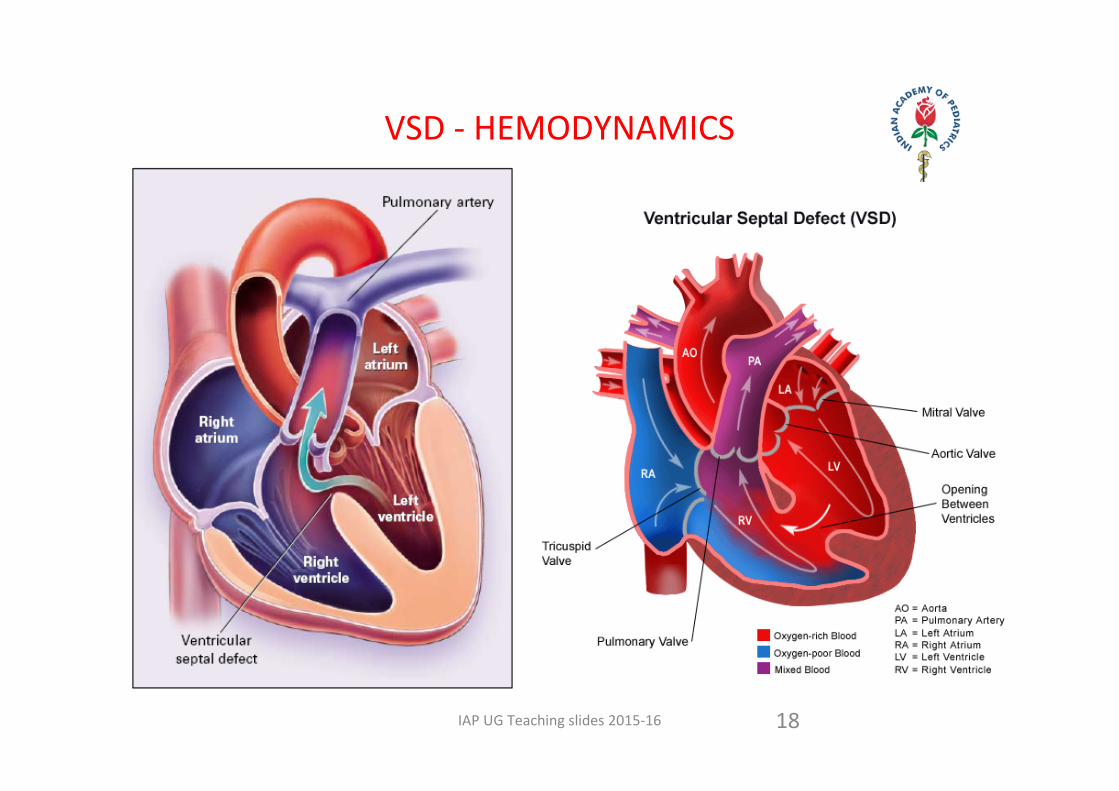
Location of the defect: • Type I: Subarterial(outlet, subpulmonic, supracristal or infundibular)
• Type II: Perimembranous (subaortic)• Type III: Inlet• Type IV: Muscular
IAP UG Teaching slides 2015‐16
VSD – TYPES (SIZE )
• Large (nonrestrictive): Diameter of the defect is approximately equal to diameter of the aortic orifice Right ventricular systolic pressure is systemicDegree of left to right shunt depends on pulmonary vascular resistance
• Moderate (restrictive): Diameter of the defect is less than that of the aortic orificeRight ventricular pressure is half to two third systemic Left to right shunt is >2:1
• Small (restrictive): Diameter of the defect is less than one third the size of the aortic orifice Right ventricular pressure is normalleft to right shunt is <2:1
17
IAP UG Teaching slides 2015‐16 18
VSD ‐ HEMODYNAMICS
IAP UG Teaching slides 2015‐16 19
CLINICAL PRESENTATION
With small VSD ‐ asymptomatic.
With large VSD, delayed growth and development, repeated pulmonary infections and CHF.
With long standing pulmonary hypertension, a history of cyanosis and a decreased activity.
IAP UG Teaching slides 2015‐16
MODE OF DIAGNOSIS
• Physical examination• ECG• X‐ray chest• Echocardiography
20
IAP UG Teaching slides 2015‐16
S 1 Normal. S 2 Loud ( P 2 )
Wide split with variable P2
Closely split when PAH develops
PSM at LLSB / MSB order
MDM at Apex . [ EDM at Aortic Area ]
21
VSD AUSCULTATION
IAP UG Teaching slides 2015‐16
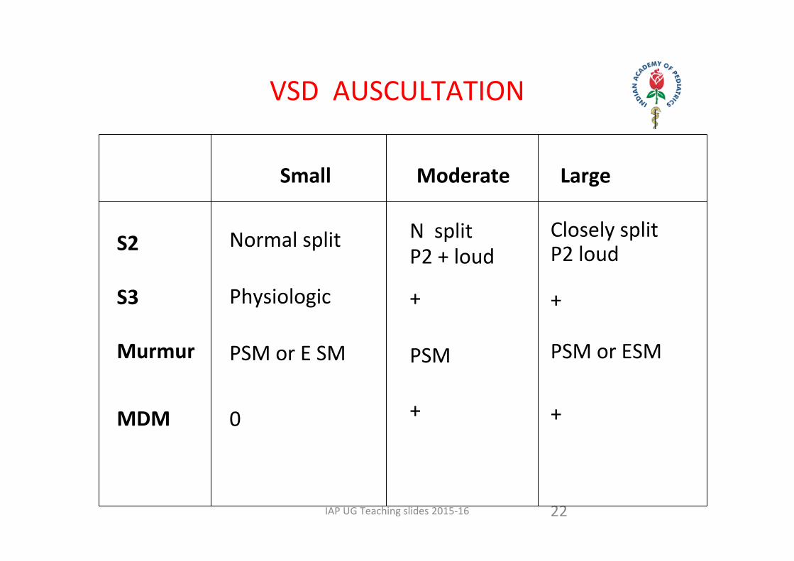
VSD AUSCULTATION
22
Small Moderate Large
S2
S3
Murmur
MDM
Normal split
Physiologic
PSM or E SM
0
N splitP2 + loud
+
PSM
+
Closely splitP2 loud
+
PSM or ESM
+
IAP UG Teaching slides 2015‐16
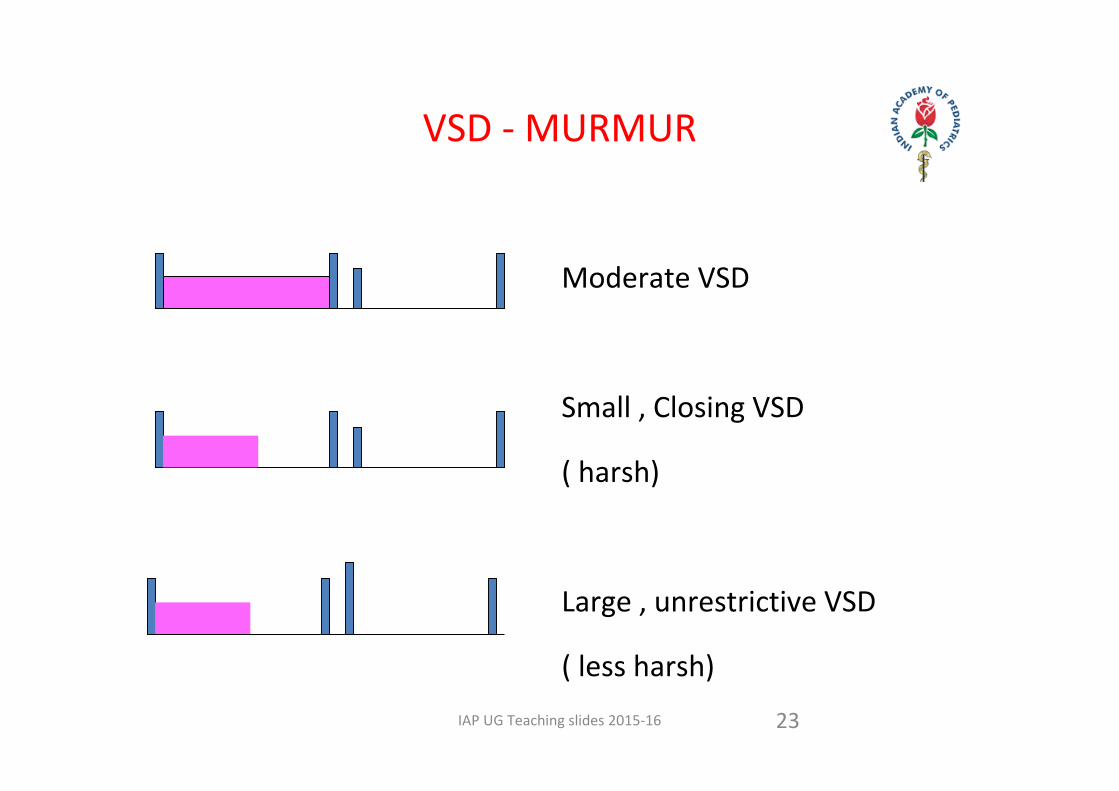
VSD ‐ MURMUR
23
Moderate VSD
Small , Closing VSD
( harsh)
Large , unrestrictive VSD
( less harsh)
IAP UG Teaching slides 2015‐16
NATURAL HISTORY
• About 10% of large nonrestrictive VSDs die in first year, primarily due to congestive heart failure.
• Spontaneous closure is uncommon in large VSDs.• 30%‐40% of moderate or small defects (restrictive) close spontaneously, majority by 3‐5 years of age.
• Decrease in size of VSD is seen in 25%.
24
IAP UG Teaching slides 2015‐16
VSD ‐TIMING OF CLOSURE
• Large VSD with uncontrolled congestive heart failure: As soon as possible.
• Large VSD with severe pulmonary artery hypertension: 3‐6 months.• Moderate VSD with pulmonary artery systolic pressure 50%‐66% of
systemic pressure: Between 1‐2 years of age, earlier if one episode of life threatening lower respiratory tract infection or FTT.
• Small sized VSD with normal pulmonary artery pressure, left to right shunt >1.5:1:Closure by 2‐4 yrs..
• Small outlet VSD (<3mm) without aortic valve prolapse: 1‐2 yearly follow up to look for development of aortic valve prolapse.
• Small outlet VSD with aortic valve prolapse without aortic regurgitation: Closure by 2‐3 years of age irrespective of the size and magnitude of left to right shunt.
25
IAP UG Teaching slides 2015‐16
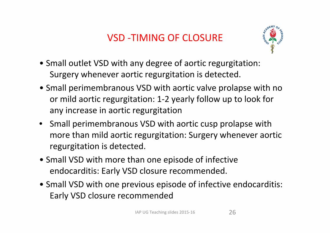
• Small outlet VSD with any degree of aortic regurgitation: Surgery whenever aortic regurgitation is detected.
• Small perimembranous VSD with aortic valve prolapse with no or mild aortic regurgitation: 1‐2 yearly follow up to look for any increase in aortic regurgitation
• Small perimembranous VSD with aortic cusp prolapse with more than mild aortic regurgitation: Surgery whenever aortic regurgitation is detected.
• Small VSD with more than one episode of infective endocarditis: Early VSD closure recommended.
• Small VSD with one previous episode of infective endocarditis: Early VSD closure recommended
26
VSD ‐TIMING OF CLOSURE
IAP UG Teaching slides 2015‐16
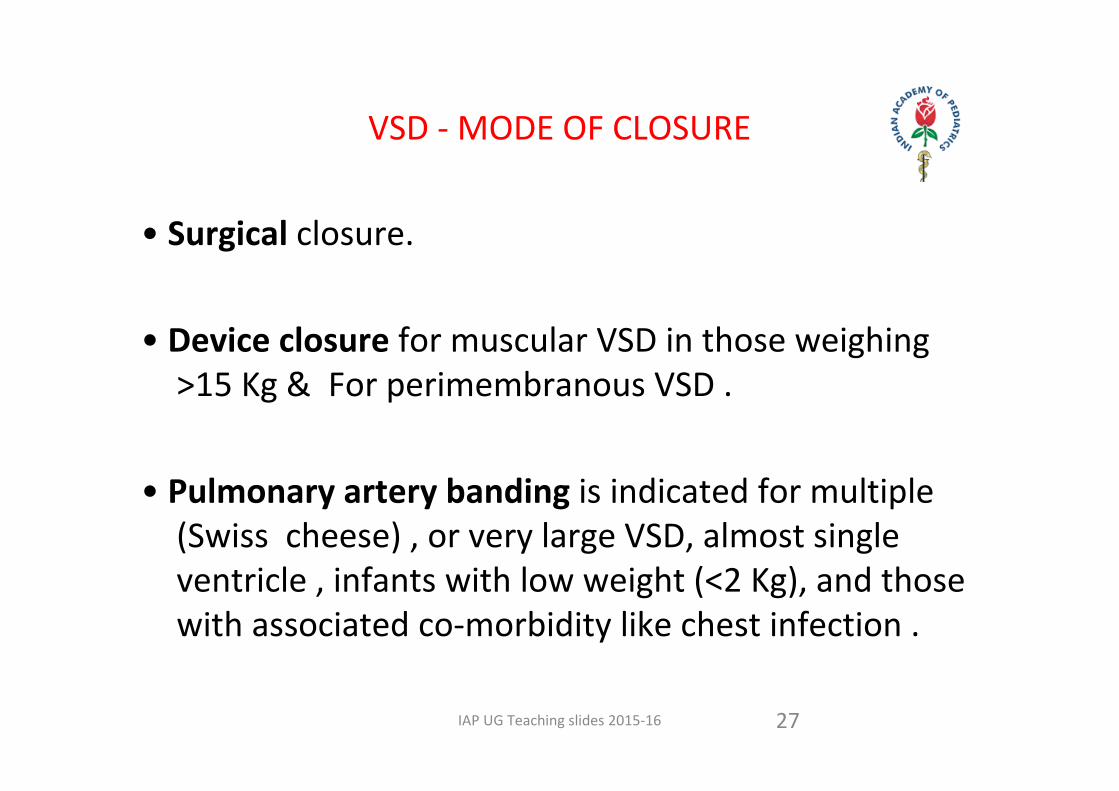
VSD ‐ MODE OF CLOSURE
• Surgical closure.
• Device closure for muscular VSD in those weighing >15 Kg & For perimembranous VSD .
• Pulmonary artery banding is indicated for multiple (Swiss cheese) , or very large VSD, almost single ventricle , infants with low weight (<2 Kg), and those with associated co‐morbidity like chest infection .
27
IAP UG Teaching slides 2015‐16 28
PDA
IAP UG Teaching slides 2015‐16 29
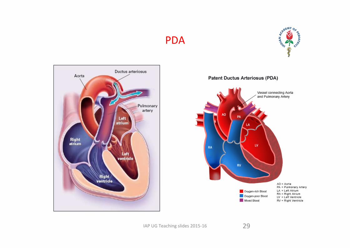
PDA
IAP UG Teaching slides 2015‐16
MODE OF DIAGNOSIS
• Physical examination• ECG• X‐ray chest • Echocardiography.
30
IAP UG Teaching slides 2015‐16 31
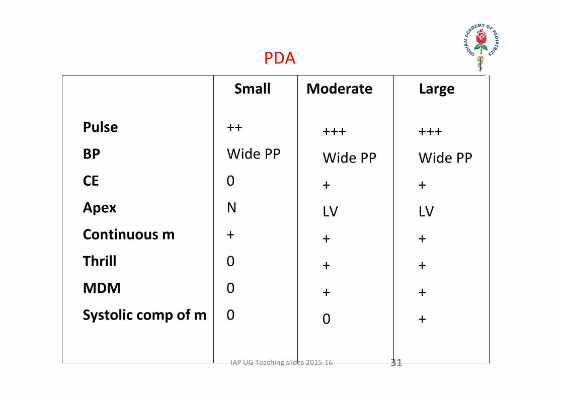
PDA
Small Moderate Large
Pulse
BP
CE
Apex
Continuous m
Thrill
MDM
Systolic comp of m
++
Wide PP
0
N
+
0
0
0
+++
Wide PP
+
LV
+
+
+
0
+++
Wide PP
+
LV
+
+
+
+
IAP UG Teaching slides 2015‐16 32
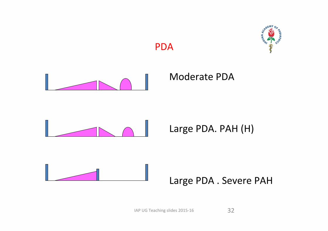
Moderate PDA
Large PDA. PAH (H)
Large PDA . Severe PAH
PDA
IAP UG Teaching slides 2015‐16 33
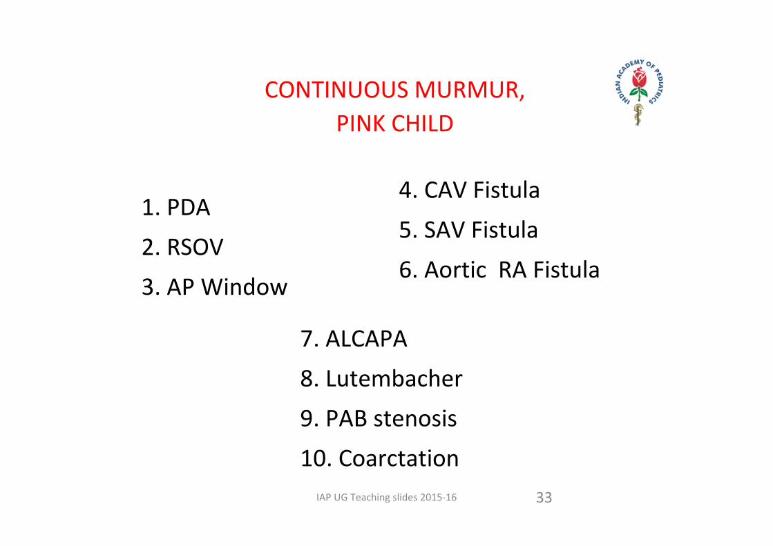
CONTINUOUS MURMUR,PINK CHILD
1. PDA 2. RSOV 3. AP Window
4. CAV Fistula5. SAV Fistula6. Aortic RA Fistula
7. ALCAPA8. Lutembacher9. PAB stenosis10. Coarctation
IAP UG Teaching slides 2015‐16
VENOUS HUM
34
Soft blowing murmurI ‐ III Medium pitchedHigh R/L Sternal border or bothNo Peaking around S2 on sitting up with neck flexed on lying down , change in neck position
D.D : PDA, AVM ,PAV fistula Collaterals
IAP UG Teaching slides 2015‐16
SIZE OF PDA
Large PDA: Associated with significant left heart volume overload, CCF, severe PAH. PDA murmur is unlikely to be loud or continuous.
Moderate PDA: Some degree of left heart overload, mild to moderate PAH, no/mild CCF. Murmur is continuous.
Small PDA: Minimal or no left heart overload. No PH / CCF. Murmur may be continuous or only systolic
Silent PDA: No murmur, no PH. Diagnosed only on echo Doppler.
Spontaneous closure: Small PDAs in full term baby may close up to 3 mo of age, large PDAs are unlikely to close.
35
IAP UG Teaching slides 2015‐16
TIMING OF CLOSURE
• Large/ moderate PDA, with congestive heart failure, pulmonary artery hypertension: Early closure (by 3‐6 months).
• Moderate PDA, no congestive heart failure: 6 months‐1 year. If failure to thrive, closure can be accomplished earlier.
• Small PDA: At 12‐18 months.• Silent PDA: Closure not recommended.
36
IAP UG Teaching slides 2015‐16
MODE OF CLOSURE
Individualized.Device closure, coils occlusion or surgical ligation in children >6 months of age.
Surgical ligation if <6 months of age. Device/ coils in <6 months . Indomethacin/ ibuprofen not to be used in term babies .
37
IAP UG Teaching slides 2015‐16
PDA IN A PRETERM BABY
• Intervene if baby in heart failure (small PDAs may close spontaneously).
• Indomethacin or Ibuprofen(20) (if no contraindication) .
• Surgical ligation if above drugs fail or are contraindicated
• Prophylactic indomethacin or ibuprofen therapy: Not recommended.
38
IAP UG Teaching slides 2015‐16 39
CONGENITAL FORMS OF LVOT OBSTRUCTION
• SubvalvularDiscrete membranous stenosis, Fibromuscular tunnel
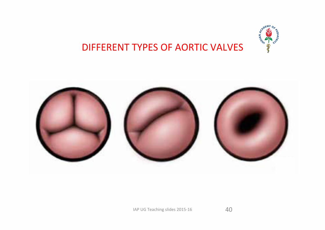
• ValvularUnicuspid , Bicuspid, Quadricuspid and Dysplastic
• SupravalvularDiscrete( membranous or hourglass)Aortic hypoplasia or atresiaInterrupted aortic archCoarctation of Aorta
IAP UG Teaching slides 2015‐16 40
DIFFERENT TYPES OF AORTIC VALVES
IAP UG Teaching slides 2015‐16 41
AV STENOSIS
Obstructive lesion. Usually asymptomatic
SCD / Syncope / Angina possible
Pulse AbnormalJVP NormalBP Near Normal
No Cardiomegaly . Heaving apex
ThrillRt USBSuprasternal
IAP UG Teaching slides 2015‐16 42
AS AUSCULTATION
S1 N S2 N Paradoxic Split?
S4 . S3 rare ( ominous )
Ejection Click ( constant )
Ejection Systolic murmur RUSB
EDM +
IAP UG Teaching slides 2015‐16 43
AS SEVERITY ASSESS
Pulse Low volumeHeaving ApexS1 E click DistanceS4Murmur Length Harshness Late PeakingSuprasternal ThrillThrill
IAP UG Teaching slides 2015‐16 44
NATURAL HISTORY OF AS
• Mild AS and Moderate AS ‐ asymptomatic.
• Severe AS ‐ heart failure in newborns, chest pain , syncope & sudden death.
• Pressure gradient increases with growth.• Worsening of AR may occur in subaortic stenosis.• SBE is 4% in valvar AS.
IAP UG Teaching slides 2015‐16
TIMING OF INTERVENTION: VALVULAR AS
45
For infants and older children:– Left ventricular dysfunction: Immediate intervention by balloon dilatation, irrespective of gradients.– Normal left ventricular function: Balloon dilatation if any of these present: (i)gradient >80 mmHg peak and 50 mmHg mean by echo‐Doppler; (ii)ST‐T changes in ECG with peak gradient of >50 mmHg; (iii)symptoms due to AS with peak gradient of >50 mmHg. I(iv)n case of doubt about severity/symptoms, an exercise test may be done for older children.• For neonates: Balloon dilatation if symptomatic or there is evidence of left ventricular dysfunction / mild left ventricular hypoplasia, or if Doppler gradient (peak) >75 mmHg.
IAP UG Teaching slides 2015‐16 46
COARCTATION OF THE AORTA
IAP UG Teaching slides 2015‐16 47
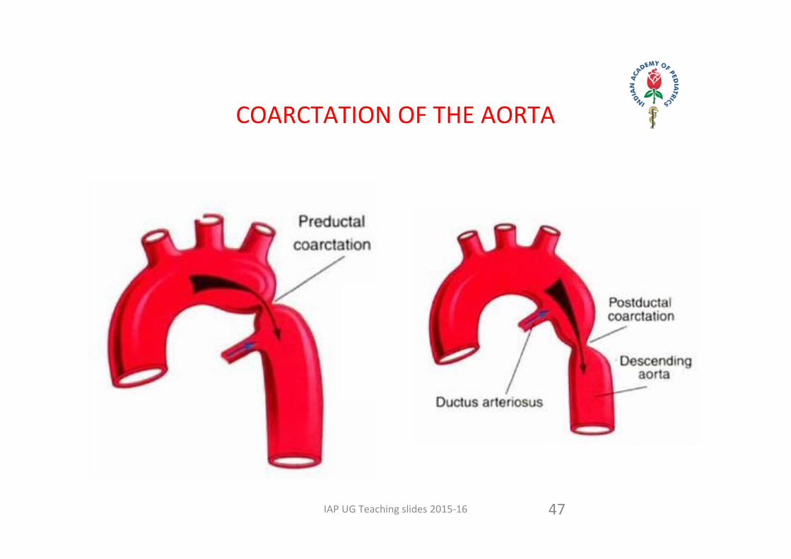
COARCTATION OF THE AORTA
IAP UG Teaching slides 2015‐16 48
COARCTATION OF AORTA (COA)
• 8 % of all CHD. • M:F = 2:1. 30% of Turner Syndrome.• 85% of COA have bicuspid valve.• Poor feeding, dyspnea & poor weight gain, & acute circulatory shock in first 6 weeks .
• 20‐30% of COA develop CHF by 3 months
IAP UG Teaching slides 2015‐16 49
COARCTATION OF AORTA
Stenotic lesion. Asymptomatic in many
Infancy to Adulthood
Normal JVP
Pulse discrepancyBP discrepancy
IAP UG Teaching slides 2015‐16 50
COA
• Radio femoral delay
Strong radials; Weak Femorals
‘Touch the Feet of Each Infant’
• Upper limb hypertension; Normotensive Lower limb
SBP of Lower limb 10 mm or more
Less than SBP of Upper limb
IAP UG Teaching slides 2015‐16 51
COA AUSCULTATION
S1 S2 N
S3 S4 not usual
Ej Click +
Ejection murmur / continuous murmur
No murmur
Clinical : Radio femoral delay ; Pulse discrepancy
Never mind the murmur !
IAP UG Teaching slides 2015‐16 52
COA CXR
Infant
Cardiomegaly. PVH
Aorta +
Child
No Cardiomegaly. PVH +
Aorta ++ & 3 signs
Rib notching
IAP UG Teaching slides 2015‐16 53
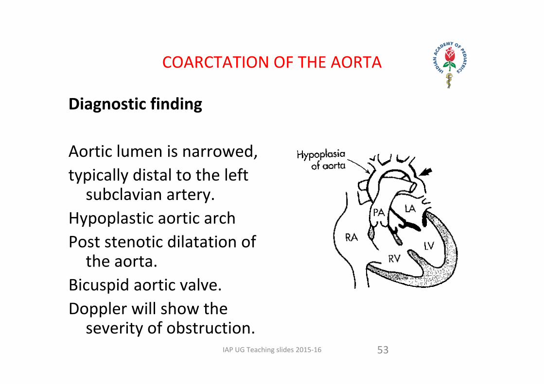
Diagnostic finding
Aortic lumen is narrowed, typically distal to the left subclavian artery.
Hypoplastic aortic archPost stenotic dilatation of the aorta.
Bicuspid aortic valve.Doppler will show the severity of obstruction.
COARCTATION OF THE AORTA
IAP UG Teaching slides 2015‐16 54
NATURAL HISTORY OF COA
• Bicuspid valve may cause stenosis or regurgitation with age.
• SBE may occur on either aortic valve or on coarctation.
• LV failure, rupture of aorta, ICH, hypertensive encephalopathy may develop during childhood.
IAP UG Teaching slides 2015‐16
TIMING & MODE OF INTERVENTION
Timing•With left ventricular dysfunction / congestive heart failure or severe
upper limb hypertension (for age): Immediate intervention.• Normal left ventricular function, no congestive heart failure and
mild upper limb hypertension: Intervention beyond 3‐6 months of age.
• No hypertension, no heart failure, normal ventricular function: Intervention at 1‐2 years
Mode of intervention• Balloon dilatation or surgery for children >6 mo of age.• Surgical repair for infants <6 mo of age.• Balloon dilatation with stent deployment can be considered in
children >10 years of age if required.• Elective endovascular stenting of aorta is contraindicated for
children <10 years of age55

IAP UG Teaching slides 2015‐16
THANK YOU
56