active-site structure and electron-transfer reactivity of plastocyanins
TRANSCRIPT

Active-Site Structure and Electron-Transfer Reactivity ofPlastocyanins
Katsuko Sato,† Takamitsu Kohzuma,‡ and Christopher Dennison*,†
Contribution from the Department of Chemistry, UniVersity of Newcastle upon Tyne,Newcastle upon Tyne NE1 7RU, U. K., and Department of Materials and Biological Science,
Faculty of Science, Ibaraki UniVersity, Mito, Ibaraki 310-8512, Japan
Received July 22, 2002; Revised Manuscript Received November 19, 2002; E-mail: [email protected]
Abstract: The active-site structures of Cu(II) plastocyanins (PCu’s) from a higher plant (parsley), a seedlessvascular plant (fern, Dryopteris crassirhizoma), a green alga (Ulva pertusa), and cyanobacteria (Anabaenavariabilis and Synechococcus) have been investigated by paramagnetic 1H NMR spectroscopy. In all casesthe spectra are similar, indicating that the structures of the cupric sites, and the spin density distributionsonto the ligands, do not differ greatly between the proteins. The active-site structure of PCu has remainedunaltered during the evolutionary process. The electron transfer (et) reactivity of these PCu’s is comparedutilizing the electron self-exchange (ESE) reaction. At moderate ionic strength (0.10 M) the ESE rate constantis dictated by the distribution of charged amino acid residues on the surface of the PCu’s. Most higherplant and the seedless vascular plant PCu’s, which have a large number of acidic residues close to thehydrophobic patch surrounding the exposed His87 ligand (the proposed recognition patch for the self-exchange process), have ESE rate constants of ∼103 M-1 s-1. Removal of some of these acidic residues,as in the parsley and green algal PCu’s, results in more favorable protein-protein association and an ESErate constant of ∼104 M-1 s-1. Complete removal of the acidic patch, as in the cyanobacterial PCu’s, leadsto ESE rate constants of ∼105-106 M-1 s-1. The ESE rate constants of the PCu’s with an acidic patchalso tend toward ∼105-106 M-1 s-1 at higher ionic strength, thus indicating that once the influence ofcharged residues has been minimized the et capabilities of the PCu’s are comparable. The cytochromesand Fe-S proteins, two other classes of redox metalloproteins, also possess ESE rate constants of ∼105-106 M-1 s-1 at high ionic strength. The effect of the protonation of the His87 ligand in PCu(I) on the ESEreactivity has been investigated. When the influence of the acidic patch is minimized, the ESE rate constantdecreases at high [H+].
Introduction
A detailed understanding of electron transfer (et) reactivity,in both biological and chemical systems, has been sought inrecent years. Biological et catalysts are commonly metallopro-teins in which the redox cofactor is protected by an elaborateprotein envelope. This biological coat not only tunes thereactivity of the redox center but also imparts particular surfaceproperties to the molecule, ensuring specificity in et reactions.The plastocyanins (PCu’s) are the best studied member of thecupredoxin family of redox metalloproteins,1,2 which possess amononuclear type 1 copper ion as the redox center. The PCu’sare involved in photosynthetic et between cytochromef of theb6f complex and P700+ of photosystem I (PSI)3-6 in plants,green algae, and cyanobacteria (blue green algae). The reactivityof PCu with its physiological partners has been investigated indetail.7-20 In this study we have analyzed the active-site structure
and et reactivity of PCu’s from a variety of sources and assessedhow these properties have been modified during evolution.
Plastocyanin was the first cupredoxin whose X-ray crystalstructure was determined21 and is one of the best structurallycharacterized et metalloproteins.22-38 PCu consists of aâ-barrel
† University of Newcastle upon Tyne.‡ Ibaraki University.
(1) Adman, E. T.AdV. Protein Chem. 1991, 42, 144-197.(2) Adman, E. T.Curr. Opin. Struct. Biol. 1991, 1, 895-904.(3) Barber, J.Plant Cell EnViron. 1983, 6, 311-322.(4) Haehnel, W.Annu. ReV. Plant Physiol.1984, 35, 659-693.(5) Barber, J.; Andersson, B.Nature1994, 370, 31-34.(6) Nugent, J. H. A.Eur. J. Biochem. 1996, 237, 519-531.
(7) He, S.; Modi, S.; Bendall, D. S.; Gray, J. C.EMBO J. 1991, 10, 4011-4016.
(8) Haehnel, W.; Jensen, T.; Gause, K.; Klo¨sgen, R. B.; Stahl, B.; Michl, D.;Huvermann, B.; Karas, M.; Herrmann, R. G.EMBO J. 1994, 13, 1028-1038.
(9) Hervas, M.; Navarro, J. A.; Dı´az, A.; Bottin, H.; De la Rosa, M. A.Biochemistry1995, 34, 11321-11326.
(10) Sigfridsson, K.; Young, S.; Hansson, O¨ . Biochemistry1996, 35, 1249-1257.
(11) Drepper, F.; Hippler, M.; Nitschke, W.; Haehnel, W.Biochemistry1996,35, 1282-1295.
(12) Hippler, M.; Reichert, J.; Sutter, M.; Zak, E.; Altschmeid, L.; Schroer, U.;Herrmann, R. G.; Haehnel, W.EMBO J. 1996, 15, 6374-6384.
(13) Ubbink, M.; Ejdeback, M.; Karlsson, B. G.; Bendall, D. S.Structure1998,6, 323-335.
(14) Olesen, K.; Ejdeba¨ck, M.; Crnogorac, M. M.; Kostic, N. M.; Hansson, O¨ .Biochemistry1999, 38, 16695-16705.
(15) Illerhaus, J.; Altschmeid, L.; Reichert, J.; Zak, E.; Herrmann, R. G.; Haehnel,W. J. Biol. Chem. 2000, 275, 17590-17595.
(16) Hope, A. B.Biochim. Biophys. Acta2000, 1456, 5-26.(17) Crowley, P. B.; Otting, G.; Schlarb-Ridley, B. G.; Canters, G. W.; Ubbink,
M. J. Am. Chem. Soc.2001, 123, 10444-10453.(18) Navarro, J. A.; Myshkin, E.; De la Rosa, M. A.; Bullerjahn, G. S.; Herva´s,
M. J. Biol. Chem. 2001, 276, 37501-37505.(19) Molina-Heredia, F. P.; Herva´s, M.; Navarro, J. A.; De la Rosa, M. A.J.
Biol. Chem. 2001, 276, 601-605.
Published on Web 01/30/2003
10.1021/ja021005u CCC: $25.00 © 2003 American Chemical Society J. AM. CHEM. SOC. 2003 , 125, 2101-2112 9 2101

with the copper ion buried approximately 6 Å from the proteinsurface in a distorted tetrahedral geometry (see Figure 1). Threeligands form strong bonds to the copper, namely the thiolatesulfur of Cys84 and the Nδ atoms of His37 and His87. Thecopper ion is slightly displaced from the plane of these three
equatorial ligands toward the weakly coordinated thioether sulfurof Met92. The structure of higher plant PCu’s reveal two surfaceareas as potential binding sites for redox partners.21,22,26,28,31
These are the hydrophobic patch surrounding the exposed His87ligand and the acidic patch which is more distant from thecopper site (see Figure 1). In the fern (ferns form a division ofthe seedless vascular plants which are among the oldestterrestrial organisms known)Dryopteris crassirhizomaPCu theacidic region has relocated and surrounds the hydrophobic patch(see Figure 1).36,37 In green algal PCu’s the acidic patch isdiminished,24,25,27,33 while in the cyanobacterial PCu’s it isnonexistent (see Figure 1).29,30,32,34,38It has been found that the
(20) Schlarb-Ridley, B. G.; Bendall, D. S.; Howe, C. J.Biochemistry2002, 41,3279-3285.
(21) Colman, P. M.; Freeman, H. C.; Guss, J. M.; Murata, M.; Norris, V. A.;Ramshaw, J. A. M.; Venkatappa, M. P.Nature (London)1978, 272, 319-324.
(22) Guss, J. M.; Freeman, H. C.J. Mol. Biol. 1983, 169, 521-563.(23) Guss, J. M.; Harrowell, P. R.; Murata, M.; Norris, V. A.; Freeman, H. C.
J. Mol. Biol. 1986, 192, 361-387.(24) Moore, J. M.; Case, D. A.; Chazin, W. J.; Gippert, G. P.; Havel, T. F.;
Powls, R.; Wright, P. E.Science1988, 240, 314-317.(25) Collyer, C. A.; Guss, J. M.; Sugimura, Y.; Yoshizaki, F.; Freeman, H. C.
J. Mol. Biol. 1990, 211, 617-632.(26) Moore, J. M.; Lepre, C. A.; Gippert, G. P.; Chazin, W. J.; Case, D. A.;
Wright, P. E.J. Mol. Biol. 1991, 221, 533-555.(27) Redinbo, M. R.; Cascio, D.; Choukair, M. K.; Rice, D.; Merchant, S.;
Yeates, T. O.Biochemistry1993, 32, 10560-10567.(28) Bagby, S.; Driscoll, P. C.; Harvey, T. S.; Hill, H. A.Biochemistry1994,
33, 6611-6622.(29) Badsberg, U.; Jorgensen, A. M.; Gesmar, H.; Led, J. J.; Hammerstad, J.
M.; Jespersen, L. L.; Ulstrup, J.Biochemistry1996, 35, 7021-7031.(30) Romero, A.; De la Cerda, B.; Varela, P. F.; Navarro, J. A.; Herva´s, M.; De
la Rosa, M. A.J. Mol. Biol. 1998, 275, 327-336.(31) Xue, Y. F.; Okvist, M.; Hansson, O.; Young, S.Protein Sci.1998, 7, 2099-
2015.(32) Bond, C. S.; Bendall, D. S.; Freeman, H. C.; Guss, J. M.; Howe, C. J.;
Waner, M. J.; Wilce, M. C.Acta Crystallogr.1999, D55, 414-421.
(33) Shibata, N.; Inoue, T.; Nagano, C.; Nishio, N.; Kohzuma, T.; Onodera,K.; Yoshizaki, F.; Sugimura, Y.; Kai, Y.J. Biol. Chem.1999, 274, 4225-4230.
(34) Inoue, T.; Sugawara, H.; Hamanaka, S.; Tsukui, H.; Suzuki, E.; Kohzuma,T.; Kai, Y. Biochemistry1999, 38, 6063-6069.
(35) Babu, C. R.; Volkman, B. F.; Bullerjahn, G. S.Biochemistry1999, 38,4988-4995.
(36) Inoue, T.; Gotowda, M.; Sugawara, H.; Kohzuma, T.; Yoshizaki, F.;Sugimura, Y.; Kai, Y.Biochemistry1999, 38, 13853-13861.
(37) Kohzuma, T.; Inoue, T.; Yoshizaki, F.; Sasakawa, Y.; Onodera, K.;Nagatomo, S.; Kitagawa, T.; Uzawa, S.; Isobe, Y.; Sugimura, Y.; Gotowda,M.; Kai, Y. J. Biol. Chem.1999, 274, 11817-11823.
(38) Bertini, I.; Bryant, D. A.; Ciurli, S.; Dikiy, A.; Fernandez, C. O.; Luchinat,C.; Safarov, N.; Vila, A. J.; Zhao, J. D.J. Biol. Chem.2001, 276, 47217-47226.
Figure 1. The structures of Cu(II) spinach (PDB entry 1AG6),31 Cu(I) parsley (PDB entry 1PLB),28 Cu(II) D. crassirhizoma(PDB entry 1KDJ),36,37Cu(II)U. pertusa(PDB entry 1IUZ),33 Cu(I) A. Variabilis (PDB entry 1NIN)29 and Cu(II)Synechococcus(PDB entry 1BXU)34 PCu’s drawn with MOLSCRIPT.39
The copper ion is shown as a black sphere in all cases, and the side chains of the coordinating amino acids are included in all proteins and are labeled inthe spinach structure. In theD. crassirhizomastructure the Phe12 residue, whose phenyl ring is involved in aπ-π interaction with the imidazole ring ofthe His87 ligand is also shown. Also included are the acidic residues which surround Tyr83 in spinach, parsley andU. pertusaPCu and which are concentratedaround the hydrophobic patch (through which the His87 ligand protrudes in all of the PCu’s) of theD. crassirhizomaprotein. In the structures of the twocyanobacterial PCu’s (A. Variabilis andSynechococcus) the charged residues which are found close to Tyr83 are included.
A R T I C L E S Sato et al.
2102 J. AM. CHEM. SOC. 9 VOL. 125, NO. 8, 2003
acidic and hydrophobic patches of higher plant PCu’s areimportant for the interaction with both physiological etpartners.7-20
The electron self-exchange (ESE) reaction is an intrinsicproperty of all redox systems.40 In the case of redox metallo-proteins it is an extremely useful reaction to study because thestructure of only one protein needs to be considered wheninterpreting the rate constants, and these values are of funda-mental importance to Marcus theory. Furthermore, the reactionhas no driving force and thus provides a relative measure ofthe et capabilities of the different members of a family of redoxproteins. Therefore, we have used the self-exchange reactionto assess the relative et capabilities of PCu’s from various(higher plant, seedless vascular plant, green algal, and cyano-bacterial) sources.
The et reactivity of metalloproteins is carefully regulated bythe environment provided by the protein for the redox center,and thus we have also assessed how the copper-site structurediffers among the PCu’s from various sources. The techniquewhich we have chosen for this purpose is paramagnetic protonnuclear magnetic resonance (1H NMR) spectroscopy as it hasrecently been shown to provide extremely detailed informationabout the active-site structure of Cu(II) cupredoxins, includingthe spin density distribution onto the ligands.41-47
Materials and Methods
Protein Isolation and Purification. Dryopteris crassirhizoma,37
UlVa pertusa,48 AnabaenaVariabilis,49 and parsley50 PCu’s were isolatedand purified as described previously. Purity ratios, which correspondto single bands on 15% sodium dodecyl sulfate-polyacrylamide gelelectrophoresis (SDS-PAGE) gels, are;A278/A590 of e1.5 for D.crassirhizomaPCu(II), A278/A595 of e2.0 for U. pertusaPCu(II), A278/A597 of e1.2 forA. Variabilis PCu(II) andA278/A597 of e1.7 for parsleyPCu(II). Protein concentrations were determined from the followingmolar absorption coefficients at the wavelength given in parentheses:4700 M-1 cm-1 (590 nm) forD. crassirhizomaPCu(II), 4900 M-1 cm-1
(595 nm) forU. pertusaPCu(II), and 4500 M-1 cm-1 (597 nm) forA.Variabilis and parsley PCu(II).
For the production ofSynechococcussp. PCC 7942 PCu,Escherichiacoli JM109 was transformed with a pKK223-3 derivative harboringthe gene for this protein under the control of thetac promoter. A 10-mL culture of LB at pH 7.0 containing 100µg/mL ampicillin wasinoculated with a single colony and grown overnight at 37°C. A 100-µL aliquot of the overnight culture was transferred into 500 mL offresh LB containing 100µM of Cu(NO3)2, 100 µg/mL of ampicillin,and 2 mL of glycerol (pH 7.0). Bacteria were grown aerobically for 6h at 37°C, and the harvested cells were frozen at-20 °C in 30 mM
tris(hydroxymethyl)aminomethane (Tris) pH 8.0 containing 20% sucroseand 1 mM ethylenediaminetetraacetic acid (EDTA). The periplasmicproteins were removed by osmotic shock, and the resulting cell solutionwas centrifuged at 27000g for 20 min at 4°C. The supernatant wasincubated with (diethylamino)ethyl (DEAE) sepharose (Pharmacia),which had been equilibrated with 10 mM Tris pH 8.0, for 60 min at 4°C with stirring. The bound proteins were eluted with 10 mM Tris pH8.0 containing 400 mM NaCl. The protein solution was exchanged into10 mM Tris pH 8.0 by ultrafiltration (Amicon stirred cell, 3 kDaMWCO membrane) and loaded onto a DEAE sepharose columnequilibrated with the same buffer. The PCu was eluted with a 0-100mM NaCl gradient in 10 mM Tris pH 8.0. The PCu-containing fractionswere concentrated to less than 1 mL and loaded onto a G-50 sephadexgel filtration column (Sigma) equilibrated with 20 mM Tris pH 8.0containing 150 mM NaCl. The PCu fractions from this column werecombined and exchanged into 2 mM phosphate buffer pH 7.1 usingultrafiltration and loaded onto a DEAE sepharose column which hadbeen equilibrated in the same buffer. PureSynechococcusPCu(II) waseluted with a 2-25 mM phosphate (pH 7.1) gradient, had aA278/A601
ratio of e2.4 and gave a single band on a 15% SDS-PAGE gel. Theprotein concentration was determined from the molar absorptioncoefficient of 4900 M-1 cm-1 at 601 nm.
Spinach PCu was expressed inE. coli TG1 using a pTrc99Aderivative harboring the gene for this protein behind thePseudomonasaeruginosaazurin transit peptide (this construct was provided byProfessor P. Schu¨rmann, Universite´ de Neuchaˆtel, Switzerland) underthe control of thelac promoter. The growth of the bacteria was carriedout as for theSynechococcusprotein (vide supra) except that 200µMisopropyl-â-D-thiogalactopyranoside (IPTG) was added to the 50-mLculture after it had reached an OD600 of ∼0.6-0.8 (ca. 2 h afterinoculation) and was then grown for a further 5 h. The isolation andpurification of spinach PCu was identical to that used for theSynechococcusprotein. PCu(II) with anA278/A597 ratio of e1.1 gave asingle band on an SDS-PAGE gel, and the protein concentration wasdetermined from the molar absorption coefficient of 4500 M-1 cm-1
at 597 nm.PCu(I) Samples for pH Titration Studies by 1H NMR Spectros-
copy.PCu was fully reduced by the addition of sodium ascorbate. Theexcess reductant was removed, and the protein was exchanged into 10mM phosphate buffer (99.9% D2O), using ultrafiltration. The sample(usually 1-2 mM) was transferred to an NMR tube and flushed withnitrogen. A small amount of sodium ascorbate (100-200 µM) wasadded to the sample to maintain the protein in the reduced form.
Adjustment of the pH of Protein Samples. The pH values ofprotein solutions were measured using a narrow pH probe (RussellKCMAW11) with an Orion 420A pH meter. The pH of the sampleswas adjusted using 1 M NaOD or DCl in deuterated solutions and 1 MNaOH and HCl in H2O solutions. The pH values quoted for deuteratedsolutions are uncorrected for the deuterium isotope effect and thus areindicated by pH*.
Sample Preparation for ESE Rate Constant Measurements.ForESE rate constant measurements the PCu’s were exchanged into 99.9%deuterated phosphate buffer (usually atI ) 0.10 M). To maintain theionic strength, as the pH* values were altered, the phosphate concentra-tion was modified in the following manner. ForA. Variabilis PCu,experiments were carried out in 73 mM phosphate at pH* 6.2, 100mM phosphate at pH* 5.1 and 4.8. ForU. pertusaPCu, measurementswere made in 35 mM phosphate at pH* 8.0, 80 mM phosphate at pH*6.0, and 100 mM phosphate at pH* 5.0. For the ESE rate constantdeterminations of spinach andSynechococcusPCu the protein was in35 mM phosphate at pH* 8.0. PCu(I) was produced as above, with theexcess reductant exchanged out by ultrafiltration. The reduced sample(usually 1-2 mM) was placed in an NMR tube, flushed with nitrogen,and sealed. Fully oxidized protein was obtained by the addition of asufficient volume of 20 mM [Fe(CN)6]3-, and the excess oxidant wasremoved by ultrafiltration. Small amounts of the oxidized protein were
(39) Kraulis, P. J.J. Appl. Crystallogr. 1991, 24, 946-950.(40) Marcus, R. A.; Sutin, N.Biochim. Biophys. Acta1985, 811, 265-322.(41) Kalverda, A. P.; Salgado, J.; Dennison, C.; Canters, G. W.Biochemistry
1996, 35, 3085-3092.(42) Bertini, I.; Ciurli, S.; Dikiy, A.; Gasanov, R.; Luchinat, C.; Martini, G.;
Safarov, N.J. Am. Chem. Soc.1999, 121, 2037-3046.(43) Dennison, C.; Kohzuma, T.Inorg. Chem.1999, 38, 1491-1497.(44) Bertini, I.; Fernandez, C. O.; Karlsson, B. G.; Leckner, J.; Luchinat, C.;
Malmstrom, B. G.; Nersissian, A. M.; Pierattelli, R.; Shipp, E.; Valentine,J. S.; Vila, A. J.J. Am. Chem. Soc.2000, 122, 3701-3707.
(45) Dennison, C.; Oda, K.; Kohzuma, T.Chem. Commun.2000, 751-752.(46) Bertini, I.; Ciurli, S.; Dikiy, A.; Fernandez, C. O.; Luchinat, C.; Safarov,
N.; Shumilin, S.; Vila, A. J.J. Am. Chem. Soc.2001, 123, 2405-2413.(47) Sato, K.; Dennison, C.Biochemistry2002, 41, 120-130.(48) Sasakawa, Y.; Onodera, K.; Karasawa, M.; Im, S. C.; Suzuki, E.; Yoshizaki,
F.; Sugimura, Y.; Shibata, N.; Inoue, T.; Kai, Y.; Nagatomo, S.; Kitagawa,T.; Kohzuma, T.Inorg. Chim. Acta1998, 283, 184-192.
(49) Dennison, C.; Kyritsis, P.; McFarlane, W.; Sykes, A. G.J. Chem. Soc.,Dalton Trans.1993, 1959-1963.
(50) Hunter, D. M.; McFarlane, W.; Sykes, A. G.; Dennison, C.Inorg. Chem.2001, 40, 354-360.
Structure and Reactivity of Plastocyanins A R T I C L E S
J. AM. CHEM. SOC. 9 VOL. 125, NO. 8, 2003 2103

added to the reduced sample. The concentration of the oxidized proteinin the sample was determined by transferring the mixed sample to a2-mm ultraviolet/visible (UV/vis) cuvette and measuring the absorbanceat the appropriate wavelength (vide supra) using a Perkin-Elmerλ35spectrophotometer. UV/vis readings were taken before and after theacquisition of NMR spectra, with an average of the two values usedfor all subsequent calculations. The ESE rate constant ofU. pertusaPCu was also measured in 10 mM phosphate buffer (in 99.9% D2O)plus 500 mM NaCl at pH* 8.0 and 5.3 (the pH* values were adjustedprior to the addition of NaCl). The ESE rate constant of spinach PCuwas also measured in 10 mM phosphate plus 2.0 M NaCl at pH* 8.0.
Samples for Paramagnetic1H NMR Studies. Paramagnetic1HNMR spectra of fully oxidized PCu’s were obtained with the proteinsin phosphate buffer at pH* 8.0 in 99.9% D2O. TheA. Variabilis andparsley PCu’s were exchanged into 10 and 20 mM phosphate bufferrespectively, whileD. crassirhizoma, Synechococcusand U. pertusaPCu’s were all exchanged into 35 mM phosphate. Spectra were alsoobtained of the PCu’s in 90% H2O/10% D2O in 10 mM phosphate (20mM phosphate in the case of parsley PCu) at pH 8.0 and 5.0 (pH 5.7for SynechococcusPCu).
To assign the paramagnetic1H NMR spectra of the PCu’s saturationtransfer difference spectra were obtained of a 1:1 mixture of PCu(I)and PCu(II) in 35 mM phosphate buffer at pH* 8.0 in 99.9% D2O.These spectra were obtained forD. crassirhizomaandU. pertusaPCu’s,and the PCu(I) and PCu(II) concentrations were typically 4-5 mM.
NMR Spectroscopy.All proton NMR spectra were acquired on aJEOL Lambda 500 spectrometer. Standard diamagnetic one-dimensional(1D) spectra were obtained by employing a spectral width of 8 kHzand presaturation of the HDO resonance during the relaxation delay.Free induction decays were accumulated into 16K data points and zero-filled to give 32K points for transformation. All chemical shifts arequoted in parts per million (ppm) relative to water using the relationshipδHDO ) -0.012t + 5.11 ppm, wheret is the temperature in°C.42 1Dand 2D spectra used for the assignment of diamagnetic spectra werecarried out as described previously.47 Spin-lattice (T1) relaxation timeswere determined using a standard inversion recovery sequence (d -180° - τD - 90° - acq, whered is the relaxation delay and acq theacquisition time). The values ofτD ranged from 0.5 ms to 13 s, withthe total relaxation delay (d + acq) always greater than 5 times theT1
of the resonances being analyzed. The solvent peak was irradiatedduringd andτD. An exponential fit of a plot of peak intensity againstτD, for a particular proton, yielded itsT1 value. Spin-spin (T2) relaxationtimes were derived from peak widths at half-height using the relationν1/2 ) (πT2)-1. All of the NMR measurements made for ESE rateconstant determinations were at 25°C.
Paramagnetic1H NMR spectra of PCu(II)s were acquired with thesuper-WEFT51 pulse sequence (d - 180° - τD - 90° - acq) whichselects fast relaxing resonances. Spectra were acquired with aτD ofca. 40 ms, a total relaxation delay (d + acq) of 45-55 ms, spectralwidths of∼100 kHz, and were processed with 40-50 Hz exponentialline broadening as apodization. Saturation-transfer experiments on 1:1mixtures of PCu(I) and PCu(II) were acquired in difference mode usinga standard one-pulse experiment with irradiation of the paramagneticsignals during the relaxation delay. Typically, a relaxation delay of 50ms was used with an acquisition time of approximately 50-100 ms.
When measuring ESE rate constants, spectra ofA. Variabilis,Synechococcus,andU. pertusaPCu were also obtained using the super-WEFT sequence. This approach can be used to observe resonances ofPCu(I) in the presence of small amounts of PCu(II) (2-20% usedtypically herein) which experience paramagnetic relaxation enhancementas a consequence of the ESE reaction (and due to their proximity tothe copper site).52,53 Typically the interpulse delay was 40 ms, and the
total relaxation delay ca. 50 ms. The number of data points was 2048,corresponding to a spectral width of 20 kHz. Free induction decayswere zero-filled to give 8-16K data points for transformation. Spin-spin (T2) relaxation times were derived from peak widths at half-heightusing the relationν1/2 ) (πT2)-1.
Electrochemistry of PCu’s. The direct measurement of the reductionpotential was carried out at ambient temperature (21( 1 °C) using anelectrochemical setup described previously.54 Measurements werecarried out at scan rates of typically 20 mV/s. All reduction potentialswere referenced to the normal hydrogen electrode (NHE) and volta-mmograms were calibrated using the [Co(phen)3]3+/2+ couple (370 mVvs NHE). The gold working electrode was modified with 2-diethy-laminoethanethiol (DEAE-SH) as described previously.54 A pH jumpmethod was used for the electrochemical studies as described previ-ously.54
Results
Paramagnetic 1H NMR Spectra of PCu(II)s and theirAssignment. The paramagnetic1H NMR spectra of the fivePCu’s studied herein in 99.9% deuterated buffer are shown inFigure 2. In all cases an additional exchangeable resonance is
(51) Inubushi, T.; Becker, E. D.J. Magn. Reson.1983, 51, 128-133.(52) Salgado, J.; Kalverda, A. P.; Canters, G. W.J. Biomol. NMR1997, 9, 299-
305.(53) Ma, L. X.; Philipp, E.; Led, J. J.J. Biomol. NMR2001, 19, 199-208.
(54) Dennison, C.; Lawler, A. T.; Kohzuma, T.Biochemistry2002, 41, 552-560.
Figure 2. 1H NMR spectra of PCu(II)s (500 MHz) in 99.9% deuteratedphosphate buffer at pH* 8.0. All of the spectra were acquired at 25°Cexcept for that ofD. crassirhizomaPCu(II) which was obtained at 30°C.The parsley PCu(II) sample was in 20 mM phosphate buffer whileD.crassirhizoma, U. pertusaand SynechococcusPCu(II)s were in 35 mMphosphate buffer andA. Variabilis PCu(II) was in 10 mM phosphate buffer.
A R T I C L E S Sato et al.
2104 J. AM. CHEM. SOC. 9 VOL. 125, NO. 8, 2003
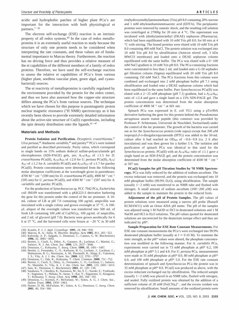
observed in 90% H2O/10% D2O at pH 8.0, with a secondexchangeable resonance present at acidic pH (data not shown).Therefore, the PCu(II)s have eight or nine directly observeddownfield-shifted resonances and one or two upfield-shiftedsignals. The temperature dependence of the positions of thehyperfine shifted signals (data not shown) indicates that all ofthese resonances exhibit Curie-type behavior (increasing shiftwith decreasing temperature). The spectrum of the higher plantPCu from parsley is very similar to that which has been assignedfor spinach PCu.42 The spectra of both of these proteins possessthe same number of directly observed isotropically shiftedresonances with very similar relaxation properties, and thus thesignals in parsley PCu(II) can be readily assigned. Furthermore,the spectra for the cyanobacterial PCu’s fromSynechococcusand A. Variabilis studied herein are comparable to that ofSynechocystisPCC6803 (also a cyanobacterium), which hasbeen assigned previously,46 and the assignment of the isotro-pically shifted resonances in the former two proteins is thereforestraightforward. A PCu(II) from a fern plant has not beeninvestigated using paramagnetic1H NMR previously; likewise,neither has that from a green algal (for exampleU. pertusa).As a result, we have assigned these spectra using saturationtransfer difference experiments on 1:1 mixtures of PCu(II) andPCu(I). The broad hyperfine shifted resonances of PCu(II) areirradiated, and saturation transfer is observed to their diamag-netic counterparts in PCu(I) as a consequence of the ESEreaction. We have assigned certain resonances in the PCu(I)susing conventional approaches (vide infra). In other cases, wherediamagnetic assignments are not available, we have used thechemical shifts observed in the reduced proteins, as comparedto typical values for PCu(I)s, for assignment of the PCu(II)signals (this approach is strongly supported by the previouslyparamagnetic NMR studies on PCu42,46). The saturation transferdifference spectra obtained when irradiating peaks a, b, g, h,and i ofD. crassirhizomaPCu(II) are shown in Figure 3. Listedin Tables S1 and S2 (see Supporting Information) are resultsof all of the saturation transfer experiments and the assignmentsthat have been made forD. crassirhizomaandU. pertusaPCu’s,respectively. The data for all of the PCu(II)s, including thosefor the spinach42 andSynechocystis46 proteins are summarizedin Table 1.
Active-Site Protonation in Reduced Plastocyanins.TheC-terminal His87 ligand in PCu(I),23,55-58 and in certain otherreduced cupredoxins,59-62 is known to protonate resulting in athree-coordinate cuprous site.23,61,62 In these studies we haveinvestigated, in detail, the effect of this pH-induced alterationat the active site on the et reactivity of PCu’s. Consequently, ithas been necessary to determine the pKa* (pKa* indicates a pKa
determined in D2O solutions for which pH readings were notcorrected for the deuterium isotope effect) of this His ligand,
using1H NMR spectroscopy, in the various PCu’s studied. TheHis ligand resonances inA. Variabilis PCu(I) have been assignedpreviously,29 and a pKa* of 5.1 for His87 has been determined.63
From the pH* dependence of the chemical shift (δ) of the His87Cε1H and Cδ2H resonances (exchange between the protonatedand deprotonated forms of His87 is fast on the NMR time scale)in this PCu(I) we obtain a pKa* of 5.0 for His87 (see ref 50 forthe equation used in the pKa determination).
The U. pertusaand SynechococcusPCu’s have not beenstudied by NMR spectroscopy previously. We have assignedthe imidazole ring resonances of both His ligands in theseproteins and have studied their pH* dependence. In the case ofU. pertusaPCu(I) pKa* values of 6.0 are obtained from thedependence on pH* of the chemical shift of the Cε1H and Cδ2Hresonances of both His ligands (His37 and His87). The His87
(55) Markley, J. L.; Ulrich, E. L.; Berg, S. P.; Krogmann, D. W.Biochemistry1975, 14, 4428-4433.
(56) Armstrong, F. A.; Hill, H. A. O.; Oliver, B. N.; Whitford, D.J. Am. Chem.Soc.1985, 107, 1473-1476.
(57) Buchi, F. N.; Bond, A. M.; Codd, R.; Huq, L. N.; Freeman, H. C.Inorg.Chem. 1992, 31, 5007-5014.
(58) McLeod, N. D. D.; Freeman, H. C.; Harvey, I.; Lay, P. A.; Bond, A. M.Inorg. Chem. 1996, 35, 7156-7165.
(59) Lommen, A.; Canters, G. W.J. Biol. Chem.1990, 265, 2768-2774.(60) Dennison, C.; Kohzuma, T.; McFarlane, W.; Suzuki, S.; Sykes, A. G.J.
Chem. Soc., Chem. Commun. 1994, 581-582.(61) Vakoufari, E.; Wilson, K. S.; Petratos, K.FEBS Lett.1994, 347, 203-
206.(62) Zhu, Z.; Cunane, L. M.; Chen, Z. W.; Durley, R. C. E.; Mathews, F. S.;
Davidson, V. L.Biochemistry1998, 37, 17128-17136. (63) Kojiro, C. L.; Markley, J. L.FEBS Lett.1983, 162, 52-56.
Figure 3. 1H NMR saturation transfer difference spectra (30°C) of a 1:1mixture ofD. crassirhizomaPCu(I) and PCu(II) in 35 mM phosphate buffer(99.9% D2O) at pH* 8. The top spectrum is that of PCu(II) and those beloware the saturation transfer difference spectra in which the peaks indicatedwere irradiated. The observed saturation transfer peaks in PCu(I) are shownby an asterisk.
Structure and Reactivity of Plastocyanins A R T I C L E S
J. AM. CHEM. SOC. 9 VOL. 125, NO. 8, 2003 2105

resonances experience much larger chemical shift differences(with the Cε1H proton being much more affected than the Cδ2Hresonance) between low and high pH*. This is consistent withHis87 being the protonating residue and the effect at His37 beinga consequence of the resulting change in active-site geometry.In theSynechococcusPCu the pH* dependence of the chemicalshift of the Cε1H resonance of His87 yields a pKa* of ∼5.3(the studies on this protein are hampered by its instability atacidic pH). It has been demonstrated previously that inD.crassirhizomaPCu(I) His87 does not become protonated in theaccessible pH range,54 while in parsley PCu(I) His87 has a pKa*of 5.6.50
The pKa* determined above for His87 in theU. pertusaprotein is high for a PCu(I). To confirm this value we studiedthe effect of pH on the reduction potential using proteinelectrochemistry.U. pertusaPCu yields a good, quasi-reversibleresponse on a DEAE-SH-modified gold electrode in the pHrange 4.1-7.7 (the variation with pH of the reduction potentialof U. pertusaPCu is shown in Figure S1 in the SupportingInformation). A reduction potential of 355 mV is found at pH7.7 and increases to a value of 470 mV at pH 4.2. The data atlow pH (<5.3) has a slope of approximately-60 mV/pH, andthus the reduction of the protein is accompanied by the uptakeof a proton at the active site (protonation of His87). The pHdependence of the reduction potential gives a pKa
red value of6.1 (see ref 54 for the equation used) in very good agreementwith that determined by NMR spectroscopy (vide supra).Attempts were also made to study the effect of pH on thereduction potential ofSynechococcusPCu. However, for thisprotein we could not obtain a good response at a gold electrodecustomized with a variety of electrode-surface modifiers withdifferent properties (anionic, cationic, and nonpolar).
It is interesting to note that in the paramagnetic1H NMRstudies of PCu(II) (vide supra) there is no evidence of His87protonating in experiments carried out at low pH. Due to proteininstability it was not possible to obtain spectra at pH values<3.5, but even in the case ofU. pertusaPCu(II) (which hasthe highest pKa* for His87 in the Cu(I) protein) there was nosign of any modification of the active-site structure under theseconditions.
Determination of the ESE Rate Constants of the PCu’sby 1H NMR Spectroscopy.Herein we determine the ESE rateconstants of the PCu’s fromU. pertusaandSynechococcusand
have reinvestigated this feature of the spinach andA. Variabilisproteins. We have assessed the dependence of the ESE rateconstant on ionic strength for spinach andU. pertusaPCu’s.Furthermore, the influence of pH* on the ESE reactivity of theU. pertusa and A. Variabilis proteins has been studied, todetermine the effect of the protonation of the His87 ligand onthe et reactivity of PCu’s.
The measurement of the ESE rate constant of a cupredoxinby 1H NMR relies on the analysis of resonances which satisfythe slow-exchange condition,64,65 in a mixture of the oxidizedand reduced proteins. It has been shown49,50,54,59,66that thiscondition is usually satisfied by the imidazole ring protons ofthe two His ligands. For these resonances, and for dilutesolutions containing only a small (<10%) proportion of theoxidized form of the protein, it can be shown that expression 1applies;49,66
whereTi (i ) 1 or 2) is the observed relaxation time of theresonance in the reduced protein in the presence of PCu(II),Ti,red is the relaxation time of the resonance in PCu(I) and [PCu-(II)] is the concentration of PCu(II). Thus, a plot ofTi
-1 against[PCu(II)] will give a straight line of slopek (k is the ESE rateconstant and we usek1 andk2 to define the slopes of the plotsof T1
-1 andT2-1 respectively, against [PCu(II)]). This approach
(the “standard method”) has been used herein and in all casesboth T1 and T2 data for His ligand resonances analyzed. Insituations where the ESE rate constant is small the effect ofincreasing [PCu(II)] on peak widths can be difficult to determineprecisely50 and in such situations thek1 values provide morereliable results (it should be noted thatT1 measurements arealso more precise when self-exchange is faster and thus allquoted ESE rate constants obtained with this method herein arean average of the availablek1 values).
It has been reported that, due to spectral overlap in thediamagnetic region of a reduced cupredoxin, a better approachfor determining the ESE rate constant is to utilize the super-WEFT sequence to select fast relaxing signals in the Cu(I)protein in the presence of small concentrations of the Cu(II)
(64) McLaughlin, A. C.; Leigh, J. S.J. Magn. Reson.1973, 9, 296-304.(65) Leigh, J. S.J. Magn. Reson.1971, 4, 308-311.(66) Groeneveld, C. M.; Canters, G. W.J. Biol. Chem.1988, 263, 167-173.
Table 1. Assigned Hyperfine Shifted Resonances in the 1H NMR Spectra of the Various PCu(II)s
δobs (ppm) in PCu(II)a
peak assignmentb spinachc parsley D. crassirhizoma U. pertusa Synechocystisd Synechococcus A. variabilis
a His87 Cδ2H 51.6 57.1 51.7 52.1 52.6 52.8 52.8b His37 Cδ2H 47.1 53.1 48.2 50.2 51.1 52.8 52.8c His37/87 Cε1H 35.6 39.1 34.9 38.0 38.5 39.3 37.8d His37/87 Cε1H 35.6 39.1 34.9 34.3 35.7 39.3 37.8e His87 Nε2H 42.2 44.3 40.6 42.9 43.8 41.9f His37 Nε2H 31.4 32.2 31.2 33.7 31.1 30.6 34.0g Met92 Cγ2H 23.5 26.1 22.3 20.7 24.0 25.9 21.7h Asn38 CRH 17.0 15.7 17.6 14.4 14.7 15.1 14.5i Met92 Cγ1H 13.0 13.1 11.0 10.6j His CâH -1.5 -1.6 -2.7 -2.2k Cys84 CRH -8.0 -7.6 -8.0 -7.5 -7.8 -7.6 -7.8
a In all cases the hyperfine shifts quoted are those at 25°C, except forD. crassirhizomaPCu(II) for which data was recorded at 30°C and theSynechocystisprotein which was measured at 22°C. Peak positions are quoted at pH 8.0 except for peak e which was observed at pH 5.7 inSynechococcusPCu(II) andat pH 5.0 in all of the other oxidized proteins, and all of theSynechocystisdata which was recorded at pH 5.2.b Small sequence differences exist betweenthe PCu’s which alters residue numbering, but herein we have numbered the proteins in all cases as in spinach PCu.c Taken from ref 42.d Taken from ref46. The peaks at 38.5 and 35.7 ppm have been assigned to the His87 Cε1H and His37 Cε1H protons respectively.
1/Ti ) (1/Ti,red) + k[PCu(II)] (1)
A R T I C L E S Sato et al.
2106 J. AM. CHEM. SOC. 9 VOL. 125, NO. 8, 2003

form.52,53This approach (the “super-WEFT” method) has beenimplemented by Ma et al. usingA. Variabilis PCu53 and alsoby Salgado et al. with amicyanin.52 The resonances which areselected are close to the copper ion and are more likely to belongto the slow-exchange regime. Due to the smaller number ofresonances observed with this approach (compared to the casein a conventional 1D spectrum) the chances of spectral overlapare greatly diminished. However, this approach is only of anyuse if the ESE rate constant is approximately 105 M-1 s-1 orlarger, as we demonstrate in this study.
Another approach for determining the ESE rate constants ofcupredoxins has also been reported very recently, and againA.Variabilis PCu has been used to test it.67 This method does notrequire assignment of peaks to the slow-exchange regime andhas been implemented using exchangeable amide protons closeto the copper site. As we can unambiguously assign all of theresonances that we have used herein to the slow-exchange limit,we have not had a need to use this approach. It should be notedthat another method for measuring the ESE rate constants byNMR spectroscopy exists which involves the analysis ofsaturation transfer experiments. However, this approach is onlyappropriate for lifetimes in the range of theT1 values, and thusthe ESE reactions of the cupredoxins are generally too fast forthis method to give reliable results.
For A. Variabilis PCu we have determined the ESE rateconstant at pH* 6.2, 5.1, and 4.8 (I ) 0.10 M) using the standardmethod and monitoring the effect of increasing [PCu(II)] ontheT1 andT2 values of the Cδ2H proton of His37, and His87 atpH* 6.2, which are well resolved. The availablek2/k1 ratios areall very close to 1 (see Table 2), highlighting the fact that theresonances used belong to the slow-exchange regime [k2/k1 ratiosin excess of 5-10 are expected for protons in the fast-exchangeregime49,66]. At pH* 6.2 and 5.1 we have also measured theESE rate constant using the super-WEFT approach, and theresults obtained are also listed in Table 2. For the cyanobacterialPCu from Synechococcuswe have determined the ESE rateconstant at pH* 8.0 (I ) 0.10 M) using only the super-WEFTmethod (see Figure 4), due to the absence of well-resolved Hisligand imidazole proton resonances in the 1D spectrum of thePCu(I). Values ofk2,WEFT (the slopes of plots ofT2
-1 obtained
from WEFT spectra against [PCu(II)]) ranging from 3.3× 10⁵
to 3.9 × 10⁵ M-1 s-1 were obtained for the ligand protonresonances labeled in Figure 4, giving an average ESE rateconstant of 3.5× 10⁵ M-1 s-1. The ESE rate constant of thisPCu could not be studied at acidic pH* values due to theinstability of the protein (both oxidation states of the proteinhave a tendency to precipitate at pH< 6.0).
The ESE rate constant of the green algal PCu fromU. pertusahas been determined at pH* 8.0, 6.0 and 5.0 (I ) 0.10 M) usingthe standard approach and utilizing the His37 Cδ2H and theHis87 Cε1H resonances. The ESE rate constants ofU. pertusaPCu were also determined at pH* 8.0 and 5.3 in 10 mMphosphate buffer plus 0.50 M NaCl and plots ofTi
-1 against[PCu(II)] for the His37 Cδ2H signal under these conditions areshown in Figure 5. The slopes of these plots (k values) are listedin Table 3 along with the results from all of the other ESEexperiments carried out on this PCu. Again the availablek2/k1
ratios are all very close to 1, which emphasizes that theresonances used are in the slow-exchange regime. We havemeasured the ESE rate constant ofU. pertusaPCu at pH* 6.0(I ) 0.10 M) at two different total protein concentrations (datanot shown). The results are almost identical, further confirmingthat the protons studied belong to the slow-exchange regime(only resonances in the fast-exchange limit will exhibit adependence on total protein concentration49,66). Attempts tostudy the ESE reactivity ofU. pertusaPCu (atI ) 0.10 M)using the super-WEFT method proved unsuccessful as no PCu-(I) signals were selected in partially oxidized samples due tothe relatively small ESE rate constant for this PCu (see Figure4).
The ESE rate constant of spinach PCu at pH* 8.0 wasmeasured at bothI ) 0.10 M (35 mM phosphate) andI ) 2.03M (10 mM phosphate plus 2.0 M NaCl) using the standardapproach and monitoring the His37 Cδ2H and His87 Cε1Hresonances. Thek2/k1 ratios are all∼1, and thek1 values forthe two resonances studied are within 10% of each other. TheESE rate constants obtained (the average of thek1 values) are2.5 × 103 M-1 s-1 at moderate ionic strength and 9.0× 10⁴
M-1 s-1 at I ) 2.03 M.All of the ESE results are summarized in Figure 6. Also
included in this Figure are data for parsley PCu50 and for thefern protein fromD. crassirhizoma.54
Discussion
Surface Properties of PCu’s from Various Sources.Thekey question which this study addresses is the influence of thestructure of the PCu’s from various sources on their et reactivity.The PCu’s from the four different types of photosynthesizing
(67) Jensen, M. R.; Hansen, D. F.; Led, J. J.J. Am. Chem. Soc.2002, 124,4093-4096.
Table 2. ESE Rate Constants (25 °C) of A. variabilis PCu atVarious pH* Values (all at I ) 0.10 M), Derived from T1 (k1) andT2 (k2) Valuesa
pH* resonances k1/M-1 s-1 k2/M-1 s-1 k2/k1 k2,WEFT/M-1 s-1
6.2 Leu12 Cδ2H ndb nd 1.7× 10⁵
His37 CRH nd nd 1.7× 10⁵
His37 Cδ2H 2.3× 10⁵ 2.1× 10⁵ 0.9 2.0× 10⁵
Cys84 CRH nd nd 2.3× 10⁵
His87 Cδ2H 2.5× 10⁵ 2.2× 10⁵ 0.9 1.6× 10⁵
Average 2.4× 10⁵ 2.2× 10⁵ 1.9× 10⁵
5.1 His37 CRH nd nd 1.2× 10⁵
His37 Cδ2H 1.4× 10⁵ 1.2× 10⁵ 0.9 1.1× 10⁵
Cys84 CRH nd nd 1.3× 10⁵
His87 Cε1H nd nd 1.2× 10⁵
Average 1.4× 10⁵ 1.2× 10⁵ 1.2× 10⁵
4.8 His37 Cδ2H 8.6× 10⁴ 7.8× 10⁴ 0.9 nd
a Also included are values derived fromT2 data in super-WEFT spectra(k2,WEFT). b Not determined.
Table 3. Summary of the ESE Rate Constants (25 °C) of U.pertusa PCu Derived from T1 (k1) and T2 (k2) Data
His37 Cδ2H His87 Cε1H
pH* k1/M-1 s-1 k2/M-1 s-1 k2/k1 k1/M-1 s-1 k2/M-1 s-1 k2/k1
8.0a 3.2× 10⁴ 3.1× 10⁴ 1.0 3.8× 10⁴ 3.1× 10⁴ 0.86.0a 3.5× 10⁴ 3.5× 10⁴ 1.0 4.6× 10⁴ 3.7× 10⁴ 0.75.0a 2.3× 10⁴ 2.8× 10⁴ 1.2 2.5× 10⁴ 2.6× 10⁴ 1.08.0b 1.2× 10⁵ 1.2× 10⁵ 1.0 1.7× 10⁵ 1.4× 10⁵ 0.85.3b 3.4× 10⁴ 4.0× 10⁴ 1.2 ndc nd
a Determined atI ) 0.10 M. b Measured in 10 mM phosphate bufferplus 0.50 M NaCl.c Not determined due to this resonance not being resolvedunder these conditions.
Structure and Reactivity of Plastocyanins A R T I C L E S
J. AM. CHEM. SOC. 9 VOL. 125, NO. 8, 2003 2107

organisms studied herein have distinct surface properties (seeFigure 1). All of the PCu’s have a hydrophobic patch surround-ing the exposed His87 ligand, which is a conserved feature ofall structurally characterized cupredoxins,1,2 and is the preferredroute for et reactions including self-exchange.8,13,16,17,68-71 In
higher plant PCu’s there is a second binding site made up ofAsp and Glu residues, which surround the solvent-exposedTyr83, and is known as the acidic patch. This surface can bedivided into upper (E59, E60, and E61) and lower (D42, E43,D44, and E45) acidic patches in higher plant PCu’s (see Figure1). In parsley PCu, the upper acidic patch consists of a singleglutamate and is thus almost nonexistent (see Figure 1) as aconsequence of deletions at positions 59 and 60 in the aminoacid sequence. A diminished upper acidic patch is also a featureof green algal PCu’s again due to deletions in this region (seeFigure 1). In the cyanobacterial proteins, both acidic patchesare absent (see Figure 1). TheD. crassirhizomaPCu has some
(68) Van de Kamp, M.; Floris, R.; Hali, F. C.; Canters, G. W.J. Am. Chem.Soc.1990, 112, 907-908.
(69) Chen, L.; Durley, R.; Poliks, B. J.; Hamada, K.; Chen, Z.; Mathews, F. S.;Davidson, V. L.; Satow, Y.; Huizinga, E.; Vellieux, F. M. D.; Hol, W. G.J. Biochemistry1992, 31, 4959-4964.
(70) Mikkelsen, K. V.; Skov, L. K.; Nar, H.; Farver, O.Proc. Natl. Acad. Sci.U.S.A.1993, 90, 5443-5445.
(71) van Amsterdam, I. M. C.; Ubbink, M.; Einsle, O.; Messerschmidt, A.; Merli,A.; Cavazzini, D.; Rossi, G. L.; Canters, G. W.Nat. Struct. Biol. 2002, 9,48-52.
Figure 4. 1H NMR spectra (25°C) of SynechococcusPCu in 35 mM phosphate buffer (99.9% D2O) at pH* 8.0 andU. pertusaPCu in 80 mM phosphatebuffer (99.9% D2O) at pH* 6.0 obtained using the super-WEFT pulse sequence (at 500 MHz). A is a spectrum ofSynechococcusPCu(I) (2.0 mM), whilein B 1.4% PCu(II) is present. The spectrum ofU. pertusaPCu(I) (1-4 mM) is shown in C and in D is a spectrum of the sample containing 4.3% PCu(II).In all cases the interpulse delay was 40 ms and the total relaxation delay ca. 50 ms.
Figure 5. Plots (25°C) of T1-1 (O) and T2
-1 (b) against [PCu(II)] for theHis37 Cδ2H resonance of theU. pertusaprotein in 10 mM phosphate buffer(99.9% D2O) plus 0.50 M NaCl at pH* 8.0. Also shown are plots (25°C)of T1
-1 (0) and T2-1 (9) against [PCu(II)] for the same resonance measured
in 10 mM phosphate buffer (99.9% D2O) plus 0.50 M NaCl at pH* 5.3.The error bars in the case of the T1
-1 data are smaller than the symbols.
Figure 6. Dependence (25°C) on pH* of log kese (kese is the ESE rateconstant) ofU. pertusaPCu in phosphate buffer atI ) 0.10 M ([), U.pertusaPCu in 10 mM phosphate buffer plus 0.50 M NaCl (]), A. VariabilisPCu in phosphate buffer atI ) 0.10 M (2), parsley PCu in phosphate bufferat I ) 0.10 M (+) andD. crassirhizomaPCu in phosphate buffer atI )0.10 M (9). Also included are data at pH* 8.0 for spinach PCu in phosphatebuffer atI ) 0.10 M (b), spinach PCu in 10 mM phosphate buffer plus 2.0M NaCl (O) andSynechococcusPCu in phosphate buffer atI ) 0.10 M(∆).
A R T I C L E S Sato et al.
2108 J. AM. CHEM. SOC. 9 VOL. 125, NO. 8, 2003

negative charge around Tyr83, but a large number of Asp andGlu residues are found in an arc around the edge of thehydrophobic patch (see Figure 1).
Paramagnetic1H NMR of the PCu(II)s and their Active-Site Structures.The 500-MHz paramagnetic1H NMR spectraof the five PCu(II)s studied in this work are very similar toeach other (see Figure 2) and also to those published previouslyfor the spinach42 andSynechocystis46 proteins (see Table 1). Inparticular the spectrum of parsley PCu(II) is almost identicalto that of the protein from spinach, and those ofA. Variabilisand SynechococcusPCu(II) are comparable to the assignedSynechocystisspectrum. We report here for the first time theassignment of the directly observed resonances in the paramag-netic 1H NMR spectrum of the PCu(II) from a green alga andfor the novel protein from a fern plant. The observed shifts (δobs)for the signals of the PCu(II)s listed in Table 1 arise from threecontributing factors as shown in the equation:
where δdia is the shift in an analogous diamagnetic system(PCu(I)),δpc is the pseudocontact (through space) contribution,andδFc is the Fermi-contact (through-bond) contribution. Thepseudocontact shifts are small for protons at a cupric type 1site,41-47 due to the small anisotropy of theg tensor, and rangefrom ∼2 to -3 ppm for the resonances listed in Table 1.42,46
Therefore, theδobs value minus theδdia value (see Tables S1and S2 in the Supporting Information for typicalδdia values)for a particular proton listed in Table 1 provide a good estimateof δFc, which is a measure of the spin density present at thatparticular proton.
In all of the proteins listed in Table 1 the ligand His signalsexhibit similarδobs (and thereforeδFc) values. This is also trueif data for other type 1 centers are included in this com-parison,41,43-45,47demonstrating that the spin density distributiononto the two His ligands is remarkably similar in all PCu’s andalso other cupredoxins. The backbone amide of Asn38, a residuewhich is completely conserved in PCu’s, is hydrogen-bondedto the thiolate sulfur of the Cys84 ligand, explaining therelatively large Fermi-contact shift of this resonance. Theδobs
of this resonance and the Cys84 CRH signal, which possessesnegative spin density, provide information about the strengthof the interaction of the metal with the Cys ligand in the differentPCu(II)s, and the similarity of their positions (see Table 1)indicates that the Cu-S(Cys) bond is comparable in all of theproteins. Further information about this interaction can beobtained from the hyperfine shifts of the Cys CâH protons,which are too broad to be observed directly. Bertini et al. havelocated these signals at 650 and 489 ppm in spinach PCu42 andat 614 and 517 ppm in theSynechocystisprotein,46 thusconfirming the similarity of the Cu-S(Cys) interaction in thevarious members of this class of metalloproteins.
The largest differences between the spectra of the PCu(II)sarises from protons associated with the axial Met92 ligand. Inall cases the Met92 Cγ2H proton is observed at∼26-20 ppm.The Met92 Cγ1H proton is only shifted outside of the diamag-netic envelope in the PCu(II)s from parsley,D. crassirhizomaand U. pertusaand also spinach42 and is not observed in thecyanobacterial PCu(II)s. The Fermi-contact shifts exhibited bythe Met92 CγH signals depends on the amount of spin densityon these protons, which is influenced by the strength of the
Cu-S(Met) bond and also the Cu-Sδ-Cγ-Hγ dihedral angles.Analysis of the available structures for the PCu’s stud-ied28,29,31,33,34,36,37shows that the bond lengths between thecopper and the thioether sulfur of the axial Met ligands rangefrom ∼2.6 to 3.0 Å. The Cu-Sδ-Cγ-Hγ1 dihedral angle rangesfrom ∼-170° to -180°, while the Cu-Sδ-Cγ-Hγ2 angle ismore varied ranging from∼-50° to -70° (note: hydrogenswere added to the crystal structures using the program InsightII). At the present time we are not able to find any directcorrelation between these bond lengths and dihedral angles, andthe δFc values of the Met CγH protons.
In summary the paramagnetic1H NMR spectra of all of thePCu(II)s studied herein are very similar. The structures of thecopper sites and the spin density distribution onto the ligandsin the different members of this subclass of the cupredoxinsare therefore alike. TheD. crassirhizomaandU. pertusaPCu-(II)s are the first members of their subclasses (seedless vascularplant and green alga respectively) to be studied by paramagneticNMR, and they both give spectra analogous to those of all ofthe other proteins. This is particularly noteworthy for theD.crassirhizomaPCu(II) which has UV/vis, electron paramagneticresonance (EPR), and resonance Raman spectra slightly differentfrom those of all other PCu(II)s.37 This has been associated withthe π-π interaction of the imidazole ring of the His87 ligandwith Phe12 (see Figure 1). It is therefore interesting to notethat the His87 resonances ofD. crassirhizomaPCu(II) exhibitδobsvalues similar to those of all of the other proteins. Therefore,the interaction between His87 and Phe12 does not affect thespin density distribution onto this ligand.
His87 Protonation in PCu(I). All PCu(I)s exhibit protonationof their His87 ligand, except for the protein fromD. crassirhi-zoma.54 The absence of His87 protonation in this PCu(I) hasbeen attributed to theπ-π interaction of the imidazole ring ofthis ligand with the phenyl ring of Phe12 (see Figure 1).37,54
Structural studies have demonstrated that the active-site proto-nation in the PCu(I)s results in a three-coordinate site,23 whichis a favored geometry for this oxidation state of copper,72 andthus leads to a dramatic increase in the reduction potential.54,56-58
The pKa* for His87 is typically in the range of 4.8-5.0 formost PCu(I)s.50,54 Herein we have determined a pKa* of ∼5.3for His87 in SynechococcusPCu(I) from NMR studies, whichis slightly higher than that for the corresponding residue in thereduced cyanobacterial protein fromA. Variabilis (pKa* 5.0).Parsley PCu(I) is unusual with its His87 possessing a pKa* of5.6.50 In theU. pertusaprotein His87 has an even higher pKa*(6.0) which is the largest reported to date for a PCu(I). It haspreviously been noted that other green algal PCu’s have quitehigh active-site pKa*’s, with a value of 5.5 reported for theScenedesmus obliquusprotein.73 It therefore appears that thePCu’s which have deletions at positions 59 and 60 in theirprimary structures, and possess a diminished upper acidic patch,also have high active-site pKa values. This is contrary to whatwould be expected on the basis of simple electrostatics.Protonation of one of the His ligands has been shown to occurin reduced pseudoazurin60,61 and amicyanin59,62 (both cupre-doxins) with pKa values of ∼5 and ∼7, respectively. Thefeatures which control the pKa value of this copper ligand are
(72) Gray, H. B.; Malmstro¨m, B. G.; Williams, R. J. P.J. Biol. Inorg. Chem.2000, 5, 551-559.
(73) McGinnis, J.; Sinclair-Day, J. D.; Sykes, A. G.; Powls, R.; Moore, J.;Wright, P. E.Inorg. Chem. 1988, 27, 2306-2312.
δobs) δdia + δpc + δFc (2)
Structure and Reactivity of Plastocyanins A R T I C L E S
J. AM. CHEM. SOC. 9 VOL. 125, NO. 8, 2003 2109

not yet completely understood [see ref 54 for a recent reviewof this subject].
ESE Reactivity of PCu’s.We have used two approaches todetermine the ESE rate constants of the PCu’s. The first is amethod (the standard method) which has been widely usedpreviously47,49,50,54,59,66,74and relies on observing the enhancedrelaxation of protons in conventional 1D spectra of PCu(I)resulting from the presence of small amounts of PCu(II), whichsatisfy the slow-exchange condition (those protons which areclose to the active site, vide supra). This method gives preciseresults, providing that suitable protons are resolved in thespectrum of the reduced protein. In all of the present studies,except in the case of theSynechococcusPCu, all PCu(I) spectrapossess sufficiently resolved resonances which we have shownbelong to the slow-exchange regime. An alternative approach,which has also been applied herein, utilizes the enhancedrelaxation of protons in partly oxidized samples to selectivelyobserve a small number of resonances using the super-WEFTsequence.52,53 The resonances which are selected by thisapproach are those which are situated close to the copper ionand thus are likely to belong to the slow-exchange limit. Thebroadening of these resonances caused by increasing theconcentration of PCu(II) has been measured. However, it shouldbe noted that, as with the standard method, signals still need tobe sufficiently resolved to provide reliable data. Furthermore,the determination of peak widths (T2 values) is prone to largererrors than the measurement ofT1 values, as the longitudinalrelaxation rates (T1
-1) of protons in proteins are usuallysignificantly smaller than the transverse relaxation rates (T2
-1).Therefore, a small change in the relaxation rates can bemeasured with higher precision forT1
-1 than forT2-1. Having
said this, the super-WEFT approach does provide a veryconvenient way of determining the ESE rate constant insituations where resolved resonances cannot be found in thestandard 1D spectrum of the reduced protein, such as in thecase of theSynechococcusPCu studied herein. However, thismethod can only be used if the ESE rate is sufficiently fast,and thus in experiments with theU. pertusa protein thediamagnetic protons in the vicinity of the active site in PCu(I)do not experience sufficient relaxation enhancement to allowthem to be selectively observed with this approach (see Figure4).
The ESE rate constants for the higher plant PCu from spinach,the green algal PCu fromU. pertusa, and the cyanobacterialproteins from A. Variabilis and Synechococcushave beendetermined. At a pH* value where His87 cannot influence theet reactivity of the proteins the observed rate constants (25°C)are 2.5× 10³, 3.5× 10⁴, 2.4× 10⁵, and 3.5× 10⁵ M-1 s-1 forthe spinach (pH* 8.0)U. pertusa(pH* 8.0), A. Variabilis (pH*6.2), andSynechococcus(pH* 8.0) proteins, respectively (allat I ) 0.10 M). These compare with rate constants (again at 25°C) of 5.0 × 10⁴ and 3.4× 10³ M-1 s-1 for parsley (at pH*7.5)49 andD. crassirhizoma(at pH* 7.9)54 PCu’s, respectively(also atI ) 0.10 M). Previous studies have reported ESE rateconstants (25°C) of ∼4 × 10³ M-1 s-1 for spinach PCu atpH* 6.0 (I ) 0.10 M)75 and 1.5× 10⁵ M-1 s-1 for A. VariabilisPCu at pH* 7.0 (I ) 0.05 M)67 in good agreement with our
results. The ESE rate constants of the PCu’s at moderate ionicstrength span 2 orders of magnitude (as do the ESE rateconstants of all cupredoxins74). The self-exchange process inPCu’s (and all cupredoxins) is thought to involve the associationof two protein molecules via the hydrophobic surfaces whichsurround the His87 ligand.43,68,70,71,74Given the comparableactive-site structures of the Cu(II) proteins which we havedemonstrated in the paramagnetic1H NMR studies and which,given the rigidity of the cupredoxin fold, also hold for the Cu-(I) forms (at a pH value well above the pKa of His87) we cananticipate that the reorganization energies of the various PCu’sare similar. Additionally, we have shown that the spin densitydistributions onto the ligands are alike in all of the PCu(II)s,and thus variations in this feature will not influence et. This isperhaps most relevant for the His87 ligand which is in theproposed et pathway for self-exchange.70,71The observed rangeof ESE rate constants for the various proteins is thereforeprobably a consequence of different association constants (Kassoc,see Scheme 1) for two PCu molecules (it is also possible thatthe structures of the encounter complexes for the ESE processdiffer in the various PCu’s and therefore et occurs over quitedifferent distances, but this is less likely especially consideringthe results obtained at elevated ionic strength). The small ESErate constants for spinach andD. crassirhizomaPCu’s can thusbe attributed to the large number of acidic residues which arepresent on the surfaces of these two proteins (see Figure 1).The smaller size of the upper acidic patch in parsley andU.pertusaPCu (see Figure 1) results in the approximately 10-fold increase in the ESE rate constant. The two cyanobacterialPCu’s exhibit ESE rate constants which are∼100-times largerthan those of the spinach andD. crassirhizomaproteins. Thisis due to the almost complete absence of an acidic patch inthese proteins (see Figure 1). A basic arginine residue adjacentto the C-terminal His ligand in all cyanobacterial PCu’s hasbeen shown to be important for the interaction with bothcytochromef 20 and PSI19 but does seem to hamper ESE (weare currently investigating this further by analyzing the influenceof ionic strength on the ESE rate constants of cyanobacterialPCu’s). The influence of the acidic patch on the ESE rateconstant is further demonstrated by measurements at high ionicstrength for spinach andU. pertusaPCu at pH* 8.0 (see Table3 and Figure 6). As expected, for two such highly chargedproteins, the ESE rate constants increase at elevated ionicstrength. The approximately 36-fold and 5-fold increases, dueto shielding of the negative charge on the proteins’ surfaces,results in spinach andU. pertusaPCu’s, respectively, havingrate constants in line with those of the cyanobacterial proteins.To summarize, the ESE process in the cyanobacterial PCu’s isgoverned mainly by hydrophobic interactions, while in the caseof the green algal and higher plant proteins a combination of
(74) Kyritsis, P.; Dennison, C.; Ingledew, W. J.; McFarlane, W.; Sykes, A. G.Inorg. Chem. 1995, 34, 5370-5374.
(75) Armstrong, F. A.; Driscoll, P. C.; Hill, H. A. O.FEBS Lett.1985, 190,242-248.
Scheme 1
A R T I C L E S Sato et al.
2110 J. AM. CHEM. SOC. 9 VOL. 125, NO. 8, 2003

hydrophobic and electrostatic interactions are important. Asimilar conclusion is emerging from studies of the interactionof PCu’s from these various sources with their physiologicalredox partners.7-20
Another family of et metalloproteins, the cytochromes, havehad their ESE reactivity studied in some detail. Cytochromecfrom both mammalian and bacterial sources has a ring of lysineresidues around the exposed heme edge.76,77 This feature isresponsible for a small ESE rate constant of∼103 M-1 s-1 atI ) 0.10 M which increases to∼105 M-1 s-1 at high ionicstrength.78-80 Cytochromeb5, which possesses a number ofacidic residues around the periphery of its exposed heme edge,81
has an ESE rate constant also of∼103 M-1 s-1 at lower ionicstrength which rises to∼105 M-1 s-1 as the NaCl concentrationis increased.78,79,82 Cytochrome c551, which lacks chargedresidues around its exposed heme edge,83 has an ESE rateconstant of∼107 M-1 s-1, which is independent of ionicstrength.78,84In the case of Fe-S proteins a more limited numberof investigations of ESE reactivity have been carried out. High-potential iron-sulfur proteins (HiPIPs) have ESE rate constantsof ∼105 M-1 s-1 which are independent of ionic strength (overthe relatively narrow range studied).85-87 The rubredoxins, whichare the simplest of the Fe-S proteins, have an ESE rate constantof ∼105 M-1 s-1 at lower ionic strength, which increases to∼106 M-1 s-1 at elevated ionic strength.88,89It therefore appearsthat all three families of redox metalloproteins (cupredoxins asrepresented by the PCu’s, cytochromes and Fe-S proteins)possess similar ESE rate constants (∼105-106 M-1 s-1) atelevated ionic strength, i.e. when the influence of surfacefeatures of the protein are minimized. This magnitude of ESErate constant (which is influenced by the reorganization energy,the distance for et, and the protein-protein association constant)therefore appears to be a consequence of a structure that hasevolved to be an efficient redox metalloprotein.
The ESE rate constant ofU. pertusaPCu is almost indepen-dent of pH* in the range 8.0-5.0 (atI ) 0.10 M, see Table 3and Figure 6). This is surprising, considering that the pKa* ofHis87 is 6.0 in the reduced protein and so at pH* 5.0,∼90%of the PCu(I) molecules will possess a three-coordinate activesite. This form of PCu(I) is known to be redox-inactive,56,59,90,91
and a smaller ESE rate constant would be expected (see Scheme
1 where [Cu(I)His87-H+] is the form of PCu(I) in which His87is protonated and is the redox inactive species). We havepreviously found that the ESE rate constant of parsley PCu isalso almost unaffected by pH* in the range 7.5-5.6 (see Figure6), with the low end of this range corresponding to the pKa* ofHis87 in this reduced PCu. The absence of any pH* dependenceof the ESE reactivity in higher plant and green algal PCu’s canbe attributed to compensation effects caused by the presenceof an acidic patch. At lower pH values the acidic residues startto become protonated, thus neutralizing the charge in this regionof the protein, leading to a largerKassoc(see Scheme 1). Thisargument is confirmed by the influence of pH* on the ESE rateconstant ofA. Variabilis PCu (see Figure 6) which lacks anacidic patch (see Figure 1). In this case the ESE rate constantis ∼3 times smaller at pH* 4.8 (the pKa* of His87 is 5.0)compared to the situation at pH* 6.2. Also, the ESE rate constantof D. crassirhizomaPCu actually increases at lower pH* values.This is due to the absence of His87 protonation in this PCu(I)plus the neutralization of the arc of acidic residues around thehydrophobic patch of this protein (see Figure 1) at lower pH*,which enhances protein-protein association. Furthermore, wehave also studied the influence of pH* on the ESE rate constantof U. pertusaPCu at high ionic strength (I ≈ 0.50 M, see Table3 and Figure 6). Under these conditions, where the influenceof the acidic patch on the association of two PCu molecules isdiminished, the ESE rate constant is∼4.5 times smaller at pH*5.3 (3.4× 10⁴ M-1 s-1) as compared to pH* 8.0 (1.5× 10⁵
M-1 s-1). This confirms that the protonation of His87 diminishesthe ESE reactivity of PCu. Thus, as the pH is lowered, more ofthe reduced protein exists as [Cu(I)His87-H+] (see Scheme 1)which is redox inactive. It would perhaps be expected that theESE process would be more dramatically affected at low pH.The fact that a decrease in ESE rate constants are observed at,and below, pH values which correspond to the pKa of His87 inthe various PCu(I)s but that the proteins are still reasonablycompetent at ESE suggests that the process has now becomegated (by the activation barrier for the transition from three-coordinate [Cu(I)His87-H+] to four-coordinate [Cu(II)His87]).From Scheme 1 it is apparent thatkoff, the rate of deprotonationof [Cu(I)His87-H+] could gate the ESE reaction at low pH.Values ofkoff have been measured for amicyanin by electro-chemistry and are very temperature-dependent (35 s-1 at 273K and >750 s-1 at 295 K).91 We are currently investigatingthe effect of temperature on the ESE reactivity of various PCu’sat low pH, and at high ionic strength, to shed more light onthis matter.
The protonation of the C-terminal His ligand in PCu(I) andother reduced cupredoxins may play an important physiologicalfunction. For example, it is thought that His87 protonation couldprovide a metabolic feedback mechanism whereby the drop inpH resulting from a high rate of photosynthesis would result ina decrease in et activity of PCu. In this context it is interestingto note that in the case of amicyanin the His ligand does notprotonate when the protein is complexed to its physiologicalelectron donor (methylamine dehydrogenase).62 In the case ofPCu(I) it has recently been shown that the protonation of His87has little effect on the interaction with cytochromef.17 Free PCu-(I) in the inner thylakoid of the chloroplast will be mainly inthe protonated (redox-inactive) form when the system isilluminated (under these conditions the pH of the inner thylakoid
(76) Timkovich, R.; Dickerson, R. E.J. Biol. Chem. 1976, 251, 4033-4046.(77) Bushnell, G. W.; Louie, G. V.; Brayer, G. D.J. Mol. Biol. 1990, 214,
585-595.(78) Dixon, D. W.; Hong, X.; Woehler, S. E.Biophys. J. 1989, 56, 339-351.(79) Andrew, S. M.; Thomasson, K. A.; Northrup, S. H.J. Am. Chem. Soc.
1993, 115, 5516-5521.(80) Ubbink, M.; Canters, G. W.Biochemistry1993, 32, 13893-13901.(81) Mathews, F. S.; Levine, M.; Argos, P. J.J. Mol. Biol. 1972, 64, 449-464.(82) Dixon, D. W.; Hong, X.; Woehler, S. E.; Mauk, A. G.; Sishta, B. P.J. Am.
Chem. Soc. 1990, 112, 1082-1088.(83) Matsuura, Y.; Takano, T,; Dickerson, R. E.J. Mol. Biol. 1982, 156, 389-
409.(84) Timkovich, R.; Cai, M. L.; Dixon, D. W.Biochem. Biophys. Res. Commun.
1988, 150, 1044-1050.(85) Sola, M.; Cowan, J. A.; Gray, H. B.J. Am. Chem. Soc. 1989, 111, 6627-
6630.(86) Bertini, I.; Gaudemer, A.; Luchinat, C.; Piccioli, M.Biochemistry1993,
32, 12887-12893.(87) Soriano, A.; Li, D.; Bian, S.; Agarwal, A.; Cowan, J. A.Biochemistry1996,
35, 12479-12486.(88) Gaillard, J.; Zhuang-Jackson, H.; Moulis, J. M.Eur. J. Biochem. 1996,
238, 346-349.(89) Kummerle, R.; Zhuang-Jackson, H.; Gaillard, J.; Moulis, J. M.Biochemistry
1997, 36, 15983-15991.(90) Jeuken, L. J. C.; van Vliet, P.; Verbeet, M. P.; Camba, R.; McEvoy, J. P.;
Armstrong, F. A.; Canters, G. W.J. Am. Chem. Soc. 2000, 122, 12186-12194.
(91) Jeuken, L. J. C.; Camba, R.; Armstrong, F. A.; Canters, G. W.J. Biol.Inorg. Chem. 2002, 7, 94-100.
Structure and Reactivity of Plastocyanins A R T I C L E S
J. AM. CHEM. SOC. 9 VOL. 125, NO. 8, 2003 2111

can drop below 5). The fact that the reduction of P700+ byPCu(I) is hardly affected at low pH92 therefore seems a littlepuzzling. It may be that the approach of a hydrophobic surfaceof PSI to the hydrophobic patch of [Cu(I)His87-H+] destabilizesthe positive charge on His87 and thus [Cu(I)His87], the redoxactive species, would be rapidly re-formed (see Scheme 1).Therefore, His87 protonation may provide a way of ensuringspecificity of photosynthetic et as the reaction with otheroxidants would be expected to be much more significantly gated.
Conclusions
In this study we have assigned the paramagnetic1H NMRspectra of the PCu(II) fromD. crassirhizoma(fern) and alsothe protein fromU. pertusa (green alga). Even though thespectroscopic properties, and active-site structure, of theD.crassirhizomaPCu(II) are slightly unusual, the paramagnetic1H NMR spectrum is very similar to those of other PCu(II)s.Paramagnetic1H NMR spectra have now been assigned for PCu-(II)s from higher plants, a seedless vascular plant, a green alga,and cyanobacteria, and in all cases they are similar. Thisindicates that the copper-site structures and the spin densitydistribution onto the ligands of this family of et proteins, whichplay a key role in photosynthesis, has changed very littlethroughout evolution. This is in contrast to the surface propertiesof the PCu’s which have been altered quite dramaticallythroughout the evolutionary process (see Figure 1).
The ESE rate constants ofSynechococcusand U. pertusaPCu’s have been determined, with the latter being the first tobe published for a green algal PCu. There are now ESE rateconstants available for a range of PCu’s with very differentsurface properties. At moderate ionic strength (I ) 0.10 M) theESE rate constant of a PCu is dictated by the presence of acidic
residues on the proteins’ surfaces. The rate constants range from∼3 × 10³ M-1 s-1 in the case of theD. crassirhizomaproteinto ∼4 × 10⁵ M-1 s-1 in the cyanobacterial proteins. Theinfluence of charged residues on protein-protein associationis further demonstrated by the larger ESE rate constant ofspinach andU. pertusaPCu’s at elevated ionic strength. Underconditions where the influence of surface charges on protein-protein association is minimized, PCu’s all possess ESE rateconstants of∼105-106 M-1 s-1. A similar conclusion can bedrawn from corresponding data for other families of redoxmetalloproteins. It therefore appears that this magnitude of ESErate constant is a consequence of a protein-encapsulated metalcenter which is a competent redox catalyst. The influence ofpH on the ESE rate constant demonstrates that His87 protonationin PCu(I) has an inhibiting effect on this reaction. Thus, wecan conclude that ligand protonation may provide a mechanismfor ensuring specificity in photosynthetic et.
Acknowledgment. We thank Dr. A. Lawler for carrying outthe electrochemistry experiments onU. pertusaPCu. We aregrateful for financial support from Newcastle University, theRoyal Society, CVCP (for an ORS award to K.S.), and EPSRC(for a grant to purchase the NMR spectrometer). We thankProfessor A. Harriman and A. Amini (University of Newcastleupon Tyne) for access to and assistance with Insight II, andProfessor P. Schu¨rmann (Universite´ de Neuchaˆtel, Switzerland)for providing the spinach PCu gene.
Supporting Information Available: Tables with results ofsaturation transfer experiments and a figure showing variationwith pH of the reduction potential ofU. pertusaPCu (PDF).This material is available free of charge via the Internet athttp://pubs.acs.org.
JA021005U(92) Sigfridsson, K.Photosynth. Res. 1998, 57, 1-28.
A R T I C L E S Sato et al.
2112 J. AM. CHEM. SOC. 9 VOL. 125, NO. 8, 2003