active contours and image segmentation: the current state of … · active contours and image...
TRANSCRIPT
© 2012. D. Baswaraj, Dr. A. Govardhan & Dr. P. Premchand. This is a research/review paper, distributed under the terms of the Creative Commons Attribution-Noncommercial 3.0 Unported License http://creativecommons.org/licenses/by-nc/3.0/), permitting all non-commercial use, distribution, and reproduction inany medium, provided the original work is properly cited.
Global Journal of Computer Science and Technology Graphics & Vision Volume 12 Issue 11 Version 1.0 Year 2012 Type: Double Blind Peer Reviewed International Research Journal Publisher: Global Journals Inc. (USA) Online ISSN: 0975-4172 & Print ISSN: 0975-4350
Active Contours and Image Segmentation: The Current State of the Art
By D. Baswaraj, Dr. A. Govardhan & Dr. P. Premchand Faculty of Engineering, OU, Hyderabad, AP, India
Abstract - Image segmentation is a fundamental task in image analysis responsible for partitioning an image into multiple sub-regions based on a desired feature. Active contours have been widely used as attractive image segmentation methods because they always produce sub-regions with continuous boundaries, while the kernel-based edge detection methods, e.g. Sobel edge detectors, often produce discontinuous boundaries. The use of level set theory has provided more flexibility and convenience in the implementation of active contours. However, traditional edge-based active contour models have been applicable to only relatively simple images whose sub-regions are uniform without internal edges. Here in this paper we attempt to brief the taxonomy and current state of the art in Image segmentation and usage of Active Contours.
Keywords : Active Contours, Snakes, Level Sets.
GJCST-F Classification: I.4.6
Active Contours and Image Segmentation The Current State of the Art
Strictly as per the compliance and regulations of:

© 2012 Global Journals Inc. (US)
Globa
l Jo
urna
l of C
ompu
ter Sc
ienc
e an
d Te
chno
logy
V
olum
e XII
Issue
XI V
ersio
n I
1
(DDDD)
F
2012
Active Contours and Image Segmentation: The Current State of the Art
D. Baswaraj α, Dr. A. Govardhan σ & Dr. P. Premchand ρ
Abstract - Image segmentation is a fundamental task in image analysis responsible for partitioning an image into multiple sub-regions based on a desired feature. Active contours have been widely used as attractive image segmentation methods because they always produce sub-regions with continuous boundaries, while the kernel-based edge detection methods, e.g. Sobel edge detectors, often produce discontinuous boundaries. The use of level set theory has provided more flexibility and convenience in the implementation of active contours. However, traditional edge-based active contour models have been applicable to only relatively simple images whose sub-regions are uniform without internal edges. Here in this paper we attempt to brief the taxonomy and current state of the art in Image segmentation and usage of Active Contours. Keywords : Active Contours, Snakes, Level Sets.
I. Introduction
n most image study operations, example classifiers need individual objects to be divided from the image, so the explanation of those objects can be
transformed into a proper structure for computer processing. Image segmentation is a basic task, responsible for the separating process. The function of segmentation is to dividing an image into its basic and disjoint sub-regions, which are identical according to their property, e.g. intensity, color, and quality. Segmentation algorithms are usually based on either discontinuity with sub regions, i.e. edges, or equality within a sub-region, though there are a few segmentation algorithms depends on both discontinuity and equality.
The difference between image segmentation and sample classification is often not clear. The purpose of segmentation is simply to divide an image into several sub-regions, while the role of sample classification is to identify the partitioned sub-regions. Thus, segmentation and sample classification generally functions as individual and sequential process as shown in table 1.1.
However, they might work as an integrated procedure as shown in table 1.2 depending on the image study problem and the performance of the segmentation process. In both way, segmentation
Author α
: Associate Professor, Department of CSE, CMRIT, Hyderabad, A.P., India. E-mail :
Author σ : Director of Evaluation, JNTU, Hyderabad, AP, India.
E-mail : [email protected]
Author ρ : Dean, Faculty of Engineering, OU, Hyderabad, AP, India.
E-mail : [email protected]
significantly affects the outcome of pattern classification, and frequently determines the ultimate success or failure of the image analysis. Since segmentation is an essential job in image analysis, it is involved in mainly image analysis applications, mostly those connected to pattern classification, e.g. medical imaging, remote sensing, security surveillance, military object detection. The stage to which segmentations carried depends on the difficulty being solved. That is, segmentation should end when the region of interest (ROI) in the function have been isolated. Due to this property of trouble dependence, independent segmentation is one of the mainly difficult tasks in image study. Noise and mixed pixels cause by the poor resolution of sensor images create the segmentation problem even more complex. In this document, we recommend novel segmentation methods with a variation framework called active contours.
Active contours are connectivity-preserving relaxation [10] methods, valid to the image segmentation problems. Active contours have been used for image segmentation and boundary tracking since the first introduction of snakes by Kass et al. [11]. The fundamental idea is to start with first boundary shapes represented in a type of closed curves, i.e. contours, and iteratively change them by applying shrink/expansion operations according to the constraints of images. Those shrink/expansion operations, called contour evolution, are done by the minimization of an energy function like fixed region-based segmentation methods or by the simulation of a geometric fractional differential equation (PDE) [12].
Table 1.1 : Medical imaging situation 1: an X-ray image of a hand segmentation and pattern classification as
sequential and separate actions
Input data: an X-ray image of a hand
1. Segmentation: separate bones from the X-ray image.
• Supervised method: qualified features or sample data of bones are provided.
• Unsupervised method: divide bright regions from the background.
• Result: bones are extracted, but we do not know what kinds of bones they are.
2. Shape description: explain the extracted bones in a form of numerical features
3. Pattern classification: recognize each bone based on the features
Output data: the character of bones, e.g. thumb, index finger, ring finger, etc.
I
Year

© 2012 Global Journals Inc. (US)
Globa
l Jo
urna
l of C
ompu
ter Sc
ienc
e an
d Te
chno
logy
V
olum
e XII
Issue
XI V
ersio
n I
2
(DDDD
)F
20
12
Table 1.2 : Medical imaging scenario 2: an MR image of a brain. Segmentation and pattern classification as an
included procedure
Input data: an MR image of a brain 1. Segmentation & pattern classification: partition white
and gray matters in the MR image. • Supervised: trained features or sample data of
white and gray Matters are provided. • Unsupervised: partition the brightest regions and
brighter regions from the background. Output data: extracted white and gray matter.
An benefit of dynamic contours as image segmentation methods is that they dividing an image into sub-regions with continuous boundaries, while the border detectors based on threshold or local filtering, e.g. Canny [13] or Sobel operator, regularly result in irregular boundaries. Apply of level set theory has provided more flexibility and convenience in the completion of active contours. Depending on the implementation method, active contours can use diverse properties used for other segmentation methods such as edges, statistics, and texture. In this paper, the proposed active contour models using the statistical information of image intensity inside a sub-region.
II. Image Segmentation using Active contours: the Taxonomy
There are two major approaches in image segmentation: edge- and region- based. Edge based segmentation partitions an image based on discontinuities with sub-regions, while region-based segmentation does the similar function based on the uniformity of a desired property within a sub-region. In this chapter, we briefly discuss existing image segmentation technologies as background.
a) Edge-based Segmentation Edge-based segmentation looks for discontinuities in the intensity of an image. It is more likely edge detection or boundary detection rather than the exact meaning of image segmentation. An edge can be defined as the border between two regions with relatively separate properties. The assumption of edge-based segmentation is that every sub-region in an image is sufficiently uniform so that the transition between two sub-regions can be determined on the basis of discontinuities alone. When this statement is not valid, region-based segmentation, discussed in the next section, regularly provides more reasonable segmentation outcome. Basically, the idea underlying most edge-detection techniques is the computation of a local derivative operator.
Edge detection by gradient operations usually works well only in the images with sharp intensity transitions and relatively low noise. Due to its sensitivity to noise, various smoothing operation is usually
essential as preprocessing, and the smoothing effect consequently blurs the edge information. However, the computational cost is comparatively lower than other segmentation methods because the computation can be complete by a local filtering operation, i.e. convolution of an image with a kernel.
b) Region-based Segmentation Region-based segmentation looks for equality inside a sub-region, based on a desired property, e.g. intensity, color, and texture. Clustering techniques encountered in pattern classification literature have related objectives and can be applied for image segmentation [14].Region rising [15] is a technique that merges pixels or small sub-regions into a bigger sub region. The simplest implementation of this approach is pixel aggregation [19], which starts with a set of seed points and grows regions from these seeds by appending nearby pixels if they satisfy the given criteria.
Additional criteria that use properties to raise the regions lead area growing into more sophisticated methods, e.g. region competition. Region competition [16, 17] merges neighboring sub-regions under criteria involving the equality of regions or sharpness of boundaries. Strong criteria tend to generate over-segmented results, while weak criteria lean to produce poor segmentation outcome by over-merging the sub-regions with blurry boundaries. An alternative of region rising is split-and-merge [18], which partitions an image firstly into a set of arbitrary, disjointed sub-regions, and then combine and/or split the sub-regions in an attempt to satisfy the segmentation criteria.
c) Active Contours The method of active contours has become
quite popular for a range of applications, mainly image segmentation and motion tracking, through the last decade. This methodology is based upon the use of deformable contours which match to various object shapes and motions. This section provides a theoretical setting of active contours and an indication of existing active contour methods. There are two main approaches in active contours based on the mathematic implementation: snakes and level sets. Snakes explicitly shift predefined snake points based on an energy minimization method, while level set approaches move contours completely as a particular level of a function.
As image segmentation methods, there are two kinds of active contour models according to the force evolving the contours: edge- and region-based. Edge-
Year
Despite the simple character of the algorithm, there are basic problems in region rising: the selection of initial seeds and suitable properties to grow the regions. Selecting initial seeds can be frequently based on the character of applications or images. For example, the ROI is generally brighter than the background in IR images. In this case, choosing bright pixels as initial seeds would be a suitable choice.

© 2012 Global Journals Inc. (US)
Globa
l Jo
urna
l of C
ompu
ter Sc
ienc
e an
d Te
chno
logy
V
olum
e XII
Issue
XI V
ersio
n I
3
(DDDD)
F
2012
based active contours apply an edge detector, typically based on the image gradient, to locate the boundaries of sub-regions and to draw the contours to the detected boundaries. Edge-based approaches are closely connected to the edge-based segmentation. Region-based active contours apply the statistical information of image intensity inside each subset instead of searching geometrical boundaries. Region-based approaches are also closely connected to the region-based segmentation.
d) Snakes The initial model of active contour was
proposed by Kass et al. [11] and named snakes suitable to the appearance of contour evolution.
Solving the problem of snakes is to locate the contour C that minimizes the total energy term E with the certain set of weightsα , β , and λ . In numerical experiments, a set of snake points residing on the image plane are defined in the first stage, and then the next location of those snake points are determined by the local minimum E. The associated form of those snake points is considered as the contour. Figure 2.1 shows an example of classic snakes [20]. There are about 70 snakes points in the image, and the snake points form a contour around the moth. The snakes points are firstly placed at more distance from the boundary of the object, i.e. the moth. Then, every point moves towards the optimum coordinates, where the energy utility converges to the minimum. The snakes points ultimately stop on the boundary of the object.
The classic snakes give an perfect location of the edges only if the first contour is given sufficiently near the edges because they make use of only the local information along the contour. Estimating a correct position of first contours without prior knowledge is a complex problem. Also, classic snakes cannot detect more than one boundary concurrently because the snakes maintain the equal topology throughout the evolution stage. That is, snakes cannot divide to several boundaries or combine from multiple first contours. Level set theory [12] has given a result for this problem.
Figure 2.1 : An example of classic snakes
e) Level Set Methods
Level set theory, a formulation to apply active contours, was proposed by Osher and Sethian [12]. They represented a contour implicitly via a two-
dimensional Lipschitz - continuous - function ( , ) :x yφ Ω→ℜ defined on the image plane. The
function ( , )x yφ is called level set function, and a
particular level, generally the zero level, of ( , )x yφ is defined as the contour.
f) Edge-based Active Contours Edge-based active contours are strongly
connected to the edge-based segmentation. Most edge based active contour models consist of two parts: the regularity part, which determines the form of contours, and the edge recognition part, which attracts the contour towards the boundaries. Edge-based active contour models have a little disadvantages compared to the region-based active contour models, discussed in the next section. Because of the constant term, edge-based active contour models evolve the contour towards only one way, each inside or outside. Therefore, an primary contour must be placed completely inside or outside of ROI, and some level of a previous knowledge is still necessary .Also, edge-based active contours inherit a few disadvantages of the edge-based segmentation methods due to the parallel method used. Since both edge-based segmentation and edge-based active contours rely on the image gradient process, edge-based active contours may omit the blurry boundaries, and they are sensitive to local minima or noise as edge-based segmentation does. Gradient vector flow quick geodesic dynamic contours [21, 22] proposed by Paragios replaced the border detection (boundary attraction) word with gradient vector field [23, 24, 25, 26, 27], that refers to a spatial diffusion of the boundary information and guides the propagation to the object boundaries from equally sides, to give extra freedom from the restriction of first contour position.
g) Region-based Active Contours Most region-based active contour models
consist of two parts: the regularity part, which determines the smooth form of contours, and the energy minimization part, which searches for equality of a preferred feature within a subset. A good characteristic of region-based active contours is that the first contours can be situated anyplace in the image as region-based segmentation relies on the global energy minimization rather than local energy minimization. Therefore, less previous knowledge is required than edge-based active contours.
Although usual region-based active contours partition an image into several sub regions, those several regions belong to only two subsets: both the inside or the outside of contours. Chan and Vese proposed multi-phase active contour model [28, 29, 30, 31, 32], which increases the amount of subsets that active contours can locate simultaneously. Multiple active contours evolve independently based on the piecewise-constant model or the piecewise-smooth
Year

© 2012 Global Journals Inc. (US)
Globa
l Jo
urna
l of C
ompu
ter Sc
ienc
e an
d Te
chno
logy
V
olum
e XII
Issue
XI V
ersio
n I
4
(DDDD
)F
20
12
model, and multiple subsets are defined by a set of disjoint combination of the level set functions.
Due to the global energy minimization; region-based active contours usually do not have any restriction on the placement of first contours. That is, region-based active contour can detect interior boundaries regardless of the position of initial contour.
That is, region-based active contour can detect inner boundaries regardless of the position of initial contours. The use of pre-defined initial contours provides a method of independent segmentation. Also, they are less responsive to local minima or noise than edge-based active contours. However, due to the supposition of uniform image intensity, most methods are relevant only to images where each subset is stand for able by a simple expression, e.g. single Gaussian distribution or a constant. If a subset, i.e. class, consists of multiple distinguishing sub-classes, these methods would produce over-segmented or under-segmented results. We propose novel region-based active contour models which produce better results using multivariate mixture density functions.
h) Active Contours integrating Edge- and Region-based Segmentation
In order to develop the segmentation performance, the integration of edge- and region based information sources using active contours has been proposed by a few authors. Geodesic active region is a supervised active contour model, proposed by Paragios [33, 34, 35], integrating edge- and region-based segmentation module in an energy function. A statistical analysis based on the Minimum Description Length (MDL) measure and the Maximum Likelihood (ML) principle for the observed density function, i.e. an image histogram, indicates the number of sub-regions and the statistical PDF within those sub-regions using a mixture of Gaussian elements. Regional probability is estimated from the statistical PDF based on previous knowledge, i.e. training samples. Then, the margin information is resolute by a probabilistic edge detector, expected from the regional probabilities of neighborhood [36, 37]. For example, an image pixel is more likely an edge pixel if the neighborhood pixels, located on the opposed sides, have high regional probabilities for a different class.
The geodesic active region model is later useful to a medical imaging problem [38, 39] with a gradient vector flow-based boundary factor. The approach was based on a joined propagation of two active contours, and integrates visual information with anatomical constraints.
Jehan-Besson et al. also proposed an active contour model [40, 41] minimizing an energy criterion concerning both region and boundary functional. These functional are consequent through a shape derivative approach as an alternative of classical calculus of variation. They focus on statistical property, i.e. the PDF
of the color histogram of a sub-region. Active contours are propagated minimizing the distance between two histograms for corresponding or tracking purposes.
III. Current State Of the Art
In order to overcome the difficulties caused by various intensity in Image segmentation, Chunming Li et
Implementing Level Set Formulation: In contrast to Level set Formulation[42], to protect the reliability of the level set function , which is necessary for exact calculation and stable level set evolution, here an approach called level set regularization introduced that is part of different level set formulation. In this level set regularization, its gradient flow is used as the level set development equation that attempts to minimize the energy functional.
Energy Minimization: The proposed model is using standard gradient descent (or steepest descent) method to minimize the energy functional.
Fitting Functions and Level Set Function regularization: The two fitting functions introduced here are different from the data
fitting functions
Year
Introduction of nonnegative kernel function with Region-Scalable Fitting Energy. The choice of the kernel function is flexible, as long as it satisfies the above three basic properties. Gaussian kernel. The fitting energy defined in the following. First, considering a weighted mean square error of the estimate of the image intensities outside and inside the contour by the fitting values for x as center point, respectively, the result of the kernel function useful on x and y coordinate difference as weight assigned to intensity at selected y-coordinate. Second, due to the localization property of the kernel function, the contribution of the intensity of y-coordinate to the fitting energy decreases and approaches to zero as y-coordinate point goes away from the center point x. Therefore, the energy is dominated by the intensities of the points in a region of. In particular, the Gaussian kernel decreases considerably to zero as y-coordinate goes away from center point x.
The authors are opted
al[1] proposed a region-based active contour model, that draws upon intensity information in local regions at a convenient scale. A data appropriate energy is defined in terms of a contour and two fitting functions that locally estimated the image intensities on the two sides of the contour. This energy is then integrated into a dissimilar level set formulation with a level set regularization term, from which a curve development equation is derived for energy minimization. Due to a kernel function in the data fitting term, intensity information in local regions isextracted to guide the motion of the contour, which thereby enables our model to cope with dissimilar intensity. The Region Scalable Fitting Model consists fallowing phases, which are
© 2012 Global Journals Inc. (US)
Globa
l Jo
urna
l of C
ompu
ter Sc
ienc
e an
d Te
chno
logy
V
olum
e XII
Issue
XI V
ersio
n I
5
(DDDD)
F
2012
observed in the PS model. This difference is due to the different natures of the data fitting energy terms in the two models. Here in this model the regularity of the level set function is naturally ensured by the level set regularization term in level set formulation described above. This term is related with the fining term as a soft constraint on the regularity of the level set function, which regularizes the developing level set function by fining its departure from a signed distance function, instead of forcing to be a signed distance function.
Observation: Chunming Li et al[1] presented a new region-based active contour model that draws upon intensity information in local regions at a convenient scale to segment images with various intensity, and has advantageous performance for images with weak object limits. To ensure exact computation and avoid expensive repeated initialization procedures in promptness of the level set function, the authors succeed by introducing the level set regularization term in the proposed level set formulation.
In the research area of multi agent IVUS image segmentation, Bovenkamp et al [2] introduced a novel User-Agent Cooperation methodology, which initiated the expert communication with a multi-agent image interpretation system using only a limited vocabulary of high-level user communications. The aim is to minimize the influence of expert’s views those encouraging the variations in image segmentation. This model is attempting to do this by keeping the total number of communications as low and simple as possible. The multi-agent image interpretation system has complicated high-level knowledge-based control over low-level image segmentation algorithms. The user, in turn, can correct, supplement, and/or confirm the results of image-processing agents. High-level communication thereby replaces more conventional contour correction methods like inserting points and/or (re)drawing contours. The system has been applied to intravascular ultrasound (IVUS) images.
semi-automatic system with low-level user communication. With comparatively few (2–3) high-level
Year
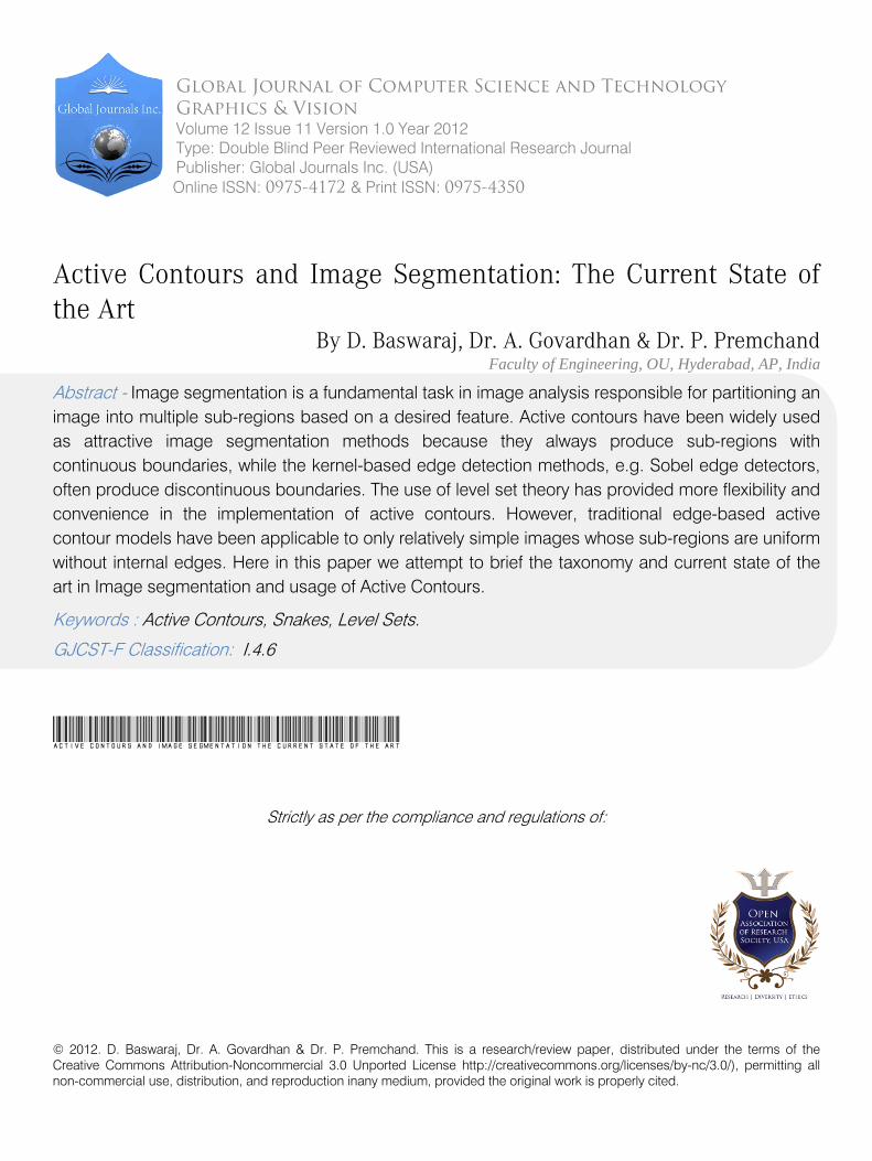
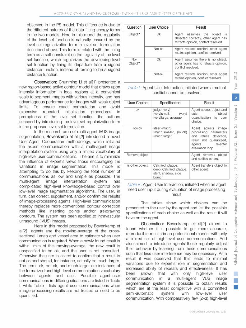
Here in this model proposed by Bovenkamp et al[2], agents use the moving-average of the cross-sectional lumen and vessel area to estimate when user communication is required. When a newly found result is within limits of this moving-average, the new result is unspecified to be ok, and the user is not consulted. Otherwise the user is asked to confirm that a result is not-ok and should, for instance, actually be much-larger. The terms ok, not-ok, and much-larger are instances of the formalized and high-level communication vocabulary between agents and user. Possible agent–user communications in differing situations are listed in Table I, while Table II lists agent–user communications when image-processing results are not trusted or need to be quantified.
The tables show which choices can be presented to the user by the agent and list the possible specifications of each choice as well as the result it will have on the agent.
Observation: Bovenkamp et al[2] aimed to found whether it is possible to get more accurate, reproducible results in an professional manner with only a limited set of high-level user communications. And also aimed to introduce agents those regularly adjust their behavior by learning from these communications such that less user interference may be necessary. As a result it was observed that this leads to minimal variations due to expert’s role in segmentation and increased ability of repeats and effectiveness. It has been shown that with only high-level user communication in a multi-agent IVUS image segmentation system it is possible to obtain results which are at the least competitive with a committed
Question User Choice Result
Object? Ok Agent assumes the object is detected correctly, other agent has retracts opinion, conflict resolved.
Not-ok Agent retracts opinion, other agent retains opinion, conflict resolved.
No-Object?
Ok Agent assumes there is no object, other agent has to retracts opinion, conflict resolved.
Not-ok Agent retracts opinion, other agent retains opinion, conflict resolved.
Table I : Agent-User Interaction, initiated when a mutual conflict cannot be resolved
User Choice Specification Result
ok judge:(very) (very)small, (very) (very)large, average
Agent accept object and sets object quantification to user choice.
not-ok steer:(much) (much)smaller, (much) (much)larger
Agent adjusts image processing parameters and retries detection, result not guaranteed, agents re-enter evaluation loop.
Remove-object Agent removes object and notifies others.
is-other object Calcified_plaque, deep_Calcified_plaque, stent, shadow, side branch
Agent transfers object to other agent.
Table II : Agent-User Interaction, initiated when an agent need user input during evaluation of image processing
result

© 2012 Global Journals Inc. (US)
Globa
l Jo
urna
l of C
ompu
ter Sc
ienc
e an
d Te
chno
logy
V
olum
e XII
Issue
XI V
ersio
n I
6
(DDDD
)F
20
12
user communications per improvement, multi-agent IVUS image segmentation can significantly be improved, while the user is initiating corrective actions in only about 43% (255 versus 594) of the cases when compared with the committed semi-automatic system. No image processing or agent knowledge is required of the user to correct image segmentation results. Experiments show that even when images are very difficult to segment, aggressive results can be obtained in this fashion. However, although enough for most cases limited control by the user over the segmentation process (only very high level) was sometimes too preventive to get to the desired result and is a source of observer errors.
Srinivasa, G et al [3] proposed an active mask algorithm for the segmentation of fluorescence microscope images of punctate patterns. In order to develop this algorithm, Srinivasa, G et al [3] considered active-contour methods for their flexibility, multi resolution methods due to their magnitude speed, multiscale methods by considering their efficiency in smoothing, and region-growing methods for their statistical modeling. The framework developed as top layer of the algorithm proposed moves from the idea of
the ldquo contour rdquo to that of ldquo inside and outside, rdquo or masks, allowing for easy multidimensional segmentation. The framework was aimed to adapt the topology of the image through the use of several masks. To claim the benefit of the algorithm proposed, Srinivasa, G et al [3] argued that since a fluorescent microscope images the cells by revealing the specimen with light of a specific wavelength, exciting the fluorescent probes to emit light of a longer wavelength; a CCD camera records photon emissions resultant in a digital image. As only some parts of the sample are tagged and the tagging is not uniform, the resulting image looks like a allocation of bright dots on a dark background, a punctate pattern. Hence they focused on images in which such patterns represent individual cells in a multi cell specimen.
The algorithm is almost invariant under initialization, allowing for random initialization, and uses a few easily tunable parameters. Experiments show that the active mask algorithm matches the ground truth well and outperforms the algorithm widely used in fluorescence microscopy, seeded watershed, both qualitatively, as well as quantitatively.
Difficulty in identifying the contour in a digital images
Updating the level set function in active-contour algorithms, which is ineffective and slow.
Difficulty in reconstructing the level set function in the multi resolution version?
Difficulty in protect topology during Updating in large increments in the multiscale version.
Observation: Srinivasa, G et al [3] worked on fluorescence microscope images of punctate patterns, and assume that: (a) the statistical properties of the foreground (cell) and background are distinct and relatively uniform; (b) the foreground is bright, while the background is dark. The first assumption is crucial, the second not at all; one can easily change the algorithm should the position be reversed in another modality (such as bright field microscopy). Thus, in this proposal, the authors are basically looking for two different statistical models in the image (foreground and background). We note, however, that the techniques existing here may be generalized to the case of more models. The proposed new algorithm termed as active mask segmentation that designed for segmentation of fluorescence microscope images of punctate patterns, a large class of data. It seems to disappear from the idea of the contour and instead uses that of a mask, as well as several masks. The algorithm easily performs multidimensional segmentation, can be initialized with random seeds, and uses a few easily tunable parameters.
Wenxian Yang et al[4] proposed a constrained random walks algorithm that facilitates the use of three types of user inputs: 1) foreground and background seed input, 2) soft constraint input, and 3) hard constraint input, as well as their combinations. To support the context of their research model Wenxian Yang et al[4] argued that one common fault in the existing interactive image segmentation algorithms is the lack of more intellectual ways to understand the intention of user inputs. The foreground and background seed input of the proposed model is meant to allow a user to draw strokes to specify foreground and background seeds. The soft constraint input is meant to allow a user to draw strokes to point out the region that the boundary should pass through. The hard constraint input meant to allow a user to specify the pixels that the boundary must align with. The proposed method attempted to support all three types of user inputs in one logical computational framework consisting of a constrained random walks and a local editing algorithm, which would allow more accurate contour refinement.
This proposed model formulates the segmentation problem on a graph, where each node represents a pixel and neighboring nodes are linked with undirected edges. In particular, a graph is represented by its vertices and edges also integrate two other types of user inputs as constraints into the random walks algorithm. We call such an extension as constrained random walks. In particular, boundary brush strokes that roughly mark parts of the boundary are introduced as the soft constraint. A vertex on which the soft constraint is forced has the property that the difference between its probability and 1/2 is within a small given range (-e, e). The second type of user inputs, boundary pixel selector,
Year
With issues rose in order to segment fluorescence microscope images such as

© 2012 Global Journals Inc. (US)
Globa
l Jo
urna
l of C
ompu
ter Sc
ienc
e an
d Te
chno
logy
V
olum
e XII
Issue
XI V
ersio
n I
7
(DDDD)
F
2012
which selects pixels on the desired contour, is introduced as the hard constraint. A vertex on which the hard constraint is imposed has a probability of 1/2.
Observation: The proposed model can be summarized as follows. First, the proposed constrained random walks algorithm together with the proposed local editing algorithm supports the three types of user inputs and their combinations in a coherent and unified framework. Second, the region prior term is integrated in the edge weights so that the proposed constrained random walks algorithm does not lose the connectivity property and is less demanding on the positions and quantities of the user input strokes than the original random walks algorithm [43]. Third, the proposed local editing algorithm also allows additional local refinement to reach a satisfactory segmentation.
Ping-Feng Chen et al[5] proposed a novel model to jointly segment and register objects of interest in layered images. Since the Layered images refer to imageries taken from different perspectives and possibly by different sensors, the registration and segmentation are therefore the two main tasks which contribute to the bottom level, data alignment, of the multi sensor data fusion hierarchical structures. In contrast to most exploitation of two layered images those assumed that scanners are at very high altitudes and that only one transformation ties the two images, the proposed model consider the data as taken at mid-range and therefore require segmentation in the process of examining different object regions in a divide-and-conquer fashion. The proposed multiphase joint segmentation by Ping- Feng Chen et al[5] is a combination of multiphase method with a combined segmentation registration practice in short that referred as MPJSR agreed out in a local moving window earlier to a global optimization. To auxiliary address layered video sequence and tracking objects in frame, MPJSR is using a trouble-free adaptation of optical flow calculation along the lively contours in a pair of layered illustration sequences.
The related kind of works introduced former to MPJSR are delineating a intention of interest [44], mosaic king scenes [45], [46], and inclusion data [47]. Techniques which jointly exploit the information from special sensors formally fall within data fusion [48]. Data fusion incorporate a well-established categorization of “fusion levels” that groups different iterative processes of opposed maturity levels. The foundation level, i.e., 0-level of “data alignment” [49], is the preprocessing, registration, and geo-registration of metaphors, which prepares the data for other blend levels. Image registration, which finds the correspondence or the transformation between two images [46], [50]–[56], therefore contributes to this stage in the data fusion hierarchical structure.
Observation: Ping-Feng Chen et al[5] have projected a joint segmentation and register method adapted to multiphase active contours (MPJSR) using a
heartrending local window. By first resembling the detected object surface within a window in the basis and the reference images by planes, and then by evolving a m-phase active contour via the proposed joint segmentation- registration technique, The proposed MPJSR would able to 1) delineate an object of interest, 2) obtain the acquired transformations between two images. This method successfully segments and registers a pair of layered images, and moreover allows us to align segmented objects from one image to another, thus, achieving a 0-level data alignment stage in the data fusion hierarchy.
Figueiredo et al[6] proposal expected to introduce a variational image segmentation method for assess the aberrant crypt foci (ACF) in the person colon captured in vivo by endoscopy. The proposed segmentation technique enhanced the active contours without edges model of Chan and Vese to account for the ACF's particular structure. Level sets to represent the segmentation boundaries and discretize in space by finite elements and in (artificial) time by fixed differences are employed. The model proposed by Figueiredo et al[6] aimed to classify the ACF, their boundaries, and some of the internal crypts' orifices. Figueiredo et al[6] suggestion focused on a fussy image processing method, for assess the ACF captured in vivo by endoscopy: image segmentation. This method consists in the dividing wall of the given image into put out of joint regions, representing distinct objects. Moreover, we use image segmentation methods based on partial differential equations, more exactly, active contours without edges (ACWEs) and level-set methods. These combine techniques of curve evolution (where the basic idea is to start with an initial curve in the image and to deform it to the boundaries of the objects in the image, and stop it there, see [57], [58], and [11]), Mumford–Shah functional for image segmentation (an optimization problem to obtain a sliding doors of the given image into different regions, see [59]) and level-set methods (essentially these consist in considering the problem in a higher measurement, such that the evolving curve is the zero level set of an unknown function; these methods allow cusps, corners, and usual topology changes, as merging and breaking curves, see [60], [61], and [62]). We note that the expression “without edges” in “ACWEs” refers to the fact that in these models it is not used any edge-detector function, based on the gradient of the given image, to identify the different objects (the “edges,” in an image, are the boundaries of the distinct objects, corresponding to the places where these objects meet). This latter property allows the model to segment images where there are no clear gradient boundaries, which is often the case for ACF endoscopic images.
Observation: The aid of the model proposed can be refer to enhancements of the Chan and Vese model and to the parallel numerical tests performed with
Year

© 2012 Global Journals Inc. (US)
Globa
l Jo
urna
l of C
ompu
ter Sc
ienc
e an
d Te
chno
logy
V
olum
e XII
Issue
XI V
ersio
n I
8
(DDDD
)F
20
12
these models to version for the in vivo endoscopic ACF segmentation. More specifically, these main issues are the following. A new numerical system for solve the weak
difference formulation of the Chan and Vese model is defined. The weak formulation has the help of requiring less functional regularity for the unknown level set function. The numerical scheme involves a finite element discretization in space and implicit finite differences in (artificial) time. It is correspondent to a L–M Newton-type optimization method.
A new ACWEs model is definite. It relies on the Chan and Vese model, but incorporates additional terms whose goal is to confine specific features of the ACF that are important to clinicians: the anomalous crypts’ restrictions stain darker than normal crypt and in general inside each focal point, the crypts’ orifices have shapes that are similar to each other.
The mixed regularize model is based on the Chan and Vese model, but involve an additional regularization term, which penalize deviations of the angle of the level set function from unity, and thus address the heterogeneity of the level-set function for a given shape. This avoids the standard line of periodically reinitializing the level-set function to a signed distance function, and permits the full power of a Newton-type optimization method to be applied to minimization of the objective (since the uniqueness constraint is automatically incorporated).
Optical coherence tomography (OCT) is a noninvasive, depth-resolved imaging modality that has become a high up ophthalmic diagnostic technique. Yazdanpanah et al[7] presented a semi-automated segmentation algorithm to detect intra-retinal layers in OCT images acquired from rodent models of retinal degeneration. The proposed segmentation technique was adapted Chan-Vese's energy-minimizing active contours without edges for the OCT images, which in turn suffered from low contrast and were highly tarnished by noise. Hence a multiphase scaffold with a circular shape prior was adopted in order to model the borders of retinal layers and educated guess the shape parameter using least squares. A related scheme was used to balance the weight of poles apart terms in the energy functional.
Observation: Earlier to the Yazdanpanah et al[7]
proposal, several robotic and semi-automated approaches have been employed in OCT segmentation [63]–[72]. Some method rely on pixel-level edge exposure algorithms [10] or are based on performing a 1-D importance peak detection procedure for each A-scan [64]–[66]. These low-level approaches could potentially lead to the finding of not working restrictions and erroneous edges. Moreover, since OCT images are highly corrupted by speckle noise, these algorithms required preprocessing to reduce the effect of noise. The de-noising procedure, however, affects the sharpness of the edges, which subsequently reduces the segmentation appearance. In [67] and [68], a Support Vector Machine (SVM) algorithm is used to perform segmentation of retinal layers. By deportment in mind the mean intensity of six neighbors at each voxel, the SVM approach can handle noisy OCT images. However, this approach is not only dependent on a user to mark a set of points for the rationale of training and segmentation but also fails to segment the layers accurately if the feature and environment points are not chosen properly. Further, SVM is computationally expensive and is not able to segment all layers at the same time. Garvin et al. [69] and Haeker et al. [70], [71] model the segmentation problem as finding the minimum s–t cut of a geometric graph. The cost function is the summation of an edge-based term and one or more region-based terms. They have developed a sequential approach to segment the intra retinal layers. First, the three easier-to-segment surfaces are found (upper surface of NFL and better and lower surfaces of OS). The position of the previous segmented surface is incorporated into the cost function to explain the remaining surfaces. The problem arises when the preceding surface are segmented inaccurately. This may result in an erroneous segmentation of the remaining surfaces. Recently, Garvin et al. [72] have proposed an extension to their algorithm. By learning the surface feasibility constraints using a training set, they can segment the layers in two stages incorporating both the image edge and true regional information in the cost function. The residential iterative algorithm[7] to segment OCT images of rodent retinal layers using a multi-phase framework with a rounded shape prior attempt to demonstrate that the approach is able to truthfully segment all of the intra-retinal layers, even when the small size and similar texture make them difficult to make a distinction visually. And also this model attempted to show that the inclusion of a shape prior constraint improves show on regions with intensity
The objective of the projected segmentation technique is to segment a given OCT image define on the image domain into R disjoint sub-regions, which exactly label the retinal layers. The decomposition of the image I will be modeled using the level set framework as a set of R−1 Signed Distance Functions (SDFs), φ. The distance function captures the distance from any point in the image province to the object limit and assigns this
distance to that point’s location. The SDF assigns opposite signs to the interior versus exterior of the object. Formally, the SDF is an implicit function with positive values in the interior region, negative values in the exterior region, and zero on the boundary with the property that ranges between 0 and 1.
Year

© 2012 Global Journals Inc. (US)
Globa
l Jo
urna
l of C
ompu
ter Sc
ienc
e an
d Te
chno
logy
V
olum
e XII
Issue
XI V
ersio
n I
9
(DDDD)
F
2012
heterogeneity. This proposed segmentation technique backed with a contextual scheme to stability the weight of different terms in the energy functional, which seems to make the algorithm more robust when the image information is not sufficient to accurately detect the layers. This method is a region-based segmentation approach combining the intensity information and the implicit use of edge information, through the shape term, to improve the final segmentation accuracy.
Delu Zeng et al[8] considered the task of object segmentation and achieve in a novel manner that backed by the Poincare map method in a defined vector field in view of dynamical systems. An interpolated swirl and attract flow (ISAF) vector field is first generated for the observed image. Then, the states on the limit cycles of the ISAF are located by the convergence of Newton-Raphson sequences on the given Poincare sections. Meanwhile, the periods of limit cycles are determined. Consequently, the objects' boundaries are represented by integral equations with the corresponding converged states and periods.
In this developed model [8], intially an interpolated swirling and attract flow (ISAF) field is generated by extending a so-called edge tangent flow (ETF) only with a nonzero value at the boundaries to the whole image domain. It is a static vector field. Different from traditional vector fields, the components in this vector field near the boundary are not making a corner but tangent to the boundary. Thus, in the proposed vector field, it is possible for evolution to be carried out along the boundaries. Then, the proposed time-invariant vector field is considered as the right-hand-side vector-valued function of an autonomous dynamical system. As a result, the segmentation problem is translated to the problem of the limit cycle location by applying the related theory in dynamical systems. ISAF is composed of two components, namely, diffused ETF (DETF; swirling component) and diffused edge perpendicular
Observation: The object segmentation is
achieved in a novel manner by the Poincaré map method in the field of dynamical systems. First, for an observed image, an ISAF vector field is proposed,
where there exist to swirling components (with fixed directions) near the object’s borders. This is a key feature of the ISAF compared with the traditional vector field utilized in the ACM method. These swirling workings treated as limit cycles in view of dynamical systems correspond to the desired objects. Then, the Poincaré section and the corresponding Poincaré map for the ISAF are defined. Accordingly, given some initial states in the vector field, they naturally belong to the basins of attraction of the corresponding limit cycles. After that, the Newton–Raphson algorithm is utilized to locate the limit cycles via locating one point on each limit cycle. In the end, the objects’ boundaries are represented by integral equations. Without using the time-consuming level-set methods like most of the ACMs, the proposed algorithm can achieve multiple-boundary extraction by placing some initial states in the vector field. In addition, it runs more competently since the Newton–Raphson algorithm is carried out in the Poincaré section, a lower dimensional subspace of the image domain, while the long-established ACMs evolve the contour in the whole image area.
IV. Conclusion
Active contour models (ACMs) integrated with various kinds of external force fields to pull the contours to the exact boundaries have shown their powerful abilities in object segmentation. However, local minimum problems still exist within these models. The current state of the art in image segmentation mostly cornered to furbish active contour models for domain specific image segmentation, more specific to medical images. The majority of the interactive approaches mainly targeting the accuracy of the segmentation process results. It is clearly evident that these interactive models probabilistic due to the role of the observers and in recent literature, it is hard to find interactive models with optimal resource utilization and computational efficiency. Hence the research scope in interactive image segmentation is optimistic. On other side the statistical and numerical analysis models introduced in recent literature are more specific to contextual issues of the domain to which the input images are belongs to. Hence it is clear evident of scope to perform research that introduce machine learning and data engineering approaches those can generalize the optimistic statistical and numerical methods to improve the computational performance and minimal resource usage in active contour based image segmentation.
References Références Referencias
1. Chunming Li; Chiu-Yen Kao; Gore, J.C.; Zhaohua Ding; , "Minimization of Region-Scalable Fitting Energy for Image Segmentation," Image Processing, IEEE Transactions on , vol.17, no.10, pp.1940-1949, Oct. 2008; doi: 10.1109/TIP.2008.2002304;
Year
flow (DEPF; attracting component). DETF is given by the steady-state solution. However, in DETF, the degree of swirl (rotation) for the streamlines away from the boundary is too high; thus, the journeys are so long for the streamlines start away from the boundary to move to the boundary. In fact, it just need a flow field bearing the properties that its components away from the boundary would point directly to the boundary and its components near the boundary are tangent to the boundary with a fixed direction (either clockwise or counterclockwise). ETF is a thin flow affecting around the objects’ boundaries with a fixed direction. It has components with a nonzero vector value near the boundaries and with a zero vector value to another place.

© 2012 Global Journals Inc. (US)
Globa
l Jo
urna
l of C
ompu
ter Sc
ienc
e an
d Te
chno
logy
V
olum
e XII
Issue
XI V
ersio
n I
10
(DDDD
)F
20
12
2. Bovenkamp, E.G.P.; Dijkstra, J.; Bosch, J.G.; Reiber, J.H.C.; , "User-Agent Cooperation in Multiagent IVUS Image Segmentation," Medical Imaging, IEEE Transactions on , vol.28, no.1, pp.94-105, Jan. 2009; doi: 10.1109/TMI.2008.927351;
3. Srinivasa, G.; Fickus, M.C.; Yusong Guo; Linstedt, A.D.; Kovacevic, J.; , "Active Mask Segmentation of Fluorescence Microscope Images," Image Processing, IEEE Transactions on , vol.18, no.8, pp.1817-1829, Aug. 2009; doi: 10.1109/TIP.2009.2021081
4. Wenxian Yang; Jianfei Cai; Jianmin Zheng; Jiebo Luo;, "User-Friendly Interactive Image Segmentation Through Unified Combinatorial User Inputs," Image Processing, IEEE Transactions on , vol.19, no.9, pp.2470-2479, Sept. 2010; doi: 10.1109/TIP.2010.2048611
5. Ping-Feng Chen; Krim, H.; Mendoza, O.L.; , "Multiphase Joint Segmentation-Registration and Object Tracking for Layered Images," Image Processing, IEEE Transactions on , vol.19, no.7, pp.1706-1719, July 2010; doi: 10.1109/TIP.2010.2045164
6. Figueiredo, I.N.; Figueiredo, P.N.; Stadler, G.; Ghattas, O.; Araujo, A.; , "Variational Image Segmentation for Endoscopic Human Colonic Aberrant Crypt Foci," Medical Imaging, IEEE Transactions on , vol.29, no.4, pp.998-1011, April 2010; doi: 10.1109/TMI.2009.2036258
7. Yazdanpanah, A.; Hamarneh, G.; Smith, B.R.; Sarunic, M.V.; , "Segmentation of Intra-Retinal Layers From Optical Coherence Tomography Images Using an Active Contour Approach," Medical Imaging, IEEE Transactions on , vol.30, no.2, pp.484-496, Feb. 2011; doi: 10.1109/TMI.2010.2087390
8. Delu Zeng; Zhiheng Zhou; Shengli Xie; , "Image Segmentation Based on the Poincaré Map Method," Image Processing, IEEE Transactions on , vol.21, no.3, pp.946-957, March 2012; doi: 10.1109/TIP.2011.2168408
9. Nguyen, T.; Cai, J.; Zhang, J.; Zheng, J.; , "Robust Interactive Image Segmentation Using Convex Active Contour," Image Processing, IEEE Transactions on , vol.PP, no.99, pp.1; doi: 10.1109/TIP.2012.2191566
10. T. Fukuda, Y. Morimoto, S. Morishita, and T. Tokuyama, “Theory of communication,” ACM Transactions on Database Systems, vol. 26, no. 2, pp. 179–213, 2001.
11. M. Kass, A. Witkin, and D. Terzopoulos, “Snakes, active contour model,” International Journal of Computer Vision, pp. 321–331, 1988
12. S. Osher and J. Sethian, “Fronts propagating with curvature dependent speed: Algorithms based on hamilton-jacobi formulations,” Journal of Computationl Physics, pp. 12–49, 1988.
13. J. Canny, “A computational approach to edge detection,” IEEE Transactions on Pattern Analysis and Machine Intelligence, no. 8, p. 769, 1986.
14. A. Jain, Fundamentals of Digital Image Processing. Prentice Hall Information and System Sciences Series, Prentice Hall, 1989
15. C. Brice and C. Fennema, Computer Method in Image Analysis, ch. Scene Analysis using Regions, Los Angeles: IEEE Computer Society, 1977
16. S. Zhu and A. Yuille, “Region competition: Unifying snakes, region growing, and bayes/MDL for multiband image segmentation,” IEEE Transactions on Pattern Analysis and Machine Intelligence, no. 9, pp. 884–200, 1996.
17. S. Zhu and A. Yuille, “Region competition and its analysis: a unified theory for image segmentation,” Tech. Rep. 7, Robotics Lab, Havard University, 1995.
18. K. Fu, R. Gonzales, and C. Lee, Robotics: Control, Sensing, Vision, and Inteligence. New York: McGraw-Hill, 1987.
19. R. Gonzales and R.Woods, Digital Image Processing. Addison-Wesley Publishing, 1st ed., 1993.
20. D. Young, “Active contour models (snakes).” http://www.cogs.susx.ac.uk/users/davidy/teachvision/vision7.html, March 1995.
21. N. Paragios, O. Mellina-Gottardo, and V. Ramesh, “Gradient vector flow fast geometric active contours,” IEEE Transactions on Pattern Analysis and Machine Intelligence, no. 3, pp. 402–407, 2004.
22. N. Paragios, O. Mellina-Gottardo, and V. Ramesh, “Gradient vector flow fast geodesic active contours,” in Proc. of IEEE International Conference on Computer Vision, 2001.
23. C. Xu and J. Prince, Handbook of Medical Imaging: Processing and Analysis Management, ch. Gradient Vector Flow Deformable Models. Academic Press, 2000
24. C. Xu, D. Pham, and J. Prince, Handbook of Medical Imaging: Processing and Analysis Management, ch. Image Segmentation Using Deformable Models, pp. 129–174. Academic Press, 2000
25. C. Xu and J. Prince, “Generalized gradient vector flow external forces for active contours,” Signal Processing, no. 3, pp. 131–139, 1998
26. C. Xu and J. Prince, “Snakes, shapes, and gradient vector flow,” IEEE Transactions on Image Processing, no. 3, pp. 359–369, 1998.
27. C. Xu and J. Prince, “Gradient vector flow: a new external force for snakes,” in Proc. of IEEE Conference on Computer Vision and Pattern Recognition, pp. 66–71, 1997.
28. L. Vese, Geometric Level Set Methods in Imaging, Vision, and Graphics, ch. Multiphase Object
Year

© 2012 Global Journals Inc. (US)
Globa
l Jo
urna
l of C
ompu
ter Sc
ienc
e an
d Te
chno
logy
V
olum
e XII
Issue
XI V
ersio
n I
11
(DDDD)
F
2012
Detection and Image Segmentation, pp. 175–194. New York: Springer-Verlog, 2003.
29. L. Vese and T. Chan, “A multiphase level set framework for image segmentation using the mumford and shah model,” International Journal of Computer Vision, no. 3, pp. 271–293, 2002.
30. T. Chan and L. Vese, “A level set algorithm for minimizing the mumford-shah functional in image processing,” in Proc. of IEEE Workshop on Variational, Geometric and Level Set Methods in Computer Vision, pp. 161–168, 2001.
31. L. Vese and T. Chan, “A multiphase level set framework for image segmentation using the mumford and shah model,” Tech. Rep. 0125, Computational Applied Math Group, UCLA, 2001.
32. T. Chan and L. Vese, “A level set algorithm for minimizing the mumford-shah functional in image processing,” Tech. Rep. 0013, Computational Applied Math Group, UCLA, 2000.
33. N. Paragios and R. Deriche, “Geodesic active regions: A new framework to deal with frame partition problems in computer vision,” Journal of Visual Communication and Image Representation, pp. 249–268, 2002.
34. N. Paragios, Geodesic Active Regions and Level Set Methods: Contributions and Applications in Artificial Vision. PhD thesis, Dept. of Electrical and Computer Engineering, INRIA, 2000
35. N. Paragios and R. Deriche, “Unifying boundary and region-based information for geodesic active tracking,” in Proc. of IEEE Conference on Computer Vision and Pattern Recognition, pp. 300–305, 1999
36. N. Paragios and R. Deriche, “Coupled geodesic active regions for image segmentation: a level set approach,” in Proc. of European Conference on Computer Vision, 2000.
37. N. Paragios and R. Deriche, “Coupled geodesic active regions for image segmentation,” Tech. Rep. 3783, INRIA, 1999
38. N. Paragios, “A variational approach for the segmentation of the left ventricle in cardiac image analysis,” International Journal of Computer Vision, no. 3, pp. 345–362, 2002
39. N. Paragios, “A variational approach for the segmentaion of the left ventricle in mr cardiac images,” in Proc. of IEEE Workshop on Variational, Geometric and Level Set Methods in Computer Vision, 2001.
40. S. Jehan-Besson, M. Barlaud, and G. Aubert, “Shape gradients for histogram segmentation using active contours,” in Proc. of IEEE International Conference on Computer Vision, 2003.
41. S. Jehan-Besson, M. Barlaud, and G. Aubert, “Region-based active contours using geometrical and statistical features for image segmentation,” in Proc. of IEEE International Conference on Image Processing, pp. 643–646, 2003.
42. S. Osher and J. Sethian, “Fronts propagating with curvature-dependent speed: algorithms based on Hamilton-Jacobi formulations,” J. Comput. Phys, vol. 79, pp. 12–49, 1988
43. L. Grady, “Random walks for image segmentation,” IEEE Trans. PAMI, vol. 28, no. 11, pp. 1768–1783, Nov. 2006
44. A. Kim and H. Krim, “Hierarchical stochastic modeling of sar imagery for segmentation/compression,” IEEE Trans. Signal Process., vol. 47, no. 2, pp. 458–468, Feb. 1999.
45. M. Irani, P. Anandan, and S. Hsu, “Mosaic based representations of video sequences and their applications,” in Proc. IEEE Int. Conf. Computer Vision, 1995, p. 605
46. L. M. G. Fonseca, D. Fedorov, B. S. Manjunath, C. Kenney, E. Castejon, and J. S. de Medeiros, “Automatic registration and mosaicking system for remotely sensed imagery,” Revista Brasileira de Cartografia, vol. 58, pp. 49–61, 2006.
47. D. L. Hall, Mathematical Techniques in Multisensor Data Fusion. Norwood, MA: Artech House, 1992
48. L. A. Klein, Sensor and Data Fusion Concepts and Applications. Bellingham, WA: SPIE, 1993
49. D. Hall and J. Llinas, “An introduction to multisensor data fusion,” Proc. IEEE, vol. 85, no. 1, pp. 6–23, Jan. 1997.
50. B. Zitova and J. Flusser, “Image registration methods:Asurvey,” Image Vis. Comput., vol. 21, no. 11, pp. 977–1000, 2003.
51. Y. He, A. Hamza, and H. Krim, “A generalized divergence measure for robust image registration,” IEEE Trans. Signal Process., vol. 51, no. 5, pp. 1211–1220, May 2003
52. Q. Zheng and R. Chellappa, “A computational vision approach to image registration,” IEEE Trans. Image Process., vol. 2, no. 3, pp. 311–326, Jul. 1993
53. C. Shekhar, V. Govindu, and R. Chellappa, Multisensor Image Registration by Feature Consensus 1996.
54. R. Kumar, H. Sawhney, J. Asmuth, A. Pope, and S. Hsu, “Registration of video to geo-referenced imagery,” in Proc. 14th International Conf. Pattern Recognition, Aug. 1998, vol. 2, pp. 1393–1400.
55. S. Jwa, U. Ozguner, and Z. Tang, “Information-theoretic data registration for uav-based sensing,” IEEE Trans. Intell, Transp. Syst,, vol. 9, no. 1, pp. 5–15, Mar. 2008.
56. R. Kumar, S. Samarasekera, S. Hsu, and K. Hanna, “Registration of highly-oblique and zoomed in aerial video to reference imagery,” in Proc. 15th Int. Conf. Pattern Recognition, 2000, vol. 4, pp. 303–307.
57. V. Caselles, F. Catté, T. Coll, and
F. Dibos, “A
geometric model for active contour in image processing” Numer, Math., vol. 66, pp. 1–31, 1993
Year

© 2012 Global Journals Inc. (US)
Globa
l Jo
urna
l of C
ompu
ter Sc
ienc
e an
d Te
chno
logy
V
olum
e XII
Issue
XI V
ersio
n I
12
(DDDD
)F
20
12
58. T. F. Chan and L. A. Vese, “Active contours without edges,” IEEE Trans. Image Process., vol. 10, no. 2, pp. 266–277, Feb. 2001
59. D. Mumford and J. Shah, “Optimal approximations by piecewise smooth functions and associated variational problems,” Comm. Pure Appl. Math., vol. 42, no. 5, pp. 577–685, 1989.
60. S. Osher and R. Fedkiw, Level Set Methods and Dynamic Implicit Surfaces, New York: Springer-Verlag, 2003, vol. 153, Applied Mathematical Sciences.
61. S. Osher and J. A. Sethian, “Fronts propagating with curvature-dependent speed: Algorithms based on Hamilton-Jacobi formulations,” J. Comput. Phys., vol. 79, no. 1, pp. 12–49, 1988
62. J. A. Sethian, Level Set Methods and Fast Marching Methods, 2nd ed. Cambridge, U.K.: Cambridge Univ. Press, 1999, vol. 3, Cambridge Monographs on Applied and Computational Mathematics.
63. D. C. Fern´andez, H. M. Salinas, and C. A. Puliafito, “Automated detection of retinal layer structures on optical coherence tomography images,” Opt. Express, vol. 13, no. 25, pp. 10 200–10 216, 2005.
64. D. C. Fern´andez, “Delineating fluid-filled region boundaries in optical coherence tomography images of the retina,” IEEE Trans. Med. Imag., vol. 24, no. 8, pp. 939 – 945, 2005.
65. A. M. Bagci, M. Shahidi, R. Ansari, M. Blair, N. P. Blair, and R. Zelkha, “Thickness profiles of retinal layers by optical coherence tomography image segmentation,” American Journal of Ophthalmology, vol. 146, no. 5, pp. 679 – 687, 2008.
66. D. Koozekanani, K. Boyer, and C. Roberts, “Retinal thickness measurements from optical coherence tomography using a Markov boundary model,” IEEE Trans. Med. Imag., vol. 20, no. 9, pp. 900–916, 2001.
67. R. J. Zawadzki, A. R. Fuller, D. F. Wiley, B. Hamann, S. S. Choi, and J. S. Werner, “Adaptation of a support vector machine algorithm for segmentation and visualization of retinal structures in volumetric optical coherence tomography data sets,” Journal of Biomedical Optics, vol. 12, no. 4, pp. 041 206, 1–8, 2007.
68. A. R. Fuller, R. J. Zawadzki, S. Choi, D. F. Wiley, J. S. Werner, and B. Hamann, “Segmentation of three-dimensional retinal image data,” Visualization and Computer Graphics, IEEE Transactions on, vol. 13, no. 6, pp. 1719–1726, 2007.
69. M. Garvin, M. Abramoff, R. Kardon, S. Russell, X. Wu, and M. Sonka, “Intraretinal layer segmentation of macular optical coherence tomography images using optimal 3-D graph search,” Medical Imaging, IEEE Transactions on, vol. 27, no. 10, pp. 1495–1505, 2008.
70. M. Haeker, M. Sonka, R. Kardon, V. A. Shah, X. Wu, and M. D. Abramoff, “Automated segmentation of intraretinal layers from macular optical coherence tomography images,” in Progress in Biomedical Optics and Imaging - Proceedings of SPIE, vol. 6512, 2007, p. 651214.
71. M. Haeker, M. Abrmoff, R. Kardon, and M. Sonka, “Segmentation of the surfaces of the retinal layer from OCT images,” in Medical Image Computing and Computer-Assisted Intervention (MICCAI), 2006, pp. 800–807.
72. M. Garvin, M. Abramoff, X. Wu, S. Russell, T. Burns, and M. Sonka, “Automated 3-D intraretinal layer segmentation of macular spectraldomain optical coherence tomography images,” IEEE Trans. Med. Imag., vol. 28, no. 9, pp. 1436 –1447, 2009.
Year