activation of beta 1 but not beta 3 integrin increases...
TRANSCRIPT

FEBS Letters 587 (2013) 763–769
journal homepage: www.FEBSLetters .org
Activation of beta 1 but not beta 3 integrin increases cell traction forces
0014-5793/$36.00 � 2013 Federation of European Biochemical Societies. Published by Elsevier B.V. All rights reserved.http://dx.doi.org/10.1016/j.febslet.2013.01.068
Abbreviations: b1KO, b1-integrin null mouse embryonic fibroblast; b3KO,b3-integrin null mouse embryonic fibroblast; BSA, bovine serum albumin; ECM,extracellular matrix; FA, focal adhesion; FN, fibronectin; LPA, lysophosphatidic acid;mPAD, micropost array detector; PDMS, polydimethylsiloxane; ROCK, Rho kinase;wtMEF, wildtype mouse embryonic fibroblast⇑ Corresponding author. Address: Department of Bioengineering, University of
Pennsylvania, 510 Skirkanich Hall, 210 South 33rd Street, Philadelphia, PA 19104,USA. Fax: +1 (215) 746 1752.
E-mail addresses: [email protected] (G.L. Lin), [email protected] (D.M. Cohen), [email protected] (R.A. Desai), [email protected] (M.T. Breckenridge), [email protected] (L. Gao), [email protected] (M.J. Humphries), [email protected] (C.S. Chen).
Grace L. Lin a, Daniel M. Cohen a, Ravi A. Desai a, Mark T. Breckenridge a, Lin Gao a, Martin J. Humphries b,Christopher S. Chen a,⇑a Department of Bioengineering, University of Pennsylvania, Philadelphia, PA, USAb Faculty of Life Sciences, Wellcome Trust Centre for Cell-Matrix Research, University of Manchester, Manchester, UK
a r t i c l e i n f o
Article history:Received 13 August 2012Revised 3 January 2013Accepted 28 January 2013Available online 8 February 2013
Edited by Michael R. Bubb
Keywords:IntegrinAdhesionCytoskeletal tensionCell traction forceMicropost arrayCell spreading
a b s t r a c t
Cell-generated traction forces induce integrin activation, leading to focal adhesion growth and cellspreading. It remains unknown, however, whether integrin activation feeds back to impact the gen-eration of cytoskeletal tension. Here, we used elastomeric micropost arrays to measure cellular trac-tion forces in wildtype and integrin-null cells. We report that activation of b1 but not b3 integrin, byeither increasing density of immobilized fibronectin or treating with manganese, elicited fibroblastspreading and cytoskeletal tension. Furthermore, this force generation required Rho kinase andmyosin activity. These findings suggest that integrin activation and cell traction forces comprise abi-directional signaling unit of cell adhesion.� 2013 Federation of European Biochemical Societies. Published by Elsevier B.V. All rights reserved.
1. Introduction
The binding of integrins to extracellular matrix (ECM) initiatescell adhesion, which can be described as a series of processesincluding cell spreading against the underlying matrix, assemblyof focal adhesions (FAs), and generation of actomyosin-mediatedcytoskeletal tension against these adhesions [1]. Each of these pro-cesses appears to be linked through several pathways. For exam-ple, the degree of cell spreading against a micropatternedsubstrate regulates RhoA activity and cytoskeletal tension [2,3],and this cytoskeletal tension is important for adhesion assembly[4,5]. Conversely, it has been shown that the clustering of integrinsrequired for adhesion assembly is critical to support cell spreading
and tension generation [6,7]. Because cell spreading, adhesionassembly, and cytoskeletal tension each have been shown to regu-late many cellular functions including proliferation, differentiation,and migration, understanding how these processes are regulated isan important question.
Integrin receptors undergo conformational activation from alow affinity to high affinity state [8,9], and these changes in inte-grin activity may contribute to the regulation of cell spreadingand FA assembly. Indeed, direct activation of integrins via manga-nese (Mn2+) [10] or conformation-modulating antibodies [11] ap-pears to enhance cell spreading and adhesion assembly [12,13].Although numerous studies have linked integrin activation to FAgrowth and superior cell adhesion and spreading on ECM, it is un-clear whether integrin activation can also directly regulate cyto-skeletal tension generation.
In this study, we found that b1 integrin activation via increasedfibronectin (FN) density or Mn2+ leads to enhanced generation ofcellular traction forces. We measured these forces by culturingcells on FN-functionalized arrays of uniformly spaced elastomericmicroposts, a system we developed previously to enable studiesof traction force dynamics [5,14]. Our data indicate that the activa-tion state of integrins is intimately connected to basic adherent cellbehaviors like contractility, which has implications for improvingour understanding of the regulation of cell shape, mechanics, andfunction.
764 G.L. Lin et al. / FEBS Letters 587 (2013) 763–769
2. Materials and methods
2.1. Cell culture
Wildtype and b3 integrin-null MEFs were provided by Dr. Rich-ard Assoian (University of Pennsylvania) and Dr. Richard Hynes(MIT), respectively. b1 Integrin-null MEFs were maintained as pre-viously described [15]. All cells were cultured in 10% FBS/DMEM(Atlanta Biologicals).
2.2. Reagents and antibodies
Reagents were obtained as follows: fibronectin (BD); vitro-nectin (Sigma); lysophosphatidic acid (Avanti Polar Lipids);Y27632 (Tocris Bioscience); blebbistatin (Calbiochem); FN block-ing antibody 16G3 (20 lg/ml; gift of Dr. Martin Schwartz,University of Virginia); b1 integrin blocking antibody BMC5 andrat control IgG (10 lg/ml; Chemicon); anti-b1 integrin (BD);anti-GAPDH (Ambion); anti-active-b1 integrin (clone 9EG7, BD);anti-vinculin (hVin1, Sigma–Aldrich); adenoviral sh-a5 integrinand scrambled sequence (gift of Dr. Rebecca Wells, Universityof Pennsylvania).
2.3. Cell attachment assay
Plates were coated overnight at 4 �C with FN in triplicate (BDBiosciences) and blocked with 50 lg/ml BSA/PBS. Cells wereseeded, gently rinsed after 1 h with warm PBS, and quantifiedusing CyQuant (Invitrogen Molecular Probes).
2.4. Substrate preparation
Micropost array detectors (mPADs) were fabricated usingPDMS-based replica-molding as previously described [5,16].Microcontact printing FN on these or flat substrates, with eithercontinuous or 625 lm2 islands, was performed as describedpreviously [17]. FN concentrations of 0.0625 or 4.0 lg/ml FN in50 lg/ml BSA are designated as low or high FN density,respectively.
2.5. Western blotting
Cells were lysed in Laemmli sample buffer (Bio-Rad), separatedvia SDS–PAGE, transferred to PVDF, immunoblotted, and detectedusing SuperSignal West Dura detection kit (Thermo Scientific).
2.6. Immunofluorescence, cell imaging, and quantitative analysis offocal adhesions and strain energies
For immunofluorescence, cells were fixed with 3.7% paraformal-dehyde (Electron Microscopy Sciences), permeabilized with 0.1%Triton X-100, and labeled using primary and then secondary anti-bodies. Quantitative analyses of adhesions and cell area were per-formed using a custom-developed MATLAB program [18]. FormPAD experiments, cells were labeled with CellTracker GreenCMFDA (Invitrogen Molecular Probes). Quantitative analyses of cellarea and total cell strain energies on mPADs were performed aspreviously described [5].
2.7. Knockdown of a5 integrin
MEFs were infected with adenovirus encoding either shRNA di-rected against a5 integrin or a scrambled sequence [19] at a MOI of50. Cells were trypsinized at 48 h post-infection and seeded onmPAD substrates.
2.8. Statistical analysis
For each box-and-whisker plot, 15 or more cells per conditionwere imaged and analyzed across 3 or more experiments. Statisti-cal comparisons between experimental conditions used eitherMann–Whitney-U tests or Wilcoxon signed-rank tests, as indicatedin individual figure legends. For all tests, statistical significancewas assigned at P-value 60.05 (ns: non-significant, ⁄P 6 0.05,⁄⁄P 6 0.01, ⁄⁄⁄P 6 0.001).
3. Results
3.1. Integrin activation enhances cell spreading and traction force
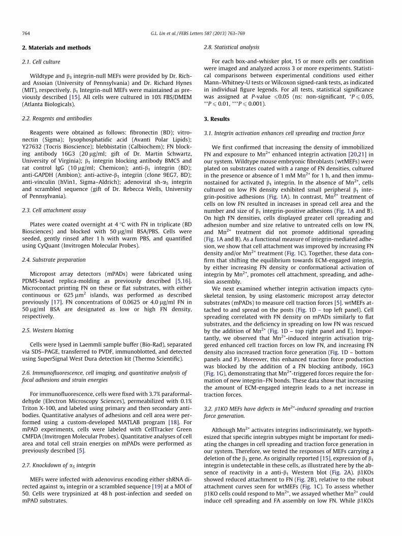
We first confirmed that increasing the density of immobilizedFN and exposure to Mn2+ enhanced integrin activation [20,21] inour system. Wildtype mouse embryonic fibroblasts (wtMEFs) wereplated on substrates coated with a range of FN densities, culturedin the presence or absence of 1 mM Mn2+ for 1 h, and then immu-nostained for activated b1 integrin. In the absence of Mn2+, cellscultured on low FN density exhibited small peripheral b1 inte-grin-positive adhesions (Fig. 1A). In contrast, Mn2+ treatment ofcells on low FN resulted in increases in spread cell area and thenumber and size of b1 integrin-positive adhesions (Fig. 1A and B).On high FN densities, cells displayed greater cell spreading andadhesion number and size relative to untreated cells on low FN,and Mn2+ treatment did not promote additional spreading(Fig. 1A and B). As a functional measure of integrin-mediated adhe-sion, we show that cell attachment was improved by increasing FNdensity and/or Mn2+ treatment (Fig. 1C). Together, these data con-firm that shifting the equilibrium towards ECM-engaged integrin,by either increasing FN density or conformational activation ofintegrin by Mn2+, promotes cell attachment, spreading, and adhe-sion assembly.
We next examined whether integrin activation impacts cyto-skeletal tension, by using elastomeric micropost array detectorsubstrates (mPADs) to measure cell traction forces [5]. wtMEFs at-tached to and spread on the posts (Fig. 1D – top left panel). Cellspreading correlated with FN density on mPADs similarly to flatsubstrates, and the deficiency in spreading on low FN was rescuedby the addition of Mn2+ (Fig. 1D – top right panel and E). Impor-tantly, we observed that Mn2+-induced integrin activation trig-gered enhanced cell traction forces on low FN, and increasing FNdensity also increased traction force generation (Fig. 1D – bottompanels and F). Moreover, this enhanced traction force productionwas blocked by the addition of a FN blocking antibody, 16G3(Fig. 1G), demonstrating that Mn2+-triggered forces require the for-mation of new integrin–FN bonds. These data show that increasingthe amount of ECM-engaged integrin leads to a net increase intraction forces.
3.2. b1KO MEFs have defects in Mn2+-induced spreading and tractionforce generation.
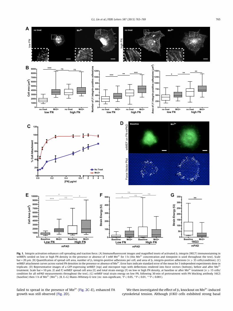
Although Mn2+ activates integrins indiscriminately, we hypoth-esized that specific integrin subtypes might be important for medi-ating the changes in cell spreading and traction force generation inour system. Therefore, we tested the responses of MEFs carrying adeletion of the b1 gene. As originally reported [15], expression of b1
integrin is undetectable in these cells, as illustrated here by the ab-sence of reactivity in a anti-b1 Western blot (Fig. 2A). b1KOsshowed reduced attachment to FN (Fig. 2B), relative to the robustattachment curves seen for wtMEFs (Fig. 1C). To assess whetherb1KO cells could respond to Mn2+, we assayed whether Mn2+ couldinduce cell spreading and FA assembly on low FN. While b1KOs
A
C D
B
E F G
low
FN
no treat Mn2+ no treat Mn2+0
1000
2000
3000
4000
5000
6000
7000
8000
9000 *** ns ns
low FN high FN
high
FN
Low FN High FNBaseline Mn2+ Baseline Mn2+
0
1000
2000
3000
4000
5000
6000
7000
8000
9000
10000 **** ns *
mPAD
0.0 0.2 0.4 0.60
20
40
60
80
100
120
No TreatMn2+
1 2 3 4 5[FN] g/ml
no treat Mn2+
Baseline Mn2+w
tMEF
on
FN-m
PAD
Forc
e Ve
ctor
Plo
t
Low FN High FNBaseline Mn2+ Baseline Mn2+
0
100
200
300
400
500 *ns ns
mPAD
ns
Baseline Mn2+0
50
100
150
200
250
300
350 **
mPAD16G3
no treat Mn2+
no treat Mn2+ no treat Mn2+0
200
400
600
800
1000
1200
*** ** ***
low FN high FNno treat Mn2+ no treat Mn2+
0
250
500
750
1000
1250
1500 *****
low FN high FN
Fig. 1. Integrin activation enhances cell spreading and traction force. (A) Immunofluorescent images and magnified insets of activated b1 integrin (9EG7) immunostaining inwtMEFs seeded on low or high FN density in the presence or absence of 1 mM Mn2+ for 1 h (this Mn2+ concentration and timepoint is used throughout the text). Scalebar = 20 lm. (B) Quantification of spread cell area, number of b1 integrin-positive adhesions per cell, and area of b1 integrin-positive adhesions (n P 35 cells/condition). (C)wtMEF attachment curves across varied FN densities in the presence or absence of Mn2+. Error bars indicate standard error of the mean for 3 independent experiments done intriplicate. (D) Representative images of a GFP-expressing wtMEF (top) and micropost tops with deflections rendered into force vectors (bottom), before and after Mn2+
treatment. Scale bar = 10 lm. (E and F) wtMEF spread cell area (E) and total strain energy (F) on low or high FN density, at baseline or after Mn2+ treatment (n P 15 cells/condition for all mPAD measurements throughout the text). (G) wtMEF total strain energy on low FN, following 30 min of pretreatment with FN blocking antibody 16G3(baseline) then 1 h of Mn2+ (Mn2+). (B, E–G) Mann–Whitney-U test (ns: non-significant, ⁄P 6 0.05, ⁄⁄P 6 0.01, ⁄⁄⁄P 6 0.001).
G.L. Lin et al. / FEBS Letters 587 (2013) 763–769 765
failed to spread in the presence of Mn2+ (Fig. 2C–E), enhanced FAgrowth was still observed (Fig. 2D).
We then investigated the effect of b1 knockout on Mn2+-inducedcytoskeletal tension. Although b1KO cells exhibited strong basal
A B C
E F
D
G H
ITGβ1
GAPDH
β1KO MEF
wtMEF
no treat Mn2+ no treat Mn2+0
1000
2000
3000
4000
5000
6000 *** ns
wtMEF 1KOno treat Mn2+ no treat Mn2+
050
100150200250300350400450500550
** *
wtMEF 1KOno treat Mn2+ no treat Mn2+
0
100
200
300
400
500
600
700
800 ** **
wtMEF 1KO
Low FN High FNBaseline Mn2+ Baseline Mn2+
0
1000
2000
3000
4000
5000 nsns
ns
1KO on mPAD
0.0 0.2 0.4 0.60
102030405060708090
100No TreatMn2+
1 2 3 4 5[FN] g/ml
wtM
EF
no treat Mn2+
β1K
O M
EF
no treat Mn2+
Low FN High FNBaseline Mn2+ Baseline Mn2+
0.1
1
10
100
1000**
ns ns
1KO on mPAD
BMC5 Control IgGBaseline Mn2+ Baseline Mn2+
0
1000
2000
3000
4000
wtMEF on mPAD
** **ns
BMC5 Control IgGBaseline Mn2+ Baseline Mn2+
0
50
100
150
200
250
300
** *
wtMEF on mPAD
**
Fig. 2. b1KO MEFs have defects in Mn2+-induced spreading and traction force generation. (A) Western blotting for b1 integrin expression in wtMEFs and b1KO MEFs. The twobands correspond to a partially glycosylated precursor protein and the mature protein. (B) b1KO attachment curves across varied FN densities in the presence or absence ofMn2+. Error bars indicate standard error of the mean for 4 independent experiments done in triplicate. (C) Vinculin immunofluorescence for wtMEFs and b1KOs in thepresence or absence of Mn2+ on low FN. Scale bar = 20 lm. (D) Quantification of spread cell area, average number of FAs per cell, and average FA area (n P 25 cells/condition).(E and F) b1KO spread cell area (E) and total strain energy (F) on low or high FN density, at baseline or after Mn2+ treatment. (G and H) wtMEF spread cell area (G) and totalstrain energy (H) on low FN, following 30 min of pretreatment with either b1 blocking antibody BMC5 or control IgG antibody (baseline) then 1 h of Mn2+ (Mn2+). (D–H)Mann–Whitney-U test (ns: non-significant, ⁄P 6 0.05, ⁄⁄P 6 0.01, ⁄⁄⁄P 6 0.001).
766 G.L. Lin et al. / FEBS Letters 587 (2013) 763–769
D EA B C
Baseline Mn2+0
5
10
15
20
25
30 **
BlebbistatinBaseline Mn2+
0
5
10
15
20
25
30
ns
Y27632Baseline Mn2+
0
20
40
60
80
100
120 ns
625 μm2 FNBaseline LPA
0
100
200
300
400
500
600
***
wtMEFBaseline LPA
0
100
200
300
400
500
600
***
1KO
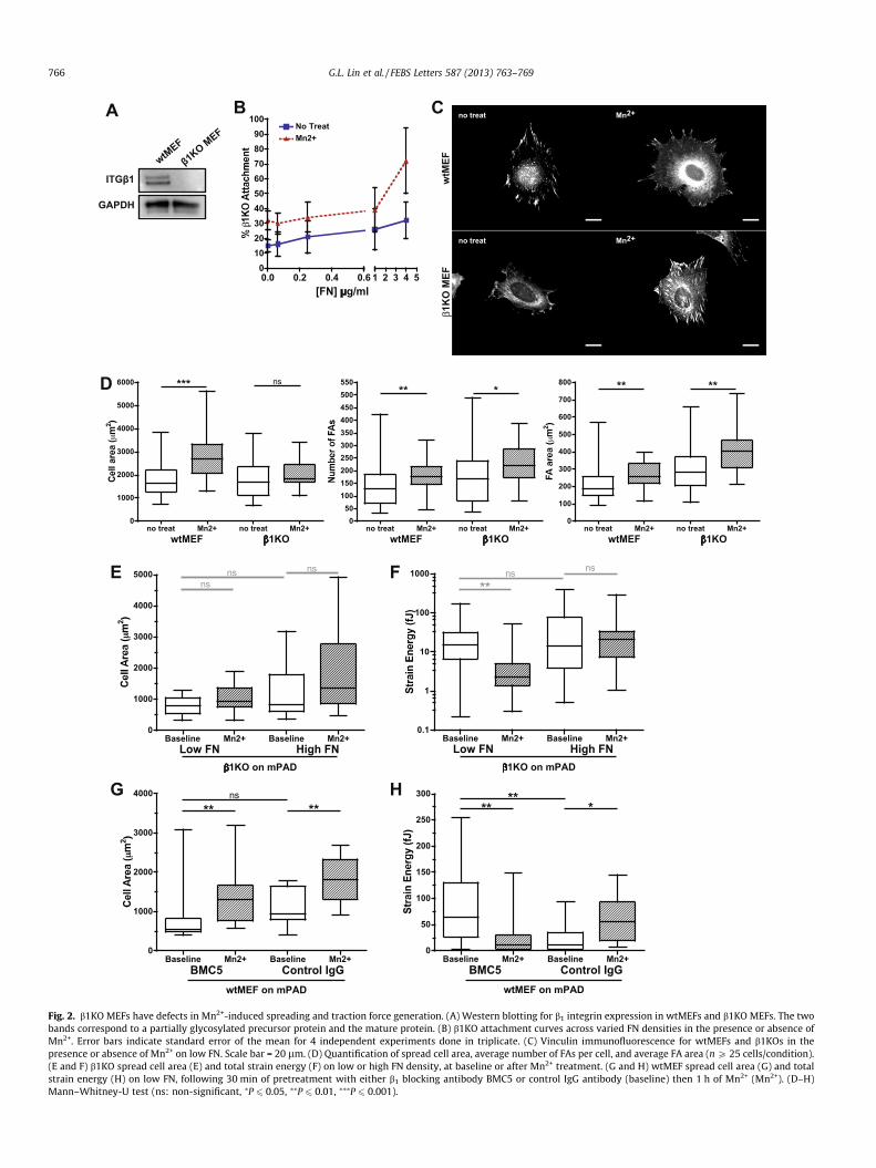
Fig. 3. b1 Integrin-dependent tractions require spread cell shape, ROCK, and myosin activity. (A and B) Total strain energy for wtMEFs pretreated with 10 lM blebbistatin (A)or 10 lM Y27632 (B) for 30 min on low FN, at baseline or after Mn2+ treatment. (C) Total strain energy for wtMEFs restricted to 625 lm2 on high FN micropatterned islands, atbaseline or after Mn2+ treatment. (D and E) Total strain energy for wtMEFs (D) and b1KOs (E) on low FN, at baseline or after 30 min of 10 lg/ml LPA treatment. (A–E) Wilcoxonsigned-rank test (ns: non-significant, ⁄⁄P 6 0.01, ⁄⁄⁄P 6 0.001).
A
E
B C
D
Baseline Mn2+0
500
1000
1500
2000
2500
3000
3500 ***
wtMEF on vitronectin
no treat Mn2+0
1000
2000
3000
4000
5000
6000
7000
8000
9000
10000 ***
3KO on FNno treat Mn2+
050
100150200250300350400450500550 ***
3KO on FNno treat Mn2+
0
100
200
300
400
500
600
700
800
***
3KO on FN
wtM
EF o
n FN
β3K
O M
EF o
n FN
no treat Mn2+
no treat Mn2+
Baseline Mn2+0
50
100
150
200
250
300 ns
wtMEF on vitronectin
Baseline Mn2+
1
10
100
1000
10000 ***
3KO on FN-mPAD
Fig. 4. b3 Integrin is dispensable for Mn2+-induced traction forces. (A and B) wtMEF spread cell area (A) and total strain energy (B) on 20 lg/ml vitronectin, at baseline or afterMn2+ treatment. (C) Vinculin immunofluorescence for wtMEFs and b3KO MEFs in the presence or absence of Mn2+ on low FN. Scale bar = 20 lm. (D) Quantification of spreadcell area, average number of FAs per cell, and average FA area (n P 25 cells/condition). (E) b3KO total strain energy on low FN density, at baseline or after Mn2+ treatment. (A,B and E) Wilcoxon signed-rank test; (D) Mann–Whitney-U test (ns: non-significant, ⁄⁄⁄P 6 0.001).
G.L. Lin et al. / FEBS Letters 587 (2013) 763–769 767
contractility compared to wildtype MEFs, the b1KOs failed tomount increased traction forces in response to either increasedFN density or Mn2+ treatment (Fig. 2E and F). In fact, Mn2+ treat-ment resulted in a statistically significant decrease in tractionforces on low FN (Fig. 2F).
Loss of b1 integrin disrupts numerous integrin heterodimers,including the principal fibronectin receptor a5b1 integrin. To testwhether a5b1 integrin was specifically required for Mn2+-induced
cytoskeletal tension, we treated wtMEFs with a function-blockinga5b1 integrin antibody, BMC5. Inhibition of a5b1 integrin trended to-ward decreased cell spreading on low FN mPADs and did not preventthe spreading response to Mn2+ treatment (Fig. 2G).In spite of thetrend toward decreased spreading, basal contractility showed anunexpected increase in response to BMC5 (Fig. 2H), whereas MEFstreated with an isotype-matched control IgG showed similar base-line contractility to untreated cells (Fig. 2H vs. 1F). Nonetheless,
768 G.L. Lin et al. / FEBS Letters 587 (2013) 763–769
upon treatment with Mn2+, BMC5-treated cells showed a statisti-cally significant decrease in strain energy that paralleled the re-sponse observed in b1KO cells (Fig. 2H vs. F). Moreover, weobserved a similar loss of strain energy in response to Mn2+ treat-ment when a5 integrin was depleted by RNA interference (Supp.Fig. 1A and B). Taken together, these data suggest that the increasedcell traction force upon stimulation of integrin activation requires b1
integrin and is likely mediated by a5b1.
3.3. b1 Integrin-dependent tractions require spread cell shape, ROCK,and myosin activity
To better understand the requirement for b1 integrin in generat-ing traction forces, we investigated whether these forces weremediated by non-muscle myosin II activity and whether b1KOswere competent to respond to other contractility agonists. Wetested the role of myosin activity in Mn2+-induced traction by pre-treating wtMEFs with Y27632 and blebbistatin, pharmacologicalinhibitors of Rho kinase (ROCK) and myosin, respectively. Bothblebbistatin (Fig. 3A) and Y27632 (Fig. 3B) prevented Mn2+-in-duced traction forces. Prior work from our group demonstratedthat cellular contractility can also be blocked by culturing cellson small micropatterned FN islands [5]. Here we observed thatmicropatterned islands (625 lm2) of high FN prevented cells frommounting traction forces in response to Mn2+ stimulation (Fig. 3C),consistent with a role for actomyosin contractile machinery inmediating Mn2+-dependent traction forces.
Restricting cell spreading by micropatterning results in a gener-alized defect in coupling extracellular stimuli with traction forceproduction, a relationship we have reported in multiple systems[5,16]. We therefore tested whether the defect in traction forceproduction in b1KOs could be attributed to a generalized defectin organizing or activating actomyosin contractility or was specificto Mn2+. Treatment of cells with lysophosphatidic acid (LPA), astrong agonist for Rho-mediated myosin activation, triggered asustained increase in traction force in both wtMEFs and b1KOswithin 1 min (Fig. 3D and E), indicating that the coupling betweensoluble agonists and contractility remains intact in the b1KOs.
3.4. b3 Integrin is dispensable for Mn2+-induced traction forces
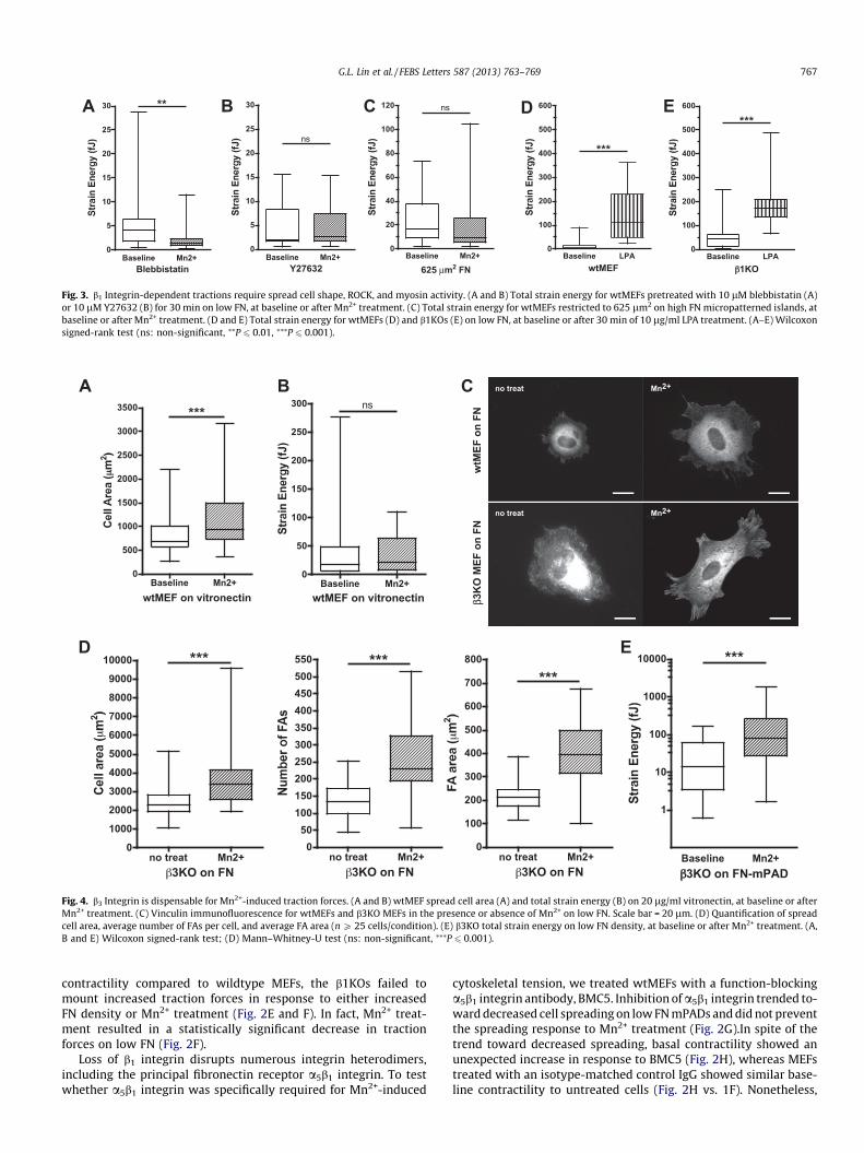
Our results suggest that, in the absence of b1 integrin, activationof integrins induces FA assembly, but fails to induce cell spreadingor traction forces on low FN. Because fibroblasts also use b3 inte-grin to bind FN, we next evaluated whether b3 integrin activationcould contribute to either enhanced cell spreading or tractionforces following Mn2+ treatment. To do so, we first plated wtMEFson vitronectin, a preferential ligand for b3 integrin [22]. Upon treat-ment with Mn2+, wtMEFs on vitronectin increased spread cell area(Fig. 4A) but generated no additional net traction force (Fig. 4B).
These data suggest that b3 integrin engagement with ECM li-gand is not sufficient to mediate Mn2+-induced traction forcesbut could support cell spreading. To test the role of b3 integrin inthe response to Mn2+ more directly, we examined the effect ofMn2+ on b3-null MEFs (b3KOs) [23]. Similar to wtMEFs, b3KOs trea-ted with Mn2+ demonstrated an increase in spread cell area and FAgrowth and number (Fig. 4C and D). Likewise, the b3KOs increasedcell traction forces after Mn2+ treatment (Fig. 4E). Thus, b3 integrinactivation can promote cell spreading; however, it is dispensablefor Mn2+-induced traction force generation.
4. Discussion
Cellular traction forces play an integral role in cell adhesion tomatrices. These forces regulate FA assembly, presumably by acting
directly on integrins to activate them through ‘‘inside-out’’ signal-ing [4,5]. Additionally, myosin-mediated contractility regulatesadhesion through recruitment of signaling proteins to FAs[24,25]. Signaling downstream from these FA proteins impacts pro-liferation [26], differentiation [27], migration [28,29], and otherhigher-level cellular functions. Here, we show ‘‘outside-in’’ signalsthat promote integrin activation (increased FN density, Mn2+) alsotrigger traction force generation. This finding clarifies how the cellmight sense matrix density through ECM-modulated integrin affin-ity that directly adjusts cytoskeletal tension to befit themicroenvironment.
Integrin activation could modulate cell traction forces throughseveral possible mechanisms. Here, we reveal that traction forcesinduced by integrin activation require Rho kinase and myosinactivity, suggesting that Rho GTPases could be involved. Alterna-tively, integrin adhesion complexes can nucleate actin polymeriza-tion via Arp2/3 in a manner dependent on the density of ligatedintegrins [30,31] and independently of Rho GTPases [32]. Actinpolymerization creates protrusive force [33] that can drive cellmotility [34] and may contribute to the integrin-mediated forcesreported in this work.
Different integrin subtypes often have overlapping functions,with some instances where distinct integrins produce unique ef-fects on cells. For example, both a5b1 and aVb3 integrins bind FN,but drive divergent migratory behavior; a5b1–FN adhesion pro-motes thin cell protrusions and random cell migration whereasaVb3–FN adhesion supports persistent migration with broad lamel-lipodia [35]. In fact, it has been proposed that these effects aremediated through a change in the balance of Rho/Rac signaling[35]. Here we show that in normal cells, Mn2+ stimulates cell trac-tion forces in a b1 integrin-dependent manner, whereas in cellsdeficient in a5 or b1, Mn2+ stimulation leads to a decrease in trac-tion forces. While the mechanism of decreased traction forces ina5 or b1 deficient cells is as yet unknown, it is interesting to spec-ulate that avb3-specific signaling, such as to Rac1 GTPase, may playa role.
Our finding that activation of b1 integrin contributes to en-hanced traction forces is consistent with previous studies inwhich force production on fibronectin substrates is disruptedby a5b1 blocking antibody in fibroblasts [36] or myocytes [37].It is interesting that the same b1 integrin subtype that inducesintracellular forces also undergoes conformational activation inresponse to extracellular forces and uniquely displays catch-bond and adhesion strength-reinforcing behavior upon forcetransmission [38–41]. In view of these previous studies, our re-sults suggest that feedback between force sensing and tractionforce generation may be a necessary component to the cellmechanotransduction system. Further studies on the regulationof basic cell functions by integrins will help shed light on howcells manage complex behavior in response to mechanical andadhesivecues.
Acknowledgements
We thank R. Hynes for his kind gift of b3 integrin-null MEFs, R.Assoian for the wildtype MEFs, M. Schwartz for the FN blockingantibody, R. Wells for the adenoviral sh-a5, M. Yang and M. Lynchfor assistance with mPADs, and J. Eyckmans and C. Choi for helpfuldiscussions. This work was supported in part by grants from theNational Institutes of Health (EB00262 and GM74048), the RESBIOTechnology Resource for Polymeric Biomaterials, and Center forEngineering Cells and Regeneration of the University of Pennsylva-nia. D.M.C. acknowledges financial support from the Ruth L. Kirsch-stein National Research Service Award, and R.A.D. was supportedby the National Science Foundation.

G.L. Lin et al. / FEBS Letters 587 (2013) 763–769 769
Appendix A. Supplementary data
Supplementary data associated with this article can be found, inthe online version, at http://dx.doi.org/10.1016/j.febslet.2013.01.068.
References
[1] Galbraith, C.G. and Sheetz, M.P. (1998) Forces on adhesive contacts affect cellfunction. Curr. Opin. Cell Biol. 10, 566–571.
[2] McBeath, R., Pirone, D.M., Nelson, C.M., Bhadriraju, K. and Chen, C.S. (2004) Cellshape, cytoskeletal tension, and RhoA regulate stem cell lineage commitment.Dev. Cell 6, 483–495.
[3] Bhadriraju, K., Yang, M., Alom Ruiz, S., Pirone, D., Tan, J. and Chen, C.S. (2007)Activation of ROCK by RhoA is regulated by cell adhesion, shape, andcytoskeletal tension. Exp. Cell Res. 313, 3616–3623.
[4] Chrzanowska-Wodnicka, M. and Burridge, K. (1996) Rho-stimulatedcontractility drives the formation of stress fibers and focal adhesions. J. CellBiol. 133, 1403–1415.
[5] Tan, J.L., Tien, J., Pirone, D.M., Gray, D.S., Bhadriraju, K. and Chen, C.S. (2003)Cells lying on a bed of microneedles: an approach to isolate mechanical force.Proc. Natl. Acad. Sci. USA 100, 1484–1489.
[6] Cavalcanti-Adam, E.A., Volberg, T., Micoulet, A., Kessler, H., Geiger, B. andSpatz, J.P. (2007) Cell spreading and focal adhesion dynamics are regulated byspacing of integrin ligands. Biophys. J. 92, 2964–2974.
[7] Selhuber-Unkel, C., Erdmann, T., Lopez-Garcia, M., Kessler, H., Schwarz, U.S.and Spatz, J.P. (2010) Cell adhesion strength is controlled by intermolecularspacing of adhesion receptors. Biophys. J. 98, 543–551.
[8] Campbell, I.D. and Humphries, M.J. (2011) Integrin structure, activation, andinteractions. Cold Spring Harb. Perspect. Biol. 3, a004994.
[9] Kim, C., Ye, F. and Ginsberg, M.H. (2011) Regulation of integrin activation.Annu. Rev. Cell Dev. Biol. 27, 321–345.
[10] Xiong, J.P., Stehle, T., Zhang, R., Joachimiak, A., Frech, M., Goodman, S.L. andArnaout, M.A. (2002) Crystal structure of the extracellular segment of integrinalphaVbeta3 in complex with an Arg-Gly-Asp ligand. Science 296, 151–155.
[11] Byron, A., Humphries, J.D., Askari, J.A., Craig, S.E., Mould, A.P. and Humphries,M.J. (2009) Anti-integrin monoclonal antibodies. J. Cell Sci. 122, 4009–4011.
[12] Edwards, J.G., Hameed, H. and Campbell, G. (1988) Induction of fibroblastspreading by Mn2+: a possible role for unusual binding sites for divalentcations in receptors for proteins containing Arg-Gly-Asp. J. Cell Sci. 89 (Pt 4),507–513.
[13] Cluzel, C., Saltel, F., Lussi, J., Paulhe, F., Imhof, B.A. and Wehrle-Haller, B. (2005)The mechanisms and dynamics of alphaVbeta3 integrin clustering in livingcells. J. Cell Biol. 171, 383–392.
[14] Yang, M.T., Reich, D.H. and Chen, C.S. (2011) Measurement and analysis oftraction force dynamics in response to vasoactive agonists. Integr. Biol.(Camb.) 3, 663–674.
[15] Parsons, M., Messent, A.J., Humphries, J.D., Deakin, N.O. and Humphries, M.J.(2008) Quantification of integrin receptor agonism by fluorescence lifetimeimaging. J. Cell Sci. 121, 265–271.
[16] Fu, J., Wang, Y.K., Yang, M.T., Desai, R.A., Yu, X., Liu, Z. and Chen, C.S. (2010)Mechanical regulation of cell function with geometrically modulatedelastomeric substrates. Nat. Methods 7, 733–736.
[17] Tan, J.L., Liu, W., Nelson, C.M., Raghavan, S. and Chen, C.S. (2004) Simpleapproach to micropattern cells on common culture substrates by tuningsubstrate wettability. Tissue Eng. 10, 865–872.
[18] Nelson, C.M., Pirone, D.M., Tan, J.L. and Chen, C.S. (2004) Vascular endothelial-cadherin regulates cytoskeletal tension, cell spreading, and focal adhesions bystimulating RhoA. Mol. Biol.Cell 15, 2943–2953.
[19] Olsen, A.L., Sackey, B.K., Marcinkiewicz, C., Boettiger, D. and Wells, R.G. (2012)Fibronectin extra domain-A promotes hepatic stellate cell motility but notdifferentiation into myofibroblasts. Gastroenterology 142, 928–937.
[20] Lenter, M., Uhlig, H., Hamann, A., Jeno, P., Imhof, B. and Vestweber, D. (1993) Amonoclonal antibody against an activation epitope on mouse integrin chain
beta 1 blocks adhesion of lymphocytes to the endothelial integrinalpha6beta1. Proc. Natl. Acad. Sci. USA 90, 9051–9055.
[21] Bazzoni, G., Shih, D.T., Buck, C.A. and Hemler, M.E. (1995) Monoclonalantibody 9EG7 defines a novel beta1 integrin epitope induced by solubleligand and manganese, but inhibited by calcium. J. Biol. Chem. 270, 25570–25577.
[22] Humphries, J.D., Byron, A. and Humphries, M.J. (2006) Integrin ligands at aglance. J. Cell Sci. 119, 3901–3903.
[23] Hodivala-Dilke, K.M. et al. (1999) Beta3-integrin-deficient mice are a modelfor Glanzmann thrombasthenia showing placental defects and reducedsurvival. J. Clin. Invest. 103, 229–238.
[24] Schiller, H.B., Friedel, C.C., Boulegue, C. and Fässler, R. (2011) Quantitativeproteomics of the integrin adhesome show a myosin II-dependent recruitmentof LIM domain proteins. EMBO Rep. 12, 259–266.
[25] Pasapera, A.M., Schneider, I.C., Rericha, E., Schlaepfer, D.D. and Waterman, C.M.(2010) Myosin II activity regulates vinculin recruitment to focal adhesionsthrough FAK-mediated paxillin phosphorylation. J. Cell Biol. 188, 877–890.
[26] Assoian, R.K. and Schwartz, M.A. (2001) Coordinate signaling by integrins andreceptor tyrosine kinases in the regulation of G1 phase cell-cycle progression.Curr. Opin. Genet. Dev. 11, 48–53.
[27] Salasznyk, R.M., Klees, R.F., Williams, W.A., Boskey, A. and Plopper, G.E. (2007)Focal adhesion kinase signaling pathways regulate the osteogenicdifferentiation of human mesenchymal stem cells. Exp. Cell Res. 313, 22–37.
[28] Sieg, D.J., Hauck, C.R. and Schlaepfer, D.D. (1999) Required role of focaladhesion kinase for integrin-stimulated cell migration. J. Cell Sci. 112 (Pt 16),2677–2691.
[29] Xu, W., Coll, J.L. and Adamson, E.D. (1998) Rescue of the mutant phenotype byreexpression of full-length vinculin in null F9 cells; effects on cell locomotionby domain deleted vinculin. J. Cell Sci. 111 (Pt 11), 1535–1544.
[30] DeMali, K.A., Barlow, C.A. and Burridge, K. (2002) Recruitment of the Arp2/3complex to vinculin: coupling membrane protrusion to matrix adhesion. J. CellBiol. 159, 881–891.
[31] Butler, B., Gao, C., Mersich, A.T. and Blystone, S.D. (2006) Purified integrinadhesion complexes exhibit actin-polymerization activity. Curr. Biol. 16, 242–251.
[32] Rohatgi, R., Nollau, P., Ho, H.Y., Kirschner, M.W. and Mayer, B.J. (2001) Nck andphosphatidylinositol 4,5-bisphosphate synergistically activate actinpolymerization through the N-WASP-Arp2/3 pathway. J. Biol. Chem. 276,26448–26452.
[33] Miyata, H., Nishiyama, S., Akashi, K. and Kinosita Jr., K. (1999) Protrusivegrowth from giant liposomes driven by actin polymerization. Proc. Natl. Acad.Sci. USA 96, 2048–2053.
[34] Mitchison, T.J. and Cramer, L.P. (1996) Actin-based cell motility and celllocomotion. Cell 84, 371–379.
[35] Danen, E.H., van Rheenen, J., Franken, W., Huveneers, S., Sonneveld, P., Jalink,K. and Sonnenberg, A. (2005) Integrins control motile strategy through a Rho-cofilin pathway. J. Cell Biol. 169, 515–526.
[36] Legate, K.R., Takahashi, S., Bonakdar, N., Fabry, B., Boettiger, D., Zent, R. andFassler, R. (2011) Integrin adhesion and force coupling are independentlyregulated by localized PtdIns(4,5)2 synthesis. EMBO J. 30, 4539–4553.
[37] Wu, X., Chakraborty, S., Heaps, C.L., Davis, M.J., Meininger, G.A. andMuthuchamy, M. (2011) Fibronectin increases the force production ofmouse papillary muscles via alpha5beta1 integrin. J. Mol. Cell Cardiol. 50,203–213.
[38] Friedland, J.C., Lee, M.H. and Boettiger, D. (2009) Mechanically activatedintegrin switch controls alpha5beta1 function. Science 323, 642–644.
[39] Garcia, A.J., Huber, F. and Boettiger, D. (1998) Force required to breakalpha5beta1 integrin–fibronectin bonds in intact adherent cells is sensitive tointegrin activation state. J. Biol. Chem. 273, 10988–10993.
[40] Garcia, A.J., Takagi, J. and Boettiger, D. (1998) Two-stage activation foralpha5beta1 integrin binding to surface-adsorbed fibronectin. J. Biol. Chem.273, 34710–34715.
[41] Roca-Cusachs, P., Gauthier, N.C., Del Rio, A. and Sheetz, M.P. (2009) Clusteringof alpha5beta1 integrins determines adhesion strength whereas alphaVbeta3and talin enable mechanotransduction. Proc. Natl. Acad. Sci. USA 106, 16245–16250.