action of valproic acid on xenopus laevis development: teratogenic effects on eyes
TRANSCRIPT
Teratogenesis, Carcinogenesis, and Mutagenesis 21:121–133 (2001)
© 2001 Wiley-Liss, Inc.
Action of Valproic Acid on Xenopus laevisDevelopment: Teratogenic Effects on Eyes
Roberta Pennati, Silvia Groppelli, Fiorenza De Bernardi, andCristina Sotgia*
Department of Biology, University of Milano, Milano, Italy
Valproic acid (VPA) is an anticonvulsive drug used in the treatment of epilepsy. Ter-atogenic effects of VPA have been described in different animal species. In this study,we investigate the effects of VPA on the development of Xenopus laevis embryos, byshort pulse treatments (4 h) with relation to the dose and the stage of exposure to thedrug. We exposed Xenopus embryos from blastula to stage 32 to three different dosesof VPA (0.25, 5, and 10 mM) and we allowed these to develop until the controlsreached stage 47. The embryos became more sensitive during the stages of neurula-tion, as observed in mouse and differently from Amblystoma, in which the more severeeffects were produced by treatments at blastula stage. The malformations observedwere similar to those described in mammals and other amphibians and consisted indevelopmental delay, perturbation of neural crest migration, and somite segmentation.We also observed abnormal development of the retina, which had never been describedfor VPA treatments. Therefore we analyzed the relation between VPA-induced eyemalformations and the expression of Pax-6. We examined VPA-treated Xenopus em-bryos by whole mount in situ hybridization for mis-expression of Pax-6 in correlationwith eye anomalies. Our results are consistent with the hypothesis that different mem-bers of Pax gene family are candidate target of VPA teratogenic action and in particu-lar the decreased level of Pax-6 expression, shown by Northern blot analysis, isresponsible for the retinal malformations we observed in VPA-treated Xenopus em-bryos. Teratogenesis Carcinog. Mutagen. 21:121–133, 2001.© 2001 Wiley-Liss, Inc.
Key words: valproic acid; Xenopus; eye malformations; Pax-6; “in situ” hybridization
INTRODUCTION
Valproic acid (VPA), 2-n-propylpentanoic acid, is an anticonvulsive drug that is usedin the treatment of different seizure disorders. Teratogenic effects of VPA have been de-scribed in different animal species. In humans fetuses, exposed in utero to VPA, craniofa-
Contract grant sponsor: Italian Ministero dell’Università e della Ricerca Scientifica e Tecnologica (40%);Contract grant sponsor: Consiglio Nazionale delle Ricerche.
*Correspondence to: Dr. Cristina Sotgia, Department of Biology, Section of Zoology S. N., Via Celoria26, 20133 Milano, Italy. E-mail [email protected]

122 Pennati et al.
cial anomalies, spina bifida, and developmental delay were observed [1–5]. Similar cran-iofacial malformations were obtained in monkeys and rats by exposure to VPA duringorganogenesis [6]. VPA induces neural tube defects (exencephaly and spina bifida) inmice but not in rats [7]. In chicken, VPA treatment affected mainly the somite develop-ment, causing somitic fusions or mis-segmentation and disorganizing somite patterning[8]. In the amphibian Amblystoma mexicanum the malformations caused by VPA con-sisted of absence of closure of the neural tube, somite irregularities, and developmentaldelay. In Xenopus, embryos, developmental delay and, only in a few cases, neural tubedefects are reported [9]. Apart from a general developmental delay, which may be due toa generalized effect of the drug, the anomalies induced by VPA treatments are different inthe different species. This can be due to an intrinsic difference of sensibility to VPA amongthe species, but the disparity of response to VPA action may be also correlated with thedifference in timing and dosage of the treatments. In fact, in mammals, VPA was givenduring somitogenesis and neurulation, both in vitro and in vivo [7,10]. Amphibians wereexposed to a pulse treatment of VPA from early blastula to late gastrula [9]. Chickenshowed the highest sensibility when treated with VPA between Hamburger and Hamilton[11] stages 12/13 to approximately H.H. stage 20 [8].
In this study, we further investigate the effects of VPA on the development of Xeno-pus laevis embryos, by short pulse treatments (4 hours), in relation to the dose and thestage of exposure to the drug, in order to better understand if there is a correlation be-tween the incidence, the kind of malformations, and the main developmental events thatoccur during the administration of the drug. The malformations observed were similar tothose described in mammals and other amphibians. We also observed eye malforma-tions, whose occurrence was occasionally reported in VPA-treated Xenopus [12].
The anomalies in development of the retina were very similar to those observed inPax-6 mutant mice [13]. Small eye mutant mice, carrying a dominant mutation of Pax-6, show folding of the anterior margins of the optic cup and infiltration of mesodermalcells [16]. The Pax genes are a family of developmental control genes. They contain thepaired box, a highly conserved sequence that encodes a DNA-binding motif of 128amino acids [15]. All Pax genes have a temporally and spatially restricted expressionpattern during embryogenesis, consistent with a regulatory role in development. Pax-6is a master control gene, which has been used also as a molecular marker of evolution-ary homologous eyes in different phyla. In Xenopus, it is first expressed at late gastrula(stage 12, N. & F. [16]) in the primordium of the neural tube and in a broad stripe thatencompasses the anterior neural plate [17]. By stage 12.5, two distinct patches of Pax-6expression are visible in the presumptive lens ectoderm.
In this report, the relation between VPA induced eye malformations and the ex-pression of Pax-6 is analyzed. We examined VPA-treated Xenopus embryos by wholemount in situ hybridization for mis-expression of Pax-6 in correlation with eye anoma-lies. Our results are consistent with the hypothesis that decreased levels of Pax-6expression may be responsible of the retinal malformations we observed in VPA-treated Xenopus embryos.
MATERIAL AND METHODSValproic Acid Treatment
Selected pairs of Xenopus laevis were injected with human chorionic gonadot-ropin (HCG) to stimulate ovulation and spawning. Males were first injected with
Teratogenic Effects of VPA on Xenopus Eyes 123
100 I.U. and, after 2 days, with a second dose of 200 I.U. The same day, femaleswere injected with a single dose of 300 I.U. Eggs were collected and manuallydejellied under a dissecting microscope. Normally developing embryos were selectedand raised in modified Barth’s saline (MBS) buffer (88 mM NaCl, 1 mM KCl, 0.7mM CaCl2, 1 mM MgSO4, 5 mM HEPES, pH 7.8, and 2.5 NaHCO3) at 21°C untilthey reached the useful developmental stages. VPA treatment was started at eightdifferent stages: 7, 10, 13, 15, 17, 20, 27, and 34, according to the normal table ofdevelopment of Nieuwkoop and Faber [16]. For each stage, three concentrations ofVPA (2-n-propylpentanoic acid sodium salt; Sigma Chemical Corp., St. Louis, MO),were applied: 0.25 mM, 5 mM and 10 mM. Dilutions were made from a stock solu-tion of 0.1 M VPA. Control embryos were maintained in MBS buffer. Twenty-fiveembryos for each combination of VPA concentration and stage were treated in plas-tic Petri dishes (60 × 15 mm) containing 20 ml of solution. Each experiment wasperformed six times (n = 150). The embryos were exposed to VPA for 4 h and thenthey were reared in MBS. The medium was changed daily using a solution freshlyprepared from a 10× stock, and dead embryos, if any, were removed. When controlsreached stage 47, all surviving embryos, except three for each treatment, were fixedin Bouin’s solution and examined under a dissecting microscope. To better analyzethe somites, the skin of some specimens was ablated using watchmaker forceps. Thenumber of dead embryos and the type and frequency of malformations were recorded.We scored the embryos according to evident abnormalities on eye development, somitesegmentation, and pigment distribution.
Histology
Three embryos for each treatment, randomly chosen, were let to develop fortwo more days, until they reached a developmental stage comparable with stage 47of control embryos and were prepared for histology. After an overnight staining inborax-carmine, the embryos were dehydrated and embedded in paraffin. Cross sec-tions of 6 µm thickness were cut and stained with aniline-blue-orange G [18].
Northern Analysis of Pax-6 Expression
Total RNA was isolated from five Xenopus embryos treated at stage 20 (N. & F.)with 10 mM VPA and from five control embryos treated with MBS solution alone(see above). Total RNA was extracted using guanidine thiocyanate [19] and analyzedby Northern blot analysis. Northern blot analysis was performed according Sambrooket al. [20]; 20 µl of total RNA for each sample were fractionated by agarose gelelectrophoresis and blotted. Prehybridization was carried at 42°C in 50% formamide,0.5 M sodium phosphate, 0.1% sodium dodecyl sulfate (SDS), and 2× Denhardt’sreagent. Hybridization was done at 42°C using 32P-labeled Pax-6 cDNA. EtBr-stainedribosomal RNA bands were used for normalization of RNA loads. The intensity ofthe hybridization signal was determined by densitometric analysis of the autoradio-gram using ImageMasterTM VDS Software (Pharmacia Biotech, Uppsala, Sweden).
Whole-Mount In Situ Hybridization
Whole-mount in situ hybridization was performed according Hemmati-Brivanlouet al. [21] with some modifications. Xenopus embryos treated with 10 mM VPA atstage 15 for 4 h and control embryos harvested in MBS were fixed in MEMFA (0.1M MOPS, pH 7.4, 2 mM EGTA, and 1 mM MgSO4) for 1 h, when the controls

124 Pennati et al.
reached stage 32. After rehydratation the embryos were treated with proteinase K (5µg/ml) for 30 min, then rinsed, and refixed in 4% paraformaldehyde for 30 min.Embryos were bleached in 50% formamide; 5×SSC;1% H2O2 under a fluorescentlight for 30 min and then were placed in prehybridization solution (50% formamide,5×SSC, 0.1% Tween-20, 50 µg/ml heparin, 5% blocking reagent, 500 µg/ml yeasttRNA) for 5 h at 45°C. Embryos were then hybridized overnight at 45°C in freshprehybridization solution containing 0.1µg/ml digoxigenin-labeled-RNA probe. Probewas prepared according in vitro trascription kit (Boehringer-Mannheim, Indianapo-lis, IN) using SP6 RNA-polymerase and Pax-6 plasmid DNA linearized with Sac IIfor antisense RNA, T7 RNA polymerase and Eco RI linearized DNA for sense RNA.
Following hybridization the embryos were treated with RNase A (20 µg/ml) for30 min. After several washes the signal was detected by the Dig-RNA Detection Kit(Boehringer-Mannheim).
RESULTS
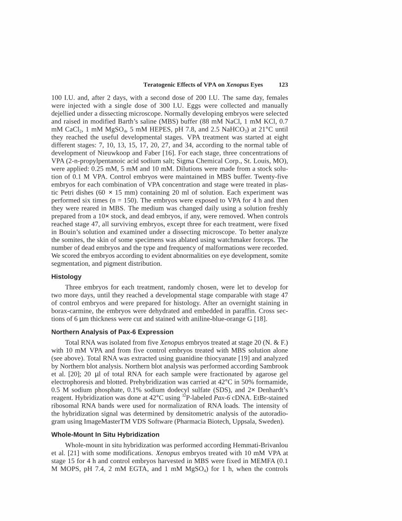
Treated embryos were first examined when controls reached stage 32. The onlyeffects of VPA exposure were the presence of edema in 50% of treated embryos anda developmental delay in 100% of treated embryos; the lethality of control and treatedembryos was similar, respectively 1% and 1.5%. The edema occurred at differentsites, principally in the head and in the heart region. “In vivo” observation of thelarvae was done daily until 6 days after fertilization, when controls reached stage 47.At this stage the edema and the developmental delay were more severe and almostall treated embryos showed anomalies in pigment distribution (Fig. 1B,C), indepen-dently from dose and stage of treatment. Frequently the anterior part of the embryoresulted shortened (Fig. 1B,C) and somites appeared duplicated or fused (Fig. 1E).Lethality, incidence of eye malformation and of abnormal somite segmentation showeda correlation with the stage of treatment.
Lethality
Data of lethality observed after 6 days from fertilization are reported in Figure2A. They were analyzed using an ANCOVA test. Lethality was significantly differ-ent in relation with the stage of treatment (F1=24.55; P<0.0001) and with the dose(F2=23.32; P<0.0001). The percentage of dead embryos for treatments at early blastulaand at late gastrula did not reach 5% for each concentration of VPA applied. Thelethality increased significantly when treatments started during neurulation stages(13, 15, 17; N. & F.). When treatments started at later stages, lethality decreasedgradually. Stage 15 resulted the more sensitive for each dose of VPA.
Incidence of Somite and Eye Malformations
The lowest dose that we tested that caused malformations in Xenopus embryoswas 0.25 mM.. The different concentrations caused the same type of malformations,but the severity of effects was dose dependent. We did not take into consideration thegravity of the effects when collecting the data but only the presence or absence ofabnormalities. Since the effects of 0.25 mM VPA were hardly detectable, we decidedto score only the embryos treated with the highest doses of VPA (5 mM and 10 mM).
We considered tail curling and back bending as symptoms of abnormalities in somitesegmentation. Data were analyzed using an ANCOVA test. The incidence of somite mal-
Teratogenic Effects of VPA on Xenopus Eyes 125
formations was significantly different in relation with the stage of treatment (F1=15.01;P<0.0001) and with the dose (F2=22.73; P=0.0002). Treatments started at blastula andgastrula stages (stage 7 and 10, N. & F.) caused somite abnormalities in a low percentageof embryos both with 5 mM and 10 mM VPA. But the incidence of somite malformationsincreased sharply in embryos treated at neurula stages (13, 15, and 17 N. & F.), reaching100% in treatment with 10 mM VPA at stage 15 (N. & F.). The percentage of somiteabnormalities decreased in embryos treated at post-neurula stages (20, 27, and 34, N. &F.) and the incidence was higher in embryos treated with 10 mM VPA (Fig. 2B).
Eyes were considered abnormal if they were different from controls in shape orin pigment distribution (data were analyzed using an ANCOVA test). The incidenceof eye malformations was significantly different in relation with the stage of treat-ment (F1=6.28; P=0.015) and with the dose (F2=33.92; P<0.0001). Exposure to VPAat stages 7 and 10 caused eye malformations in about 40% of survived embryostreated with both doses. When treatments started at neurula stages (13, 15, and 17,N. & F.) both the VPA doses caused eyes malformations in 100% of embryos. Theincidence of eye abnormalities decreased gradually in embryos treated after neuraltube closing (stages 20, 27, and 34, N. & F.): from 15% to 9% for 5 mM VPA andfrom 33% to 13% for 10 mM VPA (Fig. 2C).
Histological Analysis
In order to analyze the alterations of the tissues in the organs affected bymalformations caused by VPA, we performed a histological analysis of the em-
Fig. 1. External view of control and VPA-treated tadpoles. A: Control Xenopus laevis embryos atstage 47 (N. & F.). B,C: Embryos treated with 10 mM VPA at stage 15. B: Embryo displaying a severephenotype. Note the short and curled tail, the diffused edema, and small eyes. C: Embryo with a lesssevere phenotype. D: Detail of control embryo somites of the trunk region after skin ablation. E:Detail of malformed somites of a 10 mM VPA treated embryo. Scale bar = 500 µm.

126 Pennati et al.
Fig. 2. Lethality, eye, and somite malformations in embryos related to VPA doses and developmentalstages of treatment. Standard error is indicated.

Teratogenic Effects of VPA on Xenopus Eyes 127
bryos treated at stage 15 with 5 mM and 10 mM VPA and allowed to developuntil stage 47. All treated embryos were smaller in size than control embryos andsome of them showed edema below the epidermis that displaced connective tis-sue (Fig. 3C,4C). In treated embryos pronefric tubules had an enlarged lumenand were less convoluted than in controls; indeed in transverse sections of con-trol embryos the number of sections of tubules was higher (Fig. 3A,C).Chromatophora in treated embryos were less numerous and bigger than in con-trols. Moreover they showed an abnormal roundish shape and were abnormallydistributed; whereas in the control they formed continuum layers (Fig. 3A,B),they were absent or scarcely present in the peritoneum and in the dorsal surfaceof the CNS (Fig. 3C,D). Several treated embryos showed such a reduced adhe-sion among the cells of the somites that it was impossible to distinguish a sharpboundary between sclerotome and myotome. In the more affected embryos,somites appeared to be disorganized; they were formed by few cells with en-larged intracellular space between them (Fig. 3D).
Most of the embryos observed presented microphthalmia. Eye malformations
Fig. 3. Transverse sections at trunk level of Xenopus embryos at stage 47 (N. & F.). A,B: Controlembryo. C,D: 10 mM VPA treated embryo. ch, chromatophora; ed, edema; li, liver; nc, notochord; nt,neural tube; pt, pronephric tubules; so, somite. Scale bar = 100 µm.
128 Pennati et al.
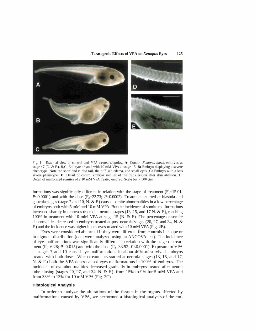
were always restricted to the optical cup and were due to an abnormal display ofneural retina and pigmented epithelium. Lens and optical nerve were always present,even if they often were smaller than in controls, perhaps as a consequence of thegeneral reduction of the size of eyes. Anterior and posterior chambers were reducedor absent. Mesodermic cells were infiltrated between the lens and the retina layers.In less severe malformed eyes, the layers of the retina were still recognizable even ifdisplaced (Fig. 4C,D); instead in the more severe affected eyes the internal organiza-tion of the retina was completely lost (Fig. 4F). In a high percentage of malformedeyes most of the tapetum nigrum was dislocated dorsally and the neural retina showednumerous folds (Fig. 4D,F). In the most drastic case, showed in Fig. 4E, one eyeprotruded into the neural tube floor.
Fig 4. Transverse sections at the eye level of Xenopus embryos at stage 47 (N. & F.). A,B: Control em-bryo. C,D,E,F: 10 mM VPA treated embryos. co, cornea; ed, edema; le, lens; nr, neural retina; nt, neuraltube; oc, optic cup; os, optic stalk; ph, pharynx; pr, pigmented layer of the retina. Scale bar = 100 µm.

Teratogenic Effects of VPA on Xenopus Eyes 129
Fig. 5. Northern blot analysis. A: Analysis of Pax-6 gene expression in VPA-treated embryos and incontrol embryos. B: EtBr staining of 18S and 28S rRNA.
Fig. 6. Xenopus embryos after in situ hybridization with Pax-6 . A: Control embryo showing a nor-mal pattern of Pax-6 expression. B: 10 mM VPA treated embryos showing a reduced expression ofPax-6 in eyes (arrow). C: 10 mM VPA treated embryo hybridized with sense probe. Scale bar = 1 mm.
130 Pennati et al.
The cartilage of the head resulted not fully developed. In all treated embryosCNS and notochord appeared normal in shape; dimension and consistence of theliver were similar to controls (Fig. 3C). We never observed necrotic tissue in any oftreated embryos.
Northern Blot Analysis
Northern blot analysis performed by total RNA isolated from VPA-treated em-bryos demonstrated a decrease in the level of Pax-6 expression compared to that incontrol embryos (Fig. 5). Densitometric estimation yield an approximately three-folddecrease in Pax-6 mRNA expression in the VPA-treated embryos.
Whole Mount In Situ Hybridization
Control embryos at stage 32 after in situ hybridization with Dig-RNA labeledPax-6 antisense probe showed the signal in the anterior CNS, with a gap in themesencephalon (optic tectum) and a more intense signal in the developing optic cup(Fig. 6A). In some embryos a weak non-specific staining of the pharyngeal pouchesappeared. Embryos treated at stage 15 with 10 mM VPA after in situ hybridizationshowed a faint signal in the optic cup and hardly visible staining in the nervoussystem (Fig. 6B). Treated embryos hybridized with sense probe showed no specificsignal (Fig. 6C). All the embryos of Figure 5 are of the same age, but treated em-bryos (B,C) frequently showed developmental delay.
DISCUSSION
In this study, we analyzed the effects of exposure to VPA on the developmentof Xenopus embryos. We observed that both timing and dosage of the drug alterthe effects of VPA treatment. The lowest concentration we tested that can inducemalformation was 0.25 mM, but the incidence and the severity of malformationswere very low (data not showed) and hard to score. The dosages used for most ofthese experiments were 5 mM and 10 mM, based on previous works on amphib-ians reported in literature [9]. Our results show that short exposures (4 h) to 5 mMand 10 mM VPA were sufficient to cause malformations in Xenopus embryos. Pulsetreatments for a short period allowed us to find the “window” of susceptibility tothe drug. The highest incidence of malformed embryos occurred, with both VPAdoses, for treatments started during neurulation, at stages 13, 15, and 17 (N. & F.).The percentage of malformed embryos increased sharply from treatments startedat stage 10 to treatments started at stage 13 and it decreased gradually from treat-ments started at stage 20. The same trend was observed for each analyzed malfor-mation. This suggests that in Xenopus embryos VPA perturbs processes active duringneurulation.
As reported in literature, we observed that VPA caused developmental retar-dation and, remarkably, we obtained this effect also with very short exposure tothe drug. Pulse treatments for 4 h were not sufficient to cause CNS malformations,which are the more frequent abnormalities reported in other species after longerexpositions [1,22,23]. Also the hepatotoxic effect described in amphibians [23] wasnot detectable after a short treatment, the liver in fact had always regular size anda normal tissue consistence. A high percentage of treated embryos showed edemain the head and in the heart region which can be a symptom of osmoregulation
Teratogenic Effects of VPA on Xenopus Eyes 131
defects. Treated embryos showed also pronefric tubules enlarged and less convo-luted then controls, compatible with malfunctioning.
Almost all treated embryos had anomalies in pigmentation, independently fromdose and stage of treatment: chromatophora were fewer than in controls and theyhad an abnormal shape and distribution. Krätke and Kirschbaum [23] reported thatVPA can affect migration or differentiation of neural crest cells. Since we observedanomalies also in other derivatives of these cells, such as the cartilage of the cranialregion, we can attribute the anomalies in pigmentation to a failure in neural crestmigration. Possibly short exposure to VPA may just retard the migration, which hap-pened after VPA removal from the medium. Some authors attributed the anomaliesof neural crest migration to the interaction of VPA with compounds of the extracel-lular matrix, such as fibronectin [24]. We considered that the influence of the drugon extracellular matrix compounds may also cause the reduced cellular adhesion thatwe observed in the somites.
Treated embryos showed disorganized somite, formed by few cells with en-larged intracellular space. These somite anomalies could be responsible for thedeformities of the tail and for back bending, and would possibly result in anoma-lies of the axial skeleton in older larvae, similar to those described in mammalsexposed to VPA gestationally [7,25]. These malformations may be correlated tothose caused by osteolathyrogenic substances [12], but we are not able to betterclarify this correlation.
Somite irregularities were also due to perturbation of the process of segmenta-tion, which lead to duplication or fusion. Irregular somite shape and distributionhave been observed also in mice and rats [7]. In chick embryos these defects wereattributed to the influence of VPA on the expression of Pax-1 gene, a member of thePax family, which is an important regulator of the axial patterning at the somite level[8]. In fact VPA treated chicken embryos showed somite anomalies and a correlatedsignificant decrease in Pax-1 expression, as observed by whole mount in situ hybrid-ization. Also in chicken the developmental stage at which VPA action is more pro-nounced corresponds to the stage of embryonic somite development, at which Pax-1expression reaches its peak, stage 15/16 H.H. [8].
We hypothesize that another member of the Pax family may be involved inthe eye malformations that we frequently observed in Xenopus embryos treatedwith VPA. At external examination the eyes appeared small and irregular in shape.The histological analysis revealed that the anomalies were principally due to anabnormal display of the layers of the retina: the neural retina appeared convolutedand presented numerous folds, the pigmented epithelium was often localized in adorsal position. These defects resemble those present in Pax-6 mutant mice [13].Pax-6 is a highly conserved gene, and it is necessary for correct retinal develop-ment in both vertebrates and invertebrates. It has been proposed that all the mem-bers of the Pax family of transcriptional factor are putative targets of theteratogenicity of VPA [8].
We analyzed by in situ hybridization the expression of Pax-6 in Xenopus em-bryos treated at stage 15 with 10 mM VPA. These embryos showed a lower signal ofhybridization in the eyes compared with controls. This result suggests that VPA hadaltered the pattern of expression of this gene and that Pax-6 is a target gene of VPAaction. We consider that decreased levels of Pax-6 expression are responsible of theretinal malformations we observed in VPA-treated Xenopus embryos.

132 Pennati et al.
CONCLUSIONS
In conclusion we found that short exposure to VPA during neurulation inducesin Xenopus embryos a high incidence of defects in neural crest migration and somitesegmentation, similar to those described in other species for longer expositions. Theseanomalies are consistent with an interaction of VPA with the compounds of the ex-tracellular matrix, as proposed by several authors. We also described a frequent eyeanomaly caused by VPA action, which had never been reported before. We considerthis anomaly as a result of decrease and of the alteration of Pax-6 gene expressionby the drug.
ACKNOWLEDGMENTS
The authors thank Dr. Thomas Hollemann and Dr. Thomas Pieler for providingPax-6 cDNA; Prof. Horst Grunz for providing histological protocols; Dr. Nicola Sainofor advice with statistical analysis; and Prof. Erminio Giavini for critical reading ofthe manuscript.
REFERENCES
1. Robert E, Guibaud P. Maternal Valproic Acid and congenital neural tube defects. Lancet 1982;2:937.2. Robert E, Rosa F. Valproate and birth defects. Lancet 1983;2:1142.3. Lindhout D, Meinhardi H. Spina bifida and in-utero exposure to valproate. Lancet 1984;2:396.4. Jager-Roman E, Deichl A, Jakob S, Hartmann AM, Koch S, Rating D, Steldinger R, Nau H, Helge
H. Fetal growth, major malformations, and minor anomalies in infants born to woman receivingvalproic acid. J Pediatr 1986;108:997–1004.
5. Lindhout D, Schmidt D. In utero exposure to valproate and neural tube defects. Lancet 1986;1:1392–1393.
6. Michejdia M, McCollough D. New animal model for the study of neural tube defects. Z Kinderchir1987;42:32–35.
7. Menegola E, Broccia ML, Nau H, Prati M, Ricolfi R, Giavini E. Teratogenic effects of sodiumvalproate in mice and rats at midgestation and at term. Teratogenesis Carcinogen Mutagen1996;16:97–108.
8. Barnes GL jr, Mariani BD, Tuan RS. Valproic Acid-induced somite teratogenesis in the chick em-bryo: relationship with Pax-1 gene expression. Teratology 1996;54:93–102.
9. Oberemm A, Kirschbaum F. Valproic acid induced abnormal development of the central nervoussystem of three species of amphibians: implications for neural tube defects and alternative experi-mental systems. Teratogenesis Carcinogen Mutagen 1992;12:251–262.
10. Naruse I, Collins MD, Scott WJ Jr. Stain differences in the teratogenicity induced by sodiumvalproate in cultured mouse embryos. Teratology 1988;38:87–96.
11. Hamburger V, Hamilton HL. A series of normal stages in development of chick embryo. J Morphol1951;88:49–67.
12. Dawson DA, Wilke TS. Initial evaluation of developmental malformation as an end point in mix-ture toxicity assessment for aquatic vertebrates. Ecotoxicol Environ Safety 1991;21:215–226.
13. Quiring R, Walldorf U, Kloter U, Gehring WJ. Homology of the eyeless gene of Drosophila to thesmall eye gene in mice and Aniridia in humans. Science 1994;265:785–789.
14. Hogan BLM, Hirst EMA, Horsburgh G, Hetherington MC. Small eye (Sey):a mouse model for thegenetic anlysis of craniofacial abnormalities. Development 1988;103:115–119.
15. Treisman J, Harris E, Desplan C. The paired box encodes a second DNA-binding domain in thepaired homeodomain protein. Genes Dev 1991;5:594–604.
16. Nieuwkoop PD, Faber J. Normal table of Xenopus laevis. Amsterdam: North Holland. 1956.17. Hirsch N, Harris WA. Xenopus Pax-6 and retinal development. J Neurobiol 1997;32:45–61.18. Grunz H. Change in the differentiation pattern of Xenopus laevis ectoderm by variation of the
incubation time and concentration of vegetalizing factor. Roux Arch Dev Biol 1983;192:130–137.
Teratogenic Effects of VPA on Xenopus Eyes 133
19. Chomcyzynski P, Sacchi N. Single-step method of RNA isolation by acid guanidinium thiocyan-ate-phenol-chloroform extraction. Anal Biochem 162:156–159.
20. Sambrook J, Fritsch EF, Maniatis T. Molecular cloning. Cold Spring Harbor, NY: Cold SpringHarbor Labs. 1982.
21. Hemmati-Brivanlou A, Frank D, Bolce ME, Brown Bd, Sive HL, Harland RM. Localization ofspecific mRNAs in Xenopus embryos by whole-mount in situ hybridization. Development1990;110:325–330.
22. Nau H. Transfer of valproic acid and its main active unsaturated metabolite to the gestationaltissue: correlation with neural tube defect formation in the mouse. Teratology 1986;33:21–27.
23. Krätke R, Kirschbaum F. Effects of the antiepileptic drug valproic acid on the development of theaxolotl (Ambystoma mexicanum): histological investigations. Teratogenesis Carcinogen Mutagen1996;16:149–167.
24. Gofflot F, van Maele-Fabry G, Picard JJ. Cranial nerves and ganglia are altered in vitro treatmentof mouse embryos with valproic acid (VPA) and 4-en-VPA. Dev Brain Res 1996;93:62–69.
25. Dansky LV, Finnell RH. Parental epilepsy, anticonvulsal drug and reproductive outcome: epide-miological and experimental findings spanning three decades. 2. Human studies. Reprod Toxicol1991;5:301–335.