actinomycetes and propionibacterium (those that form filaments)
TRANSCRIPT

Actinomycetes and Propionibacterium
(Those that form filaments)
Actinomycetes
Classification Order – Actinomycetales
Show fungus-like characteristics such as branching in tissues or in culture (look like mycelia).
The filaments frequently segment during growth to produce pleomorphic, diphtheroidal, or club shaped cells.
The cell wall and the internal structures are typical of bacteria rather than fungi.
Some are aerobic and others are anaerobic. All are slow growing

Actinomycetes
The anaerobic genera: Actinomyces, Arachnia, and Bifidobacterium
Morphology and cultural characteristics G+ branching, or diphtheroid-like bacilli Anaerobic and require CO2 for growth Non-sporing Will grow on anaerobic BA or PEA.
A. israelii, the most commonly isolated species, produces rough, granular colonies that resemble molars.
Biochemistry ID by gas liquid chromatography (GLC) of metabolic by-
products or fluorescent antibody studies

Actinomycetes
Clinical significance Are part of the NF found in the cavities of humans and
other animals. All may cause actinomycosis or “lumpy jaw” which is a
cervicofacial infection that used to occur following tooth extractions or dental surgery which provided traumatized tissue for growth of the microorganism which may also invade the bone.
This is rare today because of prophylactic antibiotic therapy.
May cause thoracic or abdominal infections May cause meningitis, endocarditis, or genital
infections

Actinomycetes
Every kind of infection is characterized by draining sinuses, usually containing characteristic granules which are colonies of bacteria that look like dense rosettes of club-shaped filaments in radial arrangement
Treatment Penicillin

Granules
Actinomycetes
The aerobic genera: Nocardia, Actinomadura, and Streptomyces. There are three clinically important species of Nocardia – N. asteroides, N. brasilensis, and N. caviae
Morphology and cultural characteristics G+ branching bacillus that may fragment to bacillary or
coccoid forms Aerobic Specimens should be inoculated onto 7H10 agar or
Lowenstein-Jensen agar and brain heart infusion agar. Colonies produced are typically orange, dry, crumbly,
and adherent. The organisms are weakly acid fast or non acid fast
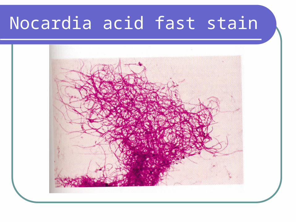
Nocardia acid fast stain

Actinomycetes
Biochemistry The organisms are identified based on sugar fermentations
and hydrolysis reactions (caseine, tyrosine, etc.) Clinical significance
Mycetoma – organism enters the body through breaks in the skin and causes a localized infection involving skin, cutaneous, and subcutaneous tissue.
The three most characteristic features seen are swelling, draining sinuses and granules.
This disease can also be caused by fungi as well as Nocardia, Actinomadura, and Streptomyces.

Actinomycetes
Nocardiosis – is a localized or disseminated disease occurring after inhalation of organisms.
Pulmonary infections resemble tuberculosis and can remain confined to the lungs or may disseminate, with a predilection for the brain and meninges.
The disease is characterized by multiple confluent abscesses and intense suppuration.
It is usually a disease of compromised hosts.Antimicrobic susceptibility/treatment
Mycetoma – aminoglycosides Nocardiosis – sulfonamides or sxt

Propionibacterium
Classification Two species P. acnes and P. granulosum. Are described as anaerobic diphtheroids, though
some can grow in CO2. Most clinical isolates are P. acnes which is part of
the NF of skin. Morphology and cultural characteristics
Pleomorphic, small G+B, may have Chinese letter configurations or may be branching.

Propionibacterium
Propionibacterium
Grow well on CBA, producing tiny translucent to opaque and white to gray colonies.
Growth may be slow. Anaerobic, though occasional strains of P.
granulosum grow in CO2
Biochemistry Catalase + Indole +/- Ferment glucose Produce caseinase

Propionibacterium
Virulance factors Protease
Clinical significance – Is part of skin NF Has been implicated in causing acne –
During adolescence more sebum is produced, and P. acnes metabolizes it to produce fatty acids.
These may contribute to the inflammatory response seen in acne.
Has also been isolated from joint infections
Propionibacterium
Antibiotic susceptibility/treatmentTetracyclineAcutane – inhibits sebum formation and is
only used in severe cases of acne because there are many side effects.