acquired ichthyosis revealing an underlying hodgkin´s … · acquired ichthyosis revealing an...
TRANSCRIPT
UHOD Number: 4 Volume: 20 Year: 2010 245
Acquired Ichthyosis Revealing an Underlying Hodgkin´s Lymphoma
Aristóteles ROSMANINHO, Ana OLIVEIRA, Inês LOBO, Madalena SANCHES, Manuela SELORES
Centro Hospitalar do Porto-HSA, Department of Dermatology, Porto, PORTUGAL
ABSTRACT
Acquired ichthyosis is a nonhereditary cutaneous disorder characterized by dry, rough skin with scaling that involves signi-ficant areas of the body. The disease most commonly manifests in adulthood and usually represents malignancy, especiallyhematologic disease. We report a 48-year-old woman who presented to the dermatology out-patient clinic because of se-vere scaling of the skin with a “dirty” appearance. The subsequent study revealed an underlying Hodgkin´s lymphoma.
Keywords: Acquired ichthyosis, Hodgkin´s disease, Lymphoma
ÖZET
Hodgkin Lenfoma Zemininde Akkiz ‹ktiyozis
Akkiz iktiyozis, vücudun büyük bölümünde pullanma, deride kuruluk ve kabalaflma ile karakterize nonherediter bir cilthastal›¤›d›r. Hastal›k, ço¤unlukla eriflkin yafl grubunda, özellikle hematolojik malignansilerin belirtisi olarak ortaya ç›kar. bizdermatoloji poliklini¤ine, ciltte “kirli” görünüm ve pullanma flikayeti ile baflvuran 48 yafl›ndaki bayan hastay› rapor ettik. Dahasonraki süreçte hastan›n Hodgkin lenfoma oldu¤u anlafl›ld›.
Anahtar Kelimeler: Akkiz iktiyozis, Hodgkin hastal›¤›, Lenfoma
ULUSLARARASı HEMATOLOJI-ONKOLOJI DERGISI CASE REPORT International Journal of Hematology and Oncology

INTRODUCTIONIchthyosis is a general term used to describe a largeand heterogeneous group of cornification disordersthat are genetic or acquired in nature and are cha-racterized clinically by the formation of visible sca-les on significant portions of the body and an exces-sively dry skin.1 In severe cases the skin resemblesfish scales ( “icthys”, which is fish from Greek) andthe patient complains about his “lizard skin”. Thecolor of the scales varies from white to gray tobrown and affects primary the trunk and limbs be-ing accentuated on the extensor surfaces and relati-vely sparing the flexures.2
Aquired ichtyosis can have a variety of underlyingetiologies, including neoplastic, infectious, drugs,endocrine, metabolic, autoimmune, and malabsorp-tive states.3-8
CASE REPORTA 48 year-old woman presented to our out-patientclinic with a diffuse symmetric scaling of the skinthat had appeared in the prior three months. Thedesquamation was more proeminent on the backand extensor surfaces of the limbs and spared theflexures. The scales were brown, fine, with curledup edges giving the skin a rough feel and a “dirty”appearance (Figure 1).
The clinical diagnosis of acquired ichthyosis wasmade. She complained about fatigue and weightloss in the last month and denied fever, night swe-ats or new drug intake.
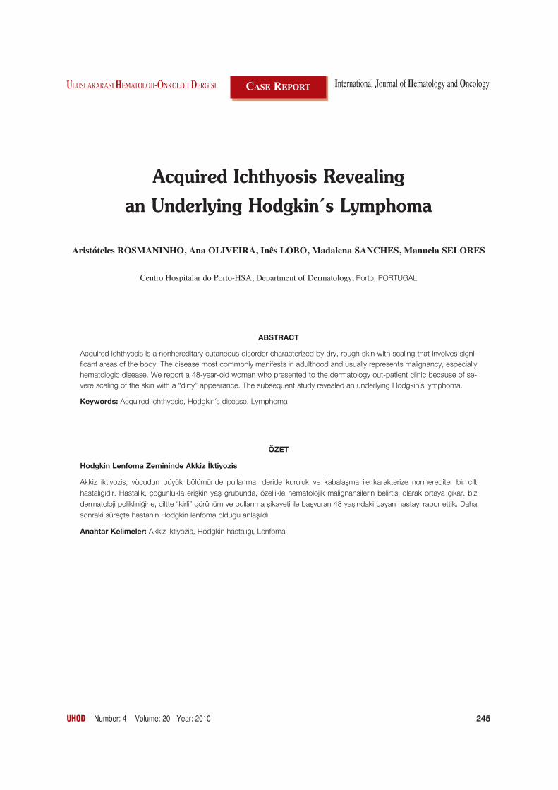
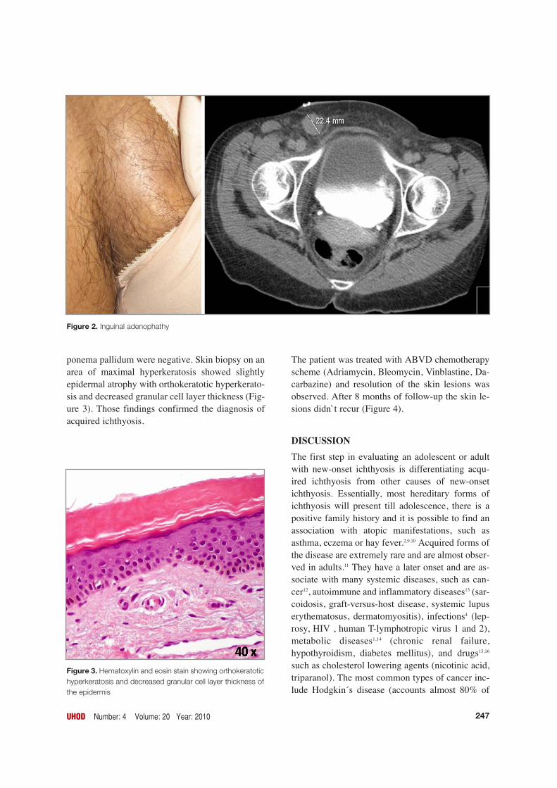
On physical examination a 3 cm right inguinal ade-nomegaly was palpable (Figure 2). Her prior medi-cal history was irrelevant.
Laboratory work-up showed an important inflam-matory syndrome with a C-reactive protein of 97mg/l (< 5), an erythrocyte sedimentation rate of 108mm/h (0-19), hemoglobin of 10.6 g/dl (11.5-16.5)and a α2-globulin of 22 g/l (3.6-13.2). Beta-2-mic-roglobulin was 2.3 mg/L (0-1.9). Electrolytes, renaland hepatic functions were normal.
Computed tomography of the chest, abdomen, andpelvis revealed multiple mediastinal, mesenteric,iliac, and inguinal lymphadenopathies. An open in-guinal lymph node biopsy was performed and thehematoxylin - eosin stain showed a neoplastic pro-liferation of Reed- Sternberg cells and a polymorp-hous cellular infiltration with eosinophils, neutrop-hils and plasma cells. Immunohistochemical sta-ining was positive for CD30, CD15, and negativefor LCA (leukocyte common antigen), CD3, andCD20. We diagnosed her as a mixed-cellularityHodgkin´s lymphoma. Bone marrow biopsy sho-wed hypoplasia with no other alterations. Serologi-cal tests for HIV, EBV, CMV, HBV, HCV, and tre-
246 UHOD Number: 4 Volume: 20 Year: 2010
Figure 1. Diffuse scaling with a “dirty” appearance.
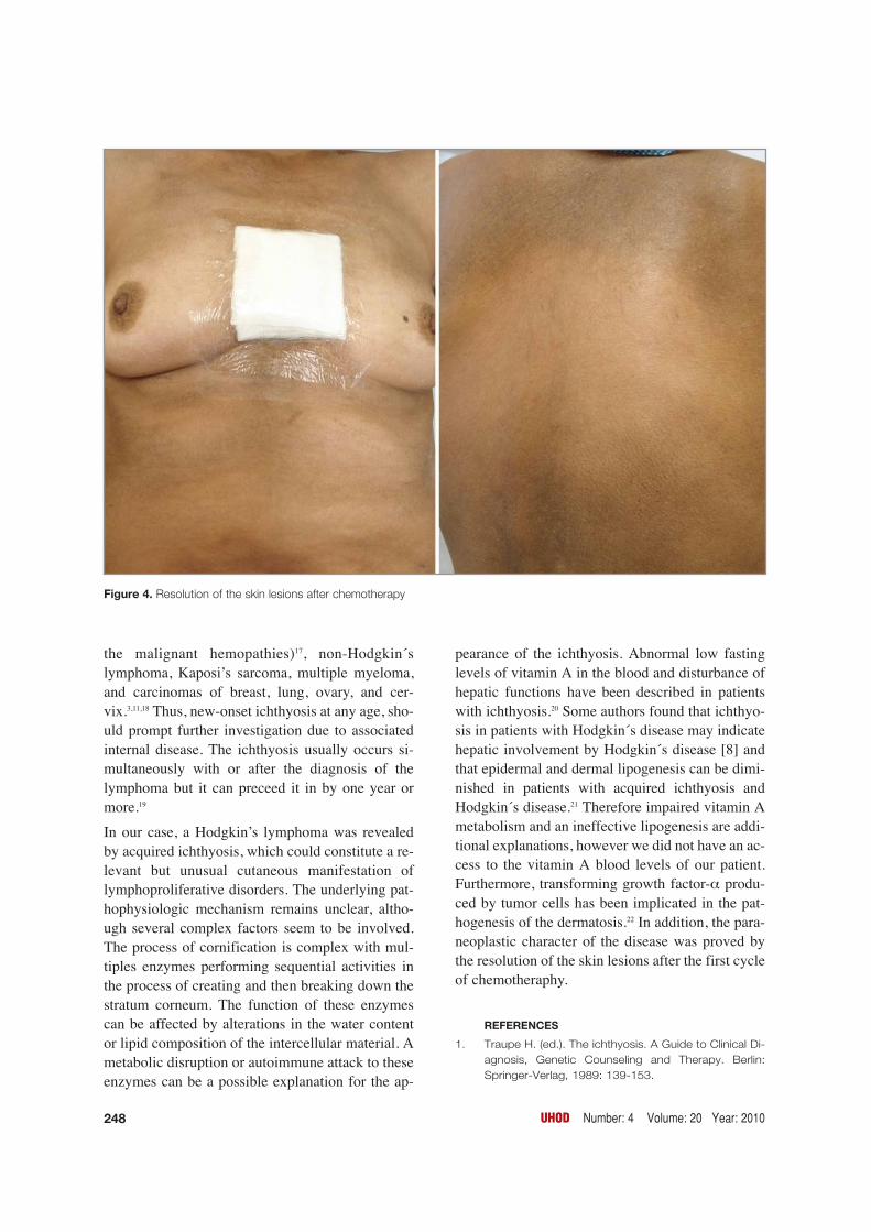
ponema pallidum were negative. Skin biopsy on anarea of maximal hyperkeratosis showed slightlyepidermal atrophy with orthokeratotic hyperkerato-sis and decreased granular cell layer thickness (Fig-ure 3). Those findings confirmed the diagnosis ofacquired ichthyosis.
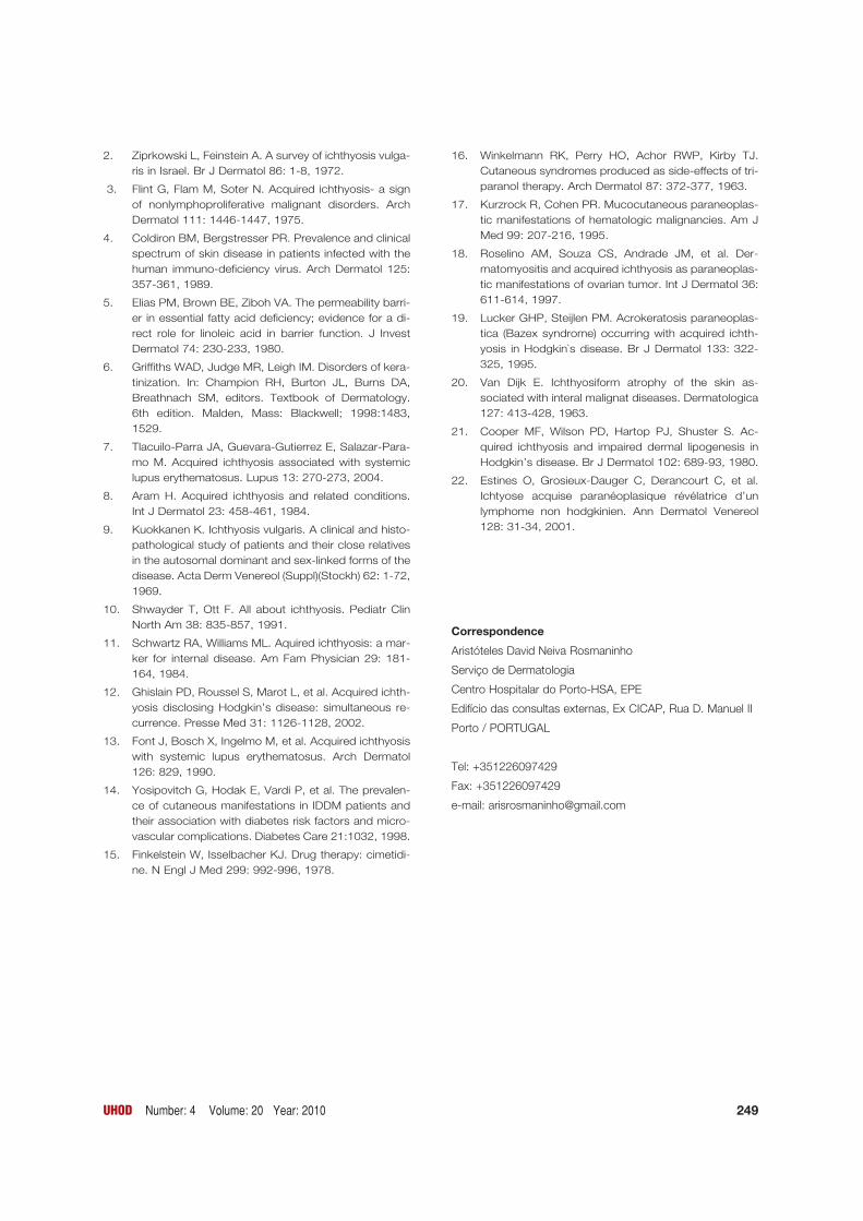
The patient was treated with ABVD chemotherapyscheme (Adriamycin, Bleomycin, Vinblastine, Da-carbazine) and resolution of the skin lesions wasobserved. After 8 months of follow-up the skin le-sions didn`t recur (Figure 4).
DISCUSSIONThe first step in evaluating an adolescent or adultwith new-onset ichthyosis is differentiating acqu-ired ichthyosis from other causes of new-onsetichthyosis. Essentially, most hereditary forms ofichthyosis will present till adolescence, there is apositive family history and it is possible to find anassociation with atopic manifestations, such asasthma, eczema or hay fever.2,9,10 Acquired forms ofthe disease are extremely rare and are almost obser-ved in adults.11 They have a later onset and are as-sociate with many systemic diseases, such as can-cer12, autoimmune and inflammatory diseases13 (sar-coidosis, graft-versus-host disease, systemic lupuserythematosus, dermatomyositis), infections4 (lep-rosy, HIV , human T-lymphotropic virus 1 and 2),metabolic diseases1,14 (chronic renal failure,hypothyroidism, diabetes mellitus), and drugs15,16
such as cholesterol lowering agents (nicotinic acid,triparanol). The most common types of cancer inc-lude Hodgkin´s disease (accounts almost 80% of
UHOD Number: 4 Volume: 20 Year: 2010 247
Figure 3. Hematoxylin and eosin stain showing orthokeratotichyperkeratosis and decreased granular cell layer thickness ofthe epidermis
Figure 2. Inguinal adenophathy
the malignant hemopathies)17, non-Hodgkin´slymphoma, Kaposi’s sarcoma, multiple myeloma,and carcinomas of breast, lung, ovary, and cer-vix.3,11,18 Thus, new-onset ichthyosis at any age, sho-uld prompt further investigation due to associatedinternal disease. The ichthyosis usually occurs si-multaneously with or after the diagnosis of thelymphoma but it can preceed it in by one year ormore.19
In our case, a Hodgkin’s lymphoma was revealedby acquired ichthyosis, which could constitute a re-levant but unusual cutaneous manifestation oflymphoproliferative disorders. The underlying pat-hophysiologic mechanism remains unclear, altho-ugh several complex factors seem to be involved.The process of cornification is complex with mul-tiples enzymes performing sequential activities inthe process of creating and then breaking down thestratum corneum. The function of these enzymescan be affected by alterations in the water contentor lipid composition of the intercellular material. Ametabolic disruption or autoimmune attack to theseenzymes can be a possible explanation for the ap-
pearance of the ichthyosis. Abnormal low fastinglevels of vitamin A in the blood and disturbance ofhepatic functions have been described in patientswith ichthyosis.20 Some authors found that ichthyo-sis in patients with Hodgkin´s disease may indicatehepatic involvement by Hodgkin´s disease [8] andthat epidermal and dermal lipogenesis can be dimi-nished in patients with acquired ichthyosis andHodgkin´s disease.21 Therefore impaired vitamin Ametabolism and an ineffective lipogenesis are addi-tional explanations, however we did not have an ac-cess to the vitamin A blood levels of our patient.Furthermore, transforming growth factor-α produ-ced by tumor cells has been implicated in the pat-hogenesis of the dermatosis.22 In addition, the para-neoplastic character of the disease was proved bythe resolution of the skin lesions after the first cycleof chemotheraphy.
REFERENCES
1. Traupe H. (ed.). The ichthyosis. A Guide to Clinical Di-agnosis, Genetic Counseling and Therapy. Berlin:Springer-Verlag, 1989: 139-153.
248 UHOD Number: 4 Volume: 20 Year: 2010
Figure 4. Resolution of the skin lesions after chemotherapy
2. Ziprkowski L, Feinstein A. A survey of ichthyosis vulga-ris in Israel. Br J Dermatol 86: 1-8, 1972.
3. Flint G, Flam M, Soter N. Acquired ichthyosis- a signof nonlymphoproliferative malignant disorders. ArchDermatol 111: 1446-1447, 1975.
4. Coldiron BM, Bergstresser PR. Prevalence and clinicalspectrum of skin disease in patients infected with thehuman immuno-deficiency virus. Arch Dermatol 125:357-361, 1989.
5. Elias PM, Brown BE, Ziboh VA. The permeability barri-er in essential fatty acid deficiency; evidence for a di-rect role for linoleic acid in barrier function. J InvestDermatol 74: 230-233, 1980.
6. Griffiths WAD, Judge MR, Leigh IM. Disorders of kera-tinization. In: Champion RH, Burton JL, Burns DA,Breathnach SM, editors. Textbook of Dermatology.6th edition. Malden, Mass: Blackwell; 1998:1483,1529.
7. Tlacuilo-Parra JA, Guevara-Gutierrez E, Salazar-Para-mo M. Acquired ichthyosis associated with systemiclupus erythematosus. Lupus 13: 270-273, 2004.
8. Aram H. Acquired ichthyosis and related conditions.Int J Dermatol 23: 458-461, 1984.
9. Kuokkanen K. Ichthyosis vulgaris. A clinical and histo-pathological study of patients and their close relativesin the autosomal dominant and sex-linked forms of thedisease. Acta Derm Venereol (Suppl)(Stockh) 62: 1-72,1969.
10. Shwayder T, Ott F. All about ichthyosis. Pediatr ClinNorth Am 38: 835-857, 1991.
11. Schwartz RA, Williams ML. Aquired ichthyosis: a mar-ker for internal disease. Am Fam Physician 29: 181-164, 1984.
12. Ghislain PD, Roussel S, Marot L, et al. Acquired ichth-yosis disclosing Hodgkin’s disease: simultaneous re-currence. Presse Med 31: 1126-1128, 2002.
13. Font J, Bosch X, Ingelmo M, et al. Acquired ichthyosiswith systemic lupus erythematosus. Arch Dermatol126: 829, 1990.
14. Yosipovitch G, Hodak E, Vardi P, et al. The prevalen-ce of cutaneous manifestations in IDDM patients andtheir association with diabetes risk factors and micro-vascular complications. Diabetes Care 21:1032, 1998.
15. Finkelstein W, Isselbacher KJ. Drug therapy: cimetidi-ne. N Engl J Med 299: 992-996, 1978.
16. Winkelmann RK, Perry HO, Achor RWP, Kirby TJ.Cutaneous syndromes produced as side-effects of tri-paranol therapy. Arch Dermatol 87: 372-377, 1963.
17. Kurzrock R, Cohen PR. Mucocutaneous paraneoplas-tic manifestations of hematologic malignancies. Am JMed 99: 207-216, 1995.
18. Roselino AM, Souza CS, Andrade JM, et al. Der-matomyositis and acquired ichthyosis as paraneoplas-tic manifestations of ovarian tumor. Int J Dermatol 36:611-614, 1997.
19. Lucker GHP, Steijlen PM. Acrokeratosis paraneoplas-tica (Bazex syndrome) occurring with acquired ichth-yosis in Hodgkin`s disease. Br J Dermatol 133: 322-325, 1995.
20. Van Dijk E. Ichthyosiform atrophy of the skin as-sociated with interal malignat diseases. Dermatologica127: 413-428, 1963.
21. Cooper MF, Wilson PD, Hartop PJ, Shuster S. Ac-quired ichthyosis and impaired dermal lipogenesis inHodgkin’s disease. Br J Dermatol 102: 689-93, 1980.
22. Estines O, Grosieux-Dauger C, Derancourt C, et al.Ichtyose acquise paranéoplasique révélatrice d’unlymphome non hodgkinien. Ann Dermatol Venereol128: 31-34, 2001.
Correspondence
Aristóteles David Neiva Rosmaninho
Serviço de Dermatologia
Centro Hospitalar do Porto-HSA, EPE
Edifício das consultas externas, Ex CICAP, Rua D. Manuel II
Porto / PORTUGAL
Tel: +351226097429
Fax: +351226097429
e-mail: [email protected]
UHOD Number: 4 Volume: 20 Year: 2010 249