acantholytic squamous cell carcinoma: pathological study ... · acantholytic squamous cell...
TRANSCRIPT

Rom J Morphol Embryol 2014, 55(2):279–283
ISSN (print) 1220–0522 ISSN (on-line) 2066–8279
OORRIIGGIINNAALL PPAAPPEERR
Acantholytic squamous cell carcinoma: pathological study of nine cases with review of literature
MARIA SAJIN1), ALINA HODOROGEA PRISĂCARU2), MIHAELA CRISTINA LUCHIAN2), OANA MARIA PĂTRAŞCU2), ADRIAN DUMITRU2), DIANA COSTACHE3), DOINA DUMITRESCU4), DANIELA VRÎNCEANU5), LILIANA MARY VOINEA6), OLGA SIMIONESCU7), MARIANA COSTACHE1)
1)Department of Pathology, “Carol Davila” University of Medicine and Pharmacy, Bucharest, Romania 2)Department of Pathology, Emergency University Hospital, Bucharest, Romania 3)Medical student, “Carol Davila” University of Medicine and Pharmacy, Bucharest, Romania 4)Department of Plastic Surgery, “Carol Davila” University of Medicine and Pharmacy, Bucharest, Romania 5)Department of ENT, Emergency University Hospital, Bucharest, Romania 6)Department of Ophthalmology, “Carol Davila” University of Medicine and Pharmacy, Bucharest, Romania 7)Department of Dermatology, “Carol Davila” University of Medicine and Pharmacy, Bucharest, Romania
Abstract Squamous cell carcinoma (SCC) is classified in many subtypes or forms; one of them is the acantholytic squamous cell carcinoma, also called pseudoglandular, adenoid, epithelioma dyskeratoticum segregans, or adenoacanthoma. Researching and analyzing nine cases of acantholytic squamous cell carcinoma, we intend to verify if the data provided by the cases studied can be validated by the scientific literature. All the cases presented lesions found on the head and neck skin, with two exceptions – one on the larynx and the other one on the tonsil, all of them ulcerated lesions. In two cases, the tumors developed on the skin, in preneoplasic lesions (actinic keratosis). The tumors had dimensions between 4/3/4 mm and 100/90/36 mm. During one year, two of the cases studied presented multiple recurrences. We also found two cases of metatypical carcinoma accompanied the acantholytic variant of squamous cell carcinoma. None of the analyzed cases presented distant metastasis. The histopathological criteria for selection were: keratinised squamous tumor cell type, adenoid structures with round spaces with a defined wall of at least one cell width, spaces with isolated or grouped dyskeratotic acantholytic cells.
Keywords: acantholytic squamous cell carcinoma, adenoid structures, immunohistochemistry, metatypical carcinoma, actinic keratosis features.
Introduction
Initially described in 1947 by Lever, the acantholytic squamous cell carcinoma (ASCC) represents a malignant epithelial tumor with gland-like and solid pattern, with extension into the dermis, also named as adenoacanthoma of sweat glands [1, 2]. Almost two decades later, Müller writes about the tumor and its microscopic details (connection with epidermis, presence of pseudoglandular elements, dyskeratosis, acantholysis, atypical mitosis, multiple aspects of cell morphology, perineural extension [3]. After that, it has been regarded as a rare variant of squamous cell carcinoma, also being named as epithelioma dyskeratoticum segregans, adenoid squamous cell carci-noma, acantholytic squamous cell carcinoma, pseudo-glandular squamous cell carcinoma. However, the most frequent locations of the tumor are the sun exposed ones, the tumor usually develops on actinic keratosis (having an increased incidence in elderly) but also, many different sites have been reported [4]. The tumor has distinct histology comparative with classic squamous cell carcinoma (SCC), being different interpreted by several authors regarded its aggressivity [5].
In this article, we choose to present nine cases of acantholytic squamous cell carcinoma driven by the limited number of cases existing in literature.
Materials and Methods
We collected pathology reports (age, gender, location of tumor, macroscopic and microscopic description) for acantholytic squamous cell carcinoma from the Archive of Laboratory of Pathology, Emergency University Hospital of Bucharest, Romania, starting with 2010 until 2012. The reports mentioned above were collected from patients in an age range between 56 and 89 years, with 5:4 gender ratio (man/woman), most of them with cutaneous lesions with two exceptions, namely a laryngeal and a tonsil lesion and they were directed for treatment to the Departments of Plastic Surgery, ENT or Ophthalmology, depending on the therapy required. After the surgical removal of the tumors, the tissue fragments were sent for processing and examination to our Department of Pathology. Samples of tissue were fixed with 10% buffered formalin and sent for the histopathological processing by conventional method using paraffin inclusion and Hematoxylin-Eosin (HE) staining. Also, immunohisto-chemical tests were performed. The paraffin blocks acquired by histopathological processing were sectioned at microtome resulting 3-μm thickness sections mounted on slides cover with poly-L-Lysine. After that, the sections were deparaffinized in toluene and alcohol successive baths, one hour, 15 minutes by bath, rehydration (three
R J M ERomanian Journal of
Morphology & Embryologyhttp://www.rjme.ro/

Maria Sajin et al.
280
successive alcohol baths with decreased concentration: 96%, 80% and 70%, 10 minutes in each bath and followed by a bath with distillated water, were the sections were hold for 10 minutes). Washing in PBS (phosphate saline buffer), incubation with normal serum, for 20 minutes, incubation with primary antibody overnight, Dako LSAB kit, washing in carbonate buffer and development in 3,3’-diaminobenzidine (DAB) hydrochloride/hydrogen peroxide and nuclear counterstaining with Mayer’s Hematoxylin. We used the following antibodies from NeoMarkers LabVision: PanCytokeratin, clone AE1/AE3 (Thermo Fisher Scientific Inc., USA, 1:100 dilution), Cytokeratin 5/6, clone D5/16 B4 (Thermo Fisher Scientific Inc., USA, 1:20 dilution), P53 protein, clone DO-7 (Thermo Fisher Scientific Inc., USA, 1:200 dilution), Vimentin, clone V9 (Thermo Fisher Scientific Inc., USA, 1:200 dilution), High Molecular Weight Cytokeratin, clone 34βE12 (Thermo Fisher Scientific Inc., USA, 1:50 dilution), Epithelial Membrane Antigen (EMA), clone E29 (Thermo Fisher Scientific Inc., USA, 1:50 dilution). The immuno-reactive cells from each cases were evaluate as follow: diffuse positive, >75% positive cells; positive, 25–75% positive cells; focal positive, <25% positive cells and negative cells.
Results
Our first case is a 70-year-old woman, presenting herself at our hospital in December 2010, with a giant ulcerated tumor on her parieto-frontal skin with a six months accelerated evolution. Macroscopically, the tumor was pedunculated, with increase consistency, lobulated surface, grey colored, of 100/90/36 mm size. Microsco-pically, we found ulcerated and infected proliferations of squamous cells characteristic of acantholytic squamous cell carcinoma and infiltrating the resection border. The same patient returns in May and after that in December 2011 at our hospital with recurrences of the tumor in medio-frontal and pre-auricular areas. Macroscopically, the tumors with sizes between 18/16/4 and 25/10/3 mm had similar appearances and microscopic diagnosis as the first tumor.
The second case is one of a 68-year-old man hospitalized for laryngeal tumor in October 2010. The tissue fragments we received were five lymphonodules and one portion of the larynx. Macroscopically, the larynx tissue had an ulceration with 60 mm width and a tumor of 13 mm width. Microscopically, the lymphonodules were hyperplasiated with sinus hystiocitosis; the lesion presented various islets of epithelial cells with high pleomorphism and frequent mitotic figures, keratotic changes and pseudoglandular pattern, therefore, the tumor was diagnosed as an acantholytic squamous cell carcinoma.
Third case represents an 84-year-old man with a history of recurrent squamous cell carcinoma of the nasal pyramid coming to our hospital for the third time, in October 2012. The macroscopic appearance was of small fragments with grey-dark color and the microscopic images showed epithelial cells in an adenoid pattern characteristic of acantholytic squamous cell carcinoma.
Case No. 4 represents a 90-year-old woman with general carcinomatosis of the cervico-frontal region admitted in our hospital in January 2011. We received
two cutaneous fragments, one with an excavated lesion and the other one with irregular borders. Both tumors were acantholytic squamous cell carcinoma; the first one had infiltrated surgical borders.
Case No. 5 is a 71-year-old man with a right tonsil tumor that we diagnosed in September 2011 with acantho-lytic squamous cell carcinoma based on the specific features for this pathology.
Case No. 6 is an 86-year-old woman with an eyelid tumor admitted in our hospital in November 2011. Macroscopically, there were two cutaneous fragments, both with a central lesion (the first one had 5 mm and the other one had 10 mm width). Microscopic diagnosis revealed actinic keratosis, acantholytic squamous cell carcinoma and fragments of a metatypical carcinoma with areas of basocellular carcinoma.
The seventh case is one of an 88-year-old man hospitalized in July 2012 for multiple tumors of the face. Macroscopically, there were three tissue fragments with irregular surface, one exophytic, sessile tumor and one cutaneous fragment with an exophytic tumor with 11/5/3 mm dimensions. Microscopically, we diagnosed a metatypical carcinoma with areas of ulcerated and infected basocellular carcinoma, a squamous cell carcinoma totally removed and an acantholytic squamous cell carcinoma, also been totally removed.
The eighth case presents the “youngest” patient, 56-year-old, admitted for an ulcerated, bleeding tumor at the base of the nose. The cutaneous fragment showed a slightly elevated tumor of 4/3 mm size. We diagnosed it as adenoid squamous cell carcinoma. The tumor was entirely removed.
Our last case is the one of a 75-year-old woman with an ulcerated, bleeding lip tumor, with two years of evolution, admitted in May 2012. The macroscopic appearance showed an exophytic tumor of 21/14/16 mm diagnosed microscopically as an ulcerated acantholytic squamous cell carcinoma. The tumor was totally removed.
Microscopic features of the cases
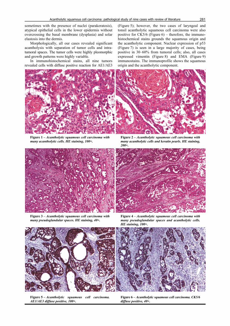
Microscopic features for diagnosing acantholytic squamous cell carcinoma of all nine cases included proli-feration of epithelial cells, islands or solid growth pattern, occasionally with central keratinized pearls (Figures 1 and 2), having the particularity of loosen cohesivity and adhesion between these cells. Tubular structures (Figures 3 and 4) containing acantholytic epithelial cells (round or polygonal in shape and with glassy eosinophilic cytoplasm) and necrotic debris were a common finding.
The tumor cells showed high nuclear pleomorphism. The nuclei varied from hyperchromatic and more or less enlarged to large and/or multinucleated nuclei with open and fine chromatin. Mitotic figures were not uncommon. The tumor cells also revealed significant cytoplasm variations. Some acantholytic tumor cells showed prominent dys-keratosis with dense eosinophilic cytoplasm (Figures 1 and 2). In some tumors, there were dispersed large cells containing large amounts of deeply eosinophilic material and peripheral vesicular nuclei mimicking high-grade sarcoma. Some dyscohesive tumor cells were also relatively small with little cytoplasm as shown in Figure 2.
Actinic keratosis features were also found in two cases: increased amount of keratin (hyperkeratosis),
Acantholytic squamous cell carcinoma: pathological study of nine cases with review of literature
281
sometimes with the presence of nuclei (parakeratosis), atypical epithelial cells in the lower epidermis without overcrossing the basal membrane (dysplasia) and solar elastosis into the dermis.
Morphologically, all our cases revealed significant acantholysis with separation of tumor cells and intra-tumoral spaces. The tumor cells were highly pleomorphic and growth patterns were highly variable.
In immunohistochemical stains, all nine tumors revealed cells with diffuse positive reaction for AE1/AE3
(Figure 5); however, the two cases of laryngeal and tonsil acantholytic squamous cell carcinoma were also positive for CK5/6 (Figure 6) – therefore, the immuno-histochemical stains grounds the squamous origin and the acantholytic component. Nuclear expression of p53 (Figure 7) is seen in a large majority of cases, being positive in 30–60% from tumoral cells; also, all cases expressed vimentin (Figure 8) and EMA (Figure 9) immunostains. The immunoprofile shows the squamous origin and the acantholytic component.
Figure 1 – Acantholytic squamous cell carcinoma with many acantholytic cells. HE staining, 100×.
Figure 2 – Acantholytic squamous cell carcinoma with many acantholytic cells and keratin pearls. HE staining, 200×.
Figure 3 – Acantholytic squamous cell carcinoma with many pseudoglandular spaces. HE staining, 40×.
Figure 4 – Acantholytic squamous cell carcinoma with many pseudoglandular spaces and acantholytic cells. HE staining, 100×.
Figure 5 – Acantholytic squamous cell carcinoma. AE1/AE3 diffuse positive, 100×.
Figure 6 – Acantholytic squamous cell carcinoma. CK5/6 diffuse positive, 40×.

Maria Sajin et al.
282
Figure 7 – Acantholytic squamous cell carcinoma. P53 positive, 40×.
Figure 8 – Acantholytic squamous cell carcinoma. Vimentin positive, 100×.
Figure 9 – Acantholytic squamous cell carcinoma. EMA positive, 40×.
Discussion
ASCC represents 2–4% of all cutaneous squamous cell carcinoma, developing mainly on elderly persons, as it can be seen in our study too, with a male predominance. It conserves the same risk factors as in the category it belongs, such as ultraviolet lights and radiation therapy and for the oral, pharyngeal and larynx location, alcohol and smoking.
Clinically, ASCC appears as an ulcerated papula with a slow growing pattern. The lesions typically arise on the head and neck as a nodule or ulcer on sun-exposed skin, particularly on and around the ears and face, de novo or may develop from an actinic keratosis, but other unusual sites have been described: larynx [6], cecum [7], gingival [8], breast [9], penis [10], vulva [11], uterine cervix [12], conjunctiva [13].
Macroscopically, the lesion appears as a tumor (nodule) consisting of thickened and/or ulcerated epithelium, usually larger than 15 mm. Microscopically, we can identify from the surface in depth, the epidermis which can be thickened, atrophic or ulcerated, but also it presents several tumor islands infiltrating the subjacent dermis; deeper, the tumor is arranged in cords and islands in a glandular (pseudo-glandular) or adenoid pattern, polygonal cells surround lumen-like spaces containing free-floating, neoplastic keratinocytes (desquamated acantholytic cells, many of which are partially or fully keratinized) [14].
At high-power field, we can identify the malignant criteria of cells: voluminous, hyperchromatic and pleo-morphic nuclei presenting atypical mitosis. We can also find in the extremity of the tumor some dilated and proli-ferated eccrine ducts, most probably due to surrounding inflammatory infiltrate, composed mainly of lymphocytes.
As regards the age of the patients, the youngest patient was 56-year-old and the oldest was 90-year-old. The sizes range from 5 mm to 100 mm width. Evolution period, were known, stretched from six months to two years. The male predominance could not be demonstrated since in our study there were five males and four women.
Morphologically, all our cases revealed significant acantholysis with separation of tumor cells and intra-tumoral spaces. The tumor cells were highly pleomorphic and growth patterns were highly variable. Two tumors emerged on actinic keratosis and other two developed on unusual regions-larynx and tonsil region. Metatypical carcinoma with basocellular features was also seen in two cases. The large majority of the tumors were ulcerated and infected; some of them were completely removed whereas others had infiltrating borders. In immunohisto-chemical stains, all tumor cells revealed positive reactions for AE1/AE3 and p53 supporting a squamous epithelial origin, as well as for HMWKs.
In contrast to conventional and aero-digestive squamous cell carcinoma, acantholytic squamous cell carcinoma indicates significant reductions of cytokeratins 5/6 and 19 as showed by our two cases of laryngeal and tonsil acantholytic squamous cell carcinoma. Also, E-cadherin and concomitant up-regulation of vimentin demonstrate a diminished expression. Both morphologic features and immunohistochemical profiles indicate that acantholytic squamous cell carcinoma has acquired an epithelial mesenchymal transition phenotype [15]. However, in contrast to other solid malignant tumors, the epithelial mesenchymal transition phenotype change in acantholytic squamous cell carcinoma is not limited to the invasive front of the peripheral tumor but, rather, diffusely involves entire tumoral lesion.
In addition, because cytokeratin immunostaining is attenuated, this would be an insensitive marker for following up or detecting disseminated neoplastic malignant cells in cases of acantholytic squamous cell carcinoma of the upper aerodigestive tract. We also have to

Acantholytic squamous cell carcinoma: pathological study of nine cases with review of literature
283
differentiate acantholytic squamous cell carcinoma from primary or metastatic adenocarcinoma. In adenocarcinoma, specialized glandular cells are arranged on the basal membrane in multiple layers of cuboidal cells with malignant criteria, forming lumens of glands. The immunohistochemical and histochemical findings of the present cases were compatible with the previous data of ASCC and exclude possibility of adenocarcinoma or adenosquamous carcinoma.
The treatment for ASCC is usually the same as for conventional SCC (surgical excision, which may be followed by skin graft for deep invaded skin tumors). It has a poor prognosis and its behavior is depth-dependent [16]. Regarding our cases, the poor prognosis is highlighted by the recurrence risks observed in three patients and the high number of tumors excised together. Furthermore, one case presented general carcinomatosis whereas two cases had more fragments excised and sent to Department of Pathology. However, no metastases were found.
Conclusions
ASCC is a rare histopathological variety of spino-cellular carcinoma, which frequently develops on sun-exposed regions of skin, usually at elderly on preexisting lesions such as solar keratosis. Expression of CK protein in ASCC are significantly down regulated with concurrent up-regulation of vimentin, advocates for the activation of epithelial mesenchymal transition in relation with pathways in carcinogenesis and tumor progression. There are still discussions between several authors, regarding its aggressivity; some say that the tumor has a well-known aggressivity and many other consider that the tumor has a low aggressivity based on direct observation of clinical evolution after surgical excision of the tumor. Reports of more cases of ASCC not only cutaneous and in the upper digestive tract, but also in uncommon sites would possibly help to elucidate the origin, clinical behavior of these rare tumors.
References [1] Boyd AS, Tumors of the epidermis. In: Barnhill LR,
Crowson AN, Magro C, Piepkorn M (eds), Dermatopathology, 3rd edition, McGraw–Hill Professional, New York, 2010, 593–594.
[2] Kirkham N, Tumors and cysts of the epidermis. In: Elder DE, Elenitsas R, Johnson BL Jr., Murphy GE, Xu G (eds), Lever’s histopathology of the skin, 10th edition, Lippincott Williams & Wilkins, 2009, 817–821.
[3] Bren T, McKee PH, Tumors of the surface epithelium. In: McKee PH, Calonje JE, Granter SR (eds), Pathology of the skin with clinical correlations, 3rd edition, Elsevier–Mosby, 2005, 1214–1216.
[4] Rinker MH, Fenske NA, Scalf LA, Glass LF, Histologic variants of squamous cell carcinoma of the skin, Cancer Control, 2001, 8(4):354–363.
[5] Müller SA, Wilhelmj CM Jr, Harrison EG Jr, Winkelmann RK, Adenoid squamous cell carcinoma (adenoacanthoma of lever). Report of seven cases and review, Arch Dermatol, 1964, 89:589–597.
[6] González-Vela MC, Val-Bernal JF, Mayorga M, Zaldumbide L, Báscones M, Adenoid squamous cell carcinoma of the larynx: an uncommon histological variant of squamous cell carcinoma, APMIS, 2006, 114(6):470–473.
[7] Jukić Z, Ledinsky I, Ulamec M, Ledinsky M, Krušlin B, Tomas D, Primary acantholytic squamous cell carcinoma of the cecum: a case report, Diagn Pathol, 2011, 6:5.
[8] Papadopoulou E, Tosios KI, Nikitakis N, Papadogeorgakis N, Sklavounou-Andrikopoulou A, Acantholytic squamous cell carcinoma of the gingiva: report of a case and review of the literature, Oral Surg Oral Med Oral Pathol Oral Radiol Endod, 2010, 109(6):e67–e71.
[9] Kamra H, Gadgil P, Chaware S, Kanade U, Acantholytic variant of squamous cell carcinoma of breast: a rare case report, Ecancermedicalscience, 2011, 5:214.
[10] Zámecník M, Mukensnabl P, Chlumská A, Pseudoglandular (adenoid, acantholytic) squamous cell carcinoma of the penis. A case report, Cesk Patol, 2011, 47(1):15–18.
[11] Horn LC, Liebert UG, Edelmann J, Höckel M, Einenkel J, Adenoid squamous carcinoma (pseudoangiosarcomatous carcinoma) of the vulva: a rare but highly aggressive variant of squamous cell carcinoma-report of a case and review of the literature, Int J Gynecol Pathol, 2008, 27(2):288–291.
[12] Aho HJ, Talve L, Mäenpää J, Acantholytic squamous cell carcinoma of the uterine cervix with amyloid deposition, Int J Gynecol Pathol, 1992, 11(2):150–155.
[13] Mauriello JA Jr, Abdelsalam A, McLean IW, AdeNOid squamous carcinoma of the conjunctiva – a clinicopathological study of 14 cases, Br J Ophthalmol, 1997, 81(11):1001–1005.
[14] Weedon D, Morgan MB, Gross C, Nagore E, Yu LL, Acantholytic squamous cell carcinoma. In: LeBoit PE, Burg G, Weedon D, Sarasin A (eds), Pathology and genetics of skin tumours, World Health Organization Classification of Tumours, 1st edition, IARC Press, Lyon, 2006, 21.
[15] Gu X, Jiang R, Fowler MR, Acantholytic squamous cell carci-noma in upper aerodigestive tract: histopathology, immuno-histochemical profile and epithelial mesenchymal transition phenotype change, Head Neck Pathol, 2012, 6(4):438–444.
[16] Garcia C, Crowson AN, Acantholytic squamous cell carcinoma: is it really a more-aggressive tumor? Dermatol Surg, 2011, 37(3):353–356.
Corresponding author Mariana Costache, Associate Professor, MD, PhD, Department of Pathology, “Carol Davila” University of Medicine and Pharmacy, 8 Eroilor Sanitari Avenue, 050474 Bucharest, Romania; Phone/Fax +4021–318 07 62, e-mail: [email protected] Received: November 21, 2013
Accepted: June 5, 2014