abstracts poster presentations (b)
Post on 02-Jul-2016
216 views
TRANSCRIPT
Journal of Inorganic Biochemistry 86 (2001) 131
Hypochlorous acid and myeloperoxidase induced alterations in phospholipids
Juergen A m h o l d a, A n a t o l y N. Os i pov b, Ho l g e r Spal teholz a, Oleg M. Panasenko c, Juergen Schil ler a
~Institute of Medical Physics and Biophysics, University of Leipzig, Liebigstr. 27, 04103 Leipzig, GERMANY (e-mail. arnj@medizin, un i-leipzig, de) bDepartment of Biophysics, Russian State Medical University, ul. Ostrovitjanova 1, 117437
Moscow, RUSSIA CResearch Institute of Physico-Chemical Medicine, ul. M. Pirogovskaya la, 119828 Moscow,
R USSIA
Interaction of exogenous hypochlorous acid as well as hypochlorous acid produced by the myeloperoxidase- hydrogen peroxide-chloride system with liposomes composed of unsaturated phosphatidylcholines was investigated by means of matrix-assisted laser desorption/ionisation time-of-flight mass spectrometry (MALDI-TOF-MS). The formation of chlorohydrins and glycols was detected according to their characteristic shifts in mass spectrum. Mainly chlorohydrins resulted upon incubation of HOC1/OC1 with phosphocholines containing oleoyl residues. The formation of glycols was only observed using phospholipids with fatty acid residues containing two or more double bonds. Lysophospholipids and lipids with fragmented fatty acids chains were additionally detected as reaction products of hypochlorous acid especially using highly unsaturated phosphocholines. The formation of lyso components was also verified by 31p NMR measurements. Bis-chlorine derivatives ofphosphocholines were found as products at high chloride concentrations (up to 3 tool/l) in the reaction buffer. Mono-chlorohydrins of 1-stearoyl-2-oleoyl-sn-glycero-3-phosphocholine were detected after the incubation of the latter phospholipid with the myeloperoxidase-hydrogen peroxide-chloride system. These chlorohydrins were not observed in the absence of chloride, hydrogen peroxide or myeloperoxidase as well as in the presence of methionine, taurine, or sodium azide.
The German Research Foundation is acknowledged for financial support.
Kinetics and mechanism of bond rearrangements in isomeric cis-[Pt(NH3)2-bis(9- methyladenine)] complexes
J o r m a Arpalaht i , Kare l D. Klika, Tar ja Ta lonen
Department of Chemistry, University of Turku, FIN-2OO14. Turku, Finland (e-mail: [email protected])
The interactions of Pt(II) with the base residues in DNA are crucial to the understanding of the biological activity of various anticancer Pt drugs. It is widely accepted that, once formed, Pt-nucleobase complexes are inert under mild conditions. However, in a few cases relatively easy Pt-N bond rearrangements have been reported in.platinated oligonucleotides (ss/ds), ~ but these particular bond rearrangements are not mechanistically well understood) In this work we have studied the intramolecular Pt-N bond rearrangements in isomeric cis-[Ptl~(NH3)z-bis(9-methyladenine)] complexes exhibiting N 1 and N7 Pt binding modes) Treatment of the bis(complexes) under basic conditions results in the migration of Pt(II) from an endocyclic nitrogen to the exocyclic amino group with concomitant loss of a n N H 2 proton. Interestingly, the N7,N7 and N1,N7 bound complexes give the same major product where one 9-methyladenine ligand is coordinated to Pt through N6 and the other through N7. In concert, the N1,N1 bound bis(complex) species gives the N1,N6 bound species (tentative assignment only) as the major product. Rate data for the bond rearrangements will be presented and the mechanism of these migration reactions will be discussed.
1. Boudvillain, M.; Dalbibs, R. and Leng, M. Met. Ions Biol. Syst., 33, 87-103 (1996). 2. Viljanen, J.; Klika, K. D.; Sillanp~i~i, R. and Arpalahti, J. Inorg. Chem., 38, 4924-4925 (1999), and references therein. 3. Arpalahti, J.; Klika, K. D. and Molander, S. Eur. J. Inorg. Chem. 1007-1013 (2000).

132 Journal of Inorganic Biochemistry 86 (2001)
New inhibitors of iron-containing nitrile hydratase
I sabe l le Ar t aud , Jul ie S tevens , Did ie r Bonne t , R o d o l p h e A l v e s D e Sousa , M a t h i e u Rat , Danie l M a n s u y
Laboratoire de Chimie et Biochimie Toxicologiques et Pharmacologiques , UMR8601, UniversitO RenO Descartes, 45 rue des St Pdres, 75270 Paris cedex 06 France
(e-mail: ar taud@biomedica le .un iv -par i s5 . f r )
There is compelling evidence in the literature emphasing the significance of the post-traductional modification of cysteine thiols to sulfenic acids (SOH) which have been found in a number of proteins 1. Crystallographic analysis of the nitrosylated form of iron-containing nitrile hydratase reveals that this enzyme retains at its active site cysteine thiolate, cysteine sulfenate and cysteine sulfmate. The presence of this unstable sulfenic group in the active site of iron nitrile hydratase is still the subject of some controversy. However, if present, it might be important for the catalytic activity.
In order to obtain an indirect evidence of the presence of this group and to test its possible involvement in the mechanism of nitrile hydration, we examined the effect of several molecules known to react with protein or non protein free sulfenic acids. Following this approach we found that thiocyanates and 7-chloro-4-nitrobenzo-1,3-diazole were good non competitive inhibitors of two iron NHases 2, Rhodococcus rhodocrous R312 for which the structure is known 3 and Comamonas testosteroni Nil for which the structure is not known (although the protein has a 50% sequence identity with R312 NHase). These results will be discussed in comparison with the reactivity of S-coordinated sulfenic groups.
1. Claibome A., Yeh J.I., Mallett T.C., Luba J., Crane E.J., Charrier V., Parsonage D., Biochemistry, 38, 15407-15416 (1999)
2. Bonnet D., Stevens J., Alves De Sousa R., Sari M.A., Mansuy D., Artaud I., J. Biochem. (2001 in press) 3. Nagashima S., Nakasako M., Dohrnae N., Tsujirama M., Takio K., Odaka M., Yohda M., Kamya N., Endo I., Nature
struct. Biol. 5,347-351 (1998).
This work was funded by the European Commission TMR research network ERBFMRXCT98-0174
Reaction of nitroprusside and thiolate revisited: effective outer-sphere reduction
Micha.el T. Ashby, Justin D. Schwane Department o f Chemistry and Biochemistry, 208, Norman, OK 73019 USA
University o f Oklahoma, 620 Parrington Oval, Room
There have been several reports in the medical literature that one-electron reduction and cyanide loss are prerequisites for release of NO from the vasodilator nitroprusside, [Fe(CN)s(NO)] 2 (NP). Endogenous thiols may serve the role of reductant in vivo. Despite earlier reports of a pentacyano species, more recent pulse radiolysis and EPR studies have suggested that C N loss produces the tetracyano species 1~ [Fe(CN)4(NO)] 2 within microseconds of the reduction of NP. We demonstrate through the use of infrared spectroscopy that the one-electron reduction [Fe(CN)sfNO)] 3 (NP) is a long o8 lived species under basic conditions. We have measured an effective pD for the 08
04 interconversion of [Fe(CN)s(NO)] 3 and [Fe(CN)4(NO)] 2 of 7.4 (inset), thus the previous o studies in unbuffered media may be erroneous. Furthermore, we provide the first experimental evidence that the infamous "red products" (RP) that are formed when thiols 4 5 6 7 8 9 ~o , 12
pD react with NP are indeed alkylthionitrite (RSNO) adducts of iron. These adducts rapidly decompose to give NP. Indeed, we demonstrate the major product of the reaction of NP and thiols (e.g., cysteine) near the pH of the human vascular system is in fact the "outer-sphere" product [Fe(CN)5(NO)] 3-.
We gratefully acknowledge the f'mancial support of the Petroleum Research Fund (ACS PRF 35088-AC3) and the Oklahoma Center for the Advancement of Science and Technology (OCAST HR98-078). The Bruker IFS 66/s FTIR and HI-TECH SF-61 were financed by the National Science Foundation (CHE-9612869) and the University of Oklahoma.
Journal of Inorganic Biochemistry 86 (2001) 133
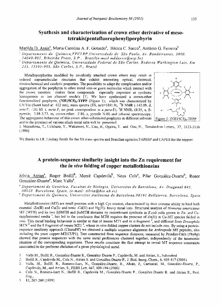
Synthesis and characterization of crown ether derivative of meso- tetrakis(pentafluorophenyl)porphyrin
Mat i lda D. Ass i s a, M a r i a Caro l ina A. F. Gota rdo a, H6t ica C. Sacco a, Ant6nio G. Ferre i ra b
'~ Depar tamento de Qu imica ,FFCLRP, Univers idade de Sdo Paulo, Av. Bandeirantes , 3900, 14040-901, Ribeirf io Preto, S.P., Braz i l ( e -mai l mddass i s@usp .br )
b Depar tamento de Quimica, Univers idade Federa l de S6o Carlos, Rodovia Wayhington Luis, Km 235, 13595-905, Silo Carlos, S.P., Braz i l
Metalloporphyrins modified by covalently attached crown ethers may result in ordered supramolecular structures that exhibit interesting optical, electrical, electrochemical and catalytic properties. The possibility to adapt the complexation and/or aggregation of the porphyrin to other metal ions or guest molecules which interact with the crown moieties makes these compounds especially important as synthetic hemoprotein or ion channel models [1]. We have synthesized a crown-ether functionalized porphyrin, (NH15CS)4-TFPP (Figure 1); which was characterized by UV/Vis (Sorer band at 422 nm), mass spectra (ES, m/z=1891.9), 19F NMR (-142.09, d, orto-F; -161.80, t, meta-F; no peak correspondent to a para-F), ~H NMR, (8.95, s, [3- pyrrole; 3.98-3.73, m, crown-ether; -2.86, s, pyrrole N-H) and infrared spectroscopy. The aggregation behaviour of this crown ether-substituted porphyrin in different solvents and in the presence of various alkali metal salts will be presented.
F F
F F
F ~
(..,o,) Figure 1: (NH 15C5)4-TFPP
1. Murashima, T., Uchihara, Y., Wakamori, N., Uno, H., Ogawa, T. and Ono, N., Tetrahedron Letters, 37, 3133-3136 (1996).
We thanks to J.R. Lindsay Smith for the ES mass spectra and Brazilian agencies FAPESP and CAPES for the support.
A protein-sequence similarity insight into the Zn requirement for the in vivo folding of copper metallothioneins
Silvia Atr ian a, R o g e r Bof i l l b, Merc6 Capdev i l a b, Neus Cols a, Pi lar Gonzf i lez-Duar te b, Rose r Gonz~ilez-Duarte a, Marc Val ls ~.
" Depar tamen t de Gen~tica, Facu l ta t de Biologia, Univers i ta t de Barcelona, Av. Diagonal 645, 08028 Barcelona, Spain, (e-mai l . s i l v ia@bio .ub .es )
b Depar tamen t de Qulmica, Univers i ta t Autdnoma de Barcelona, 08193 Bel laterra, Barcelona, Spain
Metallothioneins (MT) are small proteins with a high Cys content, characterised by their extreme ability to bind both essential -Zn(II) and Cu(I)- and toxic -Cd(II) and Hg(II)- heavy metal ions. Structural analysis of Homarus americanus MT (MTH) and its two [3]3MTH and [3ccMTH domains by recombinant synthesis in E.coli cells grown in Zn- and Cu- supplemented media J, has led to the conclusion that MTH requires the presence of Zn(II) in Cu-MT species folded in vivo. This metal binding behaviour is similar to that of mouse MT1 and its cc fragment 2, and different from Drosophila MTN 3 and the 13 fragment of mouse MT1 4, whose in-vivo-folded copper clusters do not include zinc. By using a protein- sequence similiraty approach (ClustalW) we obtained a multiple sequence alignment for Arthropoda MT peptides, also including the yeast copper-MT(CUP1). Tree constructed from sequence distances, measured by Protdist-Fitch (Phylip) showed that protein sequences with the same metal preferences clustered together, independently of the taxonomic situation of the corresponding organisms. These results constitute the first attempt to reveal MT sequence constraints associatted to the preferent chelation of a given physiological metal.
1. Vails M., Bofill R., Gonzhlez-Duarte R., Gonzfilez-Duarte P., Capdevila, M. and Atrian, S., Submitted 2. Bofill R., Capdevila M., Cols N., Atrian S. and Gonzfilez-Duarte P., J. Biol. Inorg. Chem., 6, 405-417 (2001) 3. Valls, M., Bofill, R., Romero-Isart, N., Gortzfilez-Duarte, R., Abi~n, J., Carrascal, M., Gonzfilez-Duarte, P.,
Capdevila, M., and Atrian, S., FEBS Lett. 467, 189-194 (2000) 4. Cols N., Romero-Isart N., Bofill R., Capdevila M., Gonz/dez-Duarte P., Gonzfilez-Duarte R. and Atrian S., Prot.
Eng., 5. 12, 265-269 (1999)
134 Journal of Inorganic Biochemistry 86 (2001)
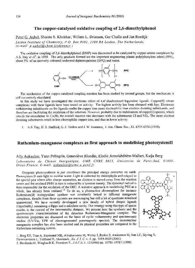
The copper-catalysed oxidative coupling of 2,6-dimethylphenol
Peter G. Aubel , Shais ta S. Khokhar , Wi l l em L. Driessen, Ger Chal la and Jan Reed i jk
Leiden Inst i tute o f Chemistry, P.O. Box 9502, 2300 RA Leiden, The Netherlands, (e-mail: p .aube l@chem. le idenun iv . )
The oxidative coupling of 2,6-dimethylphenol (DMP) was discovered to be catalysed by copper-amine complexes by A.S. Hay et al ~. in 1959. The only products formed are the important engineering plastic poly(phenylene ether) (PPE), about 5% of an intensely coloured undesired diphenoquinone (DPQ) and water.
DMP base ~ O~O DPQ
The mechanism of the copper-catalysed coupling reaction has been studied by several groups, but the mechanism is still not entirely elucidated.
In this study we have investigated the electronic effect of 4,4'-disubstituted bipyridine ligands. Copper(II) nitrate complexes with these ligands have been tested on activity. The highest activity has been obtained with bpy. Electronic withdrawing substituents on the ligands render the copper ions more electrophilic than electron donating substituents, and therefore are facilitating the oxidation of the substrate. However, probably due to stabilisation of copper(I) species, which retards the reoxidation to Cu(II), the overall reaction rate decreases with the substituents CI and NO2. The more electron donating substituents result in less electrophilic copper ions, and thus in lower activity.
1. A.S. Hay, H.S. Stafford, G.F. EndresandJ. W. Eustance, J. Am. Chem. Soc., 81, 6335-6336 (1959).
Ruthenium-manganese complexes as first approach to modelising photosystemlI
Ally Aukau loo , Y a n n Pel legr in , Genev ibve Blondin , Elodie Anxolab6h6re-Mal la r t , Kat ja Berg
Laboratoire de Chimie Inorganique, UMR CNRS 8613, Universite de Paris-Sud, Orsay, France. E-mail: at [email protected], fr
91405-
Oxygenic photosynthesis is par excellence the principal energy converter on earth. Photosystem II uses light to oxidise water. Light is collected by chlorophylls and relayed to the special pair where after charge separation, an electron is moved away from the reaction center and the oxidised P680 in turn is reduced by a tyrosine moiety. The tyrosinyl radical is then responsible for the oxidation of the OEC. A tentative approach in modelising PSII as a whole, has already been realised. ~'2 To do so, a photoactive chromophore for instance Ruthenium(II) trisbipyridine synthon was covalently linked to different manganese complexes. Results from these systems are encouraging but still a lot of questions remained unanswered. We have recently developped a new family of hybrid ditopic ligands (DppzSalH2) containing a Dppz and a salophen cavity. Our strategy using this type of spacer resides in the control of the intermetallic distance. We present here the synthesis and the spectroscopic characterisations of the dinuclear Ruthenium-Manganese complex. The electronic properties are discussed on the basis of cyclic voltammetry and spectroscopic studies (UV/Vis, EPR of electrogenerated paramagnetic species). The mononuclear manganese complex has also been studied and its physical properties are compared to the Ruthenium containing system.
~.c5. .oC~ 3+
1. Berg KE, Tran A, Raymond MK, Abrahamsson M, Wolny J, Redon S, Andersson M, Sun LC, Styring S, Hammarstrom L, Toftlund H, Akermark., Eu. J./. C, v. 4, pp. 1019-1029 (2001) 2. Burdinski D, Wieghardt K, Steenken S., J.A.C.S, v. 121(#46) pp. 10781-10787 (1999)
Journal of Inorganic Biochemistry 86 (2001) 135
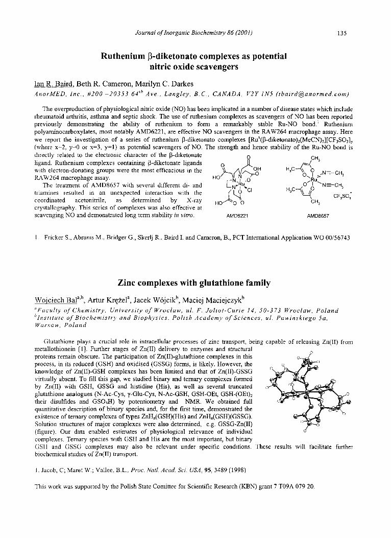
Ruthenium [3-diketonato complexes as potential nitric oxide scavengers
Ian R. Baird, Beth R. Cameron, Marilyn C. Darkes AnorMED, Inc., #200 -20353 64 th Ave., Langley, B.C., CANADA, V2Y 1N5 (ibaird@anormed. com)
The overproduction of physiological nitric oxide (NO) has been implicated in a number of disease states which include rheumatoid arthritis, asthma and septic shock. The use of ruthenium complexes as scavengers of NO has been reported previously demonstrating the ability of ruthenium to form a remarkably stable Ru-NO bond. 1 Ruthenium polyaminocarboxylates, most notably AMD6221, are effective NO scavengers in the RAW264 macrophage assay. Here we report the investigation of a series of ruthenium 13-diketonato complexes [RuX([3-diketonato)z(MeCN)2][CF3SO3]y (where x=2, y=0 or x=3, y=l) as potential scavengers of NO. The strength and hence stability of the Ru-NO bond is directly related to the electronic character of the 13-diketonate ligand. Ruthenium complexes containing 13-diketonate ligands with electron-donating groups were the most efficacious in the ILAW264 macrophage assay.
The treatment of AMD8657 with several different di- and triamines resulted in an unexpected interaction with the coordinated acetonitrile, as determined by X-ray crystallography. This series of complexes was also effective at scavenging NO and demonstrated long term stability in vitro.
O o H3c H3 r - -N. t -O ...Ru
O I ~'N_=-CH3 L-N'RU~, HaC ~--~0 CFaSO;
H 0 ..~0~0 L,I CH3
AMD6221 AMD8657
1. Fricker S., Abrams M., Bridger G., Skerlj R., Baird I. and Cameron, B., PCT International Application WO 00/56743
Zinc complexes with glutathione family
Wojc iech Bal a'b, Ar tur Kr~zel a, J acek W6jc ik b, Maciej Mac i e j czyk b
~'Faculty of Chemistry, University of Wroctaw, ul. F. Joliot-Curie 14, 50-373 Wroctaw, Poland blnstitute of Biochemistry and Biophysics, Polish Academy of Sciences, ul. Pawinskiego 5a, Warsaw, Poland
Glutathione plays a crucial role in intracellular processes of zinc transport, being capable of releasing Zn(II) from metallothionein [1]. Further stages of Zn(II) delivery to enzymes and structural proteins remain obscure. The participation of Zn(II)-glutathione complexes in this process, in its reduced (GSH) and oxidised (GSSG) forms, is likely. However, the knowledge of Zn(II)-GSH complexes has been limited and that of Zn(II)-GSSG virtually absent. To fill this gap, we studied binary and ternary complexes formed by Zn(II) with GSH, GSSG and histidine (His), as well as several truncated glutathione analogues (N-Ac-Cys, 7-Glu-Cys, N-Ac-GSH, GSH-OEt, GSH-(OEt)2 their disulfides and GSO3H) by potentiometry and NMR. We obtained full quantitative description of binary species and, for the first time, demonstrated the existence of ternary complexes of types ZnH,(GSH)(His) and ZnH.(GSH)(GSSG). Solution structures of major complexes were also determined, e.g. GSSG-Zn(II) (figure). Our data enabled estimates of physiological relevance of individual complexes. Ternary species with GSH and His are the most important, but binary GSH and GSSG complexes may also be relevant under specific conditions. biochemical studies of Zn(II) transport.
©
These results will facilitate further
1. Jacob, C; Maret W.; Vallee, B.L., Proc. Natl. Acad. Sci. USA, 95, 3489 (1998)
This work was supported by the Polish State Comittee for Scientific Research (KBN) grant 7 T09A 079 20.
136 Journal of Inorganic Biochemistry 86 (2001)
Inf luence of the spin state of Fe(III) in F e ( I I I ) O O H mode l s
V. Balland a, F. Banse a, T. Mattioli b, E. Bill c, M. Nierlich d, J-J. Gire rd a
a Laborato ire de Chimie Inorganique , UMR CNRS 8613, Universi td Paris -Sud, 91405 Orsay, France(e-mai l . vba l land@icmo .u -psud . f r ) b Sect ion de Bio~nergOtique, URA CNRS 2096, CEA Saclay, 91191 Gi f -sur-Yvet te , France c M a x - P l a n c k - I n s t i t u t f i i r S t rah lenchemie , D-45470 Mii lheim/Ruhr, Germany d DRECAM/SCM, CEA Saclay, 91191 Gi f -sur-Yvet te , France
Recently several Fe(III)OOH models have been characterized ~. They are low spin species with EPR spectra close to axiality characteristic of a (dxz)2(dyz)2(dxy) ground state with a large destabilization of the xy orbital due to strong antibonding interaction with the z~* orbital of the hydroperoxo group. The OO frequency is typically around 796 cm-~ and the FeO one around 617 cm -~ . By deprotonation the Fe(III)(qZ-OO) entity can be obtained 2'3'4. The FeO frequency decreases then to 470 cm -t and the OO frequency increases to 819 cm ~ and the Fe(III) goes high spin.
Reaction of H202 on Fe(II) complexes with bulkier ligands gives no low spin Fe(III)OOH. Indeed M6ssbauer and RR results are indicative of high spin Fe(III) either in Fe(III)-OOH at low pH or in Fe(III)OOFe(III) at high pH. Comparison between mononuclear Fe(III) peroxo groups and dinuclear ones will be proposed.
1. For a review see J.-J. Girerd, F. Banse, A.J. Simaan, Structure and Bonding 2000, 97, Metal-oxo and Metal-peroxo Species in catalytic oxidations p 146 - Volume-Ed. B. Meunier
2. J. Simaan, F. Banse, P. Mialane, A. Boussac, S. Un, T. Kargar-Grisel, G. Bouchoux, J.-J. Girerd, Eur. J. Inorg. Chem. 1999, 993
3. K.B. Jensen, C.J. MacKenzie, L.P. Nielsen, J.Z. Pedersen, H.M. Svendsen, Chem. Commun. 1999, 1313 4. R.Y.N. Ho, G. Roelfes, R. Hermant,, R. Hage, B.L. Feringa, L. Que, Jr, Chem. Commun. 1999, 2161
Ins ights into archaeal type-II N A D H dehydrogenases
T.M. Bandeiras, C.M. Gomes, M. Teixe i r a
Inst i tuto de Tecnologia Quimica e Biol6gica, Univers idade Nova de Lisboa, Rua da Quinta Grande 6, Apt 127, 2780-156, Oeiras, Por tuga l , ( e -mai l : t [email protected] .p t )
Our main concern in recent years has been the study and characterization of membrane bound respiratory complexes from thermoacidophilica archaea it-31. As a result, new type-II NADH dehydrogenases were isolated from the hyperthermoacidophilic archaea Acidianus ambivalens and Sulfolobus metallicus. These proteins have apparent molecular masses around 50kDa, no iron-sulfur clusters and contain a covalently attached flavin, a unique .and novel aspect in this type of enzymes. The high affinity for NADH is shown by apparent K m < 6 gM, and their involvement in the respiratory chain is suggested by the strong interaction between these enzymes and synthetic quinones, to which they donate electrons. Furthermore, co-reconstitution in artificial lipossomes of Acidianus ambivalens NDH-II, quinones and the aa3-type quinol oxidase resulted in a electron-transfer chain, coupling NADH oxidation to complete reduction of oxigen to water. These two proteins show a low activity upon isolation, wich can nevertheless be restored by incubation with phospholipids.
In this work we report the complete characterization of the two proteins, stressing its relevance in the respiratory chain.
1. Gomes, C.M. et al, FEBS Lett 432, 99-102 (1998) 2. Das, T.K. et al, Proc.Natl.Acad.Sci 96, 9591-9596 (1999) 3. Lemos, R.S et al, Biochem.Biophys.Res.Commun 281,141-150 (2001) 4. Gomes et al, J. Bioenerg. Biomemb. 33, (1):1-8 (2001)
Journal of Inorganic Biochemistry 86 (2001) 137
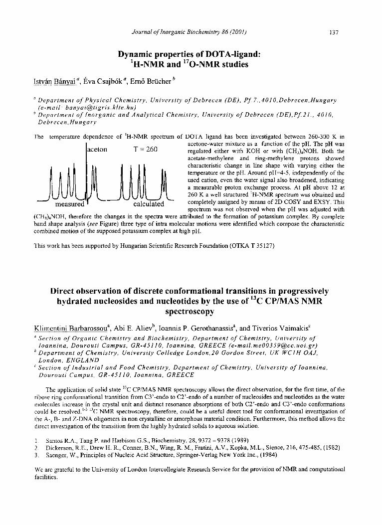
Dynamic properties of DOTA-ligand: IH-NMR and 170-NMR studies
Istvgn Bfinvai a, l~va Csajb6k a, Em6 Brticher o
a Department of Physical Chemistry, University of Debrecen (DE), Pf. 7.,4010, Debrecen,Hungary (e-mail. banyai@tigris, klte. hu)
b Department of Inorganic and Analytical Chemistry, University ofDebrecen (DE),Pf21., 4010, Debrecen, Hungary
The
aceton T = 260
temperature dependence of 1H-NMR spectrum of DOTA ligand has been investigated between 260-300 K in acetone-water mixture as a function of the pH. The pH was regulated either with KOH or with (CH3)4NOH. Both the acetate-methylene and ring-methylene protons showed characteristic change in line shape with varying either the temperature or the pH. Around pH=4-5, independently of the used cation, even the water signal also broadened, indicating a measurable proton exchange process. At pH above 12 at 260 K a well structured 1H-NMR spectrum was obtained and completely assigned by means of 2D COSY and EXSY. This spectrum was not observed when the pH was adjusted with
(CH3)4NOH, therefore the changes in the spectra were attributed to the formation of potassium complex. By complete band shape analysis (see Figure) three type of intra molecular motions were identified which compose the characteristic combined motion of the supposed potassium complex at high pH.
This work has been supported by Hungarian Scientific Research Foundation (OTKA T 35127)
Direct observation of discrete conformational transitions in progressively hydrated nucleosides and nucleotides by the use of 13C CP/MAS NMR
spectroscopy
Klimentini Barbarossou a, Abi E. Aliev b, Ioannis P. Gerothanassis a, and Tiverios Vaimakis c
a Section of Organic Chemistry and Biochemistry, Department of Chemistry, University of loannina, Dourouti Campus, GR-45110, loannina, GREECE ([email protected])
b Department of Chemistry, University Colledge London,20 Gordon Street, UK WCIH OAJ, London, ENGLAND
c Section of Industrial and Food Chemistry, Department of Chemistry, University ofloannina, Dourouti Campus, GR-45110, Ioannina, GREECE
The application of solid state 13C CP/MAS NMR spectroscopy allows the direct observation, for the first time, of the ribose ring conformational transition from C3'-endo to C2'-endo of a number of nucleosides and nucleotides as the water molecules increase in the crystal unit and distract resonance absorptions of both C2'-endo and C3'-endo conformations could be resolved.l313 C NMR spectroscopy, therefore, could be a useful direct tool for conformational investigation of the A-, B- and Z-DNA oligomers in non crystalline or amorphous material condition. Furthermore, this method allows the direct investigation of the transition from the highly hydrated solids to aqueous solution.
1. Santos R.A., Tang P. and Harbison G.S., Biochemistry, 28, 9372 - 9378 (1989) 2. Dickerson, R.E., Drew H. R., Conner, B.N., Wing, R. M., Fratini, A.V., Kopka, M.L., Sience, 216, 475-485, (1982) 3. Saenger, W., Principles of Nucleic Acid Structure, Springer-Verlag New York Inc., (1984)
We are grateful to the University of London Intercollegiate Research Service for the provision of NMR and computational facilities.
138 Journal of lnorganic Biochemistry 86 (2001)
X-Ray structural studies of Rusticyanin from Thiobacillusferroxidans
M a r k L. Bar re t t a b, J o h n F. Ha l l b, M i c h a e l A. H o u g h a, Lal j i D. K a n b i ab, S. S a m a r H a s n a i n a b
a Molecular Biophysics Group, Daresbury Laboratory, Warrington, WA4 4AD, UK (e- mail. s.hasnain@dl, ac. uk) b School of Applied Sciences, De Montfort University, Leicester, LE1 9BH, UK
Rusticyanin is a type I blue copper protein and is thought to be a principal component in the iron respiratory chain of Thiobacillus ferroxidans. It contains a single polypeptide chain with a copper atom as the prosthetic group and is composed of a core p-sandwich along with a 38 residue N-terminal extension.. Rusticyanin has exceptional acid stability and its optimal pH is between 1 and 3. In addition to this remarkable property, it has a Cu(I)/(II) redox couple of -680mV whereas more typical redox potentials for type I copper proteins are around -300mV. The copper coordination is that of a distorted trigonal planar geometry with three strong ligands, His85N ~, Cys138S Y and His 143N ~ and a relatively weaker, axial Met148S ~. A number of type I copper ligand mutants have been produced by site directed mutagenesis to probe the properties of msticyanin. The X-ray crystal structures of Met148Gln and Met148Leu have been determined offering an insight into the contribution of the axial ligand.. The structure of the His143Met mutant has been determined to atomic resolution and provides a very accurate structure of the cupredoxin fold. In both the Met148Gln and Met148Leu crystal structures two molecules are present in the crystallographic asymmetric unit. His143 from one molecule is linked via a water molecule to His 143 of the second molecule indicating a potential route of electron transfer from the redox partner.
Substrate specificity profiles of the matrix metalloproteases
Amy M. Barrios and Charles S. Craik Department of Pharmaceutical Chemistry, University of California at San Francisco, 513 Parnassus Ave. Room S-907, San Francisco, CA (USA) 94143, (e-mail. barrios@cgl, ucsf. edu)
Metalloproteases have been implicated in a number of pathogenic states including hypertension, osteoarthritis, chronic degenerative disorders and cancer. Although protease inhibitors have proven therapeutically useful in models of many of these disease states, a major barrier to their clinical use has been a lack of specificity. The active sites of many metalloproteases are so similar that it is not yet possible to design an inhibitor that is entirely specific for one protease. A detailed knowledge ofprotease substrate specificity is necessary to aid in elucidating the roles played by proteases in vivo and to develop inhibitors capable of differentiating between similar proteases. In order to address this problem, we have designed a novel method for probing the substrate specificity of the metalloproteases. Fluorogenic peptide substrate libraries have been used with much success to probe protease specificity C-terminal to the scissile bond. However, the predominant specificity determinants in metalloproteases are believed to lie N-terminal to the scissile bond, therefore existing substrate libraries are of little utility for probing the active sites of these enzymes. A fluorogenic method that is analogous to existing libraries has been developed to probe the subsites N-terminal to the scissile bond. Specificity profiles for a number of proteases and the molecular basis behind the protease-substrate recognition event will be presented.
Journal of Inorganic Biochemistry 86 (2001) 139
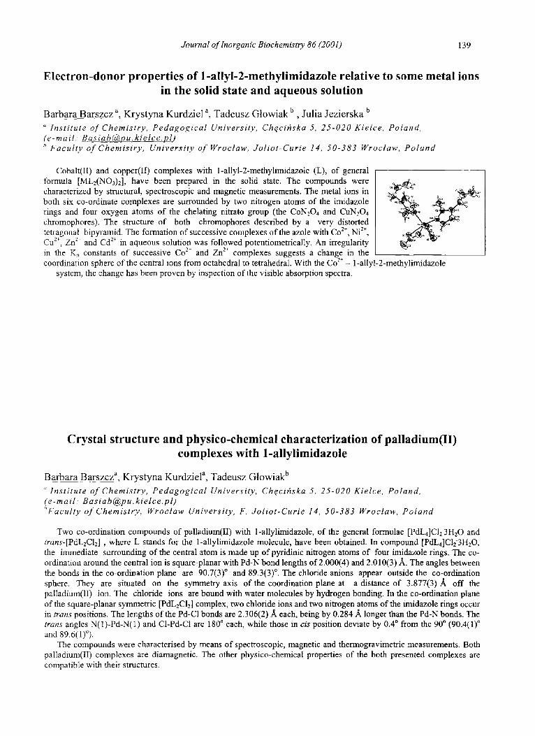
Electron-donor properties of 1-allyl-2-methylimidazole relative to some metal ions in the solid state and aqueous solution
Barbara Barszcz a, Krystyna Kurdziel a, Tadeusz Gtowiak b, Julia Jezierska b " Institute of Chemistry, Pedagogical University, Chqcifiska 5, 25-020 Kielce, Poland, (e-mail: [email protected]) b Faculty of Chemistry, University ofWroctaw, Joliot-Curie 14, 50-383 Wroctaw, Poland
Cobalt(II) and copper(II) complexes with 1-allyl-2-methylimidazole (L), of general formula [ML2(NO3)2], have been prepared in the solid state. The compounds were characterized by structural, spectroscopic and magnetic measurements. The metal ions in both six co-ordinate complexes are surrounded by two nitrogen atoms of the imidazole rings and four oxygen atoms of the chelating nitrato group (the CON204 and CuN204 chromophores). The structure of both chromophores described by a very distorted tetragonal bipyramid. The formation of successive complexes of the azole with Co 2+, Ni 2+, Cu 2÷, Zn 2~ and Cd 2÷ in aqueous solution was followed potentiometrically. An irregularity in the K, constants of successive Co 2+ and Zn z÷ complexes suggests a change in the coordination sphere of the central ions from octahedral to tetrahedral. With the Co 2+ - 1-allyl-2-methylimidazole
system, the change has been proven by inspection of the visible absorption spectra.
Crystal structure and physico-chemical characterization of palladium(II) complexes with 1-allylimidazole
B a r b a r a B a r s z c z a, K r y s t y n a K u r d z i e l a, T a d e u s z G | o w i a k b
Institute of Chemistry, Pedagogical University, Ch¢cihska 5, 25-020 Kielce, Poland, (e-mail: [email protected]) t'Faculty of Chemistry, Wroetaw University, F. Joliot-Curie 14, 50-383 Wroctaw, Poland
Two co-ordination compounds of palladium(II) with 1-allylimidazole, of the general formulae [PdL4]C123H20 and trans-[PdL2C12] , where L stands for the l-allylimidazole molecule, have been obtained. In compound [PdL4]Clf3H20, the immediate surrounding of the central atom is made up of pyridinic nitrogen atoms of four imidazole rings. The co- ordination around the central ion is square-planar with Pd-N bond lengths of 2.000(4) and 2.010(3) A. The angles between the bonds in the co-ordination plane are 90.7(3) ° and 89.3(3) ° . The chloride anions appear outside the co-ordination sphere. They are situated on the symmetry axis of the coordination plane at a distance of 3.877(3)A off the palladium(II) ion. The chloride ions are bound with water molecules by hydrogen bonding. In the co-ordination plane of the square-planar symmetric [PdL2C12] complex, two chloride ions and two nitrogen atoms of the imidazole rings occur in trans positions. The lengths of the Pd-C1 bonds are 2.306(2) A each, being by 0.284 A longer than the Pd-N bonds. The trans angles N(1)-Pd-N(1) and C1-Pd-C1 are 180 ° each, while those in cis position deviate by 0.4 ° from the 90 ° (90.4(1) ° and 89.6(1)°).
The compounds were characterised by means of spectroscopic, magnetic and thermogravimetric measurements. Both palladium(II) complexes are diamagnetic. The other physico-chemical properties of the both presented complexes are compatible with their structures.
140 Journal of Inorganic Biochemistry 86 (2001)
Cobalt (II) and zinc (II) compounds of the didentate N,O - donor ligand.
Barbara Barszcz a, Krystyna Kurdziel a, Tadeusz Gtowiak b "Institute of Chemistry, Pedagogical University, Ch~cihska .5,25-020 Kielce, POLAND (e-mail: [email protected]) bFaculty of Chemistry, University of Wroetaw, .50-383 Wroctaw, POLAND
Our aim is to provide low-molecular-weight model complex of cobalt (II) and zinc (II) ions containing the biologically relevent ligand. To obtain it, two novel cobalt (II) isomers of trans (O) - [Co (1-Bz-2-CHzOHIm)4] (NO3)2 (1) and cis - (O)- [Co (1-Bz-2-CHzOHIm)4](NOs)2.1.5 H20 (2) have been synthesized by mixing 1 : 6 (1) or 1 : 4 (2) molar ratio of Co(NO3)2 - 6 H20 and 1-benzyl - 2 hydroxymethylimidazole (1-Bz-2-CH2OHIm) in trimethylorthoformate. The crystal structure of Co (II) complex show the triclinic space group P1 (1) and C2/c (2). The present complex adopt a six co- ordinate structure and co-ordination geometry around the the Co atom is approximately octahedral (1) or very distorted octahedral (2) and is surrounded by four nitrogen atoms of the four imidazole rings and two oxygen atoms of the hydroxymethyl group. Two of the ligands act as a monodentate and two as a didentate, forming the five - membered chelate ring with the central ion. The structural data obtained for Co (II) isomers were conf'mned by the IR, UV/Vis spectroscopic methods. It is well know that cobalt(II) ion is frequently used to substitute for zinc ion in zinc proteins, and the cobalt(II)- substituted enzymes often show about as much catalytic activity as the native zinc enzymes, for that reason the structural data of Co(II) complexes were compared with the single crystal X-ray structure of [Zn (1-Bz-2-cH2OHIm)4] (NO3)2
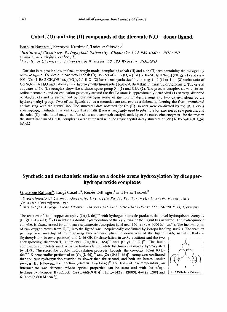
Synthetic and mechanistic studies on a double arene hydroxylation by dicopper- hydroperoxide complexes
Giuseppe Battaini a, Luigi Casella a, Ren6e Dillinger, b and Felix Tuczek b a Dipartimento di Chimica Generale, Universith Pavia, Via Taramelli 1, 27100 Pavia, Italy (e-mail: [email protected])
b lnst i tut f i ir Anorganische Chemie, Universitiit Kiel, Otto-Hahn-Platz 6/7, 24098 Kiel, German),
The reaction of the dicopper complex [Cu2(L-66)] 4+ with hydrogen peroxide produces the novel hydroquinone complex [Cu2(HO-L-66-O)] 3+ (1) in which a double hydroxylation of the xylyl ring of the ligand has occurred. The hydroquinone complex is characterized by an intense asymmetric absorption band near 350 nm (~ = 9000 M t cm-~). The incorporation of two oxygen atoms from H202 into the ligand was unequivocally confirmed by isotope labeling studies. The reaction pathway was investigated by preparing two isomeric phenolic derivatives of the ligand L-66, namely HO-L-66 (hydroxylation in meta position) and L-66-OH (hydroxylation in ortho position) and the two corresponding dicopper(II) complexes [Cu2(HO-L-66)] 4+ and [Cu2(L-66-O)] 3+. The latter complex is completely inactive in the hydroxylation, while the former is rapidly hydroxylated by H202. Therefore, the double hydroxylation proceeds through the complex [Cu2(HO-L- 66)] 4+. Kinetic studies performed on [Cu2(L-66)] 4÷ and [CuffHO-L-66)] 4÷ complexes confirmed that the first hydroxylation reaction is slower than the second, and both are intramolecular process. By following the reaction between [Cu2(L-66)] 4÷ and H202 at low temperature, an intermediate was detected whose optical properties can be associated with the rll:q 1- hydroperoxodicopper(II) adduct, [Cu2(L-66)(OOH)] 3+, [ ) ~ = 3 4 2 (e 12000), 444 (e 1200) and 610 nm (e 800 Mlcm-l)].
OH
',. 2: -°'-, .,'2". , .cu B B
1
B = 1-Methylbenzimidazole