absence of functional receptors for parathyroid hormone...
TRANSCRIPT

34
Jobert et al.
J. Clin. Invest.© The American Society for Clinical Investigation, Inc.0021-9738/98/07/0034/07 $2.00Volume 102, Number 1, July 1998, 34–40http://www.jci.org
Absence of Functional Receptors for Parathyroid Hormone and ParathyroidHormone–related Peptide in Blomstrand Chondrodysplasia
Anne-Sixtine Jobert,* Ping Zhang,* Alain Couvineau,
‡
Jacky Bonaventure,
§
Joëlle Roume,
i
Martine Le Merrer,
§
and Caroline Silve*
*
INSERM U. 426, Faculté de Médecine Xavier Bichat;
‡
INSERM U. 410, Faculté de Médecine Xavier Bichat;
§
INSERM U. 393, Hôpital Necker, Enfants Malades; and
i
Service d’Histo-Embryologie et Cytogénétique, Hôpital Saint Antoine, Paris, France
Abstract
We report the absence of functional parathyroid hormone
(PTH)/PTH-related peptide (PTHrP) receptors (PTH/PTHrPreceptor) in Blomstrand chondrodysplasia, a genetic disor-der characterized by advanced endochondral bone matura-tion. Analysis of PTH/PTHrP receptor genomic DNA froma patient with Blomstrand chondrodysplasia demonstratedthat the patient was heterozygous for a point mutation
(G
→
A substitution at nucleotide 1176) inherited from themother. Analysis of PTH/PTHrP receptor cDNA demon-
strated that: (
a
) this point mutation caused the deletion ofthe first 11 amino acids of exon M5 (encoding the fifthtransmembrane domain of the receptor), resulting from theuse of a novel splice site created by the base substitution; (
b
)the mutant receptor was well expressed in COS-7 cells, butdid not bind PTH or PTHrP, and failed to induce detectablestimulation of either cAMP or inositol phosphate produc-tion in response to these ligands; and (
c
) the paternal allelewas not expressed. Thus, only the abnormal and nonfunc-tional PTH/PTHrP receptors encoded by the maternal allelewere expressed by chondrocytes from this patient. In viewof the known role played by the PTH/PTHrP receptor inbone and cartilage development, these results strongly sup-port the conclusion that the absence of functional PTH/PTHrP receptors is responsible for the skeletal abnormali-ties seen in Blomstrand chondrodysplasia, abnormalitiesthat are the mirror image of those observed in Jansen’schondrodysplasia. These findings emphasize the impor-tance of signaling through this receptor in human fetal skel-etal development. (
J. Clin. Invest.
1998. 102:34–40.) Key
words: PTH/PTHrP receptor
•
point mutation
•
alternatesplicing
•
null allele
•
Blomstrand chondrodysplasia
Introduction
The parathyroid hormone (PTH)/PTH-related peptide (PTHrP)
1
receptor, a G protein–coupled receptor, plays a key role in theregulation of calcium and phosphorus metabolism in fetal and
adult life by mediating the actions of PTH on its target organs(1). Recent studies have demonstrated that signaling throughthe PTH/PTHrP receptor, in addition to its role in regulatingmineral metabolism, also plays an essential role in fetal devel-opment due to the important regulatory effects of PTHrP onthe development of cartilage and bone (2–4). In this regard,mice with disruptions of either the PTH/PTHrP receptor orPTHrP genes exhibit multiple, severe skeletal defects charac-terized by an advanced endochondral bone formation thatprove lethal in utero or shortly after birth (2, 4). This suggeststhat genetic abnormalities of the PTH/PTHrP receptor genemight also lead to severe developmental abnormalities in hu-mans. Consistent with this idea, the constitutive activation ofthe PTH/PTHrP receptor has been demonstrated recently tobe responsible for Jansen’s chondrodysplasia, a disease charac-terized by delayed endochondral bone formation (5, 6). Here-tofore, the effects of inhibitory mutations of the human PTH/PTHrP receptor have not been described. In particular, defectsin this receptor are not responsible for pseudohypoparathy-roidism type Ib (7–9), a condition characterized by hypocalce-mia and hyperphosphoremia due to selective renal resistanceto PTH, in which such abnormalities had been suspected previ-ously (10–12). Here, we report that Blomstrand chondrodys-plasia, a genetic disorder characterized by advanced endo-chondral bone maturation (13, 14), a defect that is the mirrorimage of that observed in Jansen’s chondrodysplasia, resultsfrom the absence of functional PTH/PTHrP receptors.
Methods
Case report.
The case evaluated in these studies has been describedrecently (15). The fetus, stillborn at 25 wk, was the result of the sec-ond pregnancy of a nonconsanguineous couple. The diagnosis ofBlomstrand chondrodysplasia was made during pregnancy by sono-graphic examination, and was confirmed after delivery by the demon-stration of characteristic radiologic and histologic findings, includinga striking increase in bone density and extremely advanced skeletalmaturation (15). 1 yr later, the mother gave birth to a healthy boy.However, her fourth pregnancy terminated spontaneously at 33 wk ofgestation with the delivery of a dead girl with the same syndrome.
Southern blot analysis of genomic DNA.
Genomic DNA from theproband and the mother was extracted from blood samples by stan-dard methods. Southern blot analysis was performed according tostandard procedures using probes prepared from plasmids containingeither PTH/PTHrP receptor cDNA (16) or the genomic sequence
corresponding to the 5
9
promoter regions as described by Schipani etal. (16). Genomic DNAs were cut by HindIII or XbaI (PTH/PTHrPreceptor cDNA probes) and by SstI (5
9
promoter and PTH/PTHrPreceptor cDNA probes) before Southern blotting. In all experiments,genomic DNA from the fetus, his mother, control fibroblasts, andnormal human kidney were analyzed in parallel.
Address correspondence to Caroline Silve, M.D., Ph.D., INSERM U.426, Faculté de Médecine Xavier Bichat, 16 rue Henri Huchard,75018 Paris, France. Phone: 33-1-44-85-62-73; FAX: 33-1-42-28-15-64;E-mail: [email protected]
Received for publication 26 January 1998 and accepted in revisedform 15 April 1998.
1.
Abbreviation used in this paper:
PTH/PTHrP receptor, PTH andPTH-related peptide receptor.

PTH/PTHrP Receptor in Blomstrand Chondrodysplasia
35
Sequencing of genomic DNA.
All coding exons of the PTH/PTHrP receptor gene were amplified by PCR using a mixture of 1 U
Taq
(GIBCO BRL, Gaithersburg, MD) and 0.06 U Pfu (Stratagene,La Jolla, CA) DNA polymerases in a final volume of 50
m
l. Primersused to amplify exons S, EL2, G, and M3, and the 3
9
untranslated re-gion are shown in Table I. Other primers were as described bySchipani et al. (7). Amplification products were sequenced in both di-rections by the dideoxy chain-termination method.
Evaluation of PTH/PTHrP receptor mRNA.
Chondrocyte cell cul-tures were established from tissue obtained from the proband at thetime of delivery. A fibroblast cell line from a normal subject, humankidney tissue, and SaOS-2 osteosarcoma cells were used as controls.Cells were maintained in Dulbecco’s modified Eagle’s medium con-taining 10% heat-inactivated fetal calf serum as described (10). TotalRNA was extracted from cells and tissue using the method of Chom-czynski and Sacchi (17), and cDNA was synthesized as described pre-viously (18). Amplification of the entire PTH/PTHrP receptor cDNAin four overlapping fragments was performed using the primers de-scribed in Table II. Sequencing of cDNA was performed as describedabove.
Construction of expression plasmids.
A vector permitting expres-sion of the wild-type PTH/PTHrP receptor with an epitope Tag (19)added at the COOH-terminal end (PTH-R/wt) was obtained by ligat-ing the dodecapeptide sequence coding for the Tag marker to theCOOH-terminal end of the full-length PTH/PTHrP receptor cDNA,and subcloning the fragment into pcDNA1-Amp (Invitrogen, LaJolla, CA). The functional properties of the tagged and native recep-tors were indistinguishable (data not shown). To obtain a vector per-mitting the expression of the abnormal PTH/PTHrP receptor identi-fied in this study (PTH-R/
D
373-383), a fragment encompassing thedeletion was produced by PCR (bases 676–1316), digested with NspIand EagI restriction enzymes, and ligated to the tagged wild-typePTH/PTHrP receptor cDNA plasmid that had been digested previ-ously with the same restriction enzymes. The construction was veri-fied by DNA sequencing.
RNase protection analysis.
A riboprobe specific for a fragment ofthe abnormal PTH/PTHrP receptor mRNA encompassing the 33-bpdeletion in exon M5 was prepared by amplifying a 477-bp fragment ofthe deleted PTH/PTHrP receptor cDNA using oligonucleotide pair 3described in Table II. The PCR fragment was cloned in the pTAgplasmid using the LigATor cloning system (R & D Systems, Abing-don, UK). The plasmid was linearized by digestion with XbaI, and theriboprobe was synthesized using T7 RNA polymerase. RNase protec-tion analysis was performed as described (10, 18). The unprotected ri-boprobe migrated at 589 bp. When the riboprobe hybridized to thedeleted receptor mRNA, a protected fragment of 477 bp was ob-served. When the riboprobe hybridized to the wild-type receptor, twofragments, one at 191 bp, the second at 286 bp, were obtained.
Evaluation of receptor expression and function by transient expres-sion.
The techniques used for transient expression of wild-type andmutated PTH/PTHrP receptors in COS-7 cells, and the evaluation ofPTH-induced cAMP production, PTH-induced inositol phosphate ac-cumulation, and binding of [
125
I]hPTHrP(1-34) by transfected cellshave been described previously (18, 20). Expression of the Tagepitope was detected by immunofluorescence on permeabilized cellsas described (19, 21). No staining was observed when untransfectedCOS-7 cells were incubated with the anti-Tag antibody.
Results
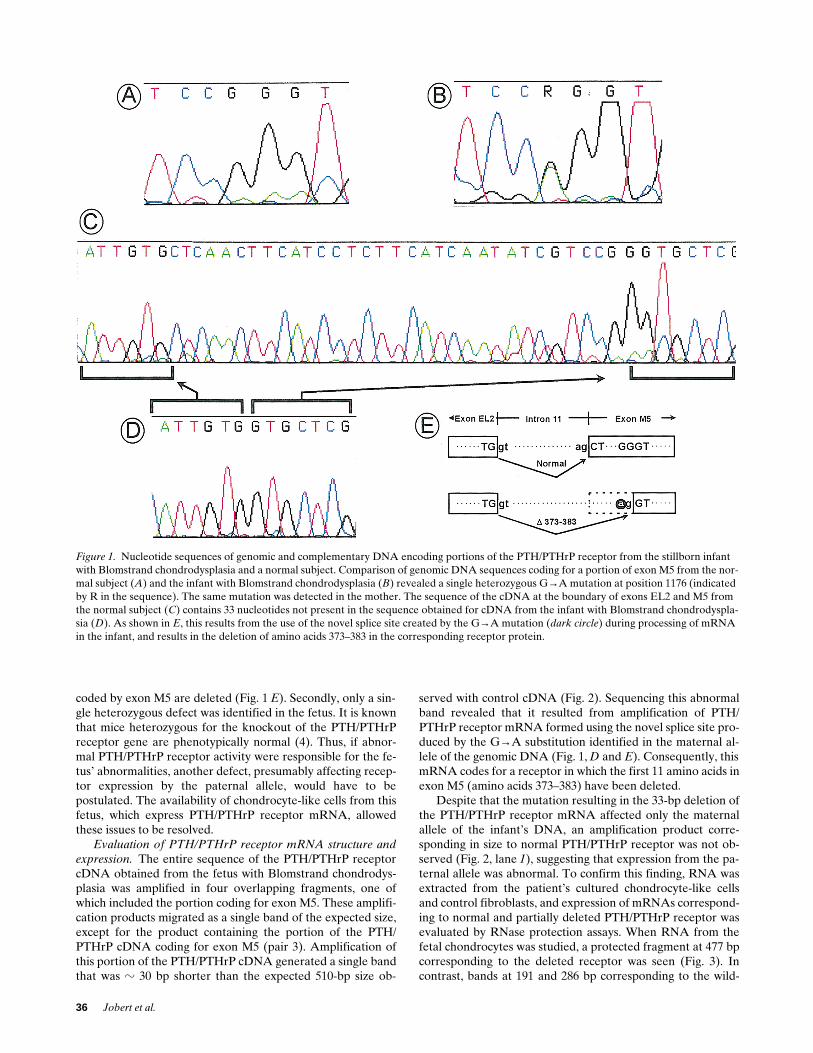
Evaluation of genomic DNA coding for the PTH/PTHrP re-ceptor.
To evaluate the possibility that defects in the PTH/PTHrP receptor gene were responsible for the abnormalitiesin the stillborn infant described above, each of the 14 codingexons and the 3
9
untranslated region of this gene were ampli-fied by PCR and sequenced. A single heterozygous basechange (G
→
A, nucleotide 1176) was detected in exon M5,which encodes the fifth transmembrane domain of the recep-tor (Fig. 1,
A
and
B
). No other base changes were identified.Amplification and sequence analysis of the mother’s genomicDNA revealed the same base substitution, indicating maternalinheritance of the trait. The base substitution removed an NciIrestriction site from the normal sequence. Amplification of ge-nomic DNA and subsequent digestion with NciI confirmed thepresence of a heterozygous base change in the affected fetusand in his mother.
These studies evaluating genomic DNA raised two impor-tant issues. First, two different abnormalities could be pro-duced by the G
→
A base substitution identified. This mutationcould result in a Arg to Gln substitution at amino acid 383 ofthe resulting PTH/PTHrP receptor. Alternatively, this substi-tution creates a novel splice acceptor site (TCC
A
G
↓
GT) 33nucleotides downstream from the normal splice acceptor sitefor exon M5 (Fig. 1,
C–E
). If this splice site were used, it wouldresult in a mutant receptor in which the first 11 amino acids
Table I. Sequences of Primers Used to Amplify Exons S, G, M3, and EL2, Their Adjacent Intron/Exon Borders, and the 3
9
Untranslated Region of the PTH/PTHrP Receptor
PCR product Primer sequence Size
bp
Exon S 5
9
-GCAGCTCTGCACCCCCTACC-3
9
2325
9
-
GACTGCGTGCCTTAGACCTACTCC
-3
9
Exon G 5
9
-TGCTGGAAGGGGTGGGGATTAC-3
9
2745
9
-
CGTGTGGGTGGGAGTGAATTTATCT
-3
9
Exon M3 5
9
-CCCCCAGCCCAGCCCTGACT-3
9
3285
9
-
GGGGCGGGATGTGCTGTGTG
-3
9
Exon EL2 5
9
-CTGGGTCTCTGTGGGCAGTCTT-3
9
1875
9
-
CGCACATCCCACCCACTCTC
-3
9
3
9
untrans- 5
9
-TCCTCAACGGCTCCTGCTCA-3
9
322lated 5
9
-
CCCTCCGCCACAGCTTTCC
-3
9
*Primers in italics are reverse primers.
Table II. Primers Used to Amplify the Entire cDNA of the PTH/PTHrP Receptor in Four Overlapping Fragments
Primers Sequence Size
bp
Pair 1: S
10
* 5
9
-GCGGCCCTAGGCGGTGG 451
G
460
5
9
-
GGCATGGCCTTTGTGATTGAA
Pair 2: G
455
5
9
-CATGCCTACCGACGCTGTGACC 595
M4
1049
5
9
-
TGACCCACACAGCCACGAAGAC
Pair 3: M3
952
5
9
-CCTCATCTTCATGGCCTTCTTCTC 510
T
1462
5
9
-
GTCCAGCGGCTCCAAGATTTC
Pair 4: M7
1409
5
9
-TTCTGCAATGGCGAGGTACAA 478
T
1885
5
9
-
GGAAATCATTCAACCACCCATCTT
Primers in italics are reverse primers. *The letter refers to the name ofthe exon on which the 5
9
end of the primer is located, and the subscriptnumber indicates the position of the 5
9
end. The expected PCR productsizes (bp) are also indicated.
36
Jobert et al.
coded by exon M5 are deleted (Fig. 1
E
). Secondly, only a sin-gle heterozygous defect was identified in the fetus. It is knownthat mice heterozygous for the knockout of the PTH/PTHrPreceptor gene are phenotypically normal (4). Thus, if abnor-mal PTH/PTHrP receptor activity were responsible for the fe-tus’ abnormalities, another defect, presumably affecting recep-tor expression by the paternal allele, would have to bepostulated. The availability of chondrocyte-like cells from thisfetus, which express PTH/PTHrP receptor mRNA, allowedthese issues to be resolved.
Evaluation of PTH/PTHrP receptor mRNA structure andexpression.
The entire sequence of the PTH/PTHrP receptorcDNA obtained from the fetus with Blomstrand chondrodys-plasia was amplified in four overlapping fragments, one ofwhich included the portion coding for exon M5. These amplifi-cation products migrated as a single band of the expected size,except for the product containing the portion of the PTH/PTHrP cDNA coding for exon M5 (pair 3). Amplification ofthis portion of the PTH/PTHrP cDNA generated a single bandthat was
z
30 bp shorter than the expected 510-bp size ob-
served with control cDNA (Fig. 2). Sequencing this abnormalband revealed that it resulted from amplification of PTH/PTHrP receptor mRNA formed using the novel splice site pro-duced by the G
→
A substitution identified in the maternal al-lele of the genomic DNA (Fig. 1,
D
and
E
). Consequently, thismRNA codes for a receptor in which the first 11 amino acids inexon M5 (amino acids 373–383) have been deleted.
Despite that the mutation resulting in the 33-bp deletion ofthe PTH/PTHrP receptor mRNA affected only the maternalallele of the infant’s DNA, an amplification product corre-sponding in size to normal PTH/PTHrP receptor was not ob-served (Fig. 2, lane
1
), suggesting that expression from the pa-ternal allele was abnormal. To confirm this finding, RNA wasextracted from the patient’s cultured chondrocyte-like cellsand control fibroblasts, and expression of mRNAs correspond-ing to normal and partially deleted PTH/PTHrP receptor wasevaluated by RNase protection assays. When RNA from thefetal chondrocytes was studied, a protected fragment at 477 bpcorresponding to the deleted receptor was seen (Fig. 3). Incontrast, bands at 191 and 286 bp corresponding to the wild-
Figure 1. Nucleotide sequences of genomic and complementary DNA encoding portions of the PTH/PTHrP receptor from the stillborn infant with Blomstrand chondrodysplasia and a normal subject. Comparison of genomic DNA sequences coding for a portion of exon M5 from the nor-mal subject (A) and the infant with Blomstrand chondrodysplasia (B) revealed a single heterozygous G→A mutation at position 1176 (indicated by R in the sequence). The same mutation was detected in the mother. The sequence of the cDNA at the boundary of exons EL2 and M5 from the normal subject (C) contains 33 nucleotides not present in the sequence obtained for cDNA from the infant with Blomstrand chondrodyspla-sia (D). As shown in E, this results from the use of the novel splice site created by the G→A mutation (dark circle) during processing of mRNA in the infant, and results in the deletion of amino acids 373–383 in the corresponding receptor protein.

PTH/PTHrP Receptor in Blomstrand Chondrodysplasia
37
type PTH/PTHrP receptor mRNA were not present. WhenRNA from the control fibroblasts was studied, the two ex-pected fragments at 191 and 286 bp were observed (Fig. 3).These studies confirm the presence of deleted, but not wild-type specific mRNA in cultured chondrocytes obtained fromthe infant, whereas only normal PTH/PTHrP receptor mRNAwas detected in control samples. Thus, expression of the short-ened PTH/PTHrP receptor mRNA encoded by the maternalallele accounted for all detectable transcripts in this patient, in-dicating that little or no mRNA from the paternal allele waspresent in the patient’s chondrocytes.
As indicated above, abnormalities in the 3
9
untranslated re-gion affecting mRNA stability are unlikely to explain thesefindings, because analysis of the fetus’ genomic DNA from thisregion was normal. Evidence for genomic rearrangement thatmight influence expression of the paternal allele was sought,but no rearrangements of the receptor gene were detected bySouthern blot analysis of genomic DNA using probes spanningthe coding exons and the 5
9
flanking promoter regions (datanot shown).
Functional properties and expression of the mutant recep-tor.
To investigate the functional consequences of the muta-tion, a sequence encoding a 12–amino acid epitope tag wasadded to the COOH-terminal end of the mutant and the wild-type cDNAs, and these sequences were cloned into an expres-sion vector and transiently expressed in COS-7 cells. Transfec-tion of COS-7 cells with increasing amounts of wild-typecDNA led to a dose-dependent increase in cAMP and inositolphosphate production induced by human (h) PTH(1-34) (max-imum response, 25–50 ng/well) (Fig. 4,
A
and
B
). By contrast,transfecting the mutant construction (up to 100 ng/well) failedto result in detectable stimulation of cAMP and inositol phos-phate production in response to hPTH(1-34) (Fig. 4,
A
and
B
).Similar results were obtained using other PTH/PTHrP recep-tor ligands [hPTH(1-84) and hPTHrP(1-34)] (data not shown).To investigate the mechanisms responsible for the absence ofreceptor function, ligand binding by the mutated receptor wasevaluated. No [
125
I]-labeled hPTHrP(1-34) ligand binding toCOS-7 cells transfected with the mutated construction wasnoted, whereas specific binding to cells transfected with thewild-type cDNA was readily detectable (Fig. 4
C
). To deter-mine whether the mutated receptor was adequately addressedto the plasma membrane, monoclonal antibodies directedagainst the 12–amino acid tag of the fusion protein were usedto detect receptor expression. Interestingly, similar signal in-tensity was obtained for cells transfected with plasmids con-taining either wild-type or mutated PTH/PTHrP receptorcDNA, indicating that the two receptors were expressed to anequivalent extent and were both addressed to the plasmamembrane (Fig. 4
D
).As would be expected for cells that did not express func-
tional PTH/PTHrP receptors, PTH-induced cAMP productionwas undetectable in chondrocyte-like cells from the infant withBlomstrand chondrodysplasia. In contrast, the patient’s chon-drocytes adequately responded to forskolin (50
m
M) (82-foldincrease of cAMP production in response to 50
m
M forskolin).Thus, the absence of PTH-induced stimulation of cAMP pro-duction in the patient’s cells could not be explained by a defi-ciency in the adenylate cyclase system. Cells from a controlsubject responded to both PTH and forskolin (6- and 50-foldincreases in cAMP, respectively, data not shown).
Discussion
We report that the absence of functional PTH/PTHrP recep-tors causes Blomstrand chondrodysplasia, a disease character-ized by advanced endochondral bone maturation and fetaldeath. Recent studies in rodents have demonstrated that, inaddition to their role in regulating calcium and phosphorusmetabolism, PTH/PTHrP receptors play a key role in fetalskeletal development by decelerating the differentiation ofgrowth plate chondrocytes into hypertrophic cells (3, 22).PTH/PTHrP receptors are abundantly expressed by growth
Figure 2. Amplification of PTH/PTHrP receptor cDNA prepared from chondrocytes from the infant with Blomstrand chondrodyspla-sia (lane 1), from human SaOS-2 osteosarcoma cells (lane 2), and from human kidney (lane 3). PCR was performed using the primer pair 3 described in Table II, which amplifies a fragment which in-cludes exon M5, and whose expected size is 510 bp. The migration of the 100-bp ladder molecular weight standard (MW lanes) and the cor-responding molecular weights (arrows) are also shown. Lane 4, Am-plification performed in the absence of template.
Figure 3. Ribonuclease protection analysis of PTH/PTHrP receptor mRNA expressed in chondrocytes from the fetus with Blomstrand chondrodysplasia (Fe) and control fibroblasts (Fb). RNase protection assay was performed as described in Methods. 50 mg of total RNA from chondrocytes from the fetus with Blom-strand chondrodyspla-sia and control fibro-blasts were hybridized with a 32P-labeled anti-sense hPTH/PTHrP re-ceptor riboprobe, as de-
scribed in Methods. The sequence of the riboprobe corresponds to that of the deleted PTH/PTHrP receptor mRNA. As expected, when RNA from the fetal chondrocytes was studied, bands at 191 and 286 bp corresponding to the wild-type PTH/PTHrP receptor mRNA were not observed (ellipses). In contrast, a protected fragment at 477 bp corresponding to the deleted receptor mRNA was seen (arrow). When PTH/PTHrP receptor mRNA from control fibroblasts was studied, the two expected fragments at 191 and 286 bp were observed. mw, Molecular weight standards; pro, unprotected riboprobe; tRNA, hybridization of the riboprobe with transfer RNA.

38 Jobert et al.
plate chondrocytes at the site of transition between prolifera-tion and hypertrophy (23). Delayed endochondral bone for-mation is observed in transgenic mice in which proliferatingchondrocytes overproduce PTHrP (3). Similarly, delayed en-dochondral bone formation also occurs in Jansen’s metaphy-seal chondrodysplasia, a disease in which mutations producingconstitutive activation of PTH/PTHrP receptors have beenidentified (5, 6). Conversely, advanced endochondral bone for-mation is observed in mice that lack either PTHrP or PTH/PTHrP receptors (2, 4). Furthermore, targeted expression ofconstitutively active PTH/PTHrP receptors delays endochon-dral bone formation and rescues mice that lack PTHrP (24).Although the abnormalities observed in the PTH/PTHrP re-ceptor and PTHrP knockout models are generally similar, thephenotype observed in mice lacking the PTH/PTHrP receptoris more severe than that of mice lacking PTHrP. Thus, althougha similar phenotype might be seen in patients with defects inthe PTHrP gene or PTH/PTHrP receptor gene, the severe ab-normalities seen in Blomstrand chondrodysplasia are morereminiscent of those occurring in PTH/PTHrP receptor geneknockout mice.
Analysis of genomic DNA and mRNA coding for the PTH/PTHrP receptor in the fetus with Blomstrand chondrodyspla-sia demonstrated that the absence of functional receptors re-sulted from the expression of a nonfunctional receptor codedby the maternal allele, associated with the absence of expres-sion of the paternal allele. Thus, the fetus was a compoundheterozygote, compatible with the recessive inheritance pat-tern described for Blomstrand chondrodysplasia (13–15, 25,26). The abnormality responsible for the absence of expressionof the paternal allele was not identified. No other mutations inthe coding exons were present, and no abnormality in mRNAsplicing other than that detected in the maternal allele wasidentified. A number of different mechanisms can result in re-duced expression, including imprinting of one allele, mutationsin the promoter region, or abnormalities reducing the stabilityof the mRNA. Results in knockout mice indicate that the geneis not imprinted. No mutations in the 39 noncoding region wereidentified. Little is known about the factors that control PTH/PTHrP receptor gene expression in fetal and adult life. Twopromoters, P1 and P2, have been described in mice and rats(27, 28). The P1 activity is mainly restricted to the adult kid-ney, while P2 activity is detected in several fetal and adult tis-sues, including cartilage and bone. The P2 promoter activity isapparently well conserved between humans and mice. In con-trast, P1 activity is weak or absent in humans, whereas a thirdpromoter P3, apparently specific to humans, appears to controlthe PTH/PTHrP receptor expression in a number of tissues in-cluding kidney and bone (29). Southern blots exploring thesepreviously described 59 promoter regions were normal in thepatient, indicating that major rearrangements or deletions ofthese sequences did not explain the absence of expression ofthe paternal allele. It is noteworthy that although PTH/PTHrPreceptor expression is regulated by factors such as cAMP,1,25(OH)2 vitamin D, and glucocorticoids, no responsive ele-
Figure 4. Functional evaluation of wild-type and mutant PTH/PTHrP receptors expressed in COS-7 cells. COS-7 cells were transfected with the indicated amounts of plasmid DNA coding for the wild-type (PTH-R/wt, left) or mutant (PTH-R/D373-383, right) PTH/PTHrP re-ceptors, and functional studies were performed 48 h later. (A) cAMP production for cells incubated in the absence (basal, open bars) or presence of 1027 M hPTH(1-34) (striped bars). (B) Accumulation of inositol phosphates for cells incubated in the absence (basal, open bars) or presence of 1027 M hPTH(1-34) (striped bars). No stimula-tion of cAMP or inositol phosphate production by PTH was detected in untransfected COS-7 cells (data not shown). (C) Binding of [125I]hPTHrP(1-34) by cells incubated with radiolabeled ligand only (open bars) or radiolabeled ligand in the presence of 1027 M unla-beled hPTH(1-34) (striped bars). No binding of [125I]hPTHrP(1-34)
by untransfected cells was detectable (data not shown). Data are pre-sented as mean6SEM for at least two independent experiments, each performed in triplicate. (D) Detection of the tagged PTH-R/wt (left) and PTH-R/D373-383 (right) proteins in transfected COS-7 cells by immunofluorescence.

PTH/PTHrP Receptor in Blomstrand Chondrodysplasia 39
ments for these factors have been found in the promoter re-gions identified to date (27–29). These findings suggest the ex-istence of unidentified enhancer or repressor sequencesparticipating in the regulation of the PTH/PTHrP gene expres-sion.
The 33-bp deletion in exon M5 identified in the maternalallele resulted from a point mutation leading to the creation ofa new 39 splice site with conservation of the reading frame. Asin our study, the previously reported cases of creation of 39splice sites have involved a G→A mutation at position 22 ofthe newly created splice site. Because their identification re-quires characterization of cDNA, the detection of mutationsresulting in the creation of a novel splice site is relatively un-common (30) and has not been described for other G protein–coupled receptors (31).
Previous work has suggested that receptor residues in thisfifth transmembrane domain are at or near a site that interactswith the NH2 terminus of PTH (32). Site-directed mutagenesisof nearby amino acids has also suggested the importance ofthis region for coupling to signal transduction (33). We foundthat the deletion in this embedded helical region strongly in-hibited the binding of ligand, without preventing receptor ex-pression. It is likely that the deletion reported here disruptsthe three-dimensional organization of the receptor, therebypreventing normal ligand recognition.
In view of the essential role played by the PTH/PTHrP re-ceptor in bone and cartilage development, this study stronglysupports the conclusion that absence of functional PTH/PTHrP receptors is responsible for Blomstrand chondrodys-plasia. This study emphasizes the importance of signalingthrough this receptor in human fetal bone development, andsuggests that abnormalities in components of this ligand/recep-tor system may contribute to the pathogenesis of other osteo-chondrodysplasias.
Note added in proof: Since the submission of this study, we haveidentified in another unrelated patient with Blomstrand chondrodys-plasia a homozygous missense inactivating mutation in the PTH/PTHrP receptor (Zhang, P., et al., manuscript submitted for publica-tion).
Acknowledgments
We thank Anne Beyou (Euro Séquences Génes Services) for herhelp with DNA sequencing.
This work was supported by grants from the Institut National dela Santé et de la Recherche Médicale, Centre National de la RechercheScientifique, Université Paris 7, Faculté Xavier Bichat, Associationpour l’Utilisation du Rein Artificiel, and Laboratoire de RecherchesPhysiologiques. Anne-Sixtine Jobert is a recipient of a fellowshipfrom the Association pour la Recherche sur le Cancer, and PingZhang is a recipient of a fellowship from la Société de Néphrologie.
References
1. Segre, G.V. 1996. Receptors for parathyroid hormone and parathyroidhormone related peptide. In Principles in Bone Biology. J.P. Bilezikian andL.G. Raisz, editors. Academic Press, New York. 377–403.
2. Karaplis, A.C., A. Luz, J. Glowacki, R.T. Bronson, V.L.J. Tybulewicz,H.M. Kronenberg, and R.C. Mulligan. 1994. Lethal skeletal dysplasia from tar-geted disruption of the parathyroid hormone-related peptide gene. Genes Dev.8:277–289.
3. Weir, E.C., W.M. Philbrick, M. Amling, L.A. Neff, R. Baron, and A.E.Broadus. 1996. Targeted overexpression of parathyroid hormone-related pep-
tide in chondrocytes causes chondrodysplasia and delayed endochondral boneformation. Proc. Natl. Acad. Sci. USA. 93:10240–10245.
4. Lanske, B., A.C. Karaplis, K. Lee, A. Luz, A. Vortkamp, A. Pirro, M.Karperien, L.H.K. Defize, C. Ho, R.C. Mulligan, et al. 1996. PTH/PTHrP re-ceptor in early development and Indian hedgehog-regulated bone growth. Sci-ence. 273:663–666.
5. Schipani, E., K. Kruse, and H. Jüppner. 1995. A constitutively active mu-tant PTH-PTHrp receptor in Jansen-type metaphyseal chondrodysplasia. Sci-ence. 268:98–100.
6. Schipani, E., C.B. Langman, A.M. Parfitt, G.S. Jensen, S. Kiküchi, S.W.Kooh, W.G. Cole, and H. Jüppner. 1996. Constitutively activated receptors forparathyroid hormone and parathyroid hormone-related peptide in Jansen’smetaphyseal chondrodysplasia. N. Engl. J. Med. 335:708–714.
7. Schipani, E., L.S. Weinstein, C. Bergwitz, A. Iida-Klein, X.F. Kong, M.Stuhrmann, K. Kruse, M.P. Whyte, T. Murray, J. Schmidtke, et al. 1995.Pseudohypothyroidism type Ib is not caused by mutations in the coding exonsof the human parathyroid hormone (PTH)/PTH-related peptide receptor gene.J. Clin. Endocrinol. Metab. 80:1611–1621.
8. Ding, C.L., T.B. Usdin, M. Labuda, and M.A. Levine. 1996. Moleculargenetic analysis of pseudohypoparathyroidism type 1b: exclusion of the genesencoding the type 1 and type 2 PTH receptors. J. Bone Miner. Res. 11:S302.(Abstr.)
9. Bettoun, J.D., M. Minagawa, M.Y. Kwan, H.S. Lee, T. Yasuda, G.N.Hendy, D. Goltzman, and J.H. White. 1997. Cloning and characterization of thepromoter regions of the human parathyroid hormone (PTH)/PTH-related pep-tide receptor gene: analysis of deoxyribonucleic acid from normal subjects andpatients with pseudohypoparathyroidism type 1b. J. Clin. Endocrinol. Metab.82:1031–1040.
10. Suarez, F., J.J. Lebrun, D. Lecossier, B. Escoubet, C. Coureau, and C.Silve. 1995. Expression and modulation of the parathyroid hormone (PTH)/PTH-related peptide receptor messenger ribonucleic acid in skin fibroblastsfrom patients with type Ib pseudohypoparathyroidism. J. Clin. Endocrinol.Metab. 80:965–970.
11. Spiegel, A.M., and L.S. Weinstein. 1995. Pseudohypoparathyroidism. InThe Metabolic and Molecular Bases of Inherited Disease. C.R. Scriver, A.L.Beaudet, W.S. Sly, and D. Valle, editors. McGraw-Hill, New York. 3073–3089.
12. Silve, C. 1995. Pseudohypoparathyroidism syndromes: the many faces ofparathyroid hormone resistance. Eur. J. Endocrinol. 133:145–146.
13. Blomstrand, S., I. Claësson, and J. Säve-Söderbergh. 1985. A case of le-thal congenital dwarfism with accelerated skeletal maturation. Pediatr. Radiol.15:141–143.
14. Spranger, J., and P. Maroteaux. 1990. The lethal osteochondrodyspla-sias. In Advances in Human Genetics. H. Harris and K. Hirschhorn, editors.141–143. Plenum Press, New York and London.
15. Loshkajian, A., J. Roume, V. Stanescu, A.L. Delezoïde, F. Stampf, andP. Maroteaux. 1997. Familial Blomstrand chondrodysplasia: further delinea-tion. Am. J. Med. Genet. 71:283–288.
16. Schipani, E., H. Karga, A.C. Karaplis, J.T. Potts, H.M. Kronenberg,G.V. Segre, A.B. Abou-Samra, and H. Jüppner. 1993. Identical complementarydeoxyribonucleic acids encode a human renal and bone parathyroid hormone(PTH)/PTH-related peptide receptor. Endocrinology. 132:2157–2165.
17. Chomczynski, P., and N. Sacchi. 1987. Single step-method of RNA isola-tion by acid-guanidium-thiocyanate-phenol-chloroform extraction. Anal. Bio-chem. 162:156–159.
18. Jobert, A.-S., C. Leroy, D. Butlen, and C. Silve. 1997. Parathyroid hor-mone-induced calcium release from intracellular stores in a human kidney cellline in the absence of stimulation of cyclic adenosine 39,59-monophosphate. En-docrinology. 138:5282–5292.
19. Couvineau, A., C. Rouyer-Fessard, D. Darmoul, J-J. Maoret, I. Carrero,E. Ogier-Denis, and M. Laburthe. 1994. Human intestinal VIP receptor: clon-ing and functional expression of two cDNA encoding proteins with differentN-terminal domains. Biochem. Biophys. Res. Commun. 200:769–776.
20. Jobert, A.-S., I. Fernandes, G. Turner, C. Coureau, D. Prie, R.A. Nis-senson, G. Friedlander, and C. Silve. 1996. Expression of alternatively splicedisoforms of the parathyroid hormone (PTH)/PTH-related peptide receptormessenger RNA in human kidney and bone cells. Mol. Endocrinol. 10:1066–1076.
21. Couvineau, A., C. Fabre, P. Gaudin, J.-J. Maoret, and M. Laburthe.1996. Mutagenesis of N-glycosylation sites in the human vasoactive intestinalpeptide 1 receptor. Evidence that asparagine 58 or 69 is crucial for correct de-livery of the receptor to plasma membrane. Biochemistry. 35:1745–1752.
22. Lee, K., B. Lanske, A.C. Karaplis, J.D. Deeds, H. Kohno, R.A. Nissen-son, H.M. Kronenberg, and G.V. Segre. 1996. Parathyroid hormone peptide de-lays terminal differentiation of chondrocytes during endochondral bone devel-opment. Endocrinology. 137:5109–5118.
23. Lee, K., J.D. Deeds, and G.V. Segre. 1994. Expression of parathyroidhormone-related peptide and its receptor messenger ribonucleic acids duringfetal development of rats. Endocrinology. 136:453–463.
24. Schipani, E., B. Lanske, J. Hunzelman, A. Luz, C.S. Kovacs, K. Lee, A.Pirro, H.M. Kronenberg, and H. Jüppner. 1997. Targeted expression of consti-tutively active receptors for parathyroid hormone and parathyroid hormone-related peptide delays endochondral bone formation and rescues mice that lack
40 Jobert et al.
parathyroid hormone-related peptide. Proc. Natl. Acad. Sci. USA. 94:13689–13694.
25. Young, I.D., J.M. Zuccollo, and N.J. Broderick. 1993. A lethal skeletaldysplasia with generalised sclerosis and advanced skeletal maturation: Blom-strand chondrodysplasia? J. Med. Genet. 30:155–157.
26. Leroy, J.G., G. Keersmaeckers, M. Coppens, J.E. Dumon, and H. Roels.1996. Blomstrand lethal osteochondrodysplasia. Am. J. Med. Genet. 63:84–89.
27. McCuaig, K.A., H.S. Lee, C.J. Clarke, H. Assa, J. Horsford, and J.H.White. 1995. Parathyroid/parathyroid hormone related peptide receptor genetranscripts are expressed from tissue-specific and ubiquitous promoters. Nucl.Acids Res. 23:1948–1955.
28. Kong, X.F., E. Schipani, B. Lanske, H. Joun, M. Karperien, L.H.K.Defize, H. Jüppner, J.T. Potts, G.V. Segre, H.M. Kronenberg, and A.B. Abou-Samra. 1995. The rat, mouse and human genes encoding the receptor for para-thyroid hormone and parathyroid hormone-related peptide are highly homolo-gous. Biochem. Biophys. Res. Commun. 200:1290–1299.
29. Bettoun, J.D., M. Minagawa, T. Yasuda, G.N. Hendy, C. Goodyer, D.Goltzman, and J.H. White. 1997. A developmental switch in expression of
human PTH/PTHrP receptor gene transcripts. J. Bone Miner. Res. 12:P243.(Abstr.)
30. Krawczak, M., J. Reiss, and D.N. Cooper. 1992. The mutational spec-trum of single base-pair substitutions in mRNA splice junctions of humangenes: causes and consequences. Hum. Genet. 90:41–54.
31. Spiegel, A.M. 1996. Genetic basis of endocrine disease. Mutations in Gproteins and G protein-coupled receptors in endocrine disease. J. Clin. Endo-crinol. Metab. 81:2434–2442.
32. Gardella, T.J., H. Jüppner, A.K. Wilson, H.T. Keutmann, A.B. Abou-Samra, G.V. Segre, F.R. Bringhurst, J.T. Potts, S.R. Nussbaum, and H.M. Kro-nenberg. 1994. Determinants of [Arg2]PTH-(1-34) binding and signaling in thetransmembrane region of the parathyroid hormone receptor. Endocrinology.135:1186–1194.
33. Huang, Z., Y. Chen, S. Pratt, T.-H. Chen, T. Bambino, R.A. Nissenson,and D.M. Shoback. 1996. The N-terminal region of the third intracellular loopof the parathyroid hormone (PTH)/PTH-related peptide receptor is critical forcoupling to cAMP and inositol phosphate/Ca21 signal transduction pathways. J.Biol. Chem. 271:33382–33389.