abdominal tuberculosis d. sidler, paediatric surgery, tygerberg children’s hospital, sun
TRANSCRIPT
ABDOMINAL TUBERCULOSIS
D. Sidler, Paediatric Surgery, Tygerberg Children’s Hospital, SUN
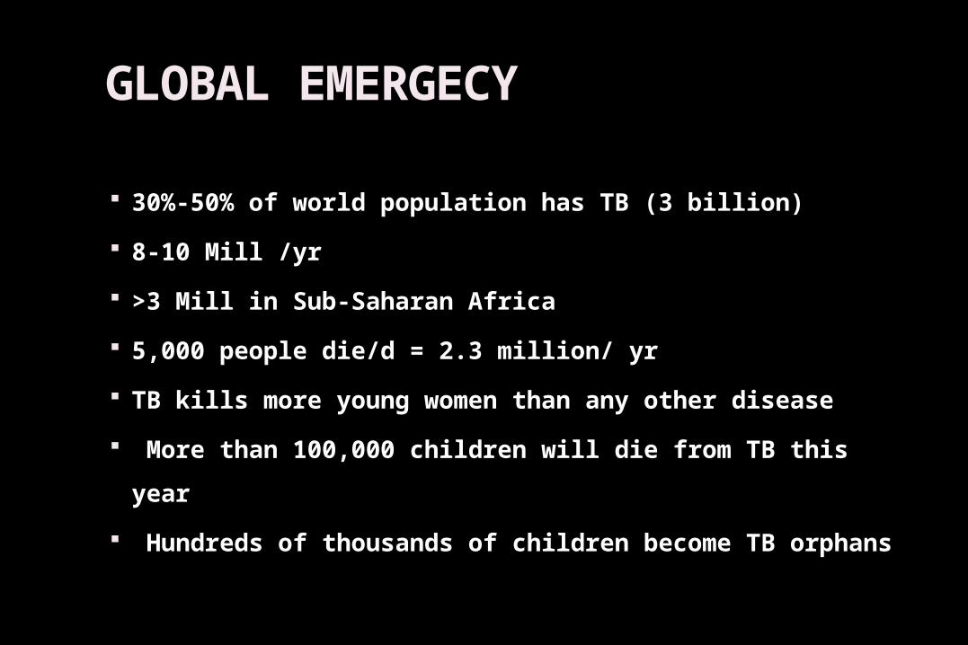
GLOBAL EMERGECY
30%-50% of world population has TB (3 billion)
8-10 Mill /yr
>3 Mill in Sub-Saharan Africa
5,000 people die/d = 2.3 million/ yr
TB kills more young women than any other disease
More than 100,000 children will die from TB this year
Hundreds of thousands of children become TB
orphans
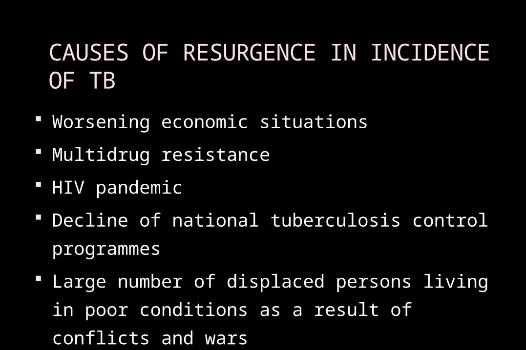
CAUSES OF RESURGENCE IN INCIDENCE OF TB
Worsening economic situations
Multidrug resistance
HIV pandemic
Decline of national tuberculosis control
programmes
Large number of displaced persons living in
poor conditions as a result of conflicts and wars
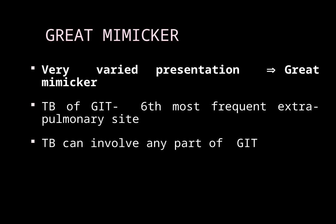
GREAT MIMICKER
Very varied presentation Great mimicker
TB of GIT- 6th most frequent extra-pulmonary site
TB can involve any part of GIT
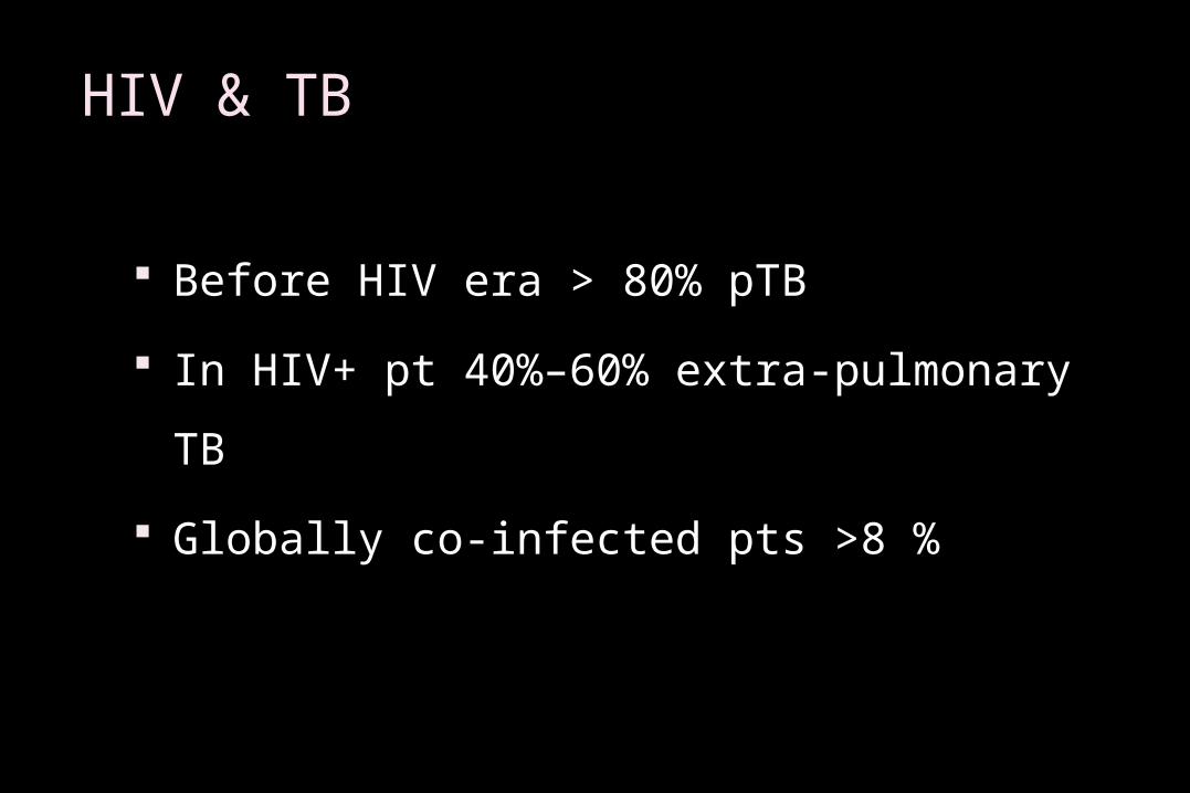
HIV & TB
Before HIV era > 80% pTB
In HIV+ pt 40%–60% extra-pulmonary TB
Globally co-infected pts >8 %
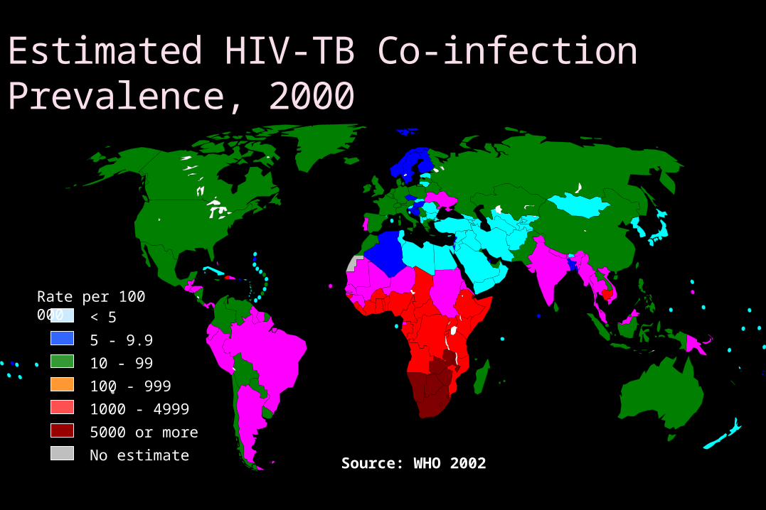
10 - 99
100 - 999
1000 - 4999
< 5
5 - 9.9
5000 or more
No estimate
Rate per 100 000
Source: WHO 2002
Estimated HIV-TB Co-infection Prevalence, 2000
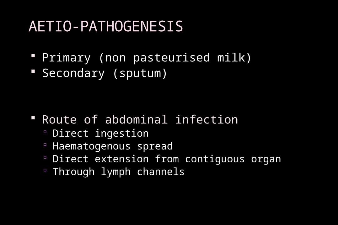
AETIO-PATHOGENESIS
Primary (non pasteurised milk) Secondary (sputum)
Route of abdominal infection Direct ingestion Haematogenous spread Direct extension from contiguous organ Through lymph channels
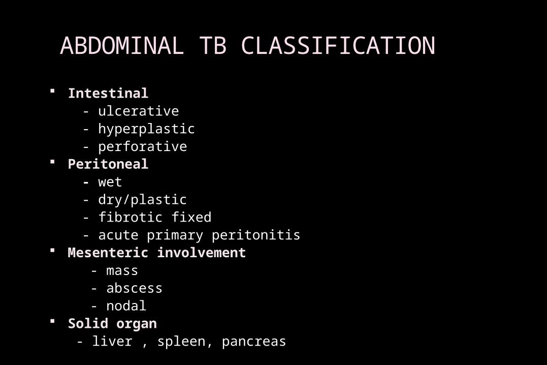
ABDOMINAL TB CLASSIFICATION
Intestinal - ulcerative - hyperplastic - perforative Peritoneal - wet - dry/plastic - fibrotic fixed - acute primary peritonitis Mesenteric involvement - mass - abscess - nodal Solid organ
- liver , spleen, pancreas
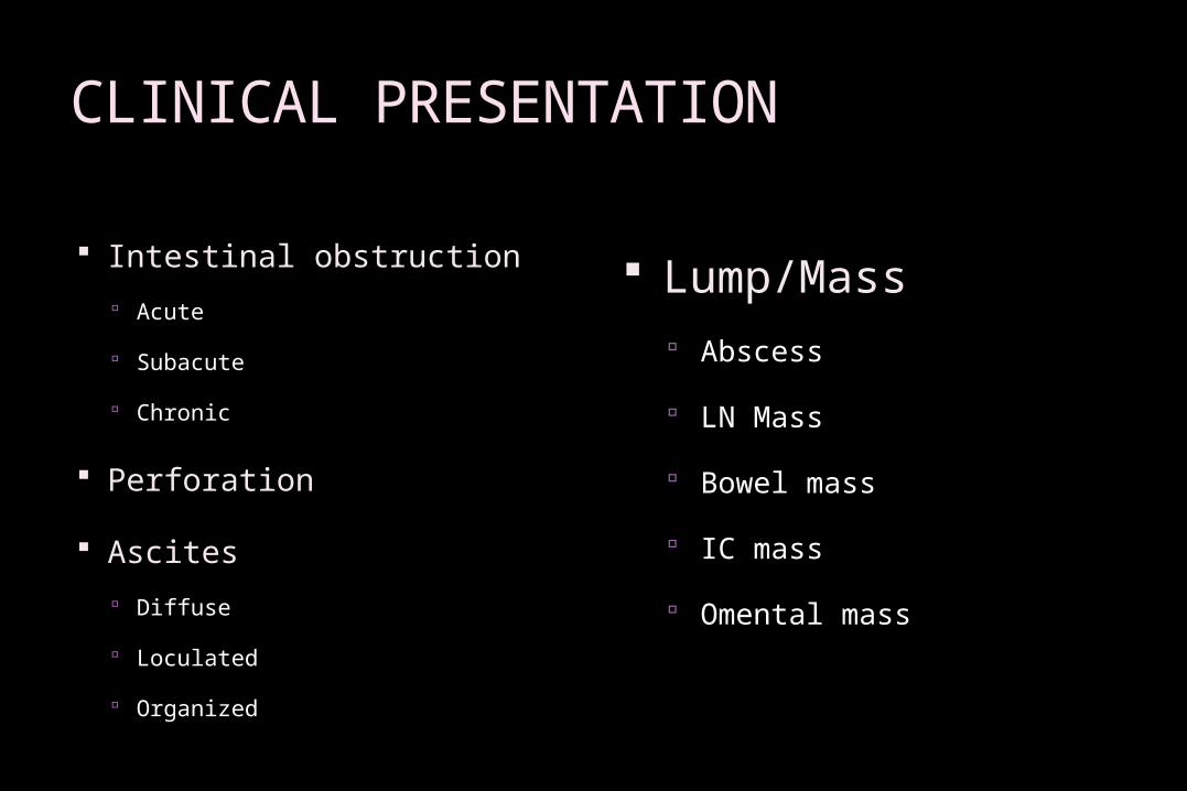
CLINICAL PRESENTATION
Intestinal obstruction
Acute
Subacute
Chronic
Perforation
Ascites
Diffuse
Loculated
Organized
Lump/Mass Abscess
LN Mass
Bowel mass
IC mass
Omental mass
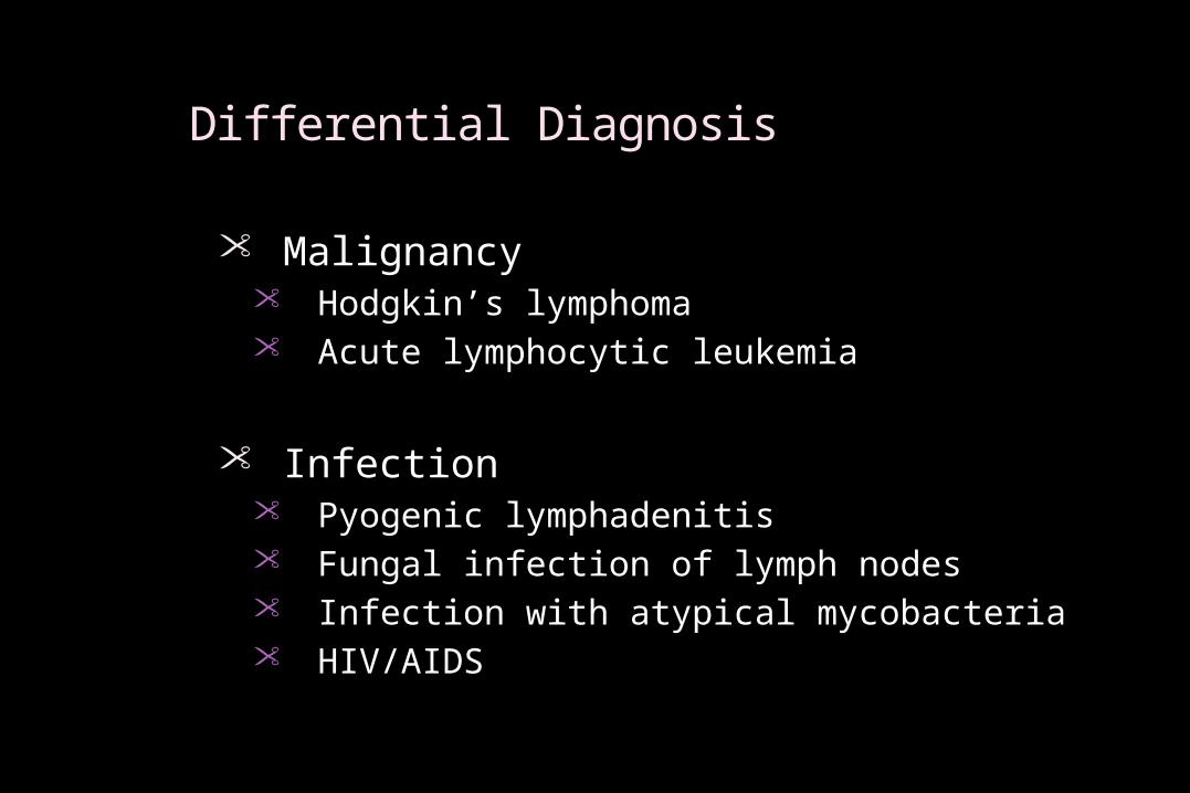
Differential Diagnosis
• Malignancy• Hodgkin’s lymphoma• Acute lymphocytic leukemia
• Infection• Pyogenic lymphadenitis• Fungal infection of lymph nodes• Infection with atypical mycobacteria• HIV/AIDS

Increased physiological stasis
Increased rate of fluid and electrolyte
absorption
Minimal digestive activity
Abundance of lymphoid tissue
Most common site - ileocaecal region
Ileum >caecum> ascending colon > jejunum>appendix > sigmoid > rectum > duodenum> stomach >oesophagus
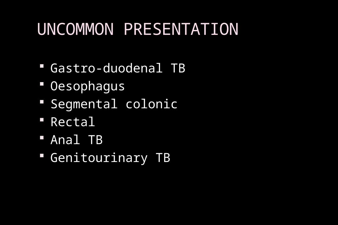
UNCOMMON PRESENTATION
Gastro-duodenal TB Oesophagus Segmental colonic Rectal Anal TB Genitourinary TB
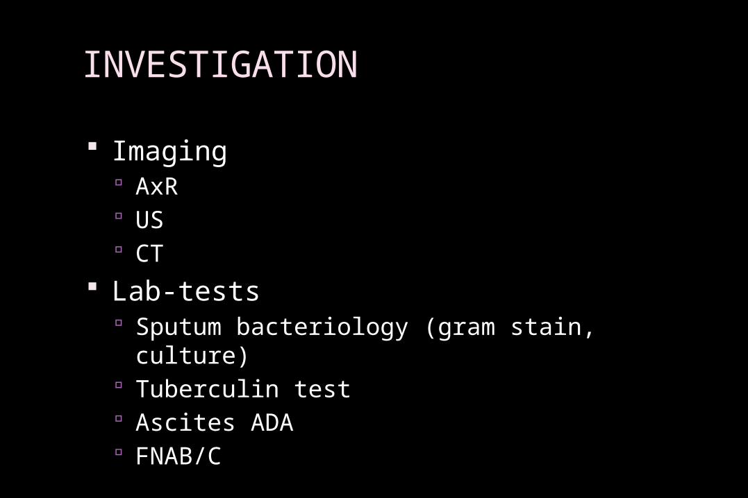
INVESTIGATION
Imaging AxR US CT
Lab-tests Sputum bacteriology (gram stain, culture) Tuberculin test Ascites ADA FNAB/C
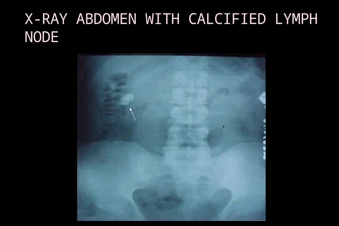
X-RAY ABDOMEN WITH CALCIFIED LYMPH NODE
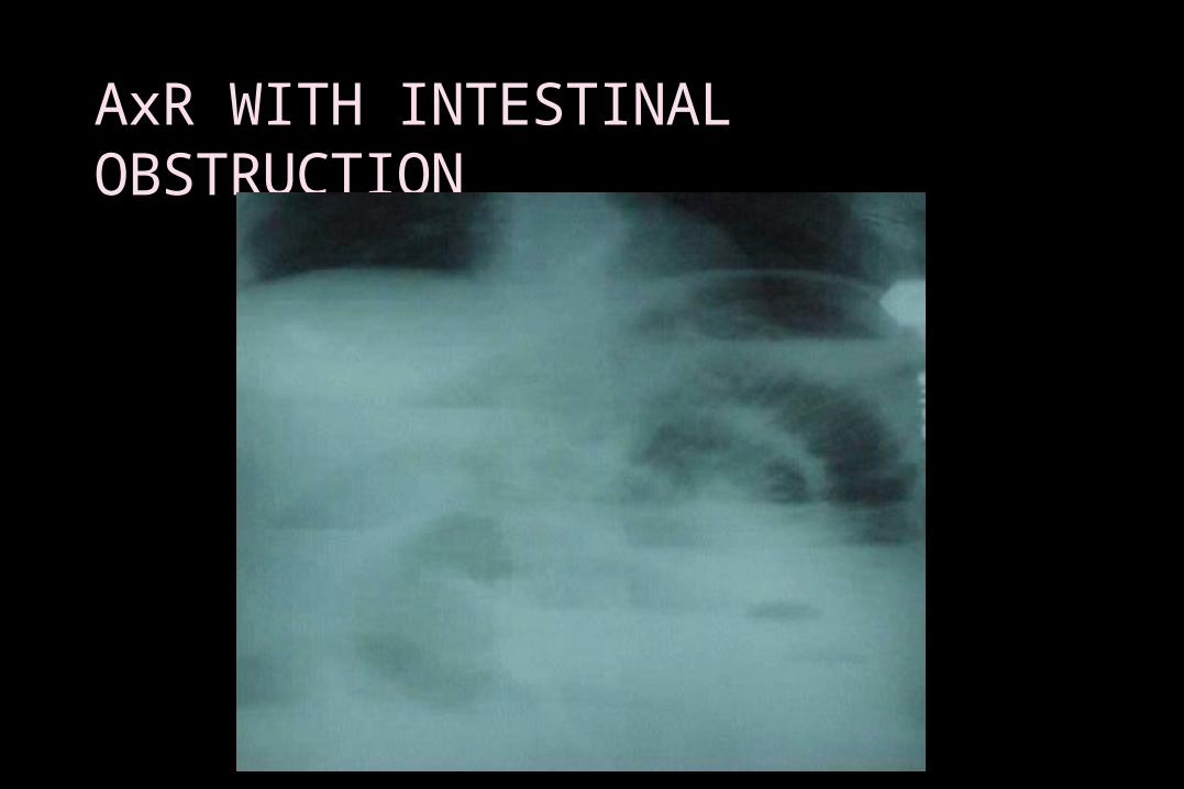
AxR WITH INTESTINAL OBSTRUCTION
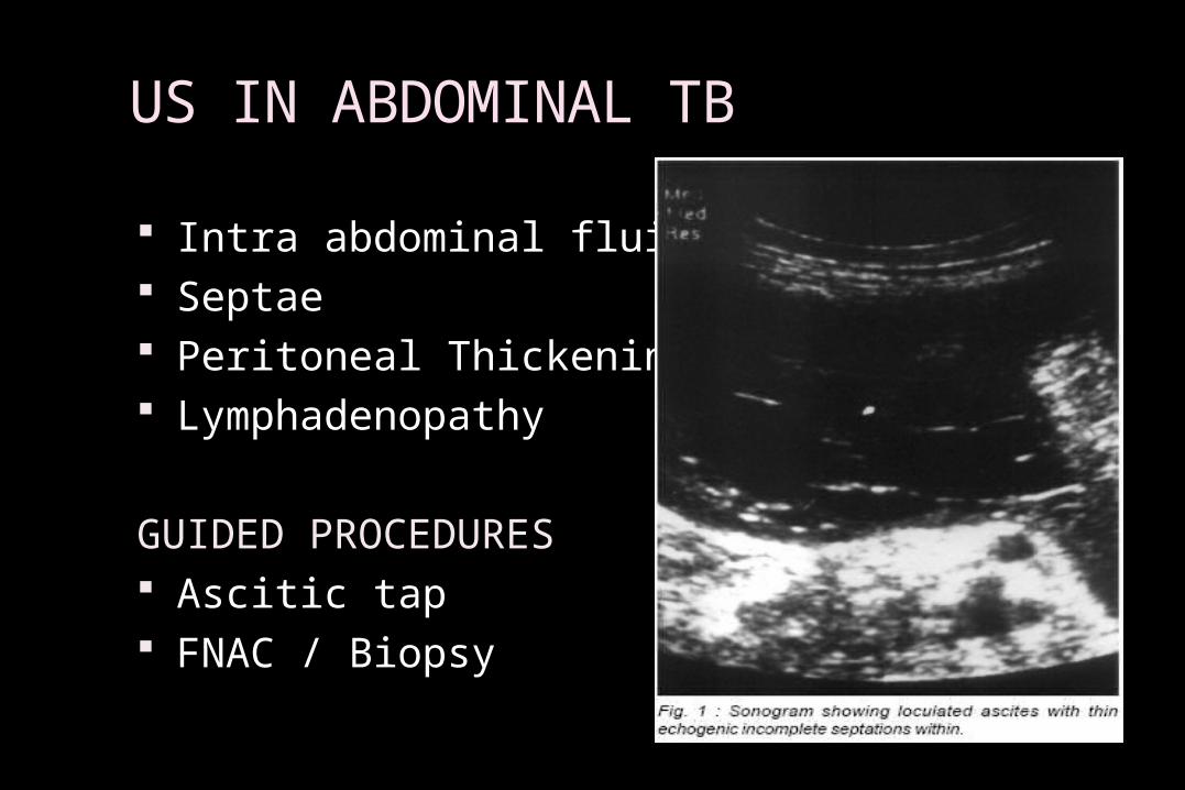
US IN ABDOMINAL TB
Intra abdominal fluid Septae Peritoneal Thickening Lymphadenopathy
GUIDED PROCEDURES Ascitic tap FNAC / Biopsy
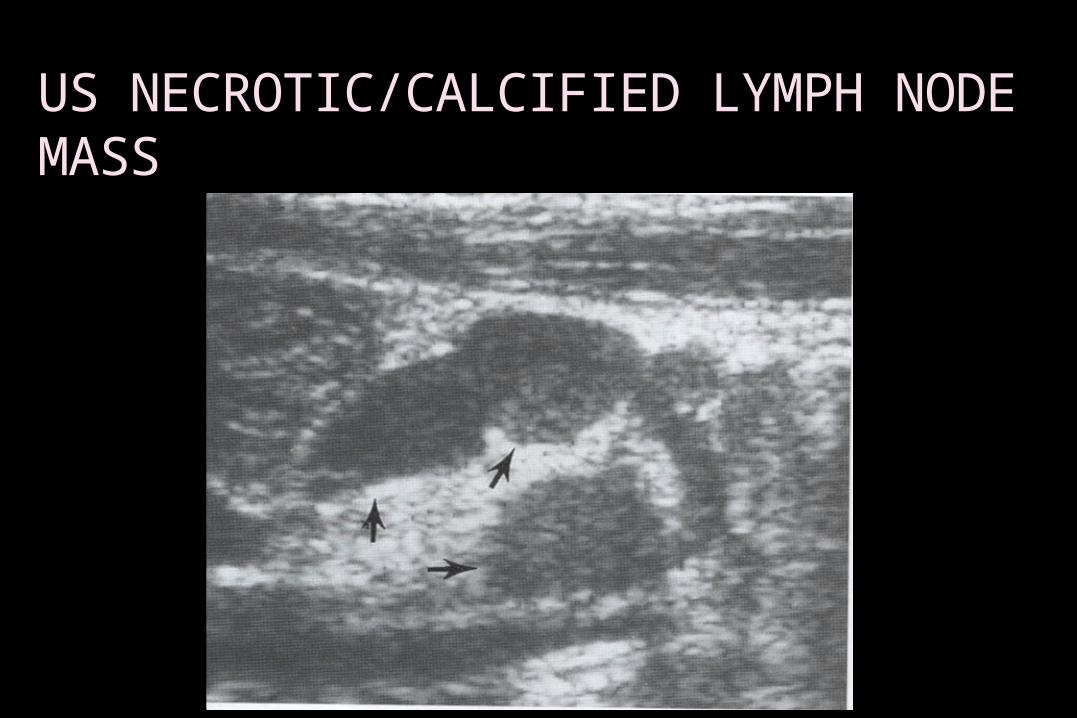
US NECROTIC/CALCIFIED LYMPH NODE MASS
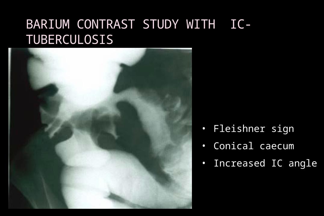
BARIUM CONTRAST STUDY WITH IC-TUBERCULOSIS
• Fleishner sign
• Conical caecum
• Increased IC angle
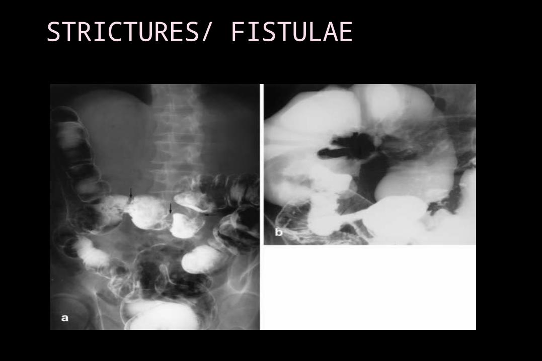
STRICTURES/ FISTULAE
CT SCAN ABDOMEN
Whenever diagnosis in doubt
Lymphadenopathy I C Mural thickening High density ascitis Irregular soft tissue densities in omental area
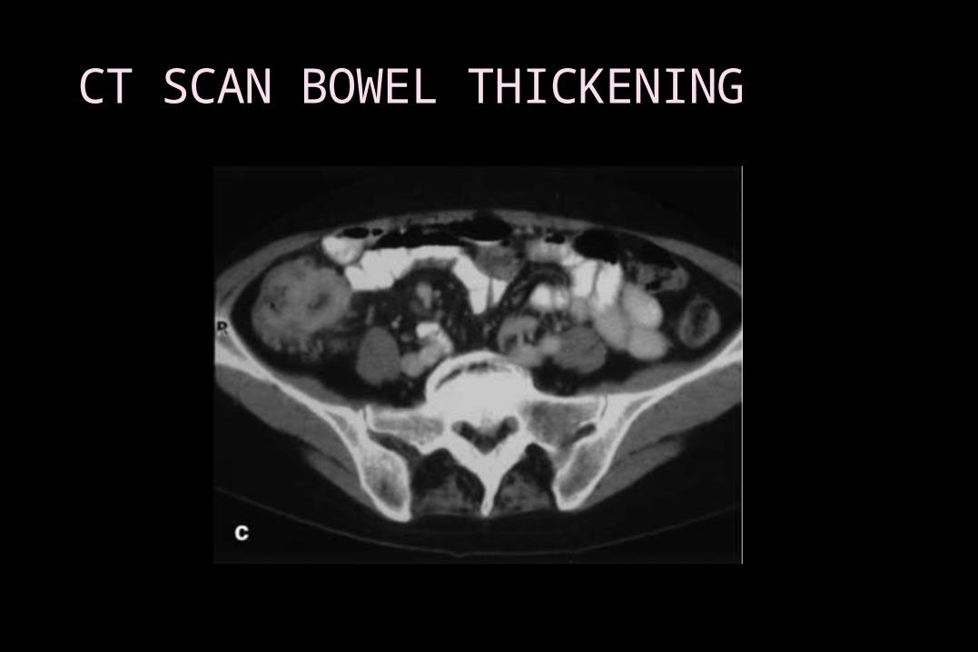
CT SCAN BOWEL THICKENING

TB peritonitis
a Axial contrast-enhanced CT • ascites paracolic gutter• thickened peritoneum (white arrow)• omental thickening (open arrow)• multiple rim-enhancing lymph nodes (black arrows)
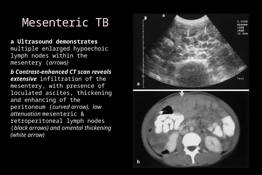
Mesenteric TB
a Ultrasound demonstrates multiple enlarged hypoechoic lymph nodes within the mesentery (arrows)
b Contrast-enhanced CT scan reveals extensive infiltration of the mesentery, with presence of loculated ascites, thickening and enhancing of the peritoneum (curved arrow), low attenuation mesenteric & retroperitoneal lymph nodes (black arrows) and omental thickening (white arrow)
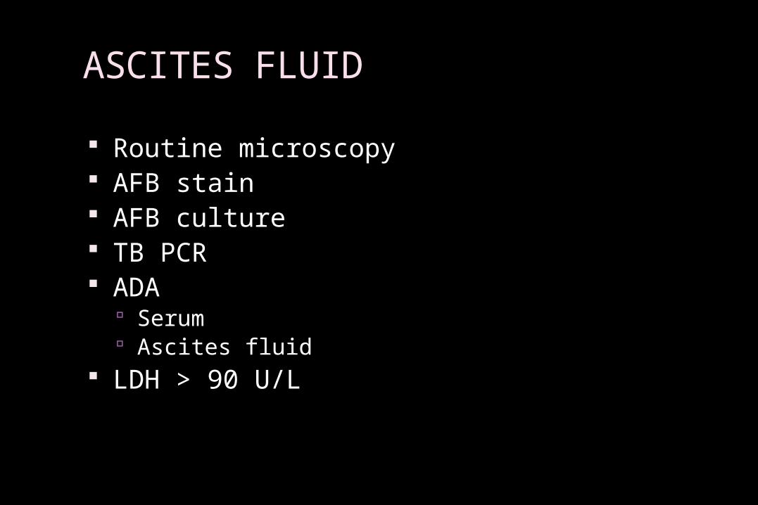
ASCITES FLUID
Routine microscopy AFB stain AFB culture TB PCR ADA
Serum Ascites fluid
LDH > 90 U/L
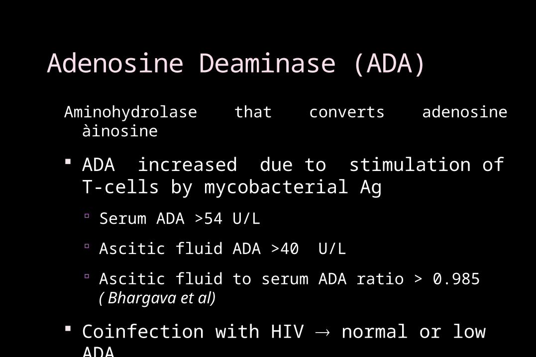
Adenosine Deaminase (ADA)
Aminohydrolase that converts adenosine àinosine
ADA increased due to stimulation of T-cells by mycobacterial Ag
Serum ADA >54 U/L
Ascitic fluid ADA >40 U/L
Ascitic fluid to serum ADA ratio > 0.985 ( Bhargava et al)
Coinfection with HIV normal or low ADA
BACTEC FAST METHOD OF TB CULTURE
Liquid (BACTEC) – results available in 10-
14 days
Solid (LJ Media) media – 4-6 wks
TB PCR
It is genetic test
Sensitive and specific
Rapid & Result available in few hours
Quantitative – 1 to 2 bacilli
LAPAROSCOPY
Advantage Diagnostic Biopsy Therapeutic May avoid empirical use of ATT
Disadvantage Invasive investigation Difficult Costly Expertise Complications
Laparoscopic Findings
Thickened peritoneum with tubercles
Multiple, yellowish white, uniform (~ 4-5mm) tubercles
Peritoneum is thickened & hyperemic
Omentum, liver, spleen also studded with tubercles
Thickened peritoneum without tubercles
Fibro adhesive peritonitis
Markedly thickened peritoneum and multiple thick adhesions
Caseatinggranulomas + in 85%-90% of Bx
TREATMENT
ATT as per dots recommendation
Ideally 6/52 before Sx
Might need increased oral dosage (malabsorption)
Empirical ATT to be condemned
Aspiration of abscess
Surgery for unrelieved obstruction
Surgery for perforation
COMPLICATIONS
Obstruction & perforation
Malnutrition and superinfection
Blind loop
Malabsorption
Enterocutaneous fistula
Short bowel syndrome
Infertility
TB AND HIV/AIDS
Children who acquire HIV by MTCT are also likely to be exposed
to TB
HIV Infection:
Increases the severity of TB in children
Prolongs Morbidity
Increases Mortality
Treatment may take longer to be effective.
HIV and TB increase the side effect from anti- TB drugs
Antiretroviral drugs interfere with anti -TB drugs
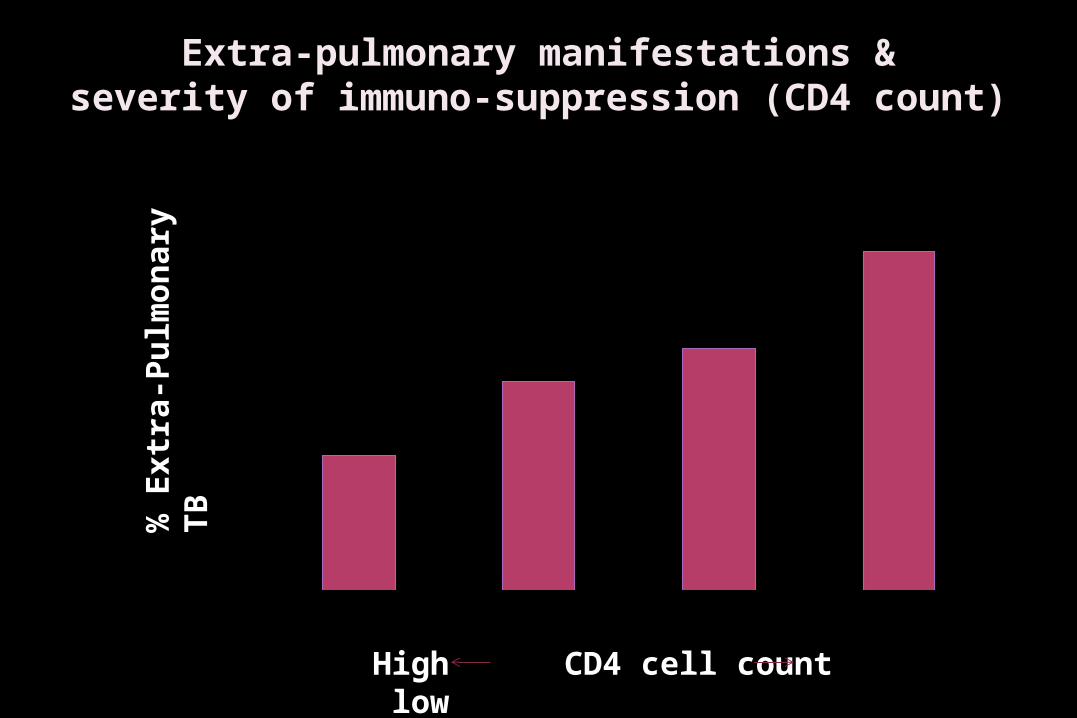
Extra-pulmonary manifestations &severity of immuno-suppression (CD4 count)
0
10
20
30
40
50
60
70
80
>300 201-300 101-200 0-100
High CD4 cell count low
% E
xtr
a-P
ulm
on
ary
TB
Immune reconstitution events in HIV - related TB
Definition - increase in manifestations of TB at prior sites
or new manifestations of disease
Closely associated with starting ARV (days to weeks)
Rarely associated with starting TB therapy
Natural history
• duration - days to months
• waxing and waning is common
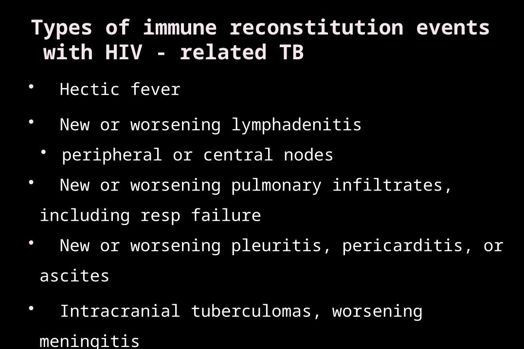
Types of immune reconstitution events with HIV - related TB
• Hectic fever
• New or worsening lymphadenitis
• peripheral or central nodes
• New or worsening pulmonary infiltrates, including resp
failure
• New or worsening pleuritis, pericarditis, or ascites
• Intracranial tuberculomas, worsening meningitis
• Disseminated skin lesions
• Epididymitis, hepatosplenomegaly, soft tissue
abscesses
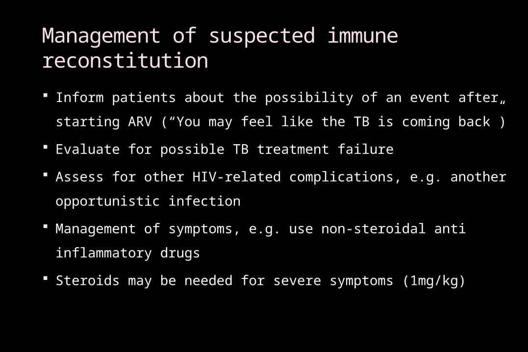
Management of suspected immune reconstitution
Inform patients about the possibility of an event after starting
ARV (“You may feel like the TB is coming back”)
Evaluate for possible TB treatment failure
Assess for other HIV-related complications, e.g. another
opportunistic infection
Management of symptoms, e.g. use non-steroidal anti
inflammatory drugs
Steroids may be needed for severe symptoms (1mg/kg)
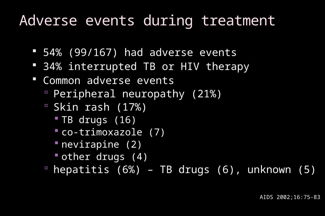
Adverse events during treatment
54% (99/167) had adverse events 34% interrupted TB or HIV therapy Common adverse events
Peripheral neuropathy (21%) Skin rash (17%)
TB drugs (16) co-trimoxazole (7) nevirapine (2) other drugs (4)
hepatitis (6%) – TB drugs (6), unknown (5)
AIDS 2002;16:75-83
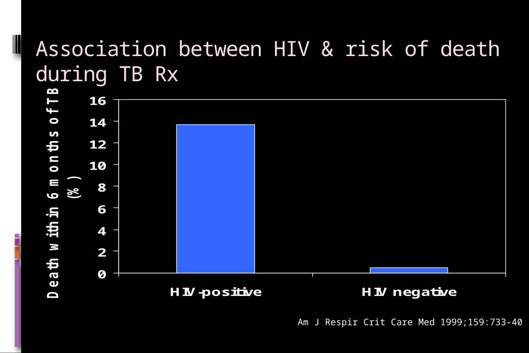
Association between HIV & risk of death during TB Rx
0
2
4
6
8
10
12
14
16
HIV-positive HIV negativeDeath
wit
hin
6 m
on
ths o
f T
B d
iag
no
sis
(%)
Am J Respir Crit Care Med 1999;159:733-40
CONCLUSIONS
Suspicion
Diagnosis is possible
FNAC/B, ADA, TB PCR are valuable tests
Empirical ATT should be avoided
Laparoscopy is an important diagnostic tool
Surgery for unavoidable reasons only
Complex issues (IRIS) with HIV

THANK YOU!