a wake-up call for resting follicles · 23.03.2018 · dormant follicles: iva, in vitro activation...
TRANSCRIPT

Director of Reproduction and Infertility CenterSt. Marianna University School of Medicine
Kazuhiro Kawamura, M.D., Ph.D.
A wake-up call for resting follicles

Contents
1. Background of POI
1. Basic and translational studies for in vitro activation of dormant follicles (IVA)
1. Clinical application of IVA
1. Future studies for IVA

Contents
1. Background of POI
1. Basic and translational studies for in vitro activation of dormant follicles (IVA)
1. Clinical application of IVA
1. Future studies for IVA
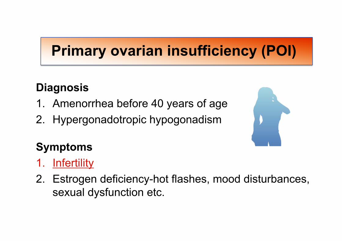
Primary ovarian insufficiency (POI)
Diagnosis1. Amenorrhea before 40 years of age2. Hypergonadotropic hypogonadism
Symptoms1. Infertility2. Estrogen deficiency-hot flashes, mood disturbances,
sexual dysfunction etc.

1. Genetic—Turner syndrome, FMR1, etc.
2. Immunological—auto-immune disease
3. Iatorogenic—extensive ovarian cystectomy, partial oophorectomy, chemo-/radiation-therapies
4. Others (unknown)
EtiologyPOI affects approx. 1% of women
Primary ovarian insufficiency (POI)

�Egg donation is the most successful treatment option, but…
�Resistant to traditional gonadotropin treatments
Treatments
�Lack of follicle growth and ovulation�Exhaustion of ovarian follicles andfew residual follicles: <1,000 follicles
(undetectable AMH levels)
Specific features
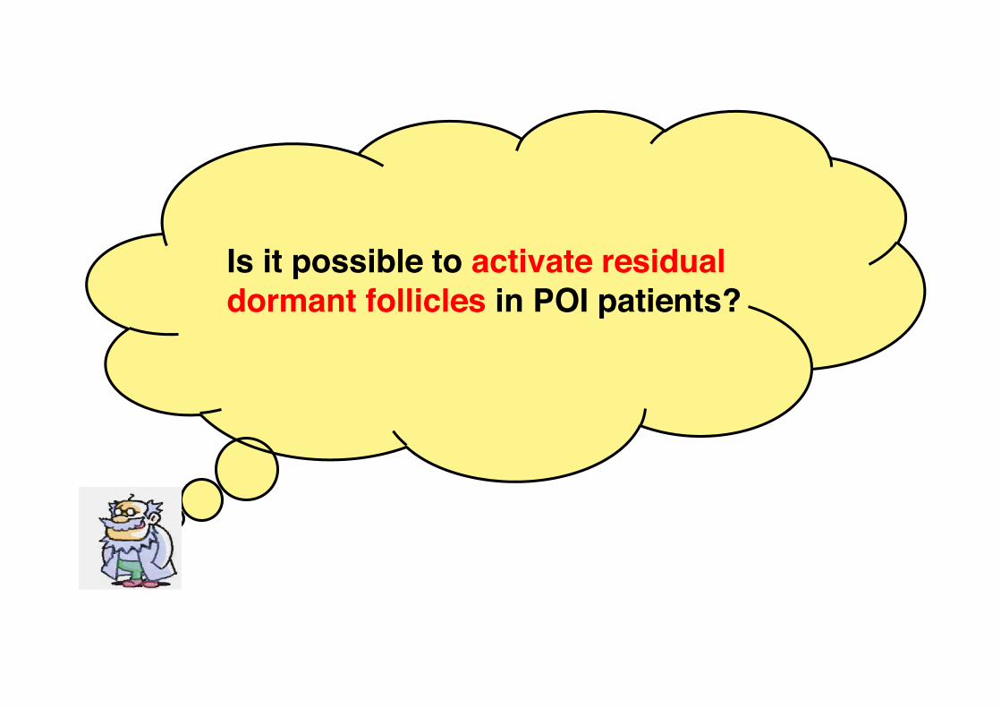
Is it possible to activate residual dormant follicles in POI patients?

Contents
1. Background of POI
1. Basic and translational studies for in vitro activation of dormant follicles (IVA)
1. Clinical application of IVA
1. Future studies for IVA
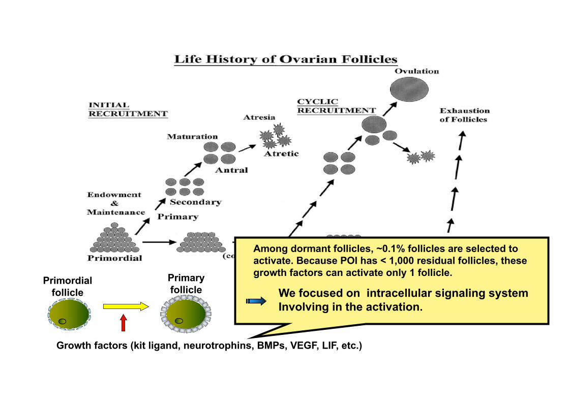
Primordial follicle
Primary follicle
Growth factors (kit ligand, neurotrophins, BMPs, VEGF, LIF, etc.)
Among dormant follicles, ~0.1% follicles are selected to activate. Because POI has < 1,000 residual follicles, these growth factors can activate only 1 follicle.
We focused on intracellular signaling system Involving in the activation.

Reddy et al. Science, 2008 Castrillon et al. Science, 2003
At early stage after birth, PTEN or FOXO3 deletion led to the activation of dormant primordial follicles and resulted in depletion of follicles within 16-18 weeks.
PTEN null mice Foxo3 null mice
PTEN PIP3PIP2 PDK1
SGKAkt
GSK3
BADp27
Apoptosis,Cell-cyclearrest
TSC2TSC1
RHEB4EBPEIF4E p70S6K
Proteinsynthesis
SGKAkt
FOXO3FOXO4
FOXO1
PI3K
Apoptosis, cell-cycle arrestNucleus
TOR
RTK
Growth factors
CRKGRB2
Ras
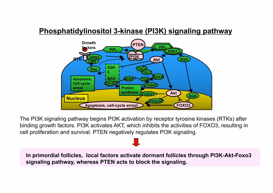
Phosphatidylinositol 3-kinase (PI3K) signaling pathway
The PI3K signaling pathway begins PI3K activation by receptor tyrosine kinases (RTKs) after binding growth factors. PI3K activates AKT, which inhibits the activities of FOXO3, resulting in cell proliferation and survival. PTEN negatively regulates PI3K signaling.
In primordial follicles, local factors activate dormant follicles through PI3K-Akt-Foxo3 signaling pathway, whereas PTEN acts to block the signaling.

Is it possible to activate residual dormant follicles in POI patients artificially by transient PTEN suppression and/or PI3K activation using drugs?
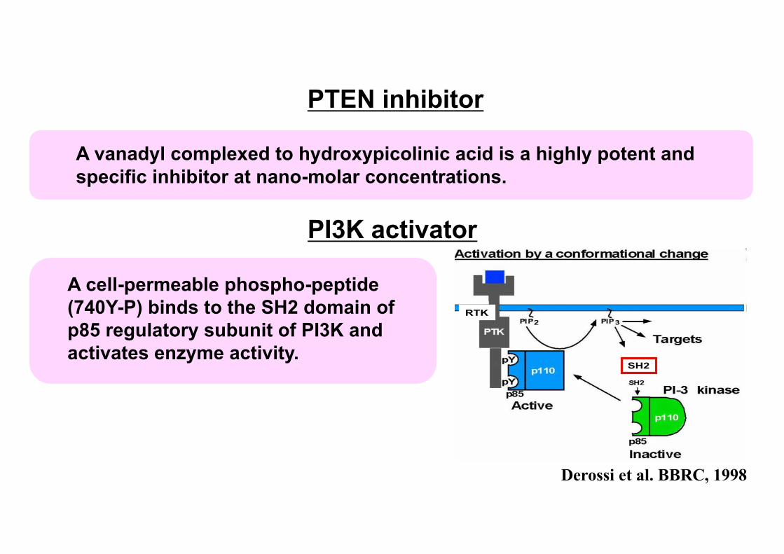
PI3K activator
PTEN inhibitor
A vanadyl complexed to hydroxypicolinic acid is a highly potent and specific inhibitor at nano-molar concentrations.
RTK
SH2
Derossi et al. BBRC, 1998
A cell-permeable phospho-peptide (740Y-P) binds to the SH2 domain of p85 regulatory subunit of PI3K and activates enzyme activity.
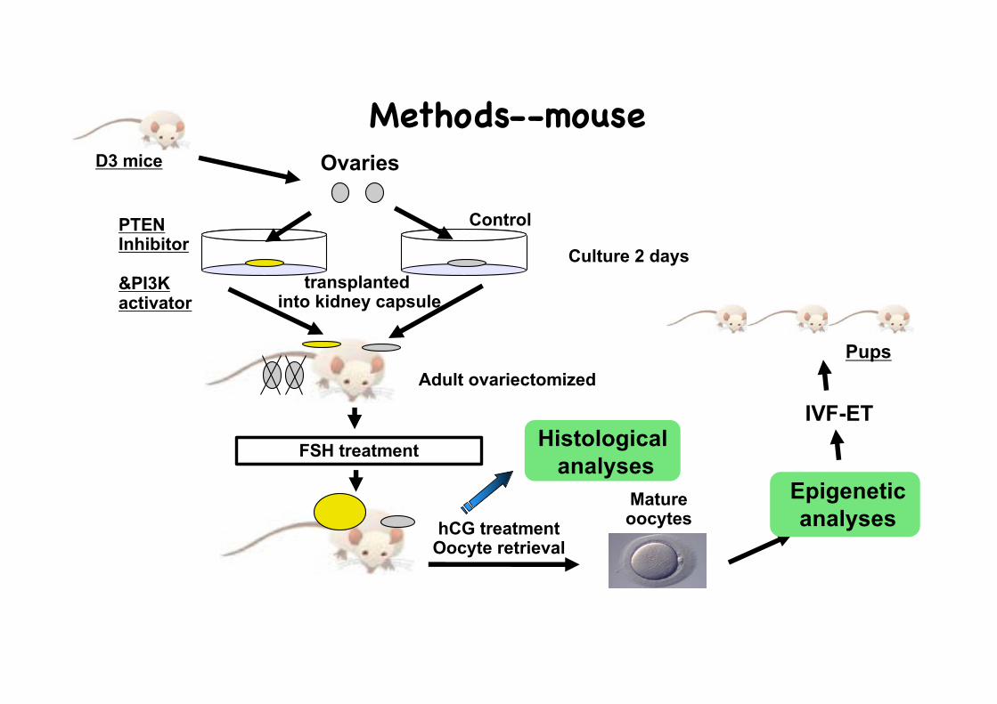
Ovaries
PTENInhibitor
&PI3Kactivator
Control
transplanted into kidney capsule
FSH treatment
hCG treatment Oocyte retrieval
Mature oocytes
Adult ovariectomized
Methods--mouseD3 mice
Epigeneticanalyses
IVF-ET
Pups
Histologicalanalyses
Culture 2 days

PTEN inhibitor
PI3K activator
control
control
controlPTEN inhibitor
+PI3K activator
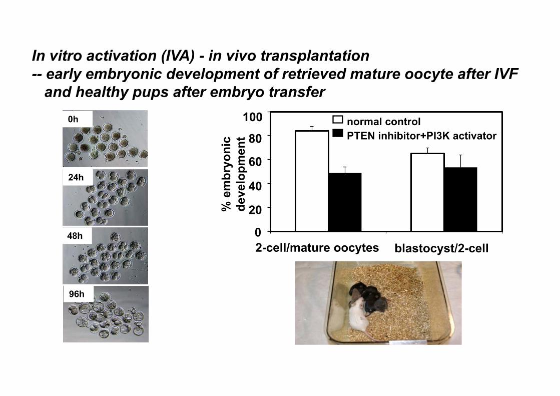
In vitro activation (IVA) - in vivo transplantation
Changes in ovarian size at day 14 after transplantaion of D3 ovaries treated with PTEN inhibitor and/or PI3K activator beneath kidney capsule of host mice.

% to
tal f
ollic
les
primordial primary preantral antral largeantral
100
80
60
40
20
0
20
15
10
5
0
Follicular dynamics at day 14 after transplantaion of activated ovaries beneath kidney capsule of host mice.
PTEN inhibitor+PI3K activator
* *
*
*
*
primordial primary preantral antral largeantral
In vitro activation (IVA) - in vivo transplantation-- ovarian histology

In vitro activation (IVA) - in vivo transplantation-- genome imprinting and meiotic spindle formation of retrieved oocyte
b-tubulin staining
Igf2r
Lit1
H19
Meiotic spindle formation was evaluated by b-tubulinstaining, whereas the integrity of genomic imprinting was confirmed by detecting methylation of CpG sites in Differentially methylated region (DMR) of some imprint genes(maternal:Igf2r, Lit1, paternal:H19).
48h
96h
0
20
40
60
80
100
1 2
normal controlbpV(pic)+740Y-P
**
% e
mbr
yoni
c de
velo
pmen
tblastocyst/2-cell
0h
In vitro activation (IVA) - in vivo transplantation-- early embryonic development of retrieved mature oocyte after IVF
and healthy pups after embryo transfer
24h
20
2-cell/mature oocytes
40
60
80100
0
normal controlPTEN inhibitor+PI3K activator
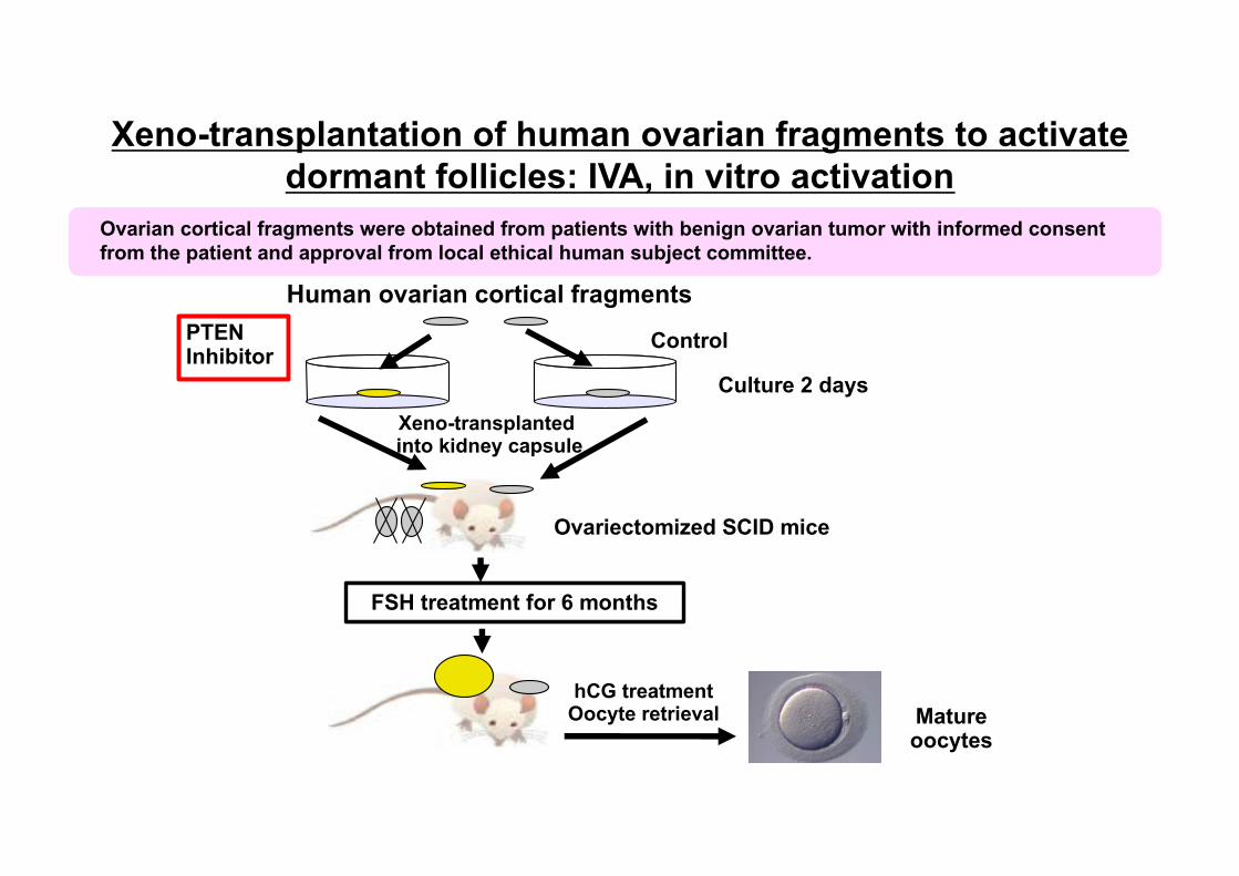
Human ovarian cortical fragmentsPTENInhibitor
Control
Xeno-transplanted into kidney capsule
FSH treatment for 6 months
hCG treatment Oocyte retrieval Mature
oocytes
Ovariectomized SCID mice
Xeno-transplantation of human ovarian fragments to activate dormant follicles: IVA, in vitro activation
Ovarian cortical fragments were obtained from patients with benign ovarian tumor with informed consent from the patient and approval from local ethical human subject committee.
Culture 2 days

Morphology of human ovarian fragments after 6 monthsof xeno-transplantation
control
PTEN inhibitor
control
PTEN inhibitor

Histology of PTEN inhibitor treated ovarian fragments
At 36 h after hCG treatment, large antral follicles in the PTEN inhibitor-treated group contained mature oocytes at metaphase II accompanied with cumulus expansion.
Li and Kawamura et al PNAS 2010

Contents
1. Background of POI
1. Basic and translational studies for in vitro activation of dormant follicles (IVA)
1. Clinical application of IVA
1. Future studies for IVA
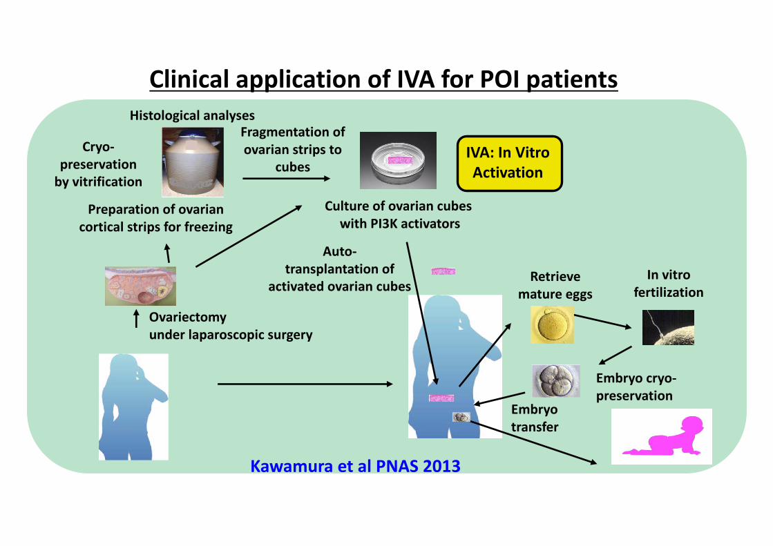
Clinical application of IVA for POI patients
Ovariectomyunder laparoscopic surgery
Cryo-preservation
by vitrificationCulture of ovarian cubes
with PI3K activators
Auto-transplantation of
activated ovarian cubes Retrieve mature eggs
In vitrofertilization
Embryo cryo-preservation
IVA: In Vitro Activation
Preparation of ovarian cortical strips for freezing
Fragmentation of ovarian strips to
cubes
Embryo transfer
Histological analyses
Kawamura et al PNAS 2013
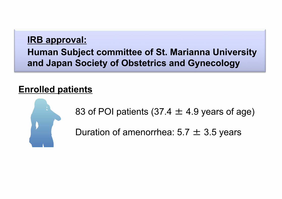
Enrolled patients
IRB approval:
83 of POI patients (37.4 � 4.9 years of age)
Duration of amenorrhea: 5.7 � 3.5 years
Human Subject committee of St. Marianna Universityand Japan Society of Obstetrics and Gynecology

ovariectomy
�Ovariectomy under laparoscopic surgery
�Minimum usage of electrocautery hemostasis to avoid damage of residual follicles.

Localization of early follicles in ovarian cortex
medulla
cortex1-2 mm

�Dissect ovarian cortices containing residual follicles by removing medulla.
�Cut into small strips (1 x 1 cm2, 1-2 mm thickness, where residual follicles are located).
�(Option: Cryo-preserve by vitrification method.)
�6-8 pieces of ovarian stripes could be obtained from one POI ovary.
�Using 10% of volume of each ovarian stripe,detect residual follicles.
histological analyses
Before dissection of medulla
After dissection of medulla
Small ovarian stripes readyfor use

�Fragment 2-3 ovarian pieces into 1-2 mm2 of cubes
�IVA drugs treatment (PTEN inhibitor and PI3K activator) for 2 days to activate dormant follicles
Culture of ovarian cubes
In Vitro Activation
Fragmentation of ovarian strips
to cubes

�Before auto-transplantation, wash cultured ovarian cubes by warmed culture media alone to avoid to introduce reagents inside of body.
�Transplant beneath the serosa of Fallopian tubes (20-40 cubes per site).
Beneath serosa of Fallopian tubes — high vascularization,
convenience for trans-vaginal ultrasound monitoring ease for oocyte retrieval
Culture of ovarian cubes
Auto-transplantation of activated ovarian
cubes
In Vitro Activation

�Monitor follicle growth weekly to biweekly: transvaginal ultrasound +serum estrogen and gonadotropin levels.
�After normalizing LH levels using EP pills and GnRHa, follicle growth was promoted by rFSH and hMG under GnRHa or GnRH AN protocols (Zhai, Kawamura, et al. JCEM 2016).
�After hCG treatment, oocyte retrieval followed by IVF was performed.
Retrieve mature eggs
In vitrofertilization
Patients’ follow up protocols
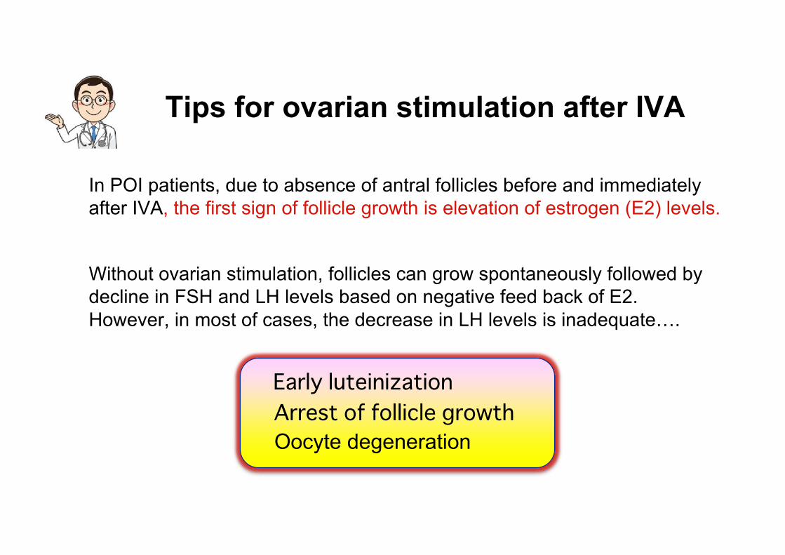
Tips for ovarian stimulation after IVA
In POI patients, due to absence of antral follicles before and immediately after IVA, the first sign of follicle growth is elevation of estrogen (E2) levels.
Early luteinizationArrest of follicle growthOocyte degeneration
Without ovarian stimulation, follicles can grow spontaneously followed by decline in FSH and LH levels based on negative feed back of E2. However, in most of cases, the decrease in LH levels is inadequate….

Orisaka 2013 Endocrinology
cAMP production after FSH stimulation
Follicle growth after FSH stimulation Preculture with LH
FSH stimulation
Effects of hyper-LH on oocyte-granulosa-theca cell interactions
Chronic LHstimulation
GDF9↓
FSHR↓
CYP17↑Preantral follicles exposed to high LH express low levels GDF-9 in oocyte and FSHR in granulosa cells, resulting in decreases in sensitivity of FSH stimulation and suppression of follicle growth.

How can we stimulate ovaries after IVA?
1. Normalize LH levels by supplementation of estrogen and estrogen + progesterone with induction of withdrawal bleeding.
2. After confirmation of normal LH levels (<10 mIU/ml), maintain its low levels using GnRHa. (Daily GnRH AN injection is too expensive)
3. Similar to short protocol, treat patients with rFSH or pure HMG (low LH content) for >2 weeks.
follicle growth (+)
follicle growth (-)
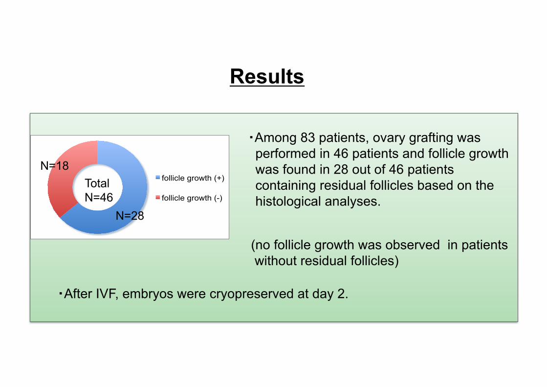
Results
�Among 83 patients, ovary grafting was performed in 46 patients and follicle growth was found in 28 out of 46 patients containing residual follicles based on the histological analyses.
(no follicle growth was observed in patients without residual follicles)
�After IVF, embryos were cryopreserved at day 2.
Total N=46
N=28
N=18
Results
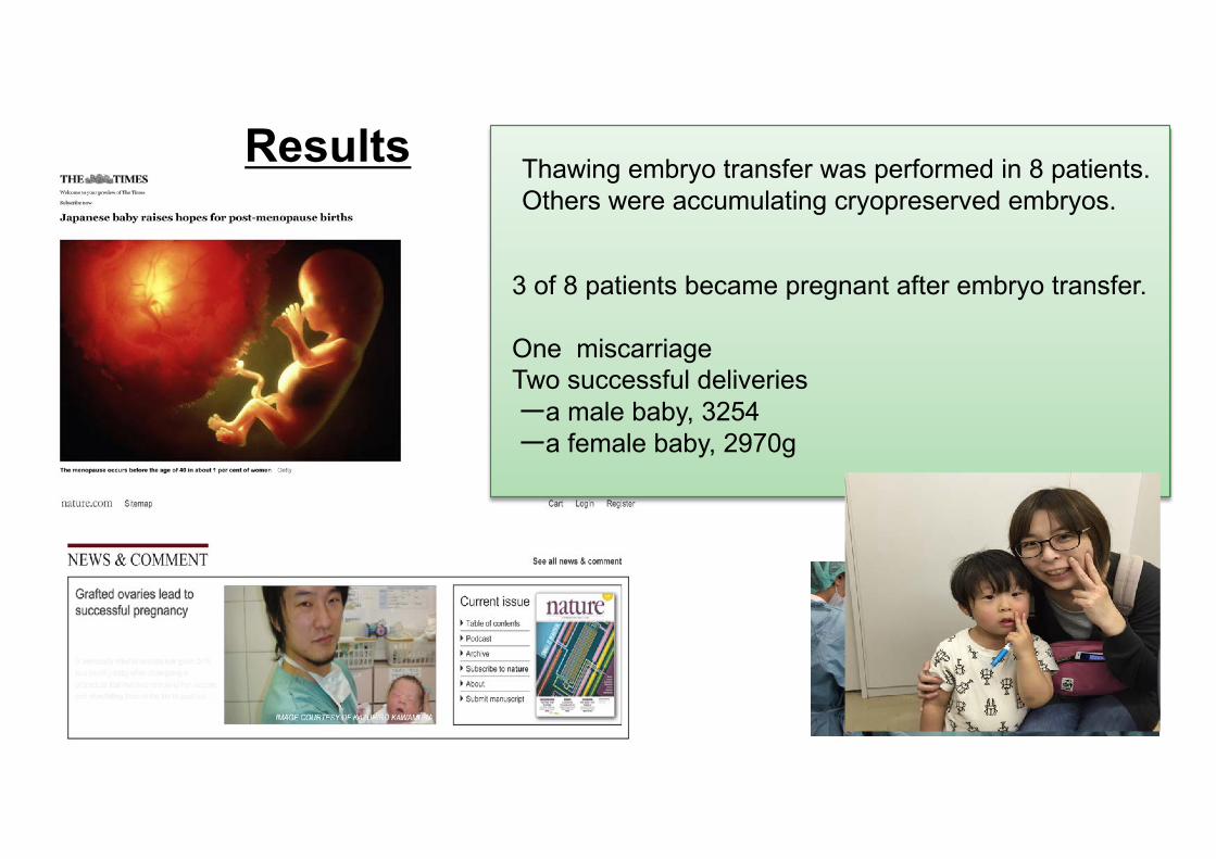
3 of 8 patients became pregnant after embryo transfer.
One miscarriage Two successful deliveries �a male baby, 3254�a female baby, 2970g
Thawing embryo transfer was performed in 8 patients. Others were accumulating cryopreserved embryos.

Current clinical outcome of IVA
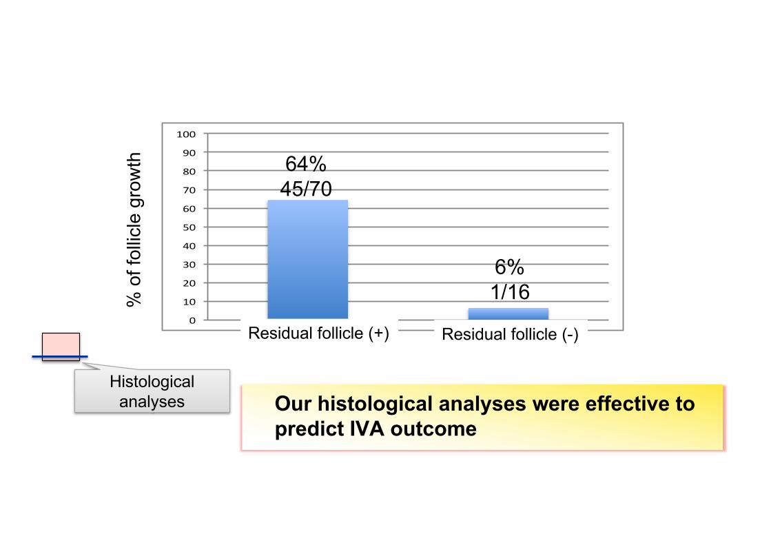
�Residual follicles based on histology:Positive: n=96Negative: n=56
�Ovariectomy: n=152
�IVA grafting with residual follicles with follow up > 6 months: n=70
Total N=152
Positive N=96
Negative N=56
�IVA grafting without residual follicles with follow up > 6 months: n=16
0"
10"
20"
30"
40"
50"
60"
70"
80"
90"
100"
64%45/70
6%1/16%
of f
ollic
le g
row
th
Our histological analyses were effective to predict IVA outcome
Histological analyses
Residual follicle (+) Residual follicle (-)

�POI/DOR patients with residual follicles.
(Young POI/DOR patients without oocyte aging).
Indication for IVA treatment

�� �$%��"�"�+��� �)�%+ ��� ��� ���
�� ��"��# �"�������!��%����
�� �$%�&&��#"��%�"����"����"�����+����
Results
��$%#�(���� �'+�#���� �*�&�� %���+��#"��%!����+����"����$��"���# �"���%#($&�("��%�#(%��(���"��� Kawamura et al Hum Reprod 2015
Zhai et al JCEM 2016
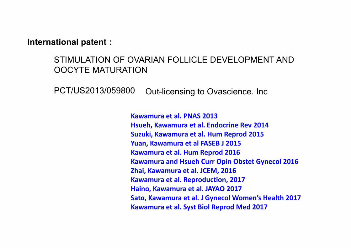
International patent:
Out-licensing to Ovascience. Inc
Kawamura et al. PNAS 2013Hsueh, Kawamura et al. Endocrine Rev 2014Suzuki, Kawamura et al. Hum Reprod 2015Yuan, Kawamura et al FASEB J 2015Kawamura et al. Hum Reprod 2016Kawamura and Hsueh Curr Opin Obstet Gynecol 2016Zhai, Kawamura et al. JCEM, 2016Kawamura et al. Reproduction, 2017Haino, Kawamura et al. JAYAO 2017Sato, Kawamura et al. J Gynecol Women’s Health 2017Kawamura et al. Syst Biol Reprod Med 2017
STIMULATION OF OVARIAN FOLLICLE DEVELOPMENT AND OOCYTE MATURATION
PCT/US2013/059800

IVA was awarded to be one of the Top 10 medical breakthrough in 2013 by TIME magazine.

Follicle growth from primordial to preovulatory stage takes more than 4-6 months.
In contrast to our expectation, we found follicle growth before 6 months after grafting.
This result suggested that our IVA method also stimulated growth of secondary follicles in grafted ovaries.
Derived from secondary follicles
Derived from primordial follicles
Temporal follicle growth in transplanted ovaries

Ovarian fragmentation led to changes in intercellular tension and facilitated the conversion of G-actin to F-actin.
Subsequent disruption of Hippo signaling decreased pYAP to total YAP ratios, leading to increased in downstream CCN growth factors.
Secretion of CCN growth factors stimulated follicle growth.
Ovarian fragmentation suppresses Hippo signaling, leading to follicle growth
Hippo pathway genes
YAP YAPP
YAP
CCNGrowthfactors
G-ActinF-Actin
14-3-3
Degradation
Anti-apoptotic
factors Secondary follicle growth
Contents
1. Background of POI
1. Basic and translational studies for in vitro activation of dormant follicles (IVA)
1. Clinical application of IVA
1. Future studies for IVA

Two-step follicle stimulation in IVA

1. Original IVA (PI3K stimulation and Hippo disruption)
Oophrectomy
Fragmentation and IVA drug treatment (2 days)
POI patients Two surgeries
Cryo-preservation
DOR/early POI patients
Fragmentation and immediate return
Partial cortex removal
One surgeryIVI workshop, Bilbao, 2017
2. Drug-free IVA (Hippo disruption only)

Limitation:
Drug-free IVA and orthologous grafting
Only applicable to DOR/early POI patients who likely have secondary follicles
Advantages:
IVI group, Spain (Bilbao workshop 2017)3 spontaneous pregnancies/14 patients
�Minimal damages to ovarian blood supply� No need to use Akt-stimulating drugs� Avoid potential follicle loss during culture� One laparoscopic surgery� Spontaneous pregnancy possible
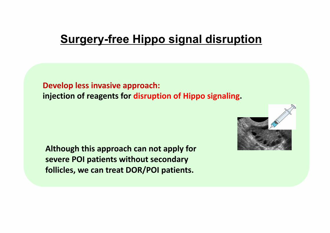
Surgery-free Hippo signal disruption
Develop less invasive approach:injection of reagents for disruption of Hippo signaling.
Although this approach can not apply for severe POI patients without secondary follicles, we can treat DOR/POI patients.
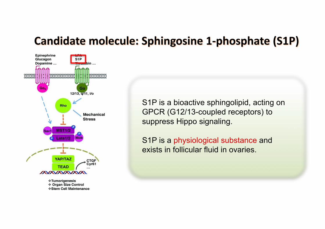
Candidate molecule: Sphingosine 1-phosphate (S1P)
S1P is a bioactive sphingolipid, acting on GPCR (G12/13-coupled receptors) to suppress Hippo signaling.
S1P is a physiological substance and exists in follicular fluid in ovaries.

Immunostaining Real-time qPCR
Effects of S1P on disruption of Hippo signaling in D10 mouse ovarian tissue culture
S1P stimulates nuclear translocation of YAP in granulosa cells followed by increase in expression of downstream CCN2 growth factor.

Histological analyses
S1P increased ovarian weight and stimulated early secondary follicle growth.
Effects of S1P on secondary follicle growth in D10 mouse ovarian tissue culture
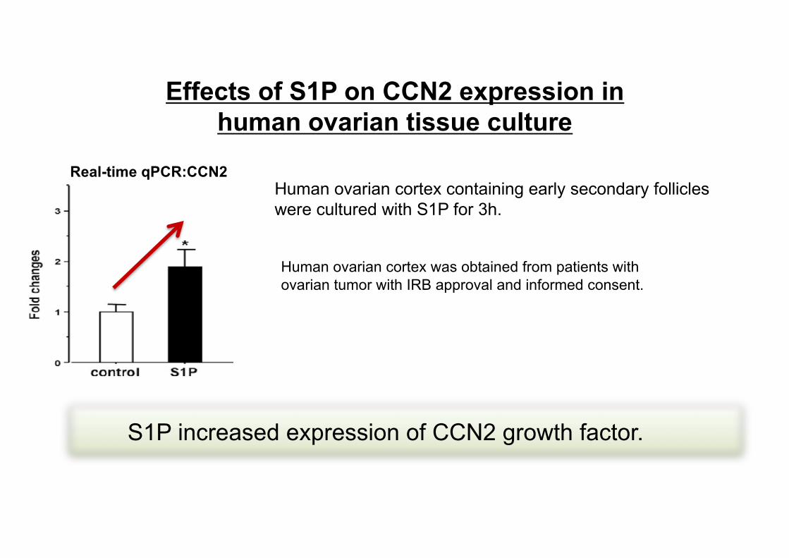
Effects of S1P on CCN2 expression in human ovarian tissue culture
Human ovarian cortex was obtained from patients with ovarian tumor with IRB approval and informed consent.
Human ovarian cortex containing early secondary follicles were cultured with S1P for 3h.
S1P increased expression of CCN2 growth factor.
Real-time qPCR:CCN2

S1P disrupts Hippo signaling in early follicles leading to stimulation of secondary follicle growth.
Yuan, Kawamura et al FASEB J 2015
Because S1P is physiological substance existing in
follicular fluid, intake or injection of S1P expects to
stimulate follicular growth in POI/DOR patients
including aging without severe adverse reactions.
Summary
Patent: PCT/US2013/059800
Collaborators
Bunpei Ishizuka, Nao SuzukiNanami Kawamura, Yorino Sato, Naoki Okamoto, Ikko Kawashima, Midori Tamura, Seido Takae, Yodo Sugishita, NobuhitoYoshioka, Yuta Kawagoe, Mariko Hoshina, Noriyuki Takahashi
Aaron JW HsuehYuan ChengJing Li
St. Marianna University School of Medicine
Stanford University School of Medicine

Questions
For questions, comments, and collaborations, please feel free contact me via email.
Thank you for your kind attention.