a study on the growth and sporulation of phytophthora ... study on the growth and sporulation of...
TRANSCRIPT
A Study on the Growth and Sporulationof Phytophthora megasperma var. sojae
H. H. Ho
Department of Biology, State University College New Paltz, New York12561, U.S.A.
Phytophthora megasperma var. sojae is the fungus causing a diseaseof soybean commonly known as root and stem rot. The growth of thefungus on agar plates has been briefly studied by some workers, e. g.,S u h o v e c k y (1955), S k o t l a n d (1955), H i l t y and S c h m i t t -h e n n e r (1962), using a few common media while the nutritionalrequirements for carbon and nitrogen were investigated by H e r r(1957). Previous workers found that sporangia never developed on solidmedia, but could be induced to form by various methods, e. g., (a) placingmycelial wefts in Petri's solution ( S k o t l a n d , 1955); (b) placingwashed, diseased tissues in semisolid Difco Lima bean agar ( K l e i n ,1959); (c) treating corn meal culture squares with Petri's solution andthen with distilled water ( H i l d e b r a n d , 1959). In this study, anattempt was made to study some factors which influence the growthand sporulation of P. megasperma var. sojae.
N a t u r e of M e d i u m
Twenty common media were tested. While many of them were pro-ducts of Difco Laboratories Inc., Detroit, Michigan, and were preparedas instructed, the others were made up as follows:Dwarf bean agar — 30 g in 1,000 ml distilled water, 20 g agar.Malt yeast agar — Difco malt broth, 0.2 per cent yeast powder, 1.8 per
cent agar.Onion agar — Peeled onion 100 g, distilled water 500 ml. Mixture auto-
claved for half hour. Decant. Add 10 g agar.Pea agar — Dried pea powder 200 g, distilled water 500 ml. Mixture
autoclaved for half hour. Decant. Add 10 g agar.Potato agar — Distilled water 500 ml, peeled and sliced potato 100 g.
Mixture autoclaved for half hour. Add 10 g agar.Soil extract agar — Extract prepared according to B a r r's method
(1965). 1.8 per cent agar.Various V-8 juice agars — M i l l e r (1955).
Medium (15 ml) was poured into 9-cm petri dishes. Discs cut fromthe margin of colonies on V8/CaCO3 medium ( M i l l e r , 1955) with a
4* 51
©Verlag Ferdinand Berger & Söhne Ges.m.b.H., Horn, Austria, download unter www.biologiezentrum.at
No. 2 cork borer were used as inoculum. Three replicates were used pertreatment and the dishes were incubated at 25 C. At the end of 6 days,colony diameters were measured along two lines drawn at right anglesthrough the centre of each colony (see Table 1).
The whole fungal colony was then cut out with a sterile scalpel toinclude as much as possible of the advancing hyphal margin. This largedisc was submerged in a 9-cm deep petri dish containing non-sterilestream water which was collected and allowed to stand overnight. After
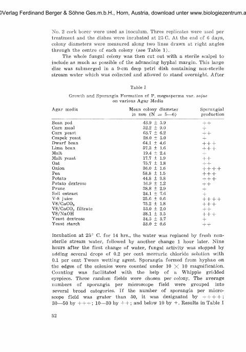
Table I
Growth and Sporangia Formation of P. megasperma var. sojaeon various Agar Media
Agar media Mean colony diameter Sporangialin mm (N = 5—6) production
Bean pod 43.9 ±5.9 + +Corn meal 52.2 ± 9.0 +Corn yeast 63.7 ±6.2 + +Czapek yeast 28.0 ± 5.0 +Dwarf bean 64.1 ±4.6 + + +Lima bean 57.3 ±1.6 + + +Malt 19.4 ± 2.4 +Malt yeast 17.7 ±1.9 + +Oat 75.7 ±1.8 + +Onion 36.0 ±1.6 + + + +Pea 58.8 ±1.5 + + +Potato 44.8 ±3.8 + + +Potato dextrose 16.9 ±1.2 + +Prune 38.8 ± 2.9 +Soil extract 24.1 ± 7.6 +V-8 juice 25.6 ±0.6 + + + +V8/CaCO5 75.2 ±1.8 + + +V8/CaCOa filtrate 53.0 ±2.0 + +V8/NaOH 38.1 ±3.5 + + +Yeast d-extrose 34.5 ± 3.7 +Yeast starch 53.0 ±0.6 + +
incubation at 25° C. for 14 hrs., the water was replaced by fresh non-sterile stream water, followed by another change 1 hour later. Ninehours after the first change of water, fungal activity was stopped byadding several drops of 0.2 per cent mercuric chloride solution with0.1 per cent Tween wetting agent. Sporangia formed from hyphae onthe edges of the colonies were counted under 10 X 10 magnification.Counting was facilitated with the help of a Whipple griddedeyepiece. Three random fields were chosen per colony. The averagenumbers of sporangia per microscope field were grouped intoseveral broad categories. If the number of sporangia per micro-scope field was grater than 50, it was designated by + + + + ;30—50 by + + + ; 10—30 by + + ; and below 10 by +. Results in Table I
52
©Verlag Ferdinand Berger & Söhne Ges.m.b.H., Horn, Austria, download unter www.biologiezentrum.at
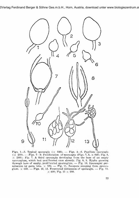
Fig-s. 1—3. Typical sporangia (x 640). — Fig-s. 4—6. Papillate sporangia(X 200). — Figs. 7—9. Proliferation of sporangia (Figs. 7, 8, X 640; Fig. 9,X 200); Fig. 7. A third sporangia developing from the base of an emptysporangium, which had proliferated once already. Fig. 8, 9. Hypha growingthrough base of empty, proliferated sporangium. — Fig. 10. Sporangial ger-mination by germ tube. X 200. — Fig. 11. Zoospore escaping from sporan-gium, x 640. — Figs. 12, 13. Frustrated dehiscence of sporangia. —• Fig. 12.
X 400; Fig. 13 x 200.
53
©Verlag Ferdinand Berger & Söhne Ges.m.b.H., Horn, Austria, download unter www.biologiezentrum.at
represent the average of two separate experiments each with threereplicates. Of all the media tested, those which supported fast colonygrowth and good sporulation, were V8/CaCO.,, lima bean, dwarf bean,and pea agars.
The sizes of sporangia were extremely variable, depending on thenature of media. The morphology of sporangia was studied in greaterdetail, using 20% V8/CaCO., as the culture medium. The sizes of sporan-gia even on this one medium varied considerably, as shown in figs. 1—13.The smallest sporangia approached the sizes of "miniature sporangia"described by D r e c h s l e r (1931) and gave rise to one or two zoosporesonly. Sporangia were typically non-papillate (figs. 1, 2, 3) thoughsometimes appearing slightly papillate to papillate (figs, 4, 5, 6). Theterm "papilla" was used to denote the nipple-like apex of sporangium( W a t e r ho u s e and B l a c k w e l l , 1954). Definite proliferation ofsporangia was observed. New sporangia may be borne on a new sporan-giophore arising from the base within the empty sporangium as describedby H i l d e b r a n d (1959) or directly on the base (figs. 7, 8, 9). In theformer case, the new sporangium may be of the same size as the oldone or the size diminishes progressively with the freqrency of proli-feration. In the latter case, the sporangia are either slightly or con-siderably smaller than those first formed. Proliferation may occur morethan once within an empty sporangium. Previous workers have describedsporangium germination both directly, by formation of germ tube(fig. 10), or indirectly, by production of zoospores (fig. 11). Presentobservations on sporangium germination on the above medium indicatedthat the latter process was by far the most predominant. Sometimes,sporangium germination was either abortive or "frustrated" — a termused by D r e c h s l e r (1931) to describe the retention of zoosporeswithin sporangia and their germination in situ (figs. 12, 13).
E f f e c t of t e m p e r a t u r eThe effect of temperature on growth of P. megasperma var. sojae
on agar plates was initially studied by S k o t l a n d (1955), whodetermined the optimum temperature for growth on oatmeal agar tobe 24° C. A similar conclusion was arrived at by H i 1 d e b r a n d (1959),who fotund that maximum growth was at 25° C on corn meal agar.However, K l e i n (1959) claimed that all of the seven isolates of soybeanPhytophthora that he studied grew best at 20° C. Cardinal temperatureshave been shown previously to be useful for species delimination inPhytophthora ( L e o n i a n , 1934). Being aware of the various draw-backs in measuring fungal growth by the current method ( H a w k e r ,1950; C o c h r a n e , 1958), the effect of temperatures was studied bothby measuring diameters of fungal colony on agar plates and bydetermining mycelial dry weight in solution culture. For directcomparison, 20% V8/CaCO3 filtrate was used in both cases.
54
©Verlag Ferdinand Berger & Söhne Ges.m.b.H., Horn, Austria, download unter www.biologiezentrum.at
E f f e c t o f t e m p e r a t u r e on c o l o n y d i a m e t e r
The V8/CaCO3 filtrate was incorporated with 1.8% Bactoagar. Suchmedium (50 ml) was poured into 140-mm diameter petri dishes, threereplicates being used for each treatment. Inoculum was taken fromedge of actively growing colonies on V8/CaCO3 agar plates using aNo. 2 cork borer. Plates were incubated at 5°, 10°, 15°, 20°, 25°,30°, and 35° C. The results are recorded in Table II. When the meancolony diameters at the end of 12 days are compared, statistical analysisproved significant differences in growth between consecutive tempera-tures (all at 0.01 level but 0.05 level between 25° and 30° C ) . Theoptimum temperature for linear growth on agar was thus 25° C.
Table II
Effect of Temperature on the Growth of P. megaspcrma var. sojaeon 20% V8/CaCO3 filtrate agar
Days
5°C
Mean colony diameter in mm (N = 3)
10° C 15° C 20° C 25° C 30c 35° C
2468
1012
000000
08.7±0.5
10.0±0.010.0±0.010.810.312.8+0.3
12.3±0.8 15.2±0.320.0±1.0 27.3±1.029.5±0.9 43.3±1.540.5 + 1.3 58.312.0
20.011.7 16.2 + 1.339.313.2 33.2+3.358.715.8 51.814.478.817.0 70.714.0
52.3+1.3 75.211.8 99.216.7 88.513.50 12.8+0.3 66.5 + 1.3 92.812.0 121.216.3 108.313.8
00
8.510.59.7+0.3
10.5+0.911.510.5
E f f e c t of t e m p e r a t u r e on m y c e l i a l d r y w e i g h t
For liquid cultures, zoospores appeared to tbe the best inoculum byvirtue of the negligible food materials they carry. It was found though,apart from the great chances of contamination, the mycelium developingfrom zoospores often stick to the bottom of the flasks so firmly thatit was difficult to wash the mycelium out for dry weight determination.Subsequently, the following procedure was adopted. From the edge ofan actively growing fungal colony on 20% V8/CaCO3 filtrate agar (15 mlin 9-cm diameter petri dishes), discs were removed with a No. 2 corkborer and transferred to 15 ml V8/CaCO3 filtrate in 250-ml Erlenmeyerflasks. The depth of solution was such that the agar disc was justsubmerged but there was no problem of aeration, due to the shallownessof solution. Mean dry weight of inoculum was determined by removingagar discs with mycelium into pre-weighed glass fiber pads and dryingat 80° C. overnight. In order to overcome the great variations amongreplicates as indicated in preliminary experiments, 20 replicates wereused per treatment. Due to the limited spaces in incubators, the experi-ment was conducted in three parts, mycelial dry weights being determinedat the end of 5, 10 and 12 days respectively. Mycelium developed in each
55
©Verlag Ferdinand Berger & Söhne Ges.m.b.H., Horn, Austria, download unter www.biologiezentrum.at

flask was filtered off using a glass filter pad in a Büchner funnel anddried at 80° C. Net increase in dry weight was obtained by subtractingthe dry weight of the incoculum. The results are recorded in Table III.
Dry weight determination at the end of 5 days showed a similargrowth/temperature relationship to that measured by colony diameter.The maximum growth was at 25° C. and was significantlty better thanthat at 15° or 30" C. (0.01 level). At the end of 10 days, maximum dryweight was at 20° and 25° C. with no significant differences betweenthem but respectively greater than that at 15° C. (0.01 level) and 30° C.(0.05 level). At the end of 12 days, though the optimum appeared to be15° C, it was found statistically non-significant when compared with20° and 25° C. but growth at 25° was still significantly better than thatat 30° C. (0.05 level). Probably, fungal growth reached maximum earlierat 25° and 20° and then autolysis developed rapidly. These resultsprovided an example of the obscurity about the real concept of a
Table IIIEffect of Temperature on Growth of P. megaspei-ma var
Temperaturein C
101520253035
in 20'/, Va/CaCO3 filtrate solution Culture
5 days(N = 16)No growth9.48± 4.26
25.43± 7.7149.10+ 7.4331.80±12.61No growth
Mean mycelial dry wt in mg10 days
(N = 19—20)0.89± 0.89
38.35±15.3151.29±11.8250.10±10.8540.80±13.581.07± 1.44
. sojae
12 days(N = 15—19)
4.26± 1.9365.70+ 2.46fi2.45± 7.18(50.42± 4.3355.08± 7.9913.52119.69
temperature-growth-optimum and the limitations of choosing only oneincubation period ( C o c h r a n e , 1958). Based on the definition that theoptimal temperature is the temperature at which the fungus growsfastest during its actively growing period, the optimum for this funguswas 25° C, whether growth was measured in terms of colony diameteror increase in mycelial dry weight.
E f f e c t of pH
The pH of liquid cultures was adjusted with N NaOH and N HC1respectively. Fifteen replicates were used for each treatment and theflasks were left a room temperature (24° to 25° C) . Mycelial dry weigthswere determined at the end of 5 days. The results are recorded inTable IV.
The optimal range was between 6.2 to 6.6 with no significant dif-ferences in the fungal growth among various pH values within theselimits. As for the other values below pH 6.2 and above 6,6, there were
56
©Verlag Ferdinand Berger & Söhne Ges.m.b.H., Horn, Austria, download unter www.biologiezentrum.at
significant differences in mycelial dry weight between successive pHvalues with the exception of the pairs of pH 4.5 and 5.5; 7.0 and 7.4.The fungal metabolites tended to lower the pH of the medium asindicated by the final pH values at the end of the experiment. Never-theless, since it took about 2 days before conspicuous mycelial growthstarted, and the experiment only lasted for 5 days, the initial pH valueswere considered more important. S u h o v e c k y (1955) studied theeffect of pH on the growth of this fungus on corn meal agar. He foundtwo optima occurring at pH 6.0 and the second at pH 9.O. He admittedthat this was an unusual case. Since different buffers were used toshift the pH towards the acid and alkaline sides respectively, his curiousresults might represent an example of chemicals on fungal growth.
Thanks are due to Dr. J. A. H a a s, Harrow Expt. Station, Ontario,for supplying the culture and to Prof. C. J. H i c k m a n, Univ. ofWestern Ontario for his constructive criticism.
Effect of pH on the GrowthInitial
pH
4.104.505.505.806.206.256.356.607.07.407.707.85
Table IV
of P. megasperma var. sojae in solution Culture
Final Mean mycelial dry wt in mgpH
4.04.404.854.905.205.305.555.605.055.455.856.20
(N = 9—15)
3.05 ± 1.1817.70 ± 5.2020.70 ± 4.1028.37 ± 6.3054.41 ± 10.0157.30 ± 6.3958.57 ± 7.6159.89 ± 2.6017.21 ± 2.4815.31 ± 2.989.72 ± 0.967.47 ± 0.62
L i t e r a t u r e c i t e dB a r r, D. J. S., 1965. Chytrids and Their relationships with freshwater algae.
Ph. D. Thesis. University Western Ontario, London, Canada. 190 p.C o c h r a n e , V. W., 1958. Physiology of fungi. John Wiley & Sons, Inc.,
New York. 524 p.D r e c h s l e r , C, 1930. Repetitional diplanetism in the genus Phytophthora.
J. Agr. Res. 40: 557—575.— 1931. A crown-rot of hollyhocks caused by Phytophthora megasperma
n. sp. J. Wash. Acad. Sei. 21: 513—526.H a w k e r , L, 1950. Physiology of fungy. University of London Press Ltd.,
London. 360 p.H e r r , L. J., 1957. Factors affecting a root rot of soybeans incited byPhyto-
phthora cactorum. Phytopathology 47: 15—16 (Abstr.).H i l d e b r a n d , A. A., 1959. A root and stalk rot of soybeans caus-ed by
Phytophthora megasperma Drechsler var. sojae var. nov. Can. J. Bo-tany 37: 927—957.
57
©Verlag Ferdinand Berger & Söhne Ges.m.b.H., Horn, Austria, download unter www.biologiezentrum.at
H i 11 y, J. W. and A. P. S c h m i t t h e n n e r, 1962. Pathogenic and culturalvariability of single zoospore isolates of Phytophthora megaspcrmavar. sojae. Phytopathology 52: 859—862.
K l e i n , H. H., 1959. Factors affecting development and morphology of re-productive structures of the soybean root and stem rot Phytophthora.Phytopathology 49: 376—379.
L e o n i a n , L. H., 1934. Identification of Phytophthora species. West Va.Univ. Agr. Expt. Sta. Bull. 262 p.
M i l l e r , P. M., 1955. V-8 juice agar as a general purpose medium for fungiand bacteria. Phytopathology 45: 461—462.
S k o 11 a n d, C. B., 1955. A Phytophthora damping-off disease of soybean.Plant Disease Reptr. 39: 682—683.
Suhovec 'ky , A. J., 1955. A Phytophthora root rot of soybeans. Ph. D.Thesis. The Ohio State Univ., Columbus, Ohio.
W a t e r h o u s e , G. M. and E. B l a c k w e l l , 1954. Key to species ofPhytophthora recorded in the British Isles. Mycological Paper no. 57,Commonwealth Mycological Institute, Kew, Surrey.
58
©Verlag Ferdinand Berger & Söhne Ges.m.b.H., Horn, Austria, download unter www.biologiezentrum.at