a senescent cell bystander effect: senescence-induced senescence
TRANSCRIPT

A senescent cell bystander effect: senescence-inducedsenescence
Glyn Nelson,1* James Wordsworth,1* Chunfang Wang,2
Diana Jurk,1 Conor Lawless,1 Carmen Martin-Ruiz1 andThomas von Zglinicki1
1Institute for Ageing and Health, Newcastle University, Campus for Ageingand Vitality, Newcastle upon Tyne NE4 5PL, UK2Department of Life Science, Walton Hall, Open University, Milton Keynes,
MK7 6AA, UK
Summary
Senescent cells produce and secrete various bioactive molecules
including interleukins, growth factors, matrix-degrading enzymes
and reactive oxygen species (ROS). Thus, it has been proposed
that senescent cells can damage their local environment, and a
stimulatory effect on tumour cell growth and invasiveness has
been documented. However, it was unknown what effect, if any,
senescent cells have on their normal, proliferation-competent
counterparts. We show here that senescent cells induce a DNA
damage response, characteristic for senescence, in neighbouring
cells via gap junction-mediated cell–cell contact and processes
involving ROS. Continuous exposure to senescent cells induced
cell senescence in intact bystander fibroblasts. Hepatocytes bear-
ing senescence markers clustered together in mice livers. Thus,
senescent cells can induce a bystander effect, spreading senes-
cence towards their neighbours in vitro and, possibly, in vivo.
Key words: aging; DNA damage; 53BP1; GFP; fluorescence; cell
signalling.
Cellular senescence is a state of irreversible cell cycle arrest, typically dri-
ven by a persistent DNA damage response (DDR) (Campisi & d’Adda di
Fagagna, 2007). Senescent cells activate downstream signalling path-
ways that trigger production and release of a host of bioactive molecules
including reactive oxygen species (ROS) (Passos et al., 2010) and a wide
variety of pro-inflammatory cytokines, chemokines and growth factors
(Coppe et al., 2008). Thus, senescent cells stimulated proliferation and
invasiveness of premalignant and malignant epithelial cells in co-culture
and co-transplantation experiments (Krtolica et al., 2001; Bavik et al.,
2006; Liu & Hornsby, 2007). However, the impact of senescent cells
upon normal cells with intact DNA damage checkpoints has not been
examined.
We hypothesized that pro-oxidant and pro-inflammatory signals from
primary senescent founder cells may trigger DNA damage and premature
senescence in surrounding primary cells, similar to the classical bystander
effect as induced by ionizing radiation (Prise & O’Sullivan, 2009). If so, this
senescence-induced bystander effect may contribute to the increasing
frequency of senescent cells with age and to the impact senescent cells
may have upon their environment.
To test this hypothesis, we examined the effects of co-culturing replica-
tively senescent fibroblasts (founder cells) with young (recipient ⁄ bystan-
der) fibroblasts in vitro. We followed the recipient cells by stably
integrating a fluorescent fusion protein (AcGFP-53BP1c), which quantita-
tively reports the number of DNA double-strand breaks (DSBs) within a
cell at any given time (Nelson et al., 2009).
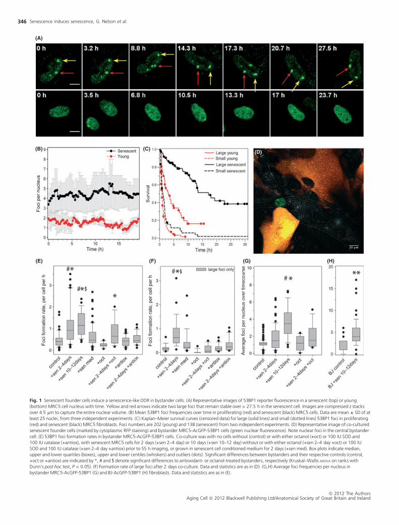
Reporter fluorescence in senescent cells differs from their young, repli-
cation-competent counterparts in various aspects: (i) Senescent cells dis-
play larger numbers of DSB foci (Fig. 1A). In senescent primary human
fibroblasts, this amounts to an average of 4–5 DSBs at any one time within
the population, compared to a maximum of 1–2 foci in proliferating cells
(Fig. 1B). (ii) DNA damage foci in senescent cells are larger (Rodier et al.,
2011). Separating 53BP1-AcGFP foci by size, we found that > 90% of
senescent MRC5 fibroblasts, but fewer than 50% of young, contained at
least one large focus (Fig. S1). (iii) Senescent cells contain long-lived,
potentially persistent foci (Passos et al., 2010). Large foci size is associated
with long foci lifespan (Fig. 1C). Therefore, foci kinetic data are ideal
markers to follow the induction of a senescent phenotype in vitro.
We next seeded unlabelled or RFP-labelled senescent founder cells in a
1:1 ratio with AcGFP-53BP1-expressing recipient fibroblasts at a PDL of
25 (Fig. 1D). Foci formation rates in young recipient MRC5 cells were sig-
nificantly increased after short (2 day) exposure to senescent cells and
remained elevated during prolonged co-culture (Fig. 1E). This increase in
focus induction was exclusively because of more frequent generation of
large 53BP1 foci (Fig. 1F); induction rates for small foci were not changed
(Fig. S2). Steady-state foci frequencies per nucleus started to increase in
the recipient cells after short exposure to senescent cells and reached lev-
els similar to those found in senescent cells after 10 days exposure
(Fig. 1G). This was confirmed in an independent foreskin fibroblast strain,
BJ (Fig. 1H). These results indicate that senescent human fibroblasts pro-
duce a DDR as bystander effect in surrounding proliferating cells.
We performed a series of experiments to clarify how the bystander
effect was mediated. Conditioned medium from senescent fibroblasts
alone was not sufficient to increase the foci formation rate (Fig. 1E,F) or
the steady-state foci frequencies (Fig. S3). Senescent inducer and young
recipient cells were also grown as separate layers sharing the same med-
ium in Transwell inserts. Growth in such vessels produced no observable
effects in terms of DNA damage foci numbers (data not shown) and had
no significant effect on growth rate of the recipient cells over 24 days
(Fig. S4). To address the role of the extracellular matrix generated by
senescent founder cells, we compared DNA foci frequencies in young
recipient cells grown for 7 days on matrix deposited by either young or
senescent cells. There was no effect of the matrix on frequencies of either
small or large foci (Fig. S5). On the contrary, blocking gap junction-medi-
ated cell–cell contact or scavenging extracellular ROS blocked the
increase of foci formation rate (Fig. 1E,F) and steady-state foci levels
(Fig. S3) in bystander cells. This is reminiscent of the weak bystander
effect induced by low dose and ⁄ or low LET irradiation, which is also
dependent on direct cell–cell contact via gap junctions (Gaillard et al.,
2009) and typically involves signalling through oxygen- and ⁄ or nitrogen-
centred radicals (Chen et al., 2009).
Correspondence
Professor Thomas von Zglinicki, Institute for Ageing and Health, Newcastle
University, Campus for Ageing and Vitality, Newcastle upon Tyne NE4 5PL, UK.
Tel.: +44 191 248 1104; fax: +44 191 248 1101; e-mail: [email protected]
*These authors contributed equally to this work.
Re-use of this article is permitted in accordance with the Terms and Conditions
set out at http://wileyonlinelibrary.com/onlineopen#onlineopen_Terms
Accepted for publication 4 January 2012
ª 2012 The AuthorsAging Cell ª 2012 Blackwell Publishing Ltd/Anatomical Society of Great Britain and Ireland
345
Aging Cell (2012) 11, pp345–349 Doi: 10.1111/j.1474-9726.2012.00795.xAg
ing
Cell
(A)
(B) (C)
(E) (F) (G) (H)
(D)
Fig. 1 Senescent founder cells induce a senescence-like DDR in bystander cells. (A) Representative images of 53BP1 reporter fluorescence in a senescent (top) or young
(bottom) MRC5 cell nucleus with time. Yellow and red arrows indicate two large foci that remain stable over > 27.5 h in the senescent cell. Images are compressed z stacks
over 4.5 lm to capture the entire nuclear volume. (B) Mean 53BP1 foci frequencies over time in proliferating (red) and senescent (black) MRC5 cells. Data are mean ± SD of at
least 25 nuclei, from three independent experiments. (C) Kaplan–Meier survival curves (censored data) for large (solid lines) and small (dotted lines) 53BP1 foci in proliferating
(red) and senescent (black) MRC5 fibroblasts. Foci numbers are 202 (young) and 138 (senescent) from two independent experiments. (D) Representative image of co-cultured
senescent founder cells (marked by cytoplasmic RFP staining) and bystander MRC5-AcGFP-53BP1 cells (green nuclear fluorescence). Note nuclear foci in the central bystander
cell. (E) 53BP1 foci formation rates in bystander MRC5-AcGFP-53BP1 cells. Co-culture was with no cells without (control) or with either octanol (+oct) or 100 IU SOD and
100 IU catalase (+antiox), with senescent MRC5 cells for 2 days (+sen 2–4 day) or 10 days (+sen 10–12 day) without or with either octanol (+sen 2–4 day +oct) or 100 IU
SOD and 100 IU catalase (+sen 2–4 day +antiox) prior to 55 h imaging, or grown in senescent cell conditioned medium for 2 days (+sen med). Box plots indicate median,
upper and lower quartiles (boxes), upper and lower centiles (whiskers) and outliers (dots). Significant differences between bystanders and their respective controls (control,
+oct or +antiox) are indicated by *, # and $ denote significant differences to antioxidant- or octanol-treated bystanders, respectively (Kruskal–Wallis ANOVA on ranks with
Dunn’s post hoc test, P < 0.05). (F) Formation rate of large foci after 2 days co-culture. Data and statistics are as in (D). (G,H) Average foci frequencies per nucleus in
bystander MRC5-AcGFP-53BP1 (G) and BJ-AcGFP-53BP1 (H) fibroblasts. Data and statistics are as in (E).
Senescence induces senescence, G. Nelson et al.
ª 2012 The AuthorsAging Cell ª 2012 Blackwell Publishing Ltd/Anatomical Society of Great Britain and Ireland
346
(A) (B) (C) (D)
(E) (F)
(G)
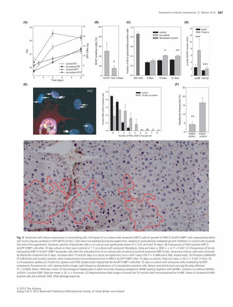
Fig. 2 Senescent cells induce senescence in surrounding cells. (A) Impact of co-culture with senescent MRC5 cells on growth of MRC5-AcGFP-53BP1 cells measured by either
cell counts (square symbols) or GFP-qPCR (circles). Cells were not replated during the experiment, leading to some density-mediated growth inhibition in control cells towards
the end of the experiment. However, growth of bystander cells in co-culture was significantly slower (P < 0.01 at 4 and 10 days). (B) Frequencies of Ki67-positive MRC5-
AcGFP-53BP1 cells after 10 day culture on their own (control) or 1:1 co-culture with senescent fibroblasts. Data are mean ± SEM, n = 4, P = 0.047. (C) Frequencies of Sen-b-
Gal-positive MRC5-AcGFP-53BP1 bystander cells after the indicated time of co-culture with or without (control) senescent MRC5 cells. Senescent inducer cells were removed
by blasticidin treatment for 6 days. Increases after 15 and 20 days co-culture are significant (ANOVA with Tukey HSD P = 0.048 and 0.008, respectively). (D) Phospho-p38MAPK
(P-p38) levels and nuclear:cytosolic ratios (measured by immunofluorescence) in MRC5-AcGFP-53BP1 after 10 days co-culture. Data are mean ± SD, P < 0.001 (T-test). (E)
Co-localization (yellow) of cH2AX foci (green) with PML bodies (red) in bystander BJ-AcGFP-53BP1 cells after 10 day co-culture with senescent cells (marked by red RFP
cytoplasmic fluorescence). Left: representative image; right frequency distributions of co-localization-positive cells. Means and distributions are significantly different
(P < 0.0000, Mann–Whitney U-test). (F) Percentage of hepatocytes in adult mice liver showing cytoplasmic 4HNE staining together with (4HNE+ cH2AX+) or without (4HNE+
cH2AX)) nuclear DDR. Data are mean ± SE, n = 4 animals. (G) Representative tiled image of mouse liver (9 months old) immunostained for 4-HNE. Areas of clustered 4-HNE-
positive cells are outlined. DDR, DNA damage response.
Senescence induces senescence, G. Nelson et al.
ª 2012 The AuthorsAging Cell ª 2012 Blackwell Publishing Ltd/Anatomical Society of Great Britain and Ireland
347
Senescent cells produce and secrete a variety of candidate signalling
molecules, including ROS (Passos et al., 2010), pro-inflammatory
interleukins (Kuilman et al., 2008), TGFb1 (Debacq-Chainiaux et al.,
2005) and various IGF-binding proteins (Kim et al., 2007; Muck et al.,
2008) that can all maintain or induce senescence in a cell autonomous or
nonautonomous fashion. Our data show that any long-lived soluble fac-
tors released by senescent cells into the medium or the matrix have little
effect on the formation of DNA damage foci. However, such factors
could be transmitted between cells via gap junctions.
While our data indicate that ROS are necessary for the induction of
DNA damage in the recipient cells, they do not allow the conclusion
that ROS released from senescent cells are directly or indirectly causal
for the damage in the recipients. Enzymatic antioxidants are essentially
confined to the extracellular space including the outer cell membrane
and will thus primarily suppress ROS in the medium. However, various
ROS species are readily interchangeable, and hydrogen peroxide is eas-
ily membrane permeable, so that extracellular antioxidants will effec-
tively reduce intracellular ROS concentrations. Our data are therefore
fully compatible with the idea that ROS production in the recipient
cells is activated by some unspecified signal(s). However, they strongly
suggest ROS as the proximal cause of DNA damage in the bystander
cells.
Normal cells with noncompromised DNA damage checkpoint function
are expected to react to persistent DNA damage by induction of a senes-
cence or apoptosis programme. To see whether the bystander effect
actually induced cell senescence, we measured multiple markers of senes-
cence in the recipient cells. Recipient MRC5 cells proliferated significantly
slower (Fig. 2A) and were less positive for the cell cycle marker Ki67
(Fig. 2B), as were recipient BJ cells (Fig. S6). The frequency of bystander
cells positive for senescence-associated b-galactosidase (Sen-b-Gal) activ-
ity was increased after co-culture for 15 or more days (Fig. 2C). It was
measured after removal of senescent inducer cells from the culture,
showing that the bystander effect induced permanent senescence. After
extended co-culture with senescent cells, MRC5 and BJ bystander cells
showed stronger p38MAPK activation (Fig. 2D) and more frequent
nuclear PML:cH2AX co-localization (Figs 2E and S7), two additional
markers of senescence. Together, these data show that senescent
cells induce permanent cell senescence as a bystander effect in their
environment.
If the bystander effect were important for the generation of cell senes-
cence in vivo, senescent cells would be expected to cluster in tissues. We
chose mouse liver for cluster analysis as a relatively homogeneous tissue
with a significant fraction of senescent hepatocytes (Krizhanovsky et al.,
2008; Wang et al., 2009). We used 4-HNE as a marker for senescent cells
because it is closely associated with other markers of senescence in
mouse liver including cH2A.X (Fig. 2F) and Sen-b-Gal (Wang et al., 2009,
2010) and allows clear cell boundary definition in the low magnification
tiled images necessary for cluster analysis. Marker-positive hepatocytes
formed closely associated clusters with essentially no negative cells
between them (Fig. 2G). This clustering is significantly higher than
expected by random chance, given the observed frequencies of marker-
positive hepatocytes in mouse livers (Fig. S8). Such clustering could in
principle be driven by focal oxidative damage. However, it should be
noted that we did not see evidence for leucocyte invasion associated with
clusters of 4-HNE-positive cells. While comprehensive proof of senes-
cence-induced senescence in tissues awaits the analysis of a senescence
reporter system in vivo, our data already indicate that senescent cells
induce a bystander effect that spreads DNA damage and, ultimately,
induces cell senescence in primary, checkpoint-competent cells in vitro
and, possibly, in vivo. This could explain how senescent cells might drive
the aging process in vivo as proposed (Tchkonia et al., 2010; Baker et al.,
2011).
Acknowledgments
The work was supported by BBSRC grant BB ⁄ C008200 ⁄ 1 (CISBAN),
MRC ⁄ Unilever Biomarker grant G0601333 and a BBSRC ⁄ Procter &
Gamble CASE studentship (JW).
Author contributions
Experimental work and analysis was carried out by GN, JW, CW, DJ,
CL and CM-R. GN, JW and TvZ wrote the manuscript. GN and TvZ
designed the study.
References
Baker DJ, Wijshake T, Tchkonia T, LeBrasseur NK, Childs BG, van de Sluis B, Kirk-
land JL, van Deursen JM (2011) Clearance of p16Ink4a-positive senescent cells
delays ageing-associated disorders. Nature 479, 232–236.
Bavik C, Coleman I, Dean JP, Knudsen B, Plymate S, Nelson PS (2006) The gene
expression program of prostate fibroblast senescence modulates neoplastic
epithelial cell proliferation through paracrine mechanisms. Cancer Res. 66,
794–802.
Campisi J, d’Adda di Fagagna F (2007) Cellular senescence: when bad things
happen to good cells. Nat. Rev. Mol. Cell Biol. 8, 729–740.
Chen S, Zhao Y, Zhao G, Han W, Bao L, Yu KN, Wu L (2009) Up-regulation of
ROS by mitochondria-dependent bystander signaling contributes to genotoxic-
ity of bystander effects. Mutat. Res. 666, 68–73.
Coppe JP, Patil CK, Rodier F, Sun Y, Munoz DP, Goldstein J, Nelson PS, Desprez
PY, Campisi J (2008) Senescence-associated secretory phenotypes reveal cell-
nonautonomous functions of oncogenic RAS and the p53 tumor suppressor.
PLoS Biol. 6, 2853–2868.
Debacq-Chainiaux F, Borlon C, Pascal T, Royer V, Eliaers F, Ninane N, Carrard G,
Friguet B, de Longueville F, Boffe S, Remacle J, Toussaint O (2005) Repeated
exposure of human skin fibroblasts to UVB at subcytotoxic level triggers pre-
mature senescence through the TGF-beta1 signaling pathway. J. Cell Sci.
118(Pt 4), 743–758.
Gaillard S, Pusset D, de Toledo SM, Fromm M, Azzam EI (2009) Propagation dis-
tance of the alpha-particle-induced bystander effect: the role of nuclear tra-
versal and gap junction communication. Radiat. Res. 171, 513–520.
Kim KS, Seu YB, Baek SH, Kim MJ, Kim KJ, Kim JH, Kim JR (2007) Induction of
cellular senescence by insulin-like growth factor binding protein-5 through a
p53-dependent mechanism. Mol. Biol. Cell 18, 4543–4552.
Krizhanovsky V, Yon M, Dickins RA, Hearn S, Simon J, Miething C, Yee H,
Zender L, Lowe SW (2008) Senescence of activated stellate cells limits liver
fibrosis. Cell 134, 657–667.
Krtolica A, Parrinello S, Lockett S, Desprez PY, Campisi J (2001) Senescent fibro-
blasts promote epithelial cell growth and tumorigenesis: a link between cancer
and aging. Proc. Natl Acad. Sci. USA 98, 12072–12077.
Kuilman T, Michaloglou C, Vredeveld LC, Douma S, van Doorn R, Desmet CJ,
Aarden LA, Mooi WJ, Peeper DS (2008) Oncogene-induced senescence
relayed by an interleukin-dependent inflammatory network. Cell 133, 1019–
1031.
Liu D, Hornsby PJ (2007) Senescent human fibroblasts increase the early growth
of xenograft tumors via matrix metalloproteinase secretion. Cancer Res. 67,
3117–3126.
Muck C, Micutkova L, Zwerschke W, Jansen-Durr P (2008) Role of insulin-like
growth factor binding protein-3 in human umbilical vein endothelial cell senes-
cence. Rejuvenation Res. 11, 449–453.
Nelson G, Buhmann M, von Zglinicki T (2009) DNA damage foci in mitosis are
devoid of 53BP1. Cell Cycle 8, 3379–3383.
Passos JF, Nelson G, Wang C, Richter T, Simillion C, Proctor CJ, Miwa S, Olijslag-
ers S, Hallinan J, Wipat A, Saretzki G, Rudolph KL, Kirkwood TB, von Zglinicki
T (2010) Feedback between p21 and reactive oxygen production is necessary
for cell senescence. Mol. Syst. Biol. 6, 347.
Prise KM, O’Sullivan JM (2009) Radiation-induced bystander signalling in cancer
therapy. Nat. Rev. Cancer 9, 351–360.
Senescence induces senescence, G. Nelson et al.
ª 2012 The AuthorsAging Cell ª 2012 Blackwell Publishing Ltd/Anatomical Society of Great Britain and Ireland
348

Rodier F, Munoz DP, Teachenor R, Chu V, Le O, Bhaumik D, Coppe JP, Campeau
E, Beausejour CM, Kim SH, Davalos AR, Campisi J (2011) DNA-SCARS: distinct
nuclear structures that sustain damage-induced senescence growth arrest and
inflammatory cytokine secretion. J. Cell Sci. 124(Pt 1), 68–81.
Tchkonia T, Morbeck DE, Von Zglinicki T, Van Deursen J, Lustgarten J, Scrable H,
Khosla S, Jensen MD, Kirkland JL (2010) Fat tissue, aging, and cellular senes-
cence. Aging Cell 9, 667–684.
Wang C, Jurk D, Maddick M, Nelson G, Martin-Ruiz C, von Zglinicki T (2009)
DNA damage response and cellular senescence in tissues of aging mice. Aging
Cell 8, 311–323.
Wang C, Maddick M, Miwa S, Jurk D, Czapiewski R, Saretzki G, Langie SA,
Godschalk RW, Cameron K, von Zglinicki T (2010) Adult-onset, short-term die-
tary restriction reduces cell senescence in mice. Aging (Albany NY) 2, 555–
566.
Supporting Information
Additional supporting information may be found in the online ver-
sion of this article:
Data S1. Experimental procedures.
Figure S1. Frequencies of cells containing small and ⁄ or large AcGFP-
53BP1 foci in proliferating and senescent MRC5 cultures.
Figure S2. AcGFP-53BP1 small foci formation rates do not differ
with treatments.
Figure S3. Average foci frequencies per nucleus in bystander MRC5-
AcGFP-53BP1 cells.
Figure S4. Growth curves of proliferating MRC5-AcGFP-53BP1 cells
co-cultured with or without senescent MRC5 cells in Transwell
dishes.
Figure S5. Extracellular matrix does not change DNA damage foci
frequencies.
Figure S6. Frequencies of Ki67-positive BJ-AcGFP-53BP1 cells after
10 day culture on their own (control) or 1:1 co-culture with senes-
cent fibroblasts.
Figure S7. Frequency distributions of cH2AX foci colocalising with
PML bodies per nucleus in bystander MRC5-AcGFP-53BP1 cells after
20 day co-culture with senescent cells and in controls.
Figure S8. Potentially senescent hepatocytes cluster in ageing mice
livers.
As a service to our authors and readers, this journal provides support-
ing information supplied by the authors. Such materials are peer-
reviewed and may be re-organized for online delivery, but are not
copy-edited or typeset. Technical support issues arising from sup-
porting information (other than missing files) should be addressed to
the authors.
Senescence induces senescence, G. Nelson et al.
ª 2012 The AuthorsAging Cell ª 2012 Blackwell Publishing Ltd/Anatomical Society of Great Britain and Ireland
349