a review of transdermal drug delivery systemrapsr.fopras.org/images/vol2no12013/32-56-ra-3.pdf ·...
TRANSCRIPT
Review Article
Journal der Pharmazie Forschung (Formerly-Recent Advances in Pharmaceutical Science
Research )
Vol-2 No-1 2013
A Review of Transdermal drug delivery system
Chinmaya Keshari Sahoo1, Prakash Kumar Nayak
2,Tanmaya Keshari Sahoo
3*, Powshya
Dasari4, Santhoshipriya Dandamundi
4
1Department of Pharmaceutics, Osmania University, Hyderabad. Andhra Pradesh, India
2Department of Clinical Research, Clinical Research Coordinator cum Pharmacist, Norwich
Clinical Services Pvt. Ltd. Sahakar Nagar, Bangalore-560092, Karnataka, India. 3Institute of Pharmacy and Technology, Salipur, Cuttack--754202, Odisha, India.
4 Princeton College of Pharmacy, Korremula, Ghatkesar-501301, Andhra Pradesh, India.
Abstract
Transdermal drug delivery system (TDDS) specialists are continuing to
search for new methods that can effectively and painlessly deliver
larger molecules in therapeutic quantities to overcome the difficulties
associated with oral route,namely poor bioavailability due to first pass
metabolism and the tendency to produce rapid blood level spikes(both
high and low). Transdermal drug delivery can improve the therapeutic
efficacy and safety of drugs by more site specificway but spatial and
temporal placement within body is recquired to reduce both the size
and number of doses necessary to achieve the objective of systemic
medication through topical application to the intact skin surface,This
review describes enhancement techniques based on drug/vehicle
optimization such as drug selection,prodrugs and ion
pairs,supersaturated drug solutions,eutectic systems,complexations,
liposomes,vescicles and particles.This review also focuses on the recent
innovations in Transdermal drug delivery system (TDDS) which can be
a platform for the research and development of pharmaceutical dosage
form for transdermal drug delivery.
Keywords: TDDS, first pass metabolism, drug selection
Review Article www.rapsr.com
33 | P a g e Journal der Pharmazie Forschung (Formerly-Recent Advances in Pharmaceutical Science Research
The transdermal route has become one of
the most successful and innovative drug
delivery system for research in
pharmaceutical sciences. Transdermal drug
delivery provides a leading edge over
injectables and oral routes by increasing
patient compliance and avoiding first pass
metabolism respectively [1]. Transdermal
drug delivery is not only provides controlled
,constant administration of the drug ,but
also allows continuous input of drugs with
short biological half lives and eliminates
pulsed entry into systemic circulation. The
success of dermtological drug to be used for
systemic drug delivery depends on ability of
the drug to penetrate through skin in
sufficient quantities to achieve the desired
therapeutic effect [2].The first transdermal
patch was approved in 1981 for the relief of
motion sickness, nausea and vomiting. The
transdermal drug delivery market was until
recently,solely based on passive patch
technology that relied on sample diffusion
across the skin. Active patches in which the
agent in someway driven through barrier
offer a wide array of capabilities allowing
delivery compounds over 500Da and those
with challenging physical properties. This
has permitted the development of active
patches to deliver pain management drugs,
proteins and vaccines. Passive patch
technology is creating smaller patches with
better adhesion[3]. Transdermal drug
delivery system (TDDS) established itself as
an integral part of novel drug delivery
systems. Hence transdermal drug delivery is
defined as self contained, discrete dosage
forms which, when applied to the intact
skin, deliver the drug, through the skin at
controlled rate to the systemic circulation.
Advantages [4]
1.Avoid the risk and inconvenience of
intravenous therapy (noninvasive).
2.Avoidance of first pass hepatic
metabolism (avoiding the deactivation by
digestive and liver enzymes) thus
increasing bioavailability and efficacy of
drugs.
3.No gastrointestinal degradation (pH,
enzymatic activity, drug interaction with
food, drink and other orally administered
drugs)
4.Substitute for oral administration of
medication when that route is unsuitable as
with vomiting and diarrohea
5.Extended therapy avoiding frequent dose
administration.
6.Reduce side effect due to optimization of
the blood concentration time profile.
7.Greater patient compliance due to
elimination of multiple dosing intervals.
8.Enhance therapeutic efficiency.
9.Minimize inter and intra patient variations.
10.Reversibility of drug delivery which
would allow the removal of drug source.
Limitations of TDDS [5]
1. Limited skin permeability.
2. Restricted to potent drug
3. Cannot use for large molecule (>500
Dalton)
4. Significant lag time
5. Difficulty for adhesion.
6. The drug undergoes degradation in the
skin.
7. Variation in absorption efficiency at
different sites of skin.
Mechanism of transdermal permeation
[6]
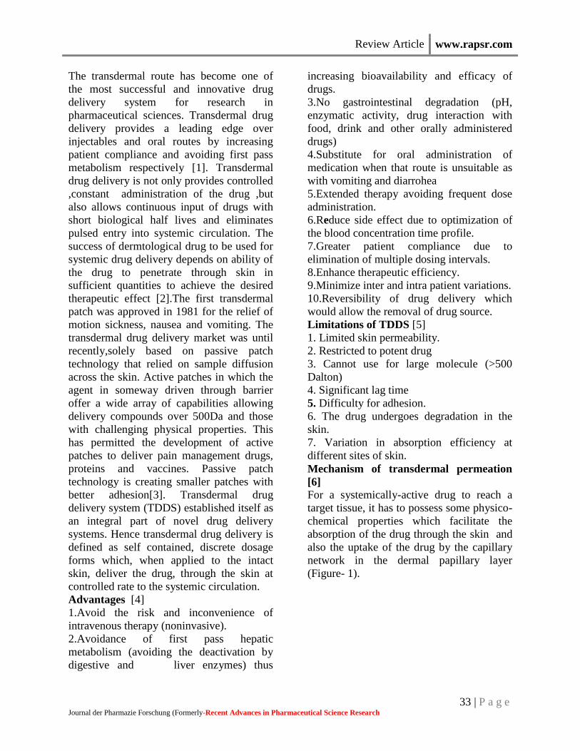
For a systemically-active drug to reach a
target tissue, it has to possess some physico-
chemical properties which facilitate the
absorption of the drug through the skin and
also the uptake of the drug by the capillary
network in the dermal papillary layer
(Figure- 1).
Review Article www.rapsr.com
34 | P a g e Journal der Pharmazie Forschung (Formerly-Recent Advances in Pharmaceutical Science Research
Figure--1: Simplified model of the human skin for mechanistic analysis of skin permeation
The rate of permeation, dQ/dt, across various layers of skin tissues can be expressed as.
dQ∕dt=Ps(Cd-Cr)……(1)
Where
Cd = the concentrations of skin penetrate in the donor phase (stratum corneum)
Cr = receptor phase(systemic circulation)
Ps = the overall permeability coefficient of the skin
The overall permeability coefficient of the skin is defined by
Ps =KsDss/hs…….(2)
Ks = Partition coefficient of the penetrant
Dss = Apparent diffusivity of penetrant
hs = Thickness of skin
Thus, permeability coefficient (PS) may be a constant since Ks; Dss and hs terms in equation (2)
are constant under the given set of conditions.
A constant rate of drug permeation achieved, if Cd>Cr, then the equation (1) may be reduced to
dQ∕dt =PsCd…..(3)
And the rate of skin permeation (dQ/dt) becomes a constant, if the Cd value remains fairly
constant throughout the course of skin permeation. To maintain the Cd at a constant value, it is
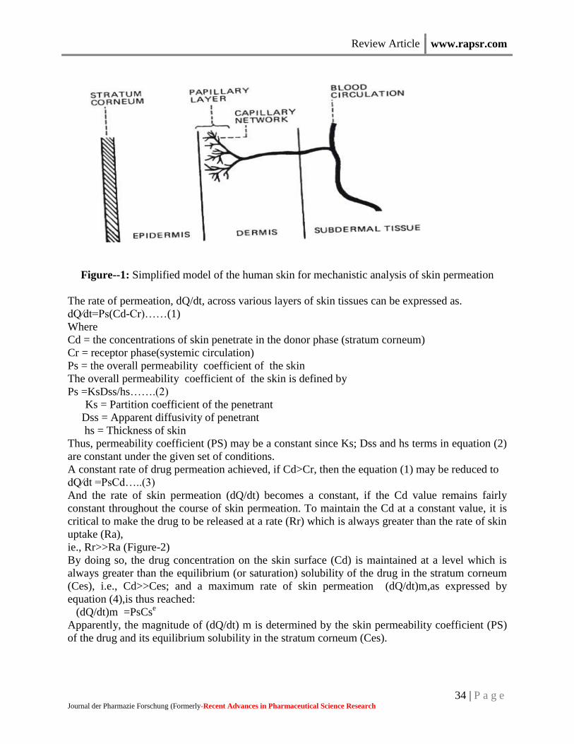
critical to make the drug to be released at a rate (Rr) which is always greater than the rate of skin
uptake (Ra),
ie., Rr>>Ra (Figure-2)
By doing so, the drug concentration on the skin surface (Cd) is maintained at a level which is
always greater than the equilibrium (or saturation) solubility of the drug in the stratum corneum
(Ces), i.e., Cd>>Ces; and a maximum rate of skin permeation (dQ/dt)m,as expressed by
equation (4),is thus reached:
(dQ/dt)m =PsCse
Apparently, the magnitude of (dQ/dt) m is determined by the skin permeability coefficient (PS)
of the drug and its equilibrium solubility in the stratum corneum (Ces).
Review Article www.rapsr.com
35 | P a g e Journal der Pharmazie Forschung (Formerly-Recent Advances in Pharmaceutical Science Research
Figure-2: Relationship between the rate of drug release (Rr) from a transdermal drug delivery
system (TDDS) and the rate of drug uptake (Ra) by the skin.
Transport through the skin [7]
Skin is structurally complex and thick
membrane. Molecules moving from the
environment must penetrate the stratum
corneum and any material of endogenous or
exogenous origin on its surface. They must
then penetrate the viable epidermis, the
papillary dermis and the capillary walls into
the blood stream or lymph channels,
whereupon they are removed from the skin
by flow of blood or lymph. To move across
the skin membrane is obviously a complex
phenomenon and challenge in analysis.
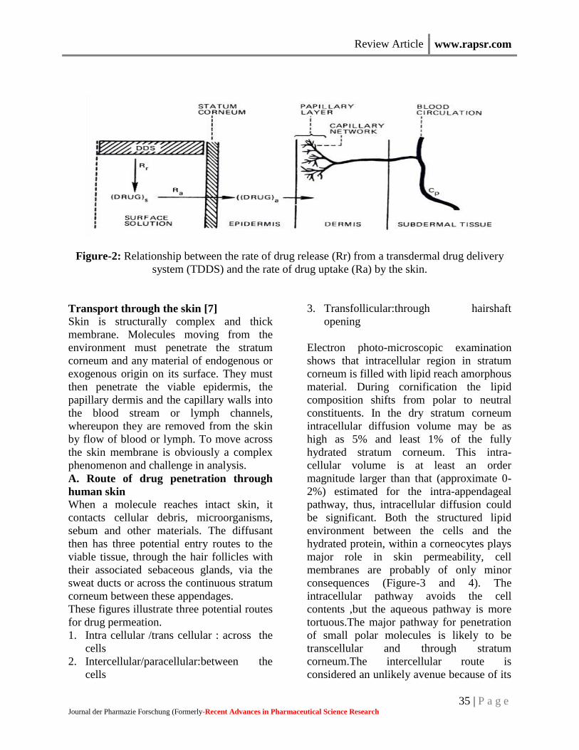
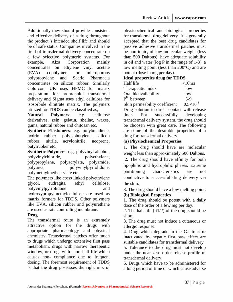
A. Route of drug penetration through
human skin
When a molecule reaches intact skin, it
contacts cellular debris, microorganisms,
sebum and other materials. The diffusant
then has three potential entry routes to the
viable tissue, through the hair follicles with
their associated sebaceous glands, via the
sweat ducts or across the continuous stratum
corneum between these appendages.
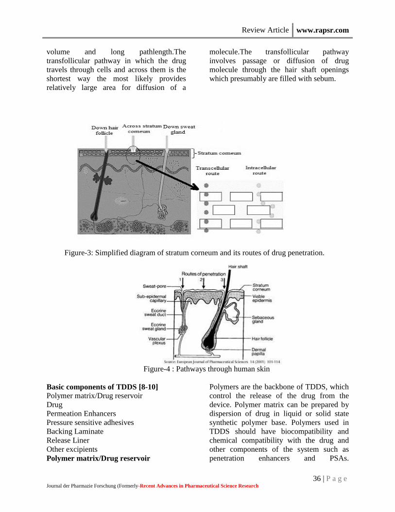
These figures illustrate three potential routes
for drug permeation.
1. Intra cellular /trans cellular : across the
cells
2. Intercellular/paracellular:between the
cells
3. Transfollicular:through hairshaft
opening
Electron photo-microscopic examination
shows that intracellular region in stratum
corneum is filled with lipid reach amorphous
material. During cornification the lipid
composition shifts from polar to neutral
constituents. In the dry stratum corneum
intracellular diffusion volume may be as
high as 5% and least 1% of the fully
hydrated stratum corneum. This intra-
cellular volume is at least an order
magnitude larger than that (approximate 0-
2%) estimated for the intra-appendageal
pathway, thus, intracellular diffusion could
be significant. Both the structured lipid
environment between the cells and the
hydrated protein, within a corneocytes plays
major role in skin permeability, cell
membranes are probably of only minor
consequences (Figure-3 and 4). The
intracellular pathway avoids the cell
contents ,but the aqueous pathway is more
tortuous.The major pathway for penetration
of small polar molecules is likely to be
transcellular and through stratum
corneum.The intercellular route is
considered an unlikely avenue because of its
Review Article www.rapsr.com
36 | P a g e Journal der Pharmazie Forschung (Formerly-Recent Advances in Pharmaceutical Science Research
volume and long pathlength.The
transfollicular pathway in which the drug
travels through cells and across them is the
shortest way the most likely provides
relatively large area for diffusion of a
molecule.The transfollicular pathway
involves passage or diffusion of drug
molecule through the hair shaft openings
which presumably are filled with sebum.
Figure-3: Simplified diagram of stratum corneum and its routes of drug penetration.
Figure-4 : Pathways through human skin
Basic components of TDDS [8-10]
Polymer matrix/Drug reservoir
Drug
Permeation Enhancers
Pressure sensitive adhesives
Backing Laminate
Release Liner
Other excipients
Polymer matrix/Drug reservoir
Polymers are the backbone of TDDS, which
control the release of the drug from the
device. Polymer matrix can be prepared by
dispersion of drug in liquid or solid state
synthetic polymer base. Polymers used in
TDDS should have biocompatibility and
chemical compatibility with the drug and
other components of the system such as
penetration enhancers and PSAs.
Review Article www.rapsr.com
37 | P a g e Journal der Pharmazie Forschung (Formerly-Recent Advances in Pharmaceutical Science Research
Additionally they should provide consistent
and effective delivery of a drug throughout
the product‟s intended shelf life and should
be of safe status. Companies involved in the
field of transdermal delivery concentrate on
a few selective polymeric systems. For
example, Alza Corporation mainly
concentrates on ethylene vinyl acetate
(EVA) copolymers or microporous
polypropylene and Searle Pharmacia
concentrates on silicon rubber. Similarly
Colorcon, UK uses HPMC for matrix
preparation for propranolol transdermal
delivery and Sigma uses ethyl cellulose for
isosorbide dinitrate matrix. The polymers
utilized for TDDS can be classified as,
Natural Polymers: e.g. cellulose
derivatives, zein, gelatin, shellac, waxes,
gums, natural rubber and chitosan etc.
Synthetic Elastomers: e.g. polybutadiene,
hydrin rubber, polyisobutylene, silicon
rubber, nitrile, acrylonitrile, neoprene,
butylrubber etc.
Synthetic Polymers: e.g. polyvinyl alcohol,
polyvinylchloride, polyethylene,
polypropylene, polyacrylate, polyamide,
polyurea, polyvinylpyrrolidone,
polymethylmethacrylate etc.
The polymers like cross linked polyethylene
glycol, eudragits, ethyl cellulose,
polyvinylpyrrolidone and
hydroxypropylmethylcellulose are used as
matrix formers for TDDS. Other polymers
like EVA, silicon rubber and polyurethane
are used as rate controlling membrane.
Drug The transdermal route is an extremely
attractive option for the drugs with
appropriate pharmacology and physical
chemistry. Transdermal patches offer much
to drugs which undergo extensive first pass
metabolism, drugs with narrow therapeutic
window, or drugs with short half life which
causes non- compliance due to frequent
dosing. The foremost requirement of TDDS
is that the drug possesses the right mix of
physicochemical and biological properties
for transdermal drug delivery. It is generally
accepted that the best drug candidates for
passive adhesive transdermal patches must
be non ionic, of low molecular weight (less
than 500 Daltons), have adequate solubility
in oil and water (log P in the range of 1-3), a
low melting point (less than 200°C) and are
potent (dose in mg per day).
Ideal properties drug for TDDS.
Half life <10hrs
Therapeutic index low
Oral bioavailability low
PH
between 5-9
Skin permeability coefficient 0.5×10⁻³
Drug solution in direct contact with release
liner. For successfully developing
transdermal delivery system, the drug should
be choosen with great care. The following
are some of the desirable properties of a
drug for transdermal delivery.
(a) Physiochemical Properties
1. The drug should have are molecular
weight less than approximately 500 Daltons.
2. The drug should have affinity for both
lipophilic and hydrophilic phases. Extreme
partitioning characteristics are not
conductive to successful drug delivery via
the skin.
3. The drug should have a low melting point.
(b) Biological Properties
1. The drug should be potent with a daily
dose of the order of a few mg per day.
2. The half life ( t1/2) of the drug should be
short.
3. The drug must not induce a cutaneous or
allergic response.
4. Drug which degrade in the G.I tract or
inactivated by hepatic first pass effect are
suitable candidates for transdermal delivery.
5. Tolerance to the drug must not develop
under the near zero order release profile of
transdermal delivery.
6. Drugs which have to be administered for
a long period of time or which cause adverse
Review Article www.rapsr.com
38 | P a g e Journal der Pharmazie Forschung (Formerly-Recent Advances in Pharmaceutical Science Research
effects to non-target tissues can also be
formulated for transdermal delivery.
Permeation Enhancers Three pathways are suggested for drug
penetration through the skin: polar, non-
polar, and polar/non-polar. The enhancers
act by altering one of these pathways. The
key to altering the polar pathway is to cause
protein conformational change or solvent
swelling. The key to altering the nonpolar
pathway is to alter the rigidity of the lipid
structure and fluidize the crystalline
pathway (this substantially increases
diffusion). The fatty acid enhancers increase
the fluidity of the lipid portion of the
Stratum Corneum. Some enhancers (binary
vehicles) act on both polar and nonpolar
pathways by altering the multilaminate
pathway for penetrants. Enhancers can
increase the drug diffusivity in the Stratum
Corneum (SC) by dissolving the skin lipids
or by denaturing skin proteins. The type of
enhancer employed has a significant impact
on the design and development of the
product. The success of dermatological drug
products that are intended for systemic drug
delivery, such as the transdermal, depends
on the ability of the drug to penetrate
through the skin in sufficient quantities to
achieve its desired therapeutic effect. The
methods employed for modifying the barrier
properties of the SC to enhance the drug
penetration (and absorption) through the
skin can be categorized as (1) Chemical and
(2) physical methods of enhancement.
Chemical Enhancers Chemicals that promote the penetration of
topically applied drugs are commonly
referred to as accelerants, absorption
promoters, or penetration enhancers. These
agents act by -increasing the drug
permeability through the skin by causing
reversible damage to the SC or by increasing
(and optimizing) thermodynamic activity of
the drug when functioning as co solvent or
by increasing the partition coefficient of the
drug to promote its release from the vehicle
into the skin or by conditioning the SC to
promote drug diffusion. Promoting
penetration and establish drug reservoir in
the SC.
Sulphoxides
Dimethyl sulphoxides (DMSO) is one of the
earliest and most widely studied penetration
enhancers. It is a powerful aportic solvent
which hydrogen bonds with itself rather than
with water. It is colourless, odourless and is
hydroscopic and is often used in many areas
of pharmaceutical sciences as a “universal
solvent”. DMSO alone has been applied
topically to treat systemic
inflammation.DMSO works rapidly as a
penetration enhancer - spillage of the
material onto the skin can be tasted in the
mouth within a second. Although DMSO is
an excellent accelerant, it does create
problems. The effect of the enhancer is
concentration-dependent and generally
cosolvents containing > 60% DMSO are
needed for optimum enhancement efficacy.
However, at these relative high
concentrations, DMSO can cause erythema
and wheal of the stratum corneum.
Denaturing of some skin proteins results in
erythema, scaling, contact uticaria ,stinging
and burning sensation.
Azone
Azone (1-dodecylazacycloheptan-2-one or
laurocapran) was the first molecule
specifically designed as a skin penetration
enhancer. Azone is a colourless, odourless
liquid with a melting point of -7 ºC and it
possesses a smooth, oily but yet non-greasy
feel. Azone is a highly lipophilic material
with a log p octanol / water of around 6.2
and it is soluble in and compatible with most
organic solvents including alcohol and
propylene glycol. Azone enhances the skin
transport of a wide variety of drugs
including steroids,antibiotics and antiviral
agents. Azone is most effective at low
concentrations, being employed typically
Review Article www.rapsr.com
39 | P a g e Journal der Pharmazie Forschung (Formerly-Recent Advances in Pharmaceutical Science Research
between 0.1- 5% but more often between 1-
3% 13. Azone partitions into a bilayer lipid
to disrupt their packing arrangement but
integration into the lipid is unlikely to be
homogeneous. Azone molecules may exist
dispersed within the barrier lipoid or
separate domains within the bilayer.
Pyrrolidones
Pyrrolidones have been used as permeation
enhancers for numerous molecules including
hydrophilic (e.g. mannitol and 5-flurouracil)
and lipophilic (progesterone and
hydrocortisone) permeants. N-methyl-2-
pyrolidone was employed with limited
success as a penetration enhancer for
captopril when formulated in a matrix-type
transdermal patch. The pyrrolidones
partition well into human stratum corneum
within the tissue and they may act by
altering the solvent nature of the membrane.
Pyrrolidones have been used to generate
reservoirs within the skin membrane. Such a
reservoir effect offers a potential for
sustained release of a permeant from the
stratum corneum over extended time
periods.
Fatty acids
Percutaneous drug absorption has been
increased by a wide variety of long-chain
fatty acids, the most popular of which is
oleic acid. It is of interest to note that many
penetration enhancers such as azone contain
saturated or unsaturated hydrocarbon chains
and some structure - activity relationships
have been drawn from the extensive studies
of Aungst who employed a range of fatty
acids, acids, alcohols, sulphoxides,
surfactants and amides as enhancers for
naloxone. Shin et al studied various
penetration enhancers like glycols
(diethylene glycol and tetraethylene glycol),
fatty acids (lauric acid, myristic acid and
capric acid) and nonic surfactant
(polyoxyethylene-2-oleyl ether, polyoxy
ethylene-2-stearly ether) on the release of
triprolidone. Lauric acid in Propylene glycol
enhanced the delivery of highly lipophilic
antiestrogen. Oleic acid greatly increased the
flux of many drugs such as increasing the
flux of salicylic acid 28-fold and 5-
flurouracil flux 56-fold through human skin
membrane in vitro. The enhancer interacts
with and modifies the lipid domains of the
stratum corneum as would be expected for a
longchain fatty acid with cis- configuration.
Essential oil, terpenes and terpenoids
Terpenes are found in essential oils, and are
compounds comprising of only carbon,
hydrogen and oxygen atoms, but which are
not aromatic. Numerous terpenes have long
been used as medicines as well as flavoring
and fragrance agents. The essential oils of
eucalyptus, chenopodium and ylang-ylang
have been found to be effective penetration
enhancers for 5-flouorouracil transversing
human skin in vivo. Cornwell et al
investigated the effect of 12 sesquiterpenes
on the permeation of 5-flurouracil in human
skin. Pretreatment of epidermal membranes
with sesquiterpene oil or using
solidsesquiterpenes saturated in dimethyl
isosorbide increased the absorption of 5-
flurouracil. L-menthol has been used to
facilitate in vitro permeation of morphine
hydrochloride through hairless rat skin as
well as diffusion of imipramine
hydrochloride across rat skin and
hydrocortisone through hairless mouse skin.
One mechanism by which this agent
operates is to modify the solvent nature of
the stratum corneum, thus improving drug
partitioning into the tissue. Many terpenes
permeate human skin well and large
amounts of terpene have been found in the
epidermis after application from a matrix-
type patch. Terpenes may also modify drug
diffusivity through the membrane. During
steady state permeation experiments using
terpenes as penetration enhancers, the lag
time for permeation was usually reduced,
indicating some increase in drug diffusivity
Review Article www.rapsr.com
40 | P a g e Journal der Pharmazie Forschung (Formerly-Recent Advances in Pharmaceutical Science Research
through the membrane following terpene
treatment.
Oxazolidinones
Oxazolidinones are a new class of chemical
agents which have the potential for use in
many cosmetic and personal care product
formulations. This is due to their ability to
localize co-administered drug in skin layers,
resulting in low systemic permeation. The
structural features of these permeation
enhancers are closely related to sphingosine
and ceramide lipids which are naturally
found in the upper skin layers.
Oxazolidinones such as 4-decyloxazolidin-
2-one has been reported to localize the
delivery of many active ingredients such as
retinoic acid and diclofenac sodium in skin
layers. This compound has a higher
molecular weight and lipophilicity than
other solvent-type enhancers, physical
characteristics that may be beneficial in
terms of a reduction in local toxicity because
of the lack of effective absorption of these
enhancers into the lower skin layers where
irritation is likely to be occur.
Urea
Urea promotes transdermal permeation by
facilitating hydration of the stratum corneum
and by the formation of hydrophilic
diffusion channels within the barrier. Cyclic
urea permeation enhancers are
biodegradable and non-toxic molecules
consisting of a polar parent moiety and a
long chain alkyl ester group. As a result,
enhancement mechanism may be a
consequence of both hydrophilicactivity and
lipid disruption mechanism4.
Physical enhancers The iontophoresis and ultra sound (also
known as phonophoresis or sonophoresis)
techniques are examples of physical means
of enhancement that have been used for
enhancing percutaneous penetration (and
absorption) of various therapeutic agents.
Pressure sensitive adhesives
A PSA is a material that helps in
maintaining an intimate contact between
transdermal system and the skin surface. It
should adhere with not more than applied
finger pressure, be aggressively and
permanently tachy, and exert a strong
holding force. Additionally, it should be
removable from the smooth surface without
leaving a residue. Polyacrylates,
polyisobutylene and silicon based adhesives
are widely used in TDDSs. The selection of
an adhesive is based on numerous factors,
including the patch design and drug
formulation. For matrix systems with a
peripheral adhesive, an incidental contact
between the adhesive and the drug and
penetration enhancer should not cause
instability of the drug, penetration enhancer
or the adhesive. In case of reservoir systems
that include a face adhesive, the diffusing
drug must not affect the adhesive. In case of
drug-in-adhesive matrix systems, the
selection will be based on the rate at which
the drug and the penetration enhancer will
diffuse through the adhesive. Ideally, PSA
should be physic chemically and
biologically compatible and should not alter
drug release.
Backing Laminate While designing a backing layer, the
consideration of chemical resistance of the
material is most important. Excipients
compatibility should also be considered
because the prolonged contact between the
backing layer and the excipients may cause
the additives to leach out of the backing
layer or may lead to diffusion of excipients,
drug or penetration enhancer through the
layer. However, an overemphasis on the
chemical resistance may lead to stiffness and
high occlusive to moisture vapor and air,
causing patches to lift and possibly irritate
the skin during long wear. The most
comfortable backing will be the one that
exhibits lowest modulus or high flexibility,
good oxygen transmission and a high
Review Article www.rapsr.com
41 | P a g e Journal der Pharmazie Forschung (Formerly-Recent Advances in Pharmaceutical Science Research
moisture vapor transmission rate. Examples
of some backing materials are vinyl,
polyethylene and polyester films.
Release Liner During storage the patch is covered by a
protective liner that is removed and
discharged immediately before the
application of the patch to skin. It is
therefore regarded as a part of the primary
packaging material rather than a part of
dosage form for delivering the drug.
However, as the liner is in intimate contact
with the delivery system, it should comply
with specific requirements regarding
chemical inertness and permeation to the
drug, penetration enhancer and water.
Typically, release liner is composed of a
base layer which may be non-occlusive (e.g.
paper fabric) or occlusive (e.g. polyethylene,
polyvinylchloride) and a release coating
layer made up of silicon or teflon. Other
materials used for TDDS release liner
include polyester foil and metallized
laminates
Other excipients Various solvents such as chloroform,
methanol, acetone, isopropanol and
dichloromethane are used to prepare drug
reservoir. In addition plasticizers such as
dibutylpthalate, triethylcitrate, polyethylene
glycol and propylene glycol are added to
provide plasticity to the transdermal patch.
APPROACHES TO DEVELOPMENT
TRANSDERMAL THERAPEUTIC
SYSTEMS [8-11]
Several technologies have been successfully
developed to provide a rate control over the
release and the transdermal permeation of
drugs. These technologies can be classified
into two major catagories as follows:
A.Rate-programmed transdermal DDS
B.Physical stimuli-activated transdermal
DDS
A.Rate-programmed transdermal DDS
1. Membrane permeation – controlled
systems
2. Adhesive dispersion – type systems.
3. Matrix diffusion – controlled systems.
4. Micro reservoir type or micro sealed
dissolution controlled systems
B.Physical stimuli-activated transdermal
DDS i.Structure based
microneedles
macroflux
MDTS
ii.Electrically based
Iontophoresis
Ultrasound
Photochemical waves
Electroporation
Electroosmosis
iii.Velocity based
Powder jet
Needle free injection
iv.Others
Transferosomes
Medicated tattoos
Skin abrasion
Heat
Laser radiation
Magnetophoresis
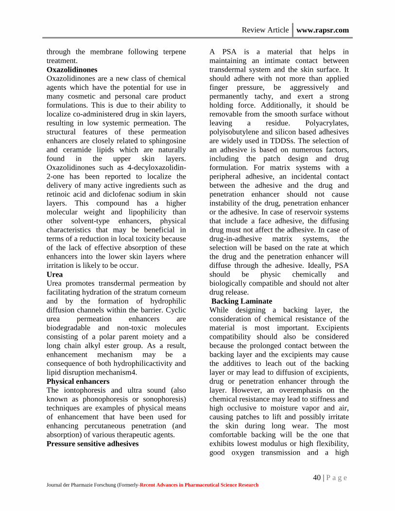
Membrane permeation – controlled
systems
In this type of system, drug reservoir is
encapsulated in a shallow compartment
moulded from a drug-impermeable metallic
plastic laminate and a rate controlling
polymeric membrane which may be micro
porous or non-porous. The drug molecules
are permitted to release only through the rate
– controlling polymeric membrane. In the
drug reservoir compartment, the drug solids
are either dispersed homogenously in a solid
polymer matrix (e.g. Polyisobutylene
adhesive) or suspended in an unbleachable,
viscous liquid medium (e.g. Silicon fluids)
to form a paste like suspension.
Review Article www.rapsr.com
42 | P a g e Journal der Pharmazie Forschung (Formerly-Recent Advances in Pharmaceutical Science Research
Figure-5: Membrane-moderated Transdermal drug delivery system.
The rate of drug release from this type of
system can be tailored by varying the
polymer composition, permeability
coefficient and thickness of the rate limiting
membrane and adhesive. The constant
release rate of the drug is the major
advantage of membrane permeation
controlled system. However, a rare risk also
exists when an accidental breakage of the
rate controlling membrane can result in dose
dumping or rapid release of entire drug
content. Examples of this system are
Transderm – Nitro Nitroglycerin – releasing transdermal system
for once a day medication in angina pectoris.
Transderm – Scop Scopolamine – releasing transdermal system
for 72 hrs. Prophylaxis of motion sickness.
Catapres Clonidine-releasing transdermal system for
7 day therapy of hypertension.
Estraderm Estradiol – releasing transdermal system for
treatment of menopausal syndrome for 3 – 4
days.
The membrane permeation-controlled
technology has also been used for controlled
percutaneous absorption of prostaglandin-
derivatives.
1. Single-layer Drug-in-Adhesive The Single-layer Drug-in-Adhesive system
is characterized by the inclusion of the drug
directly within the skin-contacting adhesive.
In this transdermal system design, the
adhesive not only serves to affix the system
to the skin, but also serves as the
formulation foundation, containing the drug
and all the excipients under a single backing
film. The rate of release of drug from this
type of system is dependent on the diffusion
across the skin. The intrinsic rate of drug
release from this type of drug delivery
system is defined by
dQ/dT =Cr∕1/Pm + 1/Pa
Where Cr is the drug concentration in the
reservoir compartment and Pa and P m are
the permeability coefficients of the adhesive
layer and the rate controlling membrane, Pm
is the sum of permeability coefficients
simultaneous penetrations across the pores
and the polymeric material. Pm and Pa,
respectively, are defined as follows.
Pm = Km/r. Dm ∕ hm
Pa = Ka/m. Da ∕ ha
where Km/r and Ka/m are the partition
coefficients for the interfacial partitioning of
drug from the reservoir to the membrane and
from the membrane to adhesive
respectively; Dm and Da are the diffusion
coefficients in the rate controlling
membrane and adhesive layer, respectively;
and hm and ha are the thicknesses of the rate
controlling membrane and adhesive layer,
respectively
2. Multi-layer Drug-in-Adhesive The Multi-layer Drug-in-Adhesive is similar
to the Single-layer Drug-in-Adhesive in that
the drug is incorporated directly into the
adhesive. However, the multi-layer
encompasses either the addition of a
membrane between two distinct drug-in-
adhesive layers or the addition of multiple
drug-in-adhesive layers under a single
Review Article www.rapsr.com
43 | P a g e Journal der Pharmazie Forschung (Formerly-Recent Advances in Pharmaceutical Science Research
backing film. The rate of drug release in this
system is defined by,
dQ/dt = Ka/r. Da Cr∕ ha
Where Ka/r is the partition coefficient for
the interfacial partitioning of the drug from
the reservoir layer to adhesive layer.
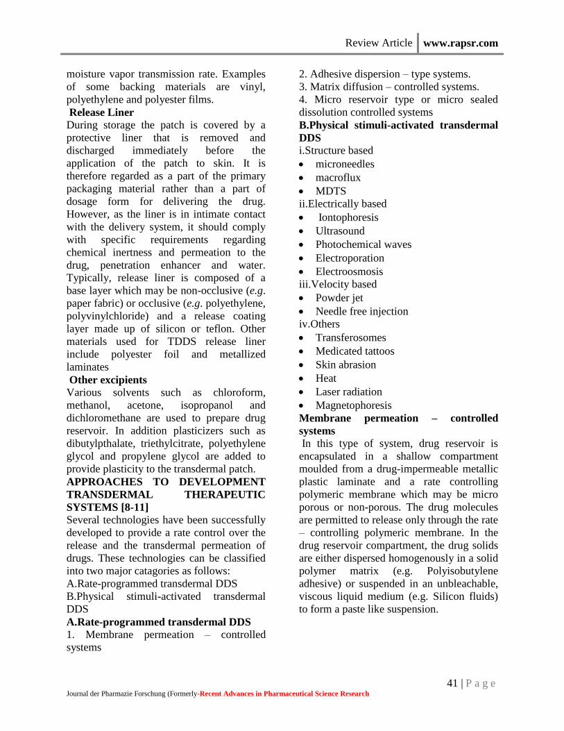
Adhesive Dispersion – Type Systems
This is a simplified form of the membrane-
permeation controlled system. As
represented in Figure-6, the drug reservoir is
formulated by directly dispersing the drug in
an adhesive polymer e.g. Poly (isobutylene)
or poly (acrylate) adhesive and then
spreading the medicated adhesive, by
solvent casting or hot melt, on to a flat sheet
of drug impermeable metallic plastic
backing to form a thin drug reservoir layer.
On the top of the drug reservoir layer, thin
layers of non-medicated, rate-controlling
adhesive polymer of a specific permeability
and constant thickness are applied to
produce an adhesive diffusion – controlled
delivery system.
Frandol tape Releases Isosorbide dinitrate for once-a-day
medication of angina pectoris.
Deponit Delivers nitroglycerine for the treatment of
angina pectoris.
Figure-6: adhesive diffusion-controlled Transdermal drug delivery system.
The Reservoir transdermal system design is
characterized by the inclusion of a liquid
compartment containing a drug solution or
suspension separated from the release liner
by a semi-permeable membrane and
adhesive. The adhesive component of the
product responsible for skin adhesion can
either be incorporated as a continuous layer
between the membrane and the release liner
or in a concentric configuration around the
membrane.
The rate of drug release from this drug
reservoir gradient controlled system is given
by,
dQ/dt = Ka/r. Da A ( ha ) ∕ha ( t )
In the above equation, the thickness of the
adhesive layer for drug molecules to diffuse
through increases with time ha (t). To
compensate for this time dependent increase
in the diffusional path due to the depletion
of drug dose by release, the drug loading
level is also increased with the thickness of
diffusional path A (ha). In the above
equation, the thickness of the adhesive layer
for drug molecules to diffuse through
increases with time ha (t). To compensate
for this time dependent increase in the
diffusional path due to the depletion of drug
dose by release, the drug loading level is
also increased with the thickness of
diffusional path A (ha
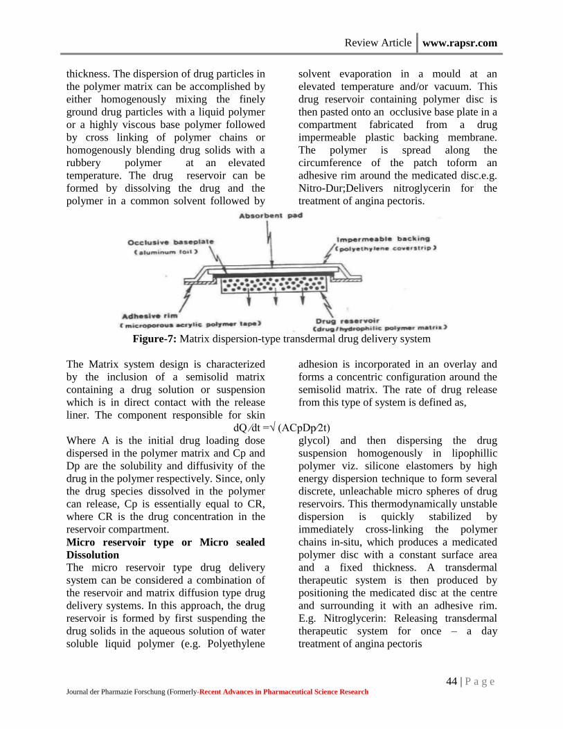
Matrix diffusion controlled systems
In this approach the drug reservoir is formed
by homogenously dispersing the drug solids
in a hydrophilic or lipophylic polymer
matrix.The resultant medicated polymer is
then molded into amedicated disc with a
defined surface area and controlled
Review Article www.rapsr.com
44 | P a g e Journal der Pharmazie Forschung (Formerly-Recent Advances in Pharmaceutical Science Research
thickness. The dispersion of drug particles in
the polymer matrix can be accomplished by
either homogenously mixing the finely
ground drug particles with a liquid polymer
or a highly viscous base polymer followed
by cross linking of polymer chains or
homogenously blending drug solids with a
rubbery polymer at an elevated
temperature. The drug reservoir can be
formed by dissolving the drug and the
polymer in a common solvent followed by
solvent evaporation in a mould at an
elevated temperature and/or vacuum. This
drug reservoir containing polymer disc is
then pasted onto an occlusive base plate in a
compartment fabricated from a drug
impermeable plastic backing membrane.
The polymer is spread along the
circumference of the patch toform an
adhesive rim around the medicated disc.e.g.
Nitro-Dur;Delivers nitroglycerin for the
treatment of angina pectoris.
Figure-7: Matrix dispersion-type transdermal drug delivery system
The Matrix system design is characterized
by the inclusion of a semisolid matrix
containing a drug solution or suspension
which is in direct contact with the release
liner. The component responsible for skin
adhesion is incorporated in an overlay and
forms a concentric configuration around the
semisolid matrix. The rate of drug release
from this type of system is defined as,
dQ ∕dt =√ (ACpDp∕2t)
Where A is the initial drug loading dose
dispersed in the polymer matrix and Cp and
Dp are the solubility and diffusivity of the
drug in the polymer respectively. Since, only
the drug species dissolved in the polymer
can release, Cp is essentially equal to CR,
where CR is the drug concentration in the
reservoir compartment.
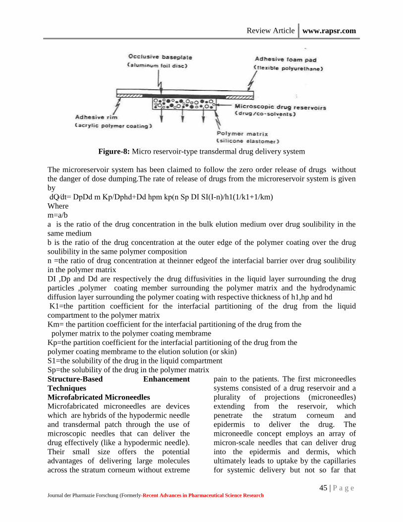
Micro reservoir type or Micro sealed
Dissolution The micro reservoir type drug delivery
system can be considered a combination of
the reservoir and matrix diffusion type drug
delivery systems. In this approach, the drug
reservoir is formed by first suspending the
drug solids in the aqueous solution of water
soluble liquid polymer (e.g. Polyethylene
glycol) and then dispersing the drug
suspension homogenously in lipophillic
polymer viz. silicone elastomers by high
energy dispersion technique to form several
discrete, unleachable micro spheres of drug
reservoirs. This thermodynamically unstable
dispersion is quickly stabilized by
immediately cross-linking the polymer
chains in-situ, which produces a medicated
polymer disc with a constant surface area
and a fixed thickness. A transdermal
therapeutic system is then produced by
positioning the medicated disc at the centre
and surrounding it with an adhesive rim.
E.g. Nitroglycerin: Releasing transdermal
therapeutic system for once – a day
treatment of angina pectoris
Review Article www.rapsr.com
45 | P a g e Journal der Pharmazie Forschung (Formerly-Recent Advances in Pharmaceutical Science Research
Figure-8: Micro reservoir-type transdermal drug delivery system
The microreservoir system has been claimed to follow the zero order release of drugs without
the danger of dose dumping.The rate of release of drugs from the microreservoir system is given
by
dQ∕dt= DpDd m Kp/Dphd+Dd hpm kp(n Sp DI SI(I-n)/h1(1/k1+1/km)
Where
m=a/b
a is the ratio of the drug concentration in the bulk elution medium over drug soulibility in the
same medium
b is the ratio of the drug concentration at the outer edge of the polymer coating over the drug
soulibility in the same polymer composition
n =the ratio of drug concentration at theinner edgeof the interfacial barrier over drug soulibility
in the polymer matrix
DI ,Dp and Dd are respectively the drug diffusivities in the liquid layer surrounding the drug
particles ,polymer coating member surrounding the polymer matrix and the hydrodynamic
diffusion layer surrounding the polymer coating with respective thickness of h1,hp and hd
K1=the partition coefficient for the interfacial partitioning of the drug from the liquid
compartment to the polymer matrix
Km= the partition coefficient for the interfacial partitioning of the drug from the
polymer matrix to the polymer coating membrame
Kp=the partition coefficient for the interfacial partitioning of the drug from the
polymer coating membrame to the elution solution (or skin)
S1=the solubility of the drug in the liquid compartment
Sp=the solubility of the drug in the polymer matrix
Structure-Based Enhancement
Techniques
Microfabricated Microneedles
Microfabricated microneedles are devices
which are hybrids of the hypodermic needle
and transdermal patch through the use of
microscopic needles that can deliver the
drug effectively (like a hypodermic needle).
Their small size offers the potential
advantages of delivering large molecules
across the stratum corneum without extreme
pain to the patients. The first microneedles
systems consisted of a drug reservoir and a
plurality of projections (microneedles)
extending from the reservoir, which
penetrate the stratum corneum and
epidermis to deliver the drug. The
microneedle concept employs an array of
micron-scale needles that can deliver drug
into the epidermis and dermis, which
ultimately leads to uptake by the capillaries
for systemic delivery but not so far that
Review Article www.rapsr.com
46 | P a g e Journal der Pharmazie Forschung (Formerly-Recent Advances in Pharmaceutical Science Research
microneedles hit the nerves. This is the
reason for the device being less painful to
patients. The most common material used
for microfabrication of needles is silicon.
These microneedles have extremely sharp
tips (radius of curvature, <1μm) that
facilitate easy piercing of the skin.
Individual silicon needles measuring
approximately 150 μm in length and with 80
μm base diameter are fabricated onto arrays
of approximately 400 microneedles (approx.
3 X 3 mm). Needles with hollow centers
have also been produced, each containing a
bore of 5-70 μm (depending on the required
design) through which drug can be
administered. A broad range of compounds
such as calcein (623 Da), insulin (6000Da),
BSA (66000Da) and polymeric
nanoparticles are delivered at significant
rates through skin permeabilized by
microfabricated microneedles.
Macroflux
Macroflux® technology is another novel
transdermal drug delivery system that
ALZA Corporation has developed to deliver
biopharmaceutical drugs in a controlled
reproducible manner that optimizes
bioavailability and efficacy without
significant discomfort for the patient. The
system incorporates a titanium
microprojection array that creates superficial
pathway through the skin barrier layer to
allow transportation of therapeutic proteins
and vaccines or access to the interstitial
fluids for sampling. Macroflux® has an area
of up to 8cm2 and contains as many as 300
microprojection per cm2 with individual
micro projection length being < 200μm. The
maximal adhesive patch size is 10 cm2. A
coating process is used to apply drug to the
tip of each microprojection in the array.
When the patch is applied to the skin, the
drug-coated microprojections penetrate
through the skin barrier layer into the
epidermis.The microcapillaries for systemic
distribution absorb the drug. The rate of
absorption is promoted by the high local
drug concentration around the
microprojections and the large surface area
provided by the patch array.
Three types of Macroflux ® have been
designed and tested in preclinical studies.
They include,
• Dry-Coated Macroflux ® system for short
duration administration that consist of a drug
coated microprojection array adhered to a
flexible polymeric adhesive backing.
• D-TRANS® Macroflux® system for short
duration administration that consist of a
microprojection array coupled with a drug
reservoir.
• E-TRANS® Macroflux ® system for
pulsatile or on demand delivery that include
a microprojection array coupled with an
electrotransport system.
Therapeutic peptides, proteins and vaccines
such as desmopressin, human growth
hormone (HGH), TH 9507 (a human growth
hormone releasing factor analog),
ovalbumin(45000 Da protein) are in the
developmental stage for transdermal
delivery by Macroflux®
Metered-Dose Transdermal Spray
(MDTS)
Metered-dose transdermal spray
(MDTSTM), originally developed at the
Victorian College of Pharmacy [Monash
University (Parkville Campus), Parkville,
Victoria, Australia] and currently being
commercialized by Acrux Limited
[Melbourne, Victoria, Australia] has the
potential to expand the growth of TDD
systems by broadening patient acceptance
and pharmaceutical applications for
enhanced TDD.MDTS relies on the
combination of a newly identified GRAS
(generally recognized as safe) chemical
penetration enhancer (AcrossTM) and the
accurate and precise topical dosing of a
volatile: nonvolatile vehicle. This MDTS
can be classified, as an enhanced, passive
TDD system. It is a topical solution made up
Review Article www.rapsr.com
47 | P a g e Journal der Pharmazie Forschung (Formerly-Recent Advances in Pharmaceutical Science Research
of a volatile cum nonvolatile vehicle
containing the drug dissolved as a single-
phase solution. A finite metered - dose
application of the formulation to intact skin
results in subsequent evaporation of the
volatile component of the vehicle, leaving
the remaining nonvolatile penetration
enhancer and drug to rapidly partition into
the stratum corneum during the first minute
after application, resulting in a stratum
corneum reservoir of drug and enhancer.
Following a once daily application of the
MDTS, a sustained and enhanced
penetration of the drug across the skin can
be achieved from the stratum corneum
reservoir. Different types of penetration
enhancers, such as ethanol and azone, are
commonly used. Clinical experience with
estradiol-MDTS to post-menopausal women
have shown increased higher plasma level of
estradiol than the baseline value measured
by radioimmunoassay.
The MDTS has the following potential
advantages:
1. Enhanced passive tdds with little or no
skin irritation primarily as a result of its
nonocclusive
nature.
2. Improved cosmetic acceptability
3. Dose flexibility
4. Simplicity of manufacture.
Electrically-Based Enhancement
Techniques
Iontophoresis:
Iontophoresis may be defined as the
facilitation of ionizable drug permeation
across the skin by an applied electrical
potential, the driving force of which may be
simply visualized as electrostatic repulsion.
A typical iontophoresis device consists of a
battery, microprocessor controller, drug
reservoir and electrodes. The technique
involves the application of a small electric
current (usually 0.5 mA/cm2) to a drug
reservoir on the skin, with the similarly
charged electrodes (on the surface of the
skin) placed together in the drug reservoir
producing a repulsion effect that effectively
drives the solute molecules away from the
electrode and into the skin.
There are three explanations of how
iontophoresis increases transdermal drug
delivery. The first, proposes that the drugs
are forced across the skin by simple
electronic repulsion of similar charges.
Anionic drugs can cross the skin by using a
negatively charged electrode. Similarly
cationic drugs enter the skin more
successfully when a positively charged
electrode is used The second, explanation
suggests that the electric current enhances
permeation by inhibiting the skin’s ability to
perform its protective barrier function. The
third, states that iontophoresis causes water,
a very effective penetration enhancer, to
enter the stratum corneum by electro-
osmosis. Dissolved drugs can be carried
across the skin along with the penetrating
water during iontophoresis. At physiological
pH, human skin has slight negative charge;
therefore, certain cationic drugs can more
easily cross the skin during iontophoresis
due to reduced resistance. Several studies
have addressed the application of
iontophoresis to the delivery of low
molecular weight solutes (< 500 Da). For
delivery ofmacromolecules, proteins and
peptides such as calcitonin, corticotrophin-
releasing hormone, δ-sleep- inducing
peptide, dextrin sulphate, inulin,
insulin, gonadotropin releasing hormone,
growth hormone releasing factor, neutral
thyrotrophinreleasing hormone, parathyroid
hormone and vasopressin iontophoresis may
also be utilized.To date, clinical studies have
been limited to smaller molecules such as
lidocaine, ketorolac dexamethasone,
etofenamate, naproxen, vincristine, cortisone
and fentanyl.

Review Article www.rapsr.com
48 | P a g e Journal der Pharmazie Forschung (Formerly-Recent Advances in Pharmaceutical Science Research
Ultrasound
Ultrasound (sonophoresis, phonophoresis
and ultraphonophoresis) is a technique for
increasing
the skin permeation of drugs using
ultrasound (20 KHZ to 16 MHZ) as a
physical force. It is a
combination of ultrasound therapy with
topical drug therapy to achieve therapeutic
drug
concentrations at selected sites in the skin.
In this technique, the drug is mixed with a
coupling
agent usually a gel but sometimes a cream or
ointment is used which transfers ultrasonic
energy from the device to the skin through
this coupling agent. Application of low –
frequency ultrasound (20 -100 KHZ)
enhances skin permeability moreeffectively
than high – frequency ultrasound (1 -16
MHZ). The mechanism of transdermal skin
permeation involves disruption of the
stratum corneum lipids, thus allowing the
drug to pass through the skin. A
corresponding reduction in skin resistance
was observed due to cavitation,
microstreaming and heat generation.Reverse
ultrasound technology may also be used for
the extraction of interstitial fluid samples for
analysis.
Photomechanical Waves
Photomechanical waves (PW’s) are the
pressure pulses produced by ablation of a
material target
(polystyrene) by Q-switched or mode-locked
lasers. Photechanical waves are able to
render the stratum corneum more permeable
to macromolecules via a possible transient
permeabilisation effect due to the formation
of transient channels. The largest molecule
that has been reported to be delivered
through the rat skin to date has a molecular
weight of 40,000Da. Suggestions have been
made that many clinically important proteins
such as insulin (6000 Da) and hematoprotien
(48000 Da) are within or close to the
delivery capability range of PW’s. However;
this relatively new technique does not yet
seem to have produced any human clinical
data.
Electroporation
This method involves the application of high
voltage pulses to the skin, which has been
suggested to induce formation of transient
pores. High voltages in the form of direct
current [DC (100 volts)] caused by electrical
pulses with short treatment durations
(milliseconds) are most frequently
employed. Other parameters that affect
delivery include pulse properties such as
wave form, rate and number. The
mechanism of penetration is the formation
of transient pores due to electric pulses that
subsequently allow the passage of
macromolecules from the outside of the cell
to the intracellular space via a combination
of possible processes such as diffusion and
local elctrophoresis.The electrical resistance
of the skin is reported to drop as much as
three orders of magnitude within
microseconds of administration of an
electric pulse. The technology has been
successfully used to enhance the skin
permeability of molecules with differing
lipophilicity and size (i.e., small
molecules,proteins, peptides and
oligonucleotides) including
biopharmaceuticals with molecular weights
greater than 7KDa.37 Increase in
transdermal penetration of up to 104 fold
have been reported in vitro for various sizes
of molecules such as metoprolol,
lidocaine,tetracaine, vitamin C, timolol and
fentanyl dyes,including calcein and
methylene blue, and macromolecules up to
40 KDa including cyclosporineA, heparin,
leutenising hormone releasing hormone,
insulin, oligonucleotides and dextrans (MW
4.4 – 39 KDa)
Review Article www.rapsr.com
49 | P a g e Journal der Pharmazie Forschung (Formerly-Recent Advances in Pharmaceutical Science Research
Electro-Osmosis
If a charged porous membrane is subjected
to a voltage difference, a bulk fluid or
volume flow,
called electro osmosis occurs without
concentration gradients, suggesting that this
flow is not diffusion. This bulk fluid flow by
electro osmosis was found to be of the order
of micro liters per hour per square
centimeter of hairless mouse skin. The
electro – osmotic flow occurs from anode to
cathode, thus enhancing the flux of
positively charged (cationic) drugs and
makingit possible to deliver neutral drugs.
Velocity Based Enhancement Techniques
Needle-Free Injections The highest value,
least developed and most technically
challenging group of needle-free
technologies is prefilled, disposable
injectors. The development of such
technologies isprimarily driven by the
demand for a convenientnon-invasive
alternative to the conventional needle and
syringe injection. The earliest needle free
injectors became available as early as 1866,
when the French company H.Galante
manufactured an “Apparatus for aqua
puncture”.18 Some of the needle free
injectors under development are:
(a) Intraject®: One of the prefilled
disposable injectors, intraject, under
development, is designed to use the nitrogen
propelled device which has a blank drug
capsule. The patient snaps off the tip, tears
off the safety end and plenus the nozzle
against the skin pressurized gas, and then
pushes the liquid formulation through a
narrow orifice into the skin.
(b)Implaject®: Implaject first pushes a
tiny,potential “Pioneer tip” thorough the
skin ahead of the drug. The tip pierces the
tissue, creating a channel through which the
therapeutic agent follows immediately.
(c)Jet Syringe®: The jet syringe, which can
deliver up to 0.5 ml; can be configured with
an adjustable dose fillable ampoule or
proprietary prefilled glass ampoule for fixed
dose applications. It is suitable for short-
term infrequent injection therapies
(d)Iject®: The design of Iject is based on
Biojector 2000. It is a light weight, hand-
held liquid NFI [Needle-free injectors]. It
can deliver 0.1 to 1.0 ml subcutaneously and
intramuscularly.
(e)Mini-ject®: The Mini–ject system utilizes
a glass drug cartridge to accommodate for
longterm drug storage and stability; a
polycarbonate syringe, to accommodate for
a wide range of pressure profiles; and a
proprietary multiphase energy system that
can deliver a specific pressure profile to
ensure that the entire drug is delivered
comfortably. It can target specific tissue
layers including the dermal, subcutaneous
and intramuscular layers.
(f)Crossjet®: It comprises three modules.
The gas generator contains the chemical
energy source and is triggered by the impact
of a syringe, the drug container and the third
module, nozzle, of polycarbonate with one
or more orifices depending on the quantity
of the formulation.The outer layers of the
skin using a suitable energy source, usually
a compact gas source, is used to propel a
pre-measured quantity of liquid medicine
through the skin and into the underlying
subcutaneous tissue, without the use of a
needle. The needle-free devices have been
developed for the delivery of drugs such as
insulin, sumatriptan and human growth
hormone.
Powderject Device
The core technology involves the high
velocity injection of particle formulated
drugs and vaccines into any physically
accessible tissue. These may be for therapy
or prevention of disease and may be small
molecules, peptides, proteins and genes. The
Powderject system involves the propulsion
of solid drug particles into the skin by means
of high-speed gas flow. This needle-free
method is painless and causes no bleeding
Review Article www.rapsr.com
50 | P a g e Journal der Pharmazie Forschung (Formerly-Recent Advances in Pharmaceutical Science Research
and damage to the skin. The use of
compressed gas to force solid drug particle
through a convergent divergent nozzle was
reported by Bellhouse et al. using
compressed helium. Drug particle velocities
of up to 800 m/s were obtained at the nozzle
exit. Adjusting the momentum density of the
particles within the gas flow optimizes the
depth of penetration of the drug particles.
Particle velocity is controlled within the
device by three parameters namely nozzle
geometry, membrane burst strength and gas
pressure. Powderject system consists of a
gas canister that allows helium gas at high
pressure to enter a chamber at the end of
which drug cassette containing powdered
drug between two polycarbonate
membranes. At the release, virtually
instantaneous rupture of both membranes
causes the gas to expand rapidly, forming a
strong shock wave that travels down the
nozzle at speed of 600–900 m/s. Powderject
device has been reported to successfully
deliver testosterone, lidocaine
hydrochloride, and macromolecules such as
calcitonin and insulin.
Other Enhancement Techniques
Transfersomes
To date, the most promising transdermal
drug carrier is the recently developed and
patented Transfersome® which penetrates
the skin barrier along the transcutaneous
moisture gradient.This leads the carriers
through the “virtual “pores between the cells
in the organ without affecting its biological
and general barrier properties. Lipid – based
suspensions, such as liposomes and
niosomes, have been proposed as low-risk
drug carriers. Transfersome carriers can
create a highly concentrated drug depot in
the systemic circulation. Liposomes are
microscopic bilayer vesicles, which are
usually made of phospholipids (mainly
phosphatidylcholine) and cholesterol,
contain both hydrophilic and lipophilic
portions and can serve as carriers for polar
and non polar drugs.Niosomes have a
similar morphology, but are made of
nonionic surfactants, typically alkyl
polyoxyethylene ethers, mixed with
cholestrol.Transfersomes contain at least
one component that controllably destabilizes
the lipid bilayers and thus makes the
vesicles very deformable. Additives useful
for this purpose are bile salts, polysorbates,
glycolipids, alkyl or acyl – poly ethoxylenes
etc. Transfersome carriers loaded with
various agents of different molecular size
and lipophilicity
(lidocaine,tetracaine,cyclosporine,
diclofenac, tamoxifen, etc.) have been
shown to cross the skin barrier. In addition,
polypeptides such as calcitonin, insulin, α-
and γ- interferon, and, Cu – Zn super oxide
dismutase, serum albumin, and dextrose
have been successfully delivered across the
skin with transfersome carriers.
Medicated Tattoos
Med-Tats is a novel means of delivering
compounds transdermally and is produced
by Lipper–Man Ltd [Morristown, N.J.].
Medicated Tattoo (Med-Tat) is a
modification of temporary tattoo which
contains an active drug substance for
trandermal delivery. Med–Tats are applied
to clean, dry skin in the same manner as
traditional temporary tattoos and, according
to Lipper –Man Ltd, are not unsightly but
rather are attractive and fun to wear. There
is no predetermined duration of therapy for
Med–Tats;instead, the manufacturer
provides a color chart that can be compared
to the color of the patient’s tattoo to
determine when the tattoo should be
removed. This visual comparison, which
relies on the dyes incorporated into the
patch, introduces a significant amount of
interpatient variability.Drugs and other
compounds used in Med-Tats prototypes
include acetaminophen and vitamin C. The
main advantage of medicated tattoos is the

Review Article www.rapsr.com
51 | P a g e Journal der Pharmazie Forschung (Formerly-Recent Advances in Pharmaceutical Science Research
delivery of drugs to children who cannot
tolerate more traditional dosage forms.
Skin Abrasion
The abrasion technique involves the direct
removal or disruption of the upper layers of
the skin to facilitate the permeation of
topically applied medicaments. Some of
these devices are based on techniques
employed by dermatologists for superficial
skin resurfacing (e.g.microdermabrasion)
which are used in the treatment of acne,
scars, hyperpigmentation and other skin
blemishes. Microscissuining is a process
which creates microchannels in the skin by
eroding the impermeable outer layers with
sharp microscopic metal granules. Carlisle
Scientific [Carlisle, MA] is currently in the
process of developing a pen – like handheld
device called the microscissioner. In
addition,Med Pharm Ltd. [Charlbury, United
Kingdom] had recently developed a novel
dermal abrasion device (D3S) for the
delivery of difficult to
formulate therapeutics ranging from
hydrophilic low molecular weight
compounds to biopharmaceuticals. In vitro
data have shown that the application of the
device can increase the penetration of
angiotensin into the skin 100- fold compared
to untreated human skin.
Controlled Heat Aided Drug Delivery
(CHADD) System
Heat increases skin temperature that leads to
increase in microcirculation and blood
vessel
permeability, thus facilitating drug transfer
to the systemic circulation. Drug solubility,
both in the patch formulation and within the
skin increase with a rise in temperature.
Zars, lnc [Salt Lake City, UT, USA] has
developed a technology that takes advantage
of heat’s ability to increase transdermal
permeation. This technology is known as
Controlled Heat-aided Drug Delivery
(CHADD) system. CHADD system is a
small heating unit that can be placed on top
of a traditional patch. An oxidation reaction
within the unit provides heat at a limited
intensity and duration.The disadvantage of
this technology is that heat can slightly
compromise the barrier function of the skin.
Laser Radiation
This method involves direct and controlled
exposure of a laser beam to the skin which
results in the ablation of the stratum
corneum without significantly damaging the
underlying epidermis. Removal of the
stratum corneum using this method has been
shown to enhance the delivery of lipophilic
and hydrophilic drugs. In 1991, Nelson et
al. reported that mid-infrared laser (1 J/cm2)
ablation of pig stratum corneum enhanced
the permeation of both hydrocortisome and
interferon. A handheld portable laser device
has been developed by Norwood Abbey Ltd.
(Victoria, Australia) that has been approved
by the U.S.and Australian regulatory bodies
for the administration of a topically applied
anaesthetic. However, the structural changes
caused by this technique still need to be
assessed for safety and reversibility,
particularly at the higher intensities that may
be needed to enhance the penetration of
large molecular weight solutes where
evidence of deeper level ablation effects
exist.
Magnetophoresis
Magnetophoresis is a novel approach in
enhancing drug delivery across biological
barriers. Benzoic acid, a diamagnetic
substance, was selected as a drug candidate.
The influence of magnetic field strength on
diffusion flux was determined and was
found to increase with increasing applied
strength.
Review Article www.rapsr.com
52 | P a g e Journal der Pharmazie Forschung (Formerly-Recent Advances in Pharmaceutical Science Research
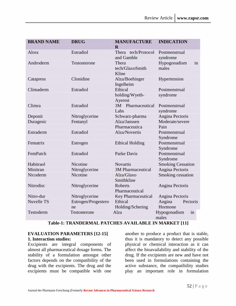
BRAND NAME DRUG MANUFACTURE
R
INDICATION
Alora Estradiol Thera tech/Protocol
and Gamble
Postmenstrual
syndrome
Androderm Testosterone Thera
tech/GlaxoSmith
Kline
Hypogonadism in
males
Catapress Clonidine Alza/Boehinger
Ingelheim
Hypertension
Climaderm Estradiol Ethical
holding/Wyeth-
Ayerest
Postmenstrual
syndrome
Climra Estradiol 3M Pharmaceutical
Labs
Postmenstrual
syndrome
Deponit Nitroglycerine Schwarz-pharma Angina Pectoris
Duragesic Fentanyl Alza/Janssen
Pharmaceutica
Moderate/severe
Pain
Estraderm Estradiol Alza/Novertis Postmenstrual
Syndrome
Fematrix Estrogen Ethical Holding Postmenstrual
Syndrome
FemPatch Estradiol Parke Davis Postmenstrual
Syndrome
Habitraol Nicotine Novartis Smoking Cessation
Minitran Nitroglycerine 3M Pharmaceutical Angina Pectoris
Nicoderm Nicotine Alza/Glaxo
Smithkline
Smoking cessation
Nitrodisc Nitroglycerine Roberts
Pharmaceutical
Angina Pectoris
Nitro-dur Nitroglycerine Key Pharmaceutical Angina Pectoris
Nuvelle TS Estrogen/Progestero
ne
Ethical
Holding/Schering
Angina Pectoris
Hormone
Testoderm Testosterone Alza Hypogonadism in
males
Table-1: TRANDERMAL PATCHES AVAILABLE IN MARKET [11]
EVALUATION PARAMETERS [12-15]
1. Interaction studies: Excipients are integral components of
almost all pharmaceutical dosage forms. The
stability of a formulation amongst other
factors depends on the compatibility of the
drug with the excipients. The drug and the
excipients must be compatible with one
another to produce a product that is stable,
thus it is mandatory to detect any possible
physical or chemical interaction as it can
affect the bioavailability and stability of the
drug. If the excipients are new and have not
been used in formulations containing the
active substance, the compatibility studies
play an important role in formulation
Review Article www.rapsr.com
53 | P a g e Journal der Pharmazie Forschung (Formerly-Recent Advances in Pharmaceutical Science Research
development. Interaction studies are
commonly carried out in Thermal analysis,
FT-IR, UV and chromatographic techniques
by comparing their physicochemical
characters such as assay, melting
endotherms, characteristic wave numbers,
absorption maxima etc.,
2. Thickness of the patch: The thickness of the drug loaded patch is
measured in different points by using a
digital micrometer and determines the
average thickness and standard deviation for
the same to ensure the thickness of the
prepared patch.
3. Weight uniformity: The prepared patches are to be dried at 60°c
for 4hrs before testing. A specified area of
patch is to be cut in different parts of the
patch and weigh in digital balance.The
average weight and standard deviation
values are to be calculated from the
individual weights.
4. Folding endurance: A strip of specific are is to be cut evenly and
repeatedly folded at the same place till it
broke. The number of times the film could
be folded at the same place without breaking
gave the value of the folding endurance.
5. Percentage Moisture content: The prepared films are to be weighed
individually and to be kept in a desiccator
containing fused calcium chloride at room
temperature for 24 hrs. After 24 hrs the
films are to be reweighed and determine the
percentage moisture content from the below
mentioned formula.
Percentage moisture content = [Initial
weight- Final weight/ Final weight] ×100.
6. Percentage Moisture uptake: The weighed films are to be kept in a
desiccator at room temperature for 24 hrs
containing saturated solution of potassium
chloride in order to maintain 84% RH. After
24 hrs the films are to be reweighed and
determine the percentage moisture uptake
from the below mentionedformula.
Percentage moisture uptake = [Final weight-
Initial weight/ initial weight] ×100.
7. Water vapour permeability (WVP)
evaluation: Water vapour permeability can be
determined with foam dressing method the
air forced oven is replaced by a natural air
circulation oven. The WVP can be
determined by the following formula
WVP=W/A
Where, WVP is expressed in gm/m2 per
24hrs,
W is the amount of vapour permeated
through the patch expressed in gm/24hrs and
A is the surface area of the exposure
samples expressed in m2.
8. Drug content:
A specified area of patch is to be dissolved
in a suitable solvent in specific volume.
Then the solution is to be filtered through a
filter medium and analyse the drug contain
with the suitable method (UV or HPLC
technique). Each value represents average of
three different samples.
9. Uniformity of dosage unit test: An accurately weighed portion of the patch
is to be cut into small pieces and transferred
to a specific volume volumetric flask,
dissolved in a suitable solvent and sonicate
for complete extraction of drug from the
patch and made up to the mark with same.
The resulting solution was allowed to settle
for about an hour, and the supernatant was
suitably diluted to give the desired
concentration with suitable solvent. The
solution was filtered using 0.2m membrane
filter and analysed by suitable analytical
technique (UV or HPLC) and the drug
content per piece will be calculated.
10. Polariscope examination: This test is to be performed to examine the
drug crystals from patch by polariscope. A
specific surface area of the piece is to be
kept on the object slide and observe for the
drugs crystals to distinguish whether the
Review Article www.rapsr.com
54 | P a g e Journal der Pharmazie Forschung (Formerly-Recent Advances in Pharmaceutical Science Research
drug is present as crystalline form or
amorphous form in the patch.
11. Shear Adhesion test: This test is to be performed for the
measurement of the cohesive strength of an
adhesive polymer. It can be influenced by
the molecular weight, the degree of
crosslinking and the composition of
polymer, type and the amount of tackifier
added. An adhesive coated tape is applied
onto a stainless steel plate; a specified
weight is hung from the tape, to affect it
pulling in a direction parallel to the plate.
Shear adhesion strength is determined by
measuring the time it takes to pull the tape
off the plate. The longer the time take for
removal, greater is the shear strength.
12. Peel Adhesion test: In this test, the force required to remove an
adhesive coating form a test substrate is
referred to as peel adhesion. Molecular
weight of adhesive polymer, the type and
amount of additives are the variables that
determined the peel adhesion properties. A
single tape is applied to a stainless steel
plate or a backing membrane of choice and
then tape is pulled from the substrate at a
180º angle, and the force required for tape
removed is measured.
13. Thumb tack test: It is a qualitative test applied for tack
property determination of adhesive. The
thumb is simply pressed on the adhesive and
the relative tack property is detected.
14. Flatness test: Three longitudinal strips are to be cut from
each film at different portion like one from
the center, other one from the left side, and
another one from the right side. The length
of each strip was measured and the variation
in length because of non-uniformity in
flatness was measured by determining
percent constriction, with 0% constriction
equivalent to 100% flatness.
15. Percentage Elongation break test:
The percentage elongation break is to be
determined by noting the length just before
the break point, the percentage elongation
can be determined from the below
mentioned formula.
Elongation percentage = L1-L2 /L2×100
Where, L1is the final length of each strip
and L2 is the initial length of each strip.
16. Rolling ball tack test: This test measures the softness of a polymer
that relates to talk. In this test, stainless steel
ball of 7/16 inches in diameter is released on
an inclined track so that it rolls down and
comes into contact with horizontal, upward
facing adhesive. The distance the ball travels
along the adhesive provides the
measurement of tack, which is expressed in
inch.
17. Quick Stick (peel-tack) test: In this test, the tape is pulled away from the
substrate at 90ºC at a speed of 12
inches/min. The peel force required to break
the bond between adhesive and substrate is
measured and recorded as tack value, which
is expressed in ounces or grams per inch
width.
18. Probe Tack test: In this test, the tip of a clean probe with a
defined surface roughness is brought into
contact with adhesive, and when a bond is
formed between probe and adhesive. The
subsequent removal of the probe
mechanically breaks it. The force required to
pull the probe away from the adhesive at
fixed rate is recorded as tack and it is
expressed in grams.
19. In vitro drug release studies: The paddle over disc method (USP
apparatus V) can be employed for
assessment of the release of the drug from
the prepared patches. Dry films of known
thickness is to be cut into definite shape,
weighed, and fixed over a glass plate with
an adhesive. The glass plate was then placed
in a 500-mL of the dissolution medium or
phosphate buffer (pH 7.4), and the apparatus
Review Article www.rapsr.com
55 | P a g e Journal der Pharmazie Forschung (Formerly-Recent Advances in Pharmaceutical Science Research
was equilibrated to 32± 0.5°C. The paddle
was then set at a distance of 2.5 cm from the
glass plate and operated at a speed of 50
rpm. Samples (5-mL aliquots) can be
withdrawn at appropriate time intervals up
to 24 h and analyzed by UV
spectrophotometer or HPLC. The
experiment is to be performed in triplicate
and the mean value can be calculated.
20. In vitro skin permeation studies: An in vitro permeation study can be carried
out by using diffusion cell. Full thickness
abdominal skin of male Wistar rats weighing
200 to 250g. Hair from the abdominal region
is to be removed carefully by using a electric
clipper; the dermal side of the skin was
thoroughly cleaned with distilled water to
remove any adhering tissues or blood
vessels, equilibrated for an hour in
dissolution medium or phosphate buffer pH
7.4 before starting the experiment and was
placed on a magnetic stirrer with a small
magnetic needle for uniform distribution of
the diffusant. The temperature of the cell
was maintained at 32 ± 0.5°C using a
thermostatically controlled heater. The
isolated rat skin piece is to be mounted
between the compartments of the diffusion
cell, with the epidermis facing upward into
the donor compartment. Sample volume of
definite volume is to be removed from the
receptor compartment at regular intervals,
and an equal volume of fresh medium is to
be replaced. Samples are to be filtered
through filtering medium and can be
analyzed spectrophotometrically or HPLC.
Flux can be determined directly as the slope
of the curve between the steady-state values
of the amount of drug permeated (mg cm-2)
vs. time in hours and permeability
coefficients were deduced by dividing the
flux by the initial drug load (mg cm-2).
21. Skin Irritation study: Skin irritation and sensitization testing can
be performed on healthy rabbits (average
weight 1.2 to 1.5 kg). The dorsal surface
(50cm2) of the rabbit is to be cleaned and
remove the hair from the clean dorsal
surface by shavingand clean the surface by
using rectified spirit and the representative
formulations can be applied over the
skin.The patch is to be removed after 24 hr
and the skin is to be observed and classified
into 5 grades on the basis of the severity of
skin injury.
22. Stability studies: Stability studies are to be conducted
according to the ICH guidelines by storing
the TDDS samples at 40±0.5°c and 75±5%
RH for 6 months. The samples were
withdrawn at 0, 30, 60, 90 and 180 days and
analyze suitably for the drug content.
CONCLUSION:
The TDDS review articles provide valuable
information regarding the transdermal drug
delivery systems and its evaluation process
details as a ready reference for the research
scientist who is involved in TDDS. The
foregoing shows that TDDS have great
potentials, being able to use for both
hydrophobic and hydrophilic active
substance into promising deliverable drugs.
To optimize this drug delivery system,
greater understanding of the different
mechanisms of biological interactions, and
polymer are required. TDDS a realistic
practical application as the next generation
of drug delivery system.
REFERENCES:
1. Jain NK. Advances in controlled and novel drug delivery, 1stEd.,CBS Publishers and
distributors, New Delhi .2001; pp 108-110
2. Barry BW,William AC. In: Swarbrick J(ed)) Boylon JC.Encyclopedia of pharmaceutical
technology vol-II, Marcel Dekker: Inc New York. 1995; pp49-93
3. Tanner T, Marks R.Delivering Drugs by the Transdermal Route: review and comment.
Skin Research and Technology .2008;14:249-260
Review Article www.rapsr.com
56 | P a g e Journal der Pharmazie Forschung (Formerly-Recent Advances in Pharmaceutical Science Research
4. Vyas SP and Khar RK .Controlled Drug Delivery Concepts and Advances : Transdermal
Drug Delivery .Chapter 10, pp 411-476
5. Allen VL, Nicholas G,Popovich and Howard C.Ansel,Ansel,s Pharmaceutical Dosage
Forms and Drug Delivery Systems. Transdermal Drug Delivery Systems. Chapter
11.pp.298-315
6. Roberts MS.Targeted drug delivery to the skin and deeper tissues :role of physiology
solute structure and disease. Clin Exp Pharmacol Physiol.1997;Nov.24(11):874-9
7. Gaur PK et al.Transdermal Drug Delivery System : A Review. Asian J Pharmaceuti Clin
Res.2009;2(1):14-20
8. Chein YW. Novel drug delivery systems ,Drugs and Pharmaceutical Sciences .Marcel
Dekker ,New York.1992;797
9. Arunachalam A et al. Transdermal Drug Delivery System : A Review. Cur Pharma Res
.2010;1(1):70-81
10. Pathan BI and Mallikarjuna SC. Chemical Penetration Enhancers for Transdermal Drug
Delivery Systems. Trop J Pharmaceut Res .2009;8(2):173-179
11. Kumar A and Philip A. Modified Transdermal Technologies :Breaking the Barriers of
Drug Permeation via the skin. Trop J Pharmaceut Res.2007;6(1):633-644
12. Singh J,Tripathy KT and Sakia TR.Effect of penetration enhancers on the in vitro
transport of ephedrine through rat skin and human epidermis from matrix based
transdermal formulations. Drug Dev Ind Pharm.1993;19:1623-1628
13. Wade A,Waller PJ. Handbook of Pharmaceutical Excipients.Washington DC:American
Publishing Association .1994;362-366
14. Ashok KJ etal. Transdermal drug delivery system :An Overview . Int J Pharmaceut Sci
Review Res.2010;3(2):49-54
15. Arati N et al.Mechanism of Oleic acid induced skin penetration enhancement in vivo in
humans. J Control Release.1995;37:299-306