a rare cause of thoracic spinal cord compression by … · 1department of neurosurgery, faculté de...
TRANSCRIPT

Copyright © 2018 Korean Neurotraumatology Society 35
Introduction
The spinal extradural arachnoid cyst (SEAC) is a rare cause of spinal cord compression. The etiology of SEAC re-mains unclear. They appear to be extradural outpouchings of arachnoid membranes that communicate with the intra-spinal subarachnoid space through small defect in the dura.7,10,17) Tarlov cyst (TC) are predominantly found at the lumbosacral level of the spine but they are known to exist at all levels of the spine. Bifocal location of SEACs, thorac-
ic and sacral, is exceedingly rarely reported in the literature. Here in we report a case of thoracic spinal cord compression by SEAC associated with asymptomatic multiple sacral cysts. The surgical management and postoperative outcome are discussed.
Case Report
A 34-year-old woman with no history of trauma was re-ferred to the emergency department for acute thoracic pain superimposed on a background of chronic long-standing back pain. The patient was complaining of walking diffi-culties. Neurological examination demonstrated an incom-plete spastic paraplegia, grade 2 motor power in both lower limbs, with sensory level in T9, with pyramidal syndrome in the inferior limbs bilateral plantar extension responses, with hyperreflexia of both Achilles tendons reflexes were present. Sensory, bladder, and bowel functions were unre-markable. Imaging a spinal magnetic resonance imaging
A Rare Cause of Thoracic Spinal Cord Compression by Multiple Large Tarlov Cysts
Ahmed-Salem Kleib1,2, Sidi-Mohamed Salihy1, Hussein Hamdi2, Romain Carron2, and Outouma Soumaré1
1Department of Neurosurgery, Faculté de Médecine, Université de Nouakchott Al-Aasriya, Nouakchott, Mauritania 2Department of Stereotactic and Functional Neurosurgery, Centre Hospitalier Universitaire La Timone, Assistance Publique Hôpitaux de Marseille, Marseille, France
Spinal extradural arachnoid cyst (SEAC) is a rare cause of spinal cord compression. Bifocal location of thoracic and sacral SEACs is rarely reported in the literature. We report a case of thoracic spinal cord compression by SEAC associated with asymptomatic multiple sacral Tarlov cysts (TC). The surgical management and postoperative outcome of the patient are discussed. A 34-year-old woman was referred to the hospital for acute thoracic pain with a history of chronic long-stand-ing back pain. She complained of walking difficulties. Neurological examination demonstrated incomplete spastic para-plegia with sensory level in T9. Magnetic resonance imaging revealed a large cystic formation from T7-11 and at the level of the sacrum. We performed laminectomies at the level of interest from T7-11. The cysts were dissected from the under-lying dura after removal of the cerebrospinal fluid. We found nerve tissue in the cysts. We excised the cyst and preserved the nerve roots. Subsequently, a duraplasty was performed with autologous grafts from the lumbar fascia. The condition of the patient improved after surgery and he was recovering well at follow-up. Although the surgical treatment of TC is controversial, especially at the sacral lumbar level, decompression at the dorsal level in this case is indisputable. (Korean J Neurotrauma 2018;14(1):35-38)
KEY WORDS: Extradural arachnoid cyst ㆍSpinal cord compression ㆍTarlov cyst.
Received: January 5, 2018 / Revised: March 16, 2018Accepted: April 12, 2018Address for correspondence: Ahmed-Salem KleibDepartment of Neurosurgery, Faculté de Médecine, Université de Nouakchott Al-Aasriya, BP 5026, Nouakchott, MauritaniaTel: +222-36712000, Fax: +222-36712000E-mail: [email protected] cc This is an Open Access article distributed under the terms of Cre-ative Attributions Non-Commercial License (http://creativecommons.org/licenses/by-nc/4.0/) which permits unrestricted noncommercial use, distribution, and reproduction in any medium, provided the original work is properly cited.
CASE REPORTKorean J Neurotrauma 2018;14(1):35-38
pISSN 2234-8999 / eISSN 2288-2243
https://doi.org/10.13004/kjnt.2018.14.1.35
36 Korean J Neurotrauma 2018;14(1):35-38
Thoracic Spinal Cord Compression by Tarlov Cysts
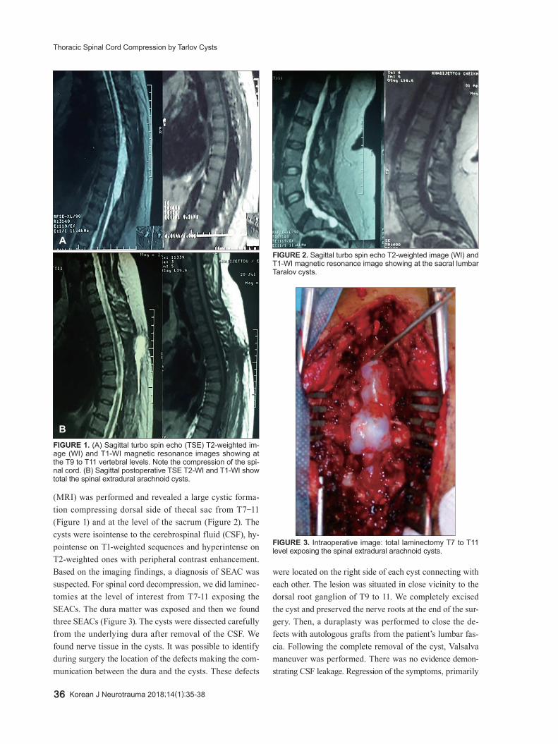
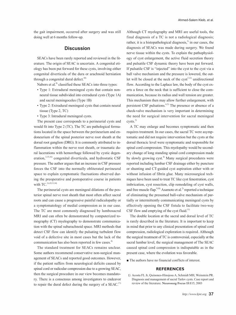
(MRI) was performed and revealed a large cystic forma-tion compressing dorsal side of thecal sac from T7-11 (Figure 1) and at the level of the sacrum (Figure 2). The cysts were isointense to the cerebrospinal fluid (CSF), hy-pointense on T1-weighted sequences and hyperintense on T2-weighted ones with peripheral contrast enhancement. Based on the imaging findings, a diagnosis of SEAC was suspected. For spinal cord decompression, we did laminec-tomies at the level of interest from T7-11 exposing the SEACs. The dura matter was exposed and then we found three SEACs (Figure 3). The cysts were dissected carefully from the underlying dura after removal of the CSF. We found nerve tissue in the cysts. It was possible to identify during surgery the location of the defects making the com-munication between the dura and the cysts. These defects
were located on the right side of each cyst connecting with each other. The lesion was situated in close vicinity to the dorsal root ganglion of T9 to 11. We completely excised the cyst and preserved the nerve roots at the end of the sur-gery. Then, a duraplasty was performed to close the de-fects with autologous grafts from the patient’s lumbar fas-cia. Following the complete removal of the cyst, Valsalva maneuver was performed. There was no evidence demon-strating CSF leakage. Regression of the symptoms, primarily
FIGURE 1. (A) Sagittal turbo spin echo (TSE) T2-weighted im-age (WI) and T1-WI magnetic resonance images showing at the T9 to T11 vertebral levels. Note the compression of the spi-nal cord. (B) Sagittal postoperative TSE T2-WI and T1-WI show total the spinal extradural arachnoid cysts.
A
B
FIGURE 2. Sagittal turbo spin echo T2-weighted image (WI) and T1-WI magnetic resonance image showing at the sacral lumbar Taralov cysts.
FIGURE 3. Intraoperative image: total laminectomy T7 to T11 level exposing the spinal extradural arachnoid cysts.
Ahmed-Salem Kleib, et al.
http://www.kjnt.org 37
the gait impairment, occurred after surgery and was still doing well at 6 months follow-up.
Discussion
SEACs have been rarely reported and reviewed in the lit-erature. The origin of SEAC is uncertain. A congenital eti-ology has been put forward for these cysts, involving either congenital diverticula of the dura or arachnoid herniation through a congenital dural defect.10)
Nabors et al.8) classified these SEACs into three types:• Type 1: Extradural meningeal cysts that contain non-
neural tissue subdivided into extradural cysts (Type 1A) and sacral meningoceles (Type 1B)
• Type 2: Extradural meningeal cysts that contain neural tissue (Type 2, TC)
• Type 3: Intradural meningeal cysts.The present case corresponds to a perineurial cysts and
would fit into Type 2 (TC). The TC are pathological forma-tions located in the space between the perineurium and en-doneurium of the spinal posterior nerve root sheath at the dorsal root ganglion (DRG). It is commonly attributed to in-flammation within the nerve root sheath, or traumatic du-ral lacerations with hemorrhage followed by cystic degen-eration,3,15,16) congenital diverticula, and hydrostatic CSF pressure. The author argues that an increase in CSF pressure forces the CSF into the normally obliterated perineural space to explain symptomatic fluctuations observed dur-ing the preoperative and postoperative course in patients with TC.3,6,9,13,14)
The perineurial cysts are meningeal dilations of the pos-terior spinal nerve root sheath that most often affect sacral roots and can cause a progressive painful radiculopathy or a symptomatology of medial compression as in our case. The TC are most commonly diagnosed by lumbosacral MRI and can often be demonstrated by computerized to-mography (CT) myelography to demonstrate communica-tion with the spinal subarachnoid space. MRI methods that detect CSF flow can identify the pulsating turbulent flow void of a defective site in most cases but the lack of the communication has also been reported in few cases.4)
The standard treatment for SEACs remains unclear. Some authors recommend conservative non-surgical man-agement of SEACs and reported good outcomes. However, if the patient suffers from neurological deficits caused by spinal cord or radicular compression due to a growing SEAC, then the surgical procedure in our view becomes mandato-ry. There is a consensus among investigators to endeavor to repair the dural defect during the surgery of a SEAC.17)
Although CT myelography and MRI are useful tools, the final diagnosis of a TC is not a radiological diagnosis; rather, it is a histopathological diagnosis,1) in our cases, the diagnosis of SEACs was made during surgery. We found nerve tissue within the cysts. To explain the pathophysiol-ogy of cyst enlargement, the active fluid secretion theory and pulsatile CSF dynamic theory have been put forward. If pulsatile CSF is “injected” into the cyst to the cyst via a ball valve mechanism and the pressure is lowered, the out-let will be closed at the neck of the cyst7,12) unidirectional flow. According to the Laplace law, the body of the cyst ex-erts a force on the neck that is sufficient to close the com-munication, because its radius and wall tension are greater. This mechanism then may allow further enlargement, with persistent CSF pulsations.4,5) The presence or absence of a check-valve mechanism is very important in determining the need for surgical intervention for sacral meningeal cysts.2)
A TC may enlarge and becomes symptomatic and then requires treatment. In our cases, the sacral TC were asymp-tomatic and did not require intervention but the cysts at the dorsal thoracic level were symptomatic and responsible for spinal cord compression. This myelopathy would be second-ary change of long standing spinal cord compression made by slowly growing cyst.4) Many surgical procedures were reported including lumbar CSF drainage either by puncture or shunting and CT-guided cyst aspiration either with or without infusion of fibrin glue. Many microsurgical tech-niques have been used to treat TC like cyst fenestration, cyst imbrication, cyst resection, clip remodeling of cyst walls, and free muscle flap.9,18) Asamoto et al.2) reported a technique of eliminating the presumed ball-valve mechanism of par-tially or intermittently communicating meningeal cysts by effectively opening the CSF fistula to facilitate two-way CSF flow and emptying of the cyst fluid.11)
The double location at the sacral and dorsal level of TC is rarely described in the literature. It is important to keep in mind that prior to any clinical presentation of spinal cord compression, radiological exploration is required. Although the surgical treatment of TC is controversial, especially at the sacral lumbar level, the surgical management of The SEAC caused spinal cord compression is indisputable as in the present case, where the evolution was favorable.
■ The authors have no financial conflicts of interest.
REFERENCES1) Acosta FL Jr, Quinones-Hinojosa A, Schmidt MH, Weinstein PR.
Diagnosis and management of sacral Tarlov cysts. Case report and review of the literature. Neurosurg Focus 15:E15, 2003

38 Korean J Neurotrauma 2018;14(1):35-38
Thoracic Spinal Cord Compression by Tarlov Cysts
2) Asamoto S, Fukui Y, Nishiyama M, Ishikawa M, Fujita N, Naka-mura S, et al. Diagnosis and surgical strategy for sacral meninge-al cysts with check-valve mechanism: technical note. Acta Neuro-chir (Wien) 155:309-313, 2013
3) Cantore G, Bistazzoni S, Esposito V, Tola S, Lenzi J, Passacantilli E, et al. Sacral Tarlov cyst: surgical treatment by clipping. World Neurosurg 79:381-389, 2013
4) Kim IS, Hong JT, Son BC, Lee SW. Noncommunicating spinal extradural meningeal cyst in thoracolumbar spine. J Korean Neu-rosurg Soc 48:534-537, 2010
5) Liu JK, Cole CD, Kan P, Schmidt MH. Spinal extradural arachnoid cysts: clinical, radiological, and surgical features. Neurosurg Fo-cus 22:E6, 2007
6) Lombardi G, Morello G. Congenital cysts of the spinal membranes and roots. Br J Radiol 36:197-205, 1963
7) McCrum C, Williams B. Spinal extradural arachnoid pouches. Report of two cases. J Neurosurg 57:849-852, 1982
8) Nabors MW, Pait TG, Byrd EB, Karim NO, Davis DO, Kobrine AI, et al. Updated assessment and current classification of spinal men-ingeal cysts. J Neurosurg 68:366-377, 1988
9) Naderi S. Surgical approaches in symptomatic tarlov cysts. World Neurosurg 86:20-21, 2016
10) Nayak R, Chaudhuri A, Sadique S, Attry S. Multiple spinal extra-
dural arachnoidal cysts: An uncommon cause of thoracic cord com-pression. Asian J Neurosurg 12:321-323, 2017
11) Potts MB, McGrath MH, Chin CT, Garcia RM, Weinstein PR. Mi-crosurgical fenestration and paraspinal muscle pedicle flaps for the treatment of symptomatic sacral tarlov cysts. World Neurosurg 86:233-242, 2016
12) Rohrer DC, Burchiel KJ, Gruber DP. Intraspinal extradural men-ingeal cyst demonstrating ball-valve mechanism of formation. Case report. J Neurosurg 78:122-125, 1993
13) Schreiber F, Haddad B. Lumbar and sacral cysts causing pain. J Neurosurg 8:504-509, 1951
14) Strully KJ, Heiser S. Lumbar and sacral cysts of meningeal ori-gin. Radiology 62:544-549, 1954
15) Tarlov IM. Perineurial cysts of the spinal nerve roots. Arch Neu-rol Psychiatry 40:1067-1074, 1938
16) Tarlov IM. Cysts, perineurial, of the sacral roots; another cause, removable, of sciatic pain. J Am Med Assoc 138:740-744, 1948
17) Woo JB, Son DW, Kang KT, Lee JS, Song GS, Sung SK, et al. Spi-nal extradural arachnoid cyst. Korean J Neurotrauma 12:185-190, 2016
18) Xu J, Sun Y, Huang X, Luan W. Management of symptomatic sacral perineural cysts. PLoS One 7:e39958, 2012