a rac-like small g-protein from brassica campestris activates a pkc-dependent phospholipase d
TRANSCRIPT

A rac-like small G-protein from Brassica campestris activates aPKC-dependent phospholipase D
Hoyeon Kima, Minyeop Nahma, Chaeoh Lima, Daejin Yuna, Mooje Choa,1,Jeongdong Bahka,b,*,1
aDivision of Applied Life Sciences, Graduate School of Gyeongsang National University, Jinju 660-701, South KoreabAgricultural Plant Stress Research Center, Chonnam National University, Gwangju 500-757, South Korea
Received 19 March 2003; received in revised form 10 September 2003
Abstract
A cDNA clone encoding a rac-like small GTP binding protein was isolated from a cDNA library of Chinese cabbage (Brassicacampestris L. ssp. pekinensis) flower buds and named Brac1. The Brac1 cDNA contains an open reading frame encoding 198 aminoacid residues with an estimated molecular mass of 21,690 Da and this coding region has conserved residues and motifs unique to theRho subfamily of proteins. The deduced amino acid sequence of the Brac1 protein is closely related to that of Arabidopsis thaliana
Arac3 (91%), but it shares relatively little homology with other members of the Ras superfamily (about 30% identity). To furthercharacterize Brac1, a pGBrac1 expression vector consisting of PCR-amplified Brac1 cDNA plus glutathione S-transferase (GST)and pBKS(+)II was used to purify the protein. Using a PEI-cellulose/TLC plate, GTPase activity of this protein was confirmed
and competition binding studies, using the guanine nucleotides, ATP, UTP and CTP, revealed that the di- and triphosphate formsof guanine nucleotides strongly bind Brac1. Membrane-bound PLD activity was synergistically enhanced by Brac1 in the presenceof protein kinase C, but not in the presence of ARF (ADP-ribosylation factor). Genomic analysis indicated that Brac1 belongs to a
multigene family. Brac1 transcripts were expressed in all the organs of Brassica, but were especially prevalent in flower buds.# 2003 Elsevier Ltd. All rights reserved.
Keywords: Brassica campestris; Cruciferae; Chinese cabbage; Rac-like small G-protein; Phospholipase D; Rho subfamily
1. Introduction
The enzyme phospholipase D (PLD, EC 3.1.4.4) iswidely distributed in plants, fungi and bacteria (Exton,1997a,b) and is generally thought to catalyze thehydrolysis of the terminal diester bond of membraneglycerophospholipids, resulting in the formation ofphosphatidic acid and a related base (Gomez-Cam-bronero and Keire, 1998). Recent studies in animal sys-tems indicate that PLD activation and formation ofphosphatidic acid are involved in transducing signalsfrom many extracellular stimuli. Various reports alsoindicate that the resulting phosphatidic acid functions inthe regulation of protein kinases, actin assembly, vesicletrafficking and secretion (Exton, 1997a,b; Ktistakis etal., 1995, 1996; Chen et al., 1997).
The structural and biochemical properties of variousplant PLDs, including castor bean (Wang et al., 1994),rice (Ueki et al., 1995), maize (Ueki et al., 1995), cowpeaand Arabidopsis (Pappan et al., 1997a,b) have beendescribed. Comparison of these properties indicated ahigh degree of similarity (except for Arabidopsis PLD)and these enzymes were named PLDa. On the otherhand Pappan et al. (1997a,b) identified a PLD isoform,named PLDb, which is different from PLDa in terms ofcDNA length, Ca2+-dependency and pH sensitivity.The first indication for the involvement of a small
G-protein in the regulation of PLD emerged from initialobservations that a cytosolic factor was involved, whichwas subsequently identified as ARF (ADP-ribosylationfactor) (Brown et al., 1993). In addition, other GTPasessuch as RhoA, Rac1 and Cdc42Hs were found to acti-vate membrane-associated PLD in the presence ofGTPgS (Malcolm et al., 1994; Siddiqui et al., 1995;Kwak et al., 1995). Among the five distinct families ofsmall GTPases (Ras, Rab, Arf/Sar, Rho and Ran), the
0031-9422/$ - see front matter # 2003 Elsevier Ltd. All rights reserved.
doi:10.1016/j.phytochem.2003.10.001
Phytochemistry 65 (2004) 71–80
www.elsevier.com/locate/phytochem
* Corresponding author. Fax: +82-55-752-7062.
E-mail address: [email protected] (J. Bahk).1 M.J. Cho and J.D. Bahk contributed equally to this work.
Rho family is known to play key roles in the regulationof focal adhesion complex and actin cytoskeleton for-mation. The Rho family can be further subdivided intothree subfamilies: Rho, Rac and Cdc42.Brown et al. (1995) showed that Rho acts synergisti-
cally with ARF to activate brain PLD. In addition, Luoet al. (1997) demonstrated using cell-free systems thatsome members of the small G-protein Ras superfamily,namely Rho, ARF, and RalA, stimulate PLD activity.Although a relatively large number of cDNAs encod-
ing Rho family small GTP-binding proteins have beenisolated in plants, such as Arabidopsis (Winge et al.,1997; Xia et al., 1996), garden pea (Yang and Watson,1993) and cotton (Delmer et al., 1995), the function ofmost of these plant genes has not been well character-ized, except in the case of Arabidopsis (Vernoud et al.,2003). Within the Rho family, the Rac proteins havebeen reported to enhance superoxide generation as aresponse to microbial infection (Abo et al., 1991; Knauset al., 1991). A similar role for Rac proteins is alsoobserved in plants during hypersensitivity responses(Kawasaki et al., 1999; Park et al., 2000). In A. thaliana,although several new members of the Rab, Ran and Arffamilies have been isolated, to date no true homologuesof the Ras-, Rho- and Cdc42-like proteins have beenfound. However, a large number of rac-like genes wereidentified in Pisum sativum (Yang and Watson, 1993),Gossypium hirsutum (Luo et al., 1997; Trainin et al.,1996) and A. thaliana (Xia et al., 1996).In order to further elucidate the mechanism by which
Rac proteins stimulate PLD activity in plants, we haveisolated a rac-like cDNA clone from Brassica campes-tris, named Brac1 (GenBank accession numberAF042330). Here we report that the isolated clone is amember of the plant-specific Rho family, and its codingregion has the ability to bind and hydrolyze GTP. Wefurther propose that Brac1 can synergistically activate aPKC (protein kinase C)-dependent rat brain PLD inthe presence of guanosine-50-O-(3-thiotriphosphate)(GTPgS).
2. Results and discussion
2.1. Isolation and sequence analysis of a rac-like cDNAclone, Brac1
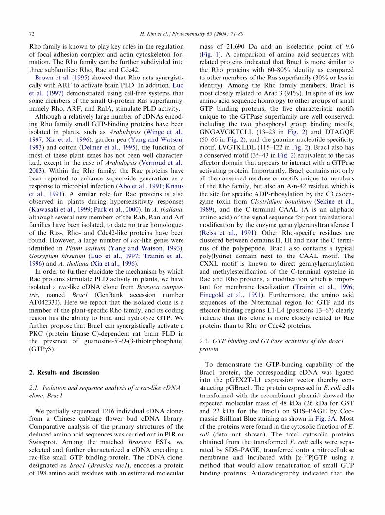
We partially sequenced 1216 individual cDNA clonesfrom a Chinese cabbage flower bud cDNA library.Comparative analysis of the primary structures of thededuced amino acid sequences was carried out in PIR orSwissprot. Among the matched Brassica ESTs, weselected and further characterized a cDNA encoding arac-like small GTP binding protein. The cDNA clone,designated as Brac1 (Brassica rac1), encodes a proteinof 198 amino acid residues with an estimated molecular
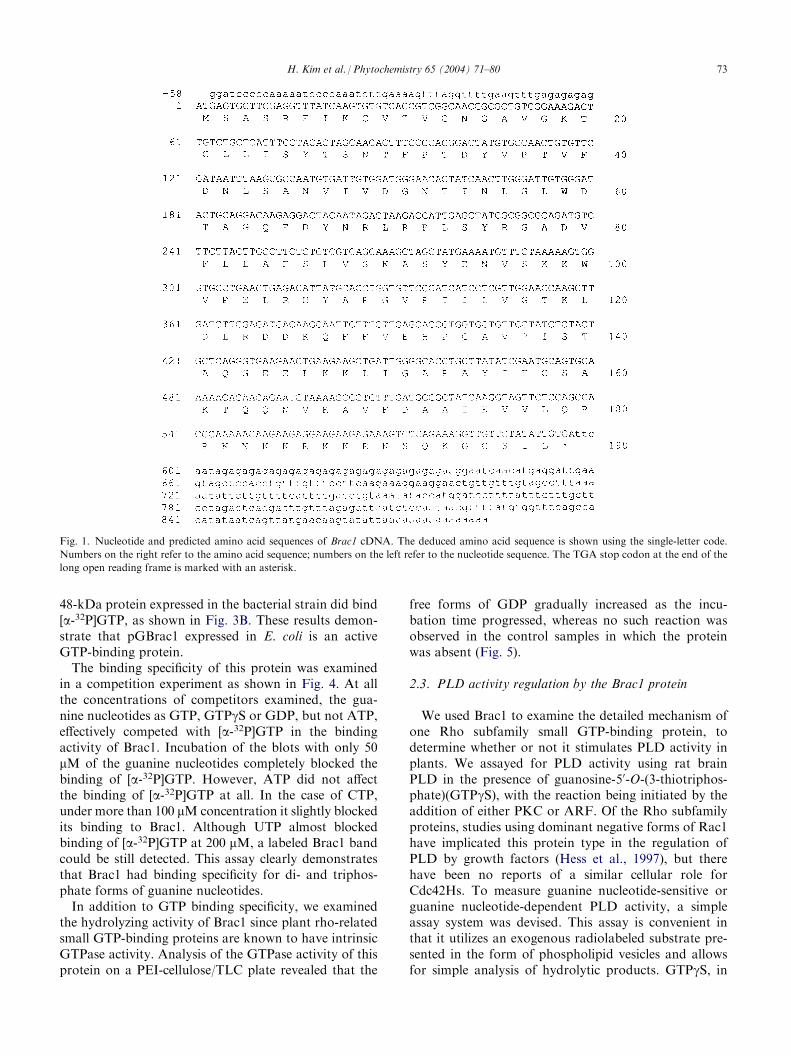
mass of 21,690 Da and an isoelectric point of 9.6(Fig. 1). A comparison of amino acid sequences withrelated proteins indicated that Brac1 is more similar tothe Rho proteins with 60–80% identity as comparedto other members of the Ras superfamily (30% or less inidentity). Among the Rho family members, Brac1 ismost closely related to Arac 3 (91%). In spite of its lowamino acid sequence homology to other groups of smallGTP binding proteins, the five characteristic motifsunique to the GTPase superfamily are well conserved,including the two phosphoryl group binding motifs,GNGAVGKTCLL (13–23 in Fig. 2) and DTAGQE(60–66 in Fig. 2), and the guanine nucleotide specificitymotif, LVGTKLDL (115–122 in Fig. 2). Brac1 also hasa conserved motif (35–43 in Fig. 2) equivalent to the raseffector domain that appears to interact with a GTPaseactivating protein. Importantly, Brac1 contains not onlyall the conserved residues or motifs unique to membersof the Rho family, but also an Asn-42 residue, which isthe site for specific ADP-ribosylation by the C3 exoen-zyme toxin from Clostridium botulinum (Sekine et al.,1989), and the C-terminal CAAL (A is an aliphaticamino acid) of the signal sequence for post-translationalmodification by the enzyme geranylgeranyltransferase I(Reiss et al., 1991). Other Rho-specific residues areclustered between domains II, III and near the C termi-nus of the polypeptide. Brac1 also contains a typicalpoly(lysine) domain next to the CAAL motif. TheCXXL motif is known to direct geranylgeranylationand methylesterification of the C-terminal cysteine inRac and Rho proteins, a modification which is impor-tant for membrane localization (Trainin et al., 1996;Finegold et al., 1991). Furthermore, the amino acidsequences of the N-terminal region for GTP and itseffector binding regions L1-L4 (positions 13–67) clearlyindicate that this clone is more closely related to Racproteins than to Rho or Cdc42 proteins.
2.2. GTP binding and GTPase activities of the Brac1protein
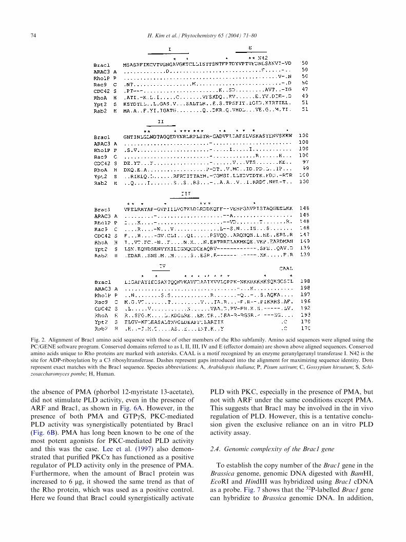
To demonstrate the GTP-binding capability of theBrac1 protein, the corresponding cDNA was ligatedinto the pGEX2T-L1 expression vector thereby con-structing pGBrac1. The protein expressed in E. coli cellstransformed with the recombinant plasmid showed theexpected molecular mass of 48 kDa (26 kDa for GSTand 22 kDa for the Brac1) on SDS–PAGE by Coo-massie Brilliant Blue staining as shown in Fig. 3A. Mostof the proteins were found in the cytosolic fraction of E.coli (data not shown). The total cytosolic proteinsobtained from the transformed E. coli cells were sepa-rated by SDS–PAGE, transferred onto a nitrocellulosemembrane and incubated with [a-32P]GTP using amethod that would allow renaturation of small GTPbinding proteins. Autoradiography indicated that the
72 H. Kim et al. / Phytochemistry 65 (2004) 71–80
48-kDa protein expressed in the bacterial strain did bind[a-32P]GTP, as shown in Fig. 3B. These results demon-strate that pGBrac1 expressed in E. coli is an activeGTP-binding protein.The binding specificity of this protein was examined
in a competition experiment as shown in Fig. 4. At allthe concentrations of competitors examined, the gua-nine nucleotides as GTP, GTPgS or GDP, but not ATP,effectively competed with [a-32P]GTP in the bindingactivity of Brac1. Incubation of the blots with only 50mM of the guanine nucleotides completely blocked thebinding of [a-32P]GTP. However, ATP did not affectthe binding of [a-32P]GTP at all. In the case of CTP,under more than 100 mM concentration it slightly blockedits binding to Brac1. Although UTP almost blockedbinding of [a-32P]GTP at 200 mM, a labeled Brac1 bandcould be still detected. This assay clearly demonstratesthat Brac1 had binding specificity for di- and triphos-phate forms of guanine nucleotides.In addition to GTP binding specificity, we examined
the hydrolyzing activity of Brac1 since plant rho-relatedsmall GTP-binding proteins are known to have intrinsicGTPase activity. Analysis of the GTPase activity of thisprotein on a PEI-cellulose/TLC plate revealed that the
free forms of GDP gradually increased as the incu-bation time progressed, whereas no such reaction wasobserved in the control samples in which the proteinwas absent (Fig. 5).
2.3. PLD activity regulation by the Brac1 protein
We used Brac1 to examine the detailed mechanism ofone Rho subfamily small GTP-binding protein, todetermine whether or not it stimulates PLD activity inplants. We assayed for PLD activity using rat brainPLD in the presence of guanosine-50-O-(3-thiotriphos-phate)(GTPgS), with the reaction being initiated by theaddition of either PKC or ARF. Of the Rho subfamilyproteins, studies using dominant negative forms of Rac1have implicated this protein type in the regulation ofPLD by growth factors (Hess et al., 1997), but therehave been no reports of a similar cellular role forCdc42Hs. To measure guanine nucleotide-sensitive orguanine nucleotide-dependent PLD activity, a simpleassay system was devised. This assay is convenient inthat it utilizes an exogenous radiolabeled substrate pre-sented in the form of phospholipid vesicles and allowsfor simple analysis of hydrolytic products. GTPgS, in
Fig. 1. Nucleotide and predicted amino acid sequences of Brac1 cDNA. The deduced amino acid sequence is shown using the single-letter code.
Numbers on the right refer to the amino acid sequence; numbers on the left refer to the nucleotide sequence. The TGA stop codon at the end of the
long open reading frame is marked with an asterisk.
H. Kim et al. / Phytochemistry 65 (2004) 71–80 73
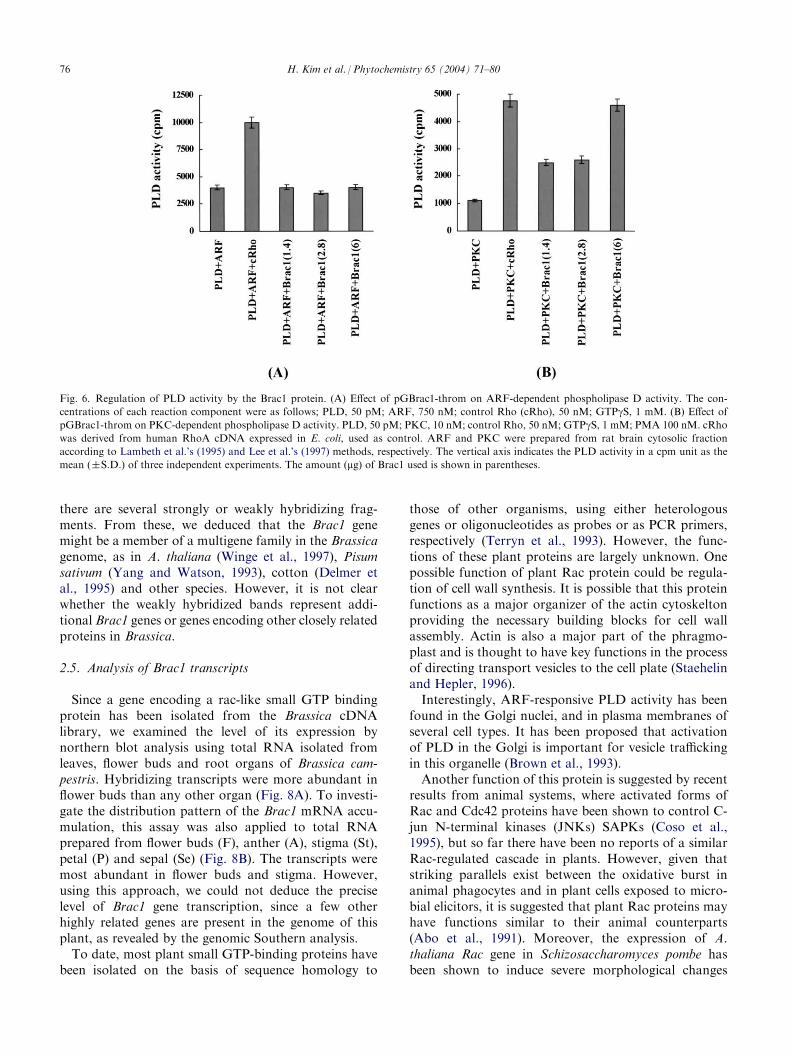
the absence of PMA (phorbol 12-myristate 13-acetate),did not stimulate PLD activity, even in the presence ofARF and Brac1, as shown in Fig. 6A. However, in thepresence of both PMA and GTPgS, PKC-mediatedPLD activity was synergistically potentiated by Brac1(Fig. 6B). PMA has long been known to be one of themost potent agonists for PKC-mediated PLD activityand this was the case. Lee et al. (1997) also demon-strated that purified PKCa has functioned as a positiveregulator of PLD activity only in the presence of PMA.Furthermore, when the amount of Brac1 protein wasincreased to 6 mg, it showed the same trend as that ofthe Rho protein, which was used as a positive control.Here we found that Brac1 could synergistically activate
PLD with PKC, especially in the presence of PMA, butnot with ARF under the same conditions except PMA.This suggests that Brac1 may be involved in the in vivoregulation of PLD. However, this is a tentative conclu-sion given the exclusive reliance on an in vitro PLDactivity assay.
2.4. Genomic complexity of the Brac1 gene
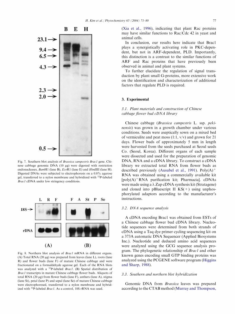
To establish the copy number of the Brac1 gene in theBrassica genome, genomic DNA digested with BamHI,EcoRI and HindIII was hybridized using Brac1 cDNAas a probe. Fig. 7 shows that the 32P-labelled Brac1 genecan hybridize to Brassica genomic DNA. In addition,
Fig. 2. Alignment of Brac1 amino acid sequence with those of other members of the Rho subfamily. Amino acid sequences were aligned using the
PC/GENE software program. Conserved domains referred to as I, II, III, IV and E (effector domain) are shown above aligned sequences. Conserved
amino acids unique to Rho proteins are marked with asterisks. CAAL is a motif recognized by an enzyme geranylgeranyl transferase I. N42 is the
site for ADP-ribosylation by a C3 ribosyltransferase. Dashes represent gaps introduced into the alignment for maximizing sequence identity. Dots
represent exact matches with the Brac1 sequence. Species abbreviations: A, Arabidopsis thaliana; P, Pisum sativum; C, Gossypium hirsutum; S, Schi-
zosaccharomyces pombe; H, Human.
74 H. Kim et al. / Phytochemistry 65 (2004) 71–80
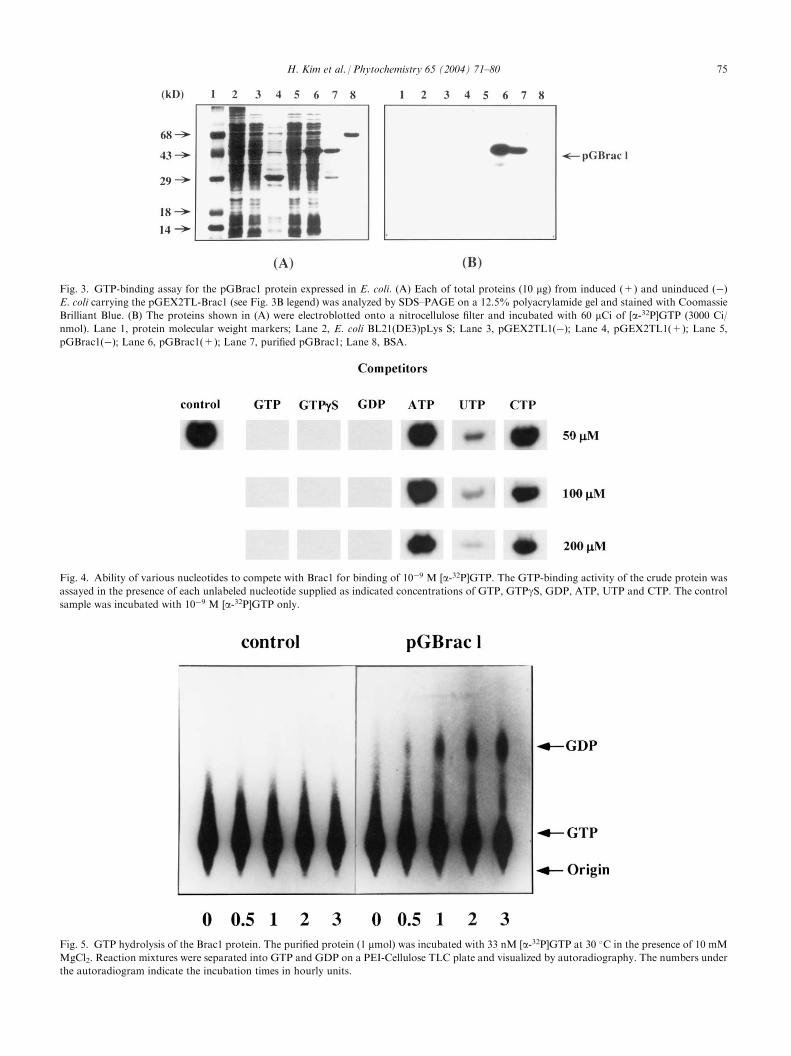
Fig. 4. Ability of various nucleotides to compete with Brac1 for binding of 10�9 M [a-32P]GTP. The GTP-binding activity of the crude protein wasassayed in the presence of each unlabeled nucleotide supplied as indicated concentrations of GTP, GTPgS, GDP, ATP, UTP and CTP. The controlsample was incubated with 10�9 M [a-32P]GTP only.
Fig. 3. GTP-binding assay for the pGBrac1 protein expressed in E. coli. (A) Each of total proteins (10 mg) from induced (+) and uninduced (�)
E. coli carrying the pGEX2TL-Brac1 (see Fig. 3B legend) was analyzed by SDS–PAGE on a 12.5% polyacrylamide gel and stained with Coomassie
Brilliant Blue. (B) The proteins shown in (A) were electroblotted onto a nitrocellulose filter and incubated with 60 mCi of [a-32P]GTP (3000 Ci/nmol). Lane 1, protein molecular weight markers; Lane 2, E. coli BL21(DE3)pLys S; Lane 3, pGEX2TL1(�); Lane 4, pGEX2TL1(+); Lane 5,
pGBrac1(�); Lane 6, pGBrac1(+); Lane 7, purified pGBrac1; Lane 8, BSA.
Fig. 5. GTP hydrolysis of the Brac1 protein. The purified protein (1 mmol) was incubated with 33 nM [a-32P]GTP at 30 �C in the presence of 10 mM
MgCl2. Reaction mixtures were separated into GTP and GDP on a PEI-Cellulose TLC plate and visualized by autoradiography. The numbers under
the autoradiogram indicate the incubation times in hourly units.
H. Kim et al. / Phytochemistry 65 (2004) 71–80 75
there are several strongly or weakly hybridizing frag-ments. From these, we deduced that the Brac1 genemight be a member of a multigene family in the Brassicagenome, as in A. thaliana (Winge et al., 1997), Pisumsativum (Yang and Watson, 1993), cotton (Delmer etal., 1995) and other species. However, it is not clearwhether the weakly hybridized bands represent addi-tional Brac1 genes or genes encoding other closely relatedproteins in Brassica.
2.5. Analysis of Brac1 transcripts
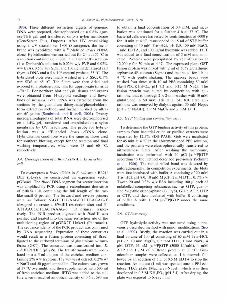
Since a gene encoding a rac-like small GTP bindingprotein has been isolated from the Brassica cDNAlibrary, we examined the level of its expression bynorthern blot analysis using total RNA isolated fromleaves, flower buds and root organs of Brassica cam-pestris. Hybridizing transcripts were more abundant inflower buds than any other organ (Fig. 8A). To investi-gate the distribution pattern of the Brac1 mRNA accu-mulation, this assay was also applied to total RNAprepared from flower buds (F), anther (A), stigma (St),petal (P) and sepal (Se) (Fig. 8B). The transcripts weremost abundant in flower buds and stigma. However,using this approach, we could not deduce the preciselevel of Brac1 gene transcription, since a few otherhighly related genes are present in the genome of thisplant, as revealed by the genomic Southern analysis.To date, most plant small GTP-binding proteins have
been isolated on the basis of sequence homology to
those of other organisms, using either heterologousgenes or oligonucleotides as probes or as PCR primers,respectively (Terryn et al., 1993). However, the func-tions of these plant proteins are largely unknown. Onepossible function of plant Rac protein could be regula-tion of cell wall synthesis. It is possible that this proteinfunctions as a major organizer of the actin cytoskeltonproviding the necessary building blocks for cell wallassembly. Actin is also a major part of the phragmo-plast and is thought to have key functions in the processof directing transport vesicles to the cell plate (Staehelinand Hepler, 1996).Interestingly, ARF-responsive PLD activity has been
found in the Golgi nuclei, and in plasma membranes ofseveral cell types. It has been proposed that activationof PLD in the Golgi is important for vesicle traffickingin this organelle (Brown et al., 1993).Another function of this protein is suggested by recent
results from animal systems, where activated forms ofRac and Cdc42 proteins have been shown to control C-jun N-terminal kinases (JNKs) SAPKs (Coso et al.,1995), but so far there have been no reports of a similarRac-regulated cascade in plants. However, given thatstriking parallels exist between the oxidative burst inanimal phagocytes and in plant cells exposed to micro-bial elicitors, it is suggested that plant Rac proteins mayhave functions similar to their animal counterparts(Abo et al., 1991). Moreover, the expression of A.thaliana Rac gene in Schizosaccharomyces pombe hasbeen shown to induce severe morphological changes
Fig. 6. Regulation of PLD activity by the Brac1 protein. (A) Effect of pGBrac1-throm on ARF-dependent phospholipase D activity. The con-
centrations of each reaction component were as follows; PLD, 50 pM; ARF, 750 nM; control Rho (cRho), 50 nM; GTPgS, 1 mM. (B) Effect ofpGBrac1-throm on PKC-dependent phospholipase D activity. PLD, 50 pM; PKC, 10 nM; control Rho, 50 nM; GTPgS, 1 mM; PMA 100 nM. cRhowas derived from human RhoA cDNA expressed in E. coli, used as control. ARF and PKC were prepared from rat brain cytosolic fraction
according to Lambeth et al.’s (1995) and Lee et al.’s (1997) methods, respectively. The vertical axis indicates the PLD activity in a cpm unit as the
mean (�S.D.) of three independent experiments. The amount (mg) of Brac1 used is shown in parentheses.
76 H. Kim et al. / Phytochemistry 65 (2004) 71–80
(Xia et al., 1996), indicating that plant Rac proteinsmay have similar functions to Rac/Cdc 42 in yeast andanimal cells.In conclusion, our results here indicate that Brac1
plays a synergistically activating role in PKC-depen-dent, but not in ARF-dependent, PLD. Importantly,this distinction is a contrast to the similar functions ofARF and Rac proteins that have previously beenobserved in animal and plant systems.To further elucidate the regulation of signal trans-
duction by plant small G-proteins, more extensive workon the identification and characterization of additionalfactors that regulate PLD is required.
3. Experimental
3.1. Plant materials and construction of Chinesecabbage flower bud cDNA library
Chinese cabbage (Brassica campestris L. ssp. peki-nensis) was grown in a growth chamber under variousconditions. Seeds were aseptically sown on a mixed bedof vermiculite and peat moss (1:1, v/v) and grown for 21days. Flower buds of approximately 5 mm in lengthwere harvested from the seeds purchased at Seoul seedsCo. (Seoul, Korea). Different organs of each samplewere dissected and used for the preparation of genomicDNA, RNA and a cDNA library. To construct a cDNAlibrary we extracted total RNA from flower buds asdescribed previously (Ausubel et al., 1991). Poly(A)+
RNA was obtained using a commercially available kit[poly(A)+RNA purification kit; Pharmacia]. cDNAswere made using a lZap cDNA synthesis kit (Stratagene)and cloned into pBluescript II KS(+) using unphos-phorylated adaptors according to the manufacturer’sinstructions.
3.2. DNA sequence analysis
A cDNA encoding Brac1 was obtained from ESTs ofa Chinese cabbage flower bud cDNA library. Nucleo-tide sequences were determined from both strands ofcDNA using a Taq dye primer cycling sequencing kit ona 373A automatic DNA Sequencer (Applied BiosystemsInc.). Nucleotide and deduced amino acid sequenceswere analyzed using the GCG sequence analysis pro-gram. The phylogenetic relationship of Brac1 and otherknown genes encoding small GTP binding proteins wasanalyzed using the PCGENE software program (Higginsand Sharp, 1988).
3.3. Southern and northern blot hybridization
Genomic DNA from Brassica leaves was preparedaccording to the CTABmethod (Murray and Thompson,
Fig. 7. Southern blot analysis of Brassica campestris Brac1 gene. Chi-
nese cabbage genomic DNA (10 mg) were digested with restrictionendonucleases, BamH1 (lane B), EcoR1 (lane E) and HindIII (lane H).
Digested DNAs were subjected to electrophoresis on a 0.8% agarose
gel, transferred to a nylon membrane and hybridized with 32P-labeled
Brac1 cDNA under low stringency conditions.
Fig. 8. Northern blot analysis of Brac1 mRNA in different organs.
(A) Total RNA (20 mg) was prepared from leaves (lane L), roots (laneR) and flower buds (lane F) of mature Chinese cabbage and were
fractionated on a formaldehyde agarose gel. Each of the RNA blots
was analyzed with a 32P-labeled Brac1. (B) Spatial distribution of
Brac1 transcripts in mature Chinese cabbage flower buds. Aliquots of
total RNA (20 mg) from flower buds (lane F), anthers (lane A), stigma(lane St), petal (lane P) and sepal (lane Se) of mature Chinese cabbage
were electrophoresed, transferred to a nylon membrane and hybrid-
ized with 32P-labeled Brac1. As a control, 18S rRNA was used.
H. Kim et al. / Phytochemistry 65 (2004) 71–80 77
1980). Three different restriction digests of genomicDNA were prepared, electrophoresed on a 0.8% agar-ose-TBE gel, and transferred onto a nylon membrane(GeneScreen Plus, Dupont). After UV crosslinkingusing a UV stratalinker 1800 (Stratagene), the mem-brane was hybridized with a 32P-labeled Brac1 cDNAclone. Hybridization was carried out for 24 h at 55 �C ina solution containing 6 � SSC, 5 � Denhardt’s solution(1 � Denhardt’s solution is 0.02% w/v PVP and 0.02%w/v BSA), 0.5% w/v SDS, and 100 mg/ml denatured calfthymus DNA and a 5 � 106 cpm/ml probe at 55 �C. Thehybridized blots were finally washed in 2 � SSC, 0.1%w/v SDS at 65 �C. The filters were then dried andexposed to a photographic film for appropriate times at�70 �C. For northern blot analysis, tissues and organswere obtained from 14-day-old seedlings and flowerbuds of Brassica. Total RNA was extracted from thesections by the guanidium thiocyanate/phenol/chloro-form extraction method, and further purified by ultra-centrifugation (Sambrook and Russell, 2001). Twentymicrogram-aliquots of total RNA were electrophoresedon a 1.4% gel, transferred and crosslinked to a nylonmembrane by UV irradiation. The probe for hybrid-ization was a 32P-labeled Brac1 cDNA clone.Hybridization conditions were the same as those usedfor southern blotting, except for the reaction and finalwashing temperatures, which were 53 and 60 �C,respectively.
3.4. Overexpression of a Brac1 cDNA in Escherichiacoli
To overexpress a Brac1 cDNA in E. coli strain BL21/DE3 (pLysS), we constructed an expression vectorpGBrac1. The Brac1 DNA segment for vector insertionwas amplified by PCR using a recombinant derivativeof pBKS(+)II containing the full length of the rac-like small G-protein. The forward and reverse primerswere as follows; 50-GTTTTGAAGCTTTGAGAG-30
(designed to create a HindIII restriction site) and 50-ATTAACCCTCACTAAAG-30 (T3 primer), respec-tively. The PCR product digested with HindIII waspurified and ligated into the same restriction site of themulticloning region of pGEX2T Linker1 (Pharmacia).The sequence fidelity of the PCR product was confirmedby DNA sequencing. Expression of these constructswould result in a fusion protein consisting of Brac1ligated to the carboxyl terminus of glutathione S-trans-ferase (GST). The construct was transformed into E.coli BL21/DE3 (pLysS). The transformants were inocu-lated into a 5-ml aliquot of the enriched medium con-taining 2% w/v tryptone, 1% w/v yeast extract, 0.2% w/v NaCl and 50 mg/ml ampicillin. The culture was grownat 37 �C overnight, and then supplemented with 500 mlof fresh enriched medium. IPTG was added to the cul-ture when it reached an optical density of 0.6 at 590 nm
to obtain a final concentration of 0.4 mM, and incu-bation was continued for a further 4 h at 37 �C. Thebacterial cells were harvested by centrifugation at 6000 gfor 10 min at 4 �C, resuspended in 15 ml of STE bufferconsisting of 10 mM Tris–HCl, pH 8.0, 150 mM NaCl,1 mM EDTA, and 100 mg/ml lysozyme was added. DTTwas added to a final concentration of 5 mM and soni-cated. Proteins were precipitated by centrifugation at12,000 g for 30 min at 4 �C. The expressed plant GSTfusion protein was mixed with 2 ml of 50% glutathionesepharose-4B column (Sigma) and incubated for 1 h at4 �C with gentle shaking. The agarose beads werewashed four times with 10 ml PBS containing 50 mMNa2HPO4/KH2PO4, pH 7.2 and 0.12 M NaCl. Thefusion protein was eluted by competition with glu-tathione, that is, through 2 � 2-min-washes with 10 mMglutathione in 50 mM Tris–HCl, pH 8.0. Free glu-tathione was removed by dialysis against 50 mM Hepes(pH 7.5/ NaOH), 2 mM MgCl2 and 2 mM DTT.
3.5. GTP binding and competition assay
To determine the GTP binding activity of this protein,samples from bacterial crude or purified extracts wereseparated by 12.5% SDS–PAGE. Gels were incubatedfor 45 min at 4 �C in the aforementioned PBS solution,and the proteins were electrophoretically transferred tonitrocellulose filters. After washing the membrane,incubation was performed with 60 mCi [a-32P]GTPaccording to the method described previously (Schmittet al., 1986). The radiolabelled band was detected byautoradiography. In competition experiments, the blotswere first incubated with buffer A consisting of 20 mMTris–HCl, pH 8.0, 10 mMMgCl2, 2 mM DTT, 0.1% v/vTween 20 and 0.3% w/v BSA including 100 ml each ofunlabelled competing substances such as GTP, guano-sine 50-(g-thio)triphosphate (GTPgS), GDP, ATP, UTPor CTP, and then incubated with buffer B consistingof buffer A with 1 nM [a-32P]GTP under the sameconditions.
3.6. GTPase assay
GTP hydrolytic activity was measured using a pre-viously described method with minor modifications (Seoet al., 1997). Briefly, the reaction was carried out in afinal volume of 100 ml consisting of 65 mM Tris–HCl,pH 7.5, 10 mM MgCl2, 0.5 mM DTT, 1 mM NaN3, 4mM GTP, 33 nM [a-32P]GTP (3000 Ci/mM), 1 mMATP and 1 mM of pGBrac1 protein at 30 �C. Five-microliter samples were collected at 1-h intervals fol-lowed by an addition of 5 ml of 0.5 M EDTA to stop thereaction. An aliquot (1 ml) was spotted onto a PEI-cel-lulose TLC/ plate (Macherey-Nagel), which was thendeveloped in 0.5 M KH2PO4 (pH 3.4). After drying, theplate was exposed to X-ray film.
78 H. Kim et al. / Phytochemistry 65 (2004) 71–80
3.7. Assay of PLD activity
The fusion protein (�2 mg) was incubated withthrombin (4 mg) at 37 �C for 20 min in 5 ml reactionbuffer (50 mM Tris–HCl, pH 8.0, 150 mM NaCl, 2.5mM CaCl2 and 0.1% b-mercaptoethanol). The fusionprotein cleaved by thrombin was used to assay PLDactivity. This was determined by the method of Brownet al. (1993) with minor modifications. Phospholipidvesicles were prepared by mixing phospholipids in amolar ratio of PE: PIP2: PC=16: 1.4: 1, upon dryingunder a stream of nitrogen and sonication in a vesiclebuffer (50 mM Hepes/NaOH, pH 7.5, 80 mM KCl, 3mM EGTA). [Choline-methyl-3H]dipalmitoyl-PC wasadded to allow about 150,000 cpm/0.25 nM per assay.The reaction was carried out at 37 �C for 15–20 min in avolume of 150 ml containing 50 mM Hepes/NaOH, pH7.5, 5 mM MgCl2, 3 mM CaCl2, 3 mM EGTA, 80 mMKCl, phospholipid vesicles and PLD enzyme prepar-ation. Other PLD regulating proteins such as ARF orthe resolved cytosolic fractions and regulating ligands,for example, GTPgS, PMA or ATP, were also includedas indicated in the figure legends (Fig. 6). The reactionswere quenched by addition of 1 ml CHCl3:MeOH:HCl=50:50:0.3 and 0.35 ml of 1 N HCl. The mixturewas shaken vigorously, and a 0.5-ml aliquot of thesupernatant was withdrawn for liquid scintillationcounting after centrifugation at 2000 g for 5 min.
Acknowledgements
PLD is a kind gift from Professor S.H. Ryu’s lab atPOSTEC in Korea. This work was supported by a grant(No. 2000-015-DP0314) from KRF to the ResearchInstitute of Natural Sciences (RINS) at GyeongsangNational University and in part, by the AgriculturalPlant Stress Research Center at Chonnam NationalUniversity.
References
Abo, A., Pick, E., Hall, A., Totty, N., Teahan, C.G., Segal, A.W.,
1991. Activation of the NADPH oxidase involves the small GTP-
binding protein p21rac1. Nature 353, 668–670.
Ausubel, F.M., Brent, R., Kingston, R.E., Moore, D.D., Seidman,
J.G., Smith, J.A., Struhl, K., 1991. Short Protocols in Molecular
Biology, second ed. John Wiley & Sons, New York, NY., pp. 4.8–4.9.
Brown, H.A., Gutowski, S., Kahn, R.A., Sternweis, P.C., 1995. Partial
purification and characterization of Arf-sensitive phospholipase D
from porcine brain. J. Biol. Chem. 270, 14935–14943.
Brown, H.A., Gutowski, S., Moomaw, C.R., Slaughter, C., Sternweis,
P.C., 1993. ADP-ribosylation factor, a small GTP-dependent reg-
ulatory protein, stimulates phospholipase D activity. Cell 75, 1137–
1144.
Chen, Y.G., Siddhanta, A., Austin, C.D., Hammond, S.M., Sung,
T.C., Frohman, M.A., Morris, A.J., Shields, D., 1997. Phospho-
lipase D stimulates release of nascent secretory vesicles from the
trans-Golgi network. J. Cell Biol. 138, 495–504.
Coso, O.A., Chiariello, M., Yu, J., Teramoto, H., Crespo, P., Xu, N.,
Miki, T., Gutkind, J.S., 1995. The small GTP-binding proteins Rac1
and Cdc42 regulate the activity of the JUN/SAPK signaling path-
way. Cell 81, 1137–1146.
Delmer, D.P., Pear, J.R., Andrawis, A., Stalker, D.M., 1995. Genes
encoding small GTP-binding proteins analogous to mammalian Rac
are preferentially expressed in developing cotton fibers. Mol. Gen.
Genet. 248, 43–51.
Exton, J.H., 1997a. New developments in phospholipase D. J. Biol.
Chem. 272, 15579–15582.
Exton, J.H., 1997b. Phospholipase D: enzymology, mechanisms of
regulation, and function. Physiol. Rev. 77, 303–320.
Finegold, A.A., Johnson, D.I., Farnsworth, C.C., Gelb, M.H., Judd,
S.R., Glomset, J.A., Tamanori, F., 1991. Protein geranylgeranyl
transferase of Saccharomyces cerevisiae is specific for Cys-Xaa-Xaa-
Leu motif proteins and requires the CDC43 gene product but not
the DPR1 gene product. Proc. Natl. Acad. Sci. U.S.A. 88, 4448–
4452.
Gomez-Cambronero, J., Keire, P., 1998. Phospholipase D: a novel
major player in signal transduction. Cell. Signal 10, 387–397.
Hess, J.A., Ross, A.H., Qiu, R.G., Symons, M., Exton, J.H., 1997.
Role of Rho family proteins in phospholipase D activation by
growth factors. J. Biol. Chem. 272, 1615–1620.
Higgins, D.G., Sharp, P.M., 1988. CLUSTAL: a package for per-
forming multiple sequence alignment on a microcomputer. Gene 73,
237–244.
Kawasaki, T., Henmi, K., Ono, E., Hatakeyama, S., Iwano, M.,
Satoh, H., Shimamoto, K., 1999. The small GTP-binding protein
rac is a regulator of cell death in plants. Proc. Natl. Acad. Sci.
U.S.A. 96, 10922–10926.
Knaus, U.G., Heyworth, P.G., Evans, T., Curnutte, J.T., Bokoch,
G.M., 1991. Regulation of phagocyte oxygen radical production by
the GTP-binding protein Rac2. Science 254, 1512–1515.
Ktistakis, N.T., Brown, H.A., Sternweis, P.C., Roth, M.G., 1995.
Phospholipase D is present on Golgi-enriched membranes and its
activation by ADP ribosylation factor is sensitive to brefeldin A.
Proc. Natl. Acad. Sci. U.S.A. 92, 4952–4956.
Ktistakis, N.T., Brown, H.A., Waters, M.G., Sternweis, P.C., Roth,
M.G., 1996. Evidence that phospholipase D mediates ADP ribosy-
lation factor-dependent formation of Golgi coated vesicles. J. Cell
Biol. 134, 295–306.
Kwak, J.Y., Lopez, I., Uhlinger, D.J., Ryu, S.H., Lambeth, J.D.,
1995. RhoA and a cytosolic 50-kDa factor reconstitute GTPgS-dependent phospholipase D activity in human neutrophil sub-
cellular fractions. J. Biol. Chem. 270, 27093–27098.
Lambeth, J.D., Kwak, J.-Y., Bowman, E.P., Perry, D., Uhlinger, D.J.,
Lopez, I., 1995. ADP-ribosylation factor functions synergistically
with a 50-kDa cystolic factor in cell-free activation of human neu-
trophil phospholipase D. J. Biol. Chem. 270, 2431–2434.
Lee, T.G., Park, J.B., Lee, S.D., Hong, S., Kim, J.H., Kim, Y., Yi,
K.S., Bae, S., Hannun, Y.A., Obeid, L.M., Suh, P-G., Ryu, S.H.,
1997. Phorbol myristate acetate-dependent association of protein
kinase Ca with phospholipase D1 in intact cells. Biochim. Biophys.Acta 1347, 199–204.
Luo, J.Q., Liu, X., Hammond, S.M., Colley, W.C., Feig, L.A., Froh-
man, M.A., Morris, A.J., Foster, D.A., 1997. RalA interacts directly
with the Arf-responsive, PIP2-dependent phospholipase D1. Bio-
chem. Biophys. Res. Commun. 235, 854–859.
Malcolm, K.C., Ross, A.H., Qin, R.G., Symons, M., Exton, J.H.,
1994. Activation of rat liver phospholipase D by the small GTP-
binding protein RhoA. J. Biol. Chem. 269, 25951–25954.
Murray, M.G., Thompson, W.F., 1980. Rapid isolation of high mol-
ecular weight plant DNA. Nucleic. Acids. Res. 8, 4321–4325.
Pappan, K., Qin, W., Dyer, J.H., Zheng, L., Wang, X., 1997a. Mol-
ecular cloning and functional analysis of polyphosphoinositide-
H. Kim et al. / Phytochemistry 65 (2004) 71–80 79

dependent phospholipase D, PLDb, from Arabidopsis. J. Biol.
Chem. 272, 7055–7061.
Pappan, K., Zheng, S., Wang, X., 1997b. Identification and char-
acterization of a novel plant phospholipase D that requires poly-
phosphoinositides and submicromolar calcium for activity in
Arabidopsis. J. Biol. Chem. 272, 7048–7054.
Park, J., Choi, H.J., Lee, S., Lee, T., Yang, Z., Lee, Y., 2000. Rac-
related GTP-binding protein in elicitor-induced reactive oxygen
generation by suspension-cultured soybean cells. Plant Physiol. 124,
725–732.
Reiss, Y., Stradley, S.J., Gierasch, L.M., Brown, M.S., Goldstein,
J.L., 1991. Sequence requirement for peptide recognition by rat
brain p21ras protein farnesyltransferase. Proc. Natl. Acad. Sci.
U.S.A. 88, 732–736.
Sambrook, J., Russell, D.W., 2001. Molecular Cloning: A Laboratory
Manual, third ed. Cold Spring Harbor Laboratory Press., Vol. 1, pp.
7.4–7.8.
Schmitt, H.D., Wagner, P., Pfaff, E., Gallwitz, D., 1986. The ras-rela-
ted YPT1 gene product in yeast: a GTP binding protein that might
be involved in microtubule organization. Cell 47, 401–412.
Seo, H.S., Choi, C.H., Kim, H.Y., Jeong, J.Y., Lee, S.Y., Cho, M.J.,
Bahk, J.D., 1997. Guanine-nucleotide binding and hydrolyzing
kinetics of ORrab2, a rice small GTP-binding protein expressed in
Escherichia coli. Eur. J. Biochem. 249, 293–300.
Sekine, A., Fujiwara, M., Narumiya, S., 1989. Asparagine residue in
the rho gene product is the modification site for botulinum ADP-
ribosyltransferase. J. Biol. Chem. 264, 8602–8605.
Siddiqui, A.R., Smith, J.L., Ross, A.H., Qin, R.G., Symons, M., Exton,
J.H., 1995. Regulation of phospholipase D in HL60 cells. Evidence
for a cytosolic phospholipase D. J. Biol. Chem. 270, 8466–8473.
Staehelin, L.A., Hepler, P.K., 1996. Cytokinesis in higher plants. Cell
84, 821–824.
Terryn, N., van Montagu, M., Inze, D., 1993. GTP-binding proteins
in plants. Plant Mol. Biol. 22, 143–152.
Trainin, T., Shmuel, M., Delmer, D.P., 1996. In vitro prenylation of
the small GTPase Rac13 of cotton. Plant Physiol. 112, 1491–1497.
Ueki, J., Morioka, S., Komari, T., Kumashiro, T., 1995. Purification
and characterization of phospholipase D (PLD) from rice (Oryza
sativa L.) and cloning of cDNA for PLD from rice and maize (Zea
mays L.). Plant Cell Physiol. 36, 903–914.
Vernoud, V., Horton, A.C., Yang, Z., Nielsen, E., 2003. Analysis of
the small GTPase gene superfamily of Arabidopsis. Plant Physiol.
131, 1191–1208.
Wang, X., Xu, L., Zheng, L., 1994. Cloning and expression of phos-
phatidylcholine-hydrolyzing phospholipase D from Ricinus commu-
nis L. J. Biol. Chem. 269, 20312–20317.
Winge, P., Brembu, T., Bones, A.M., 1997. Cloning and characteriza-
tion of rac-like cDNAs from Arabidopsis thaliana. Plant Mol. Biol.
35, 483–495.
Xia, G., Ramachandran, S., Hong, Y., Chan, Y.S., Simanis, V., Chua,
N.H., 1996. Identification of plant cytoskeletal, cell cycle-related
and polarity-related proteins using Schizosaccharomyces pombe.
Plant J. 10, 761–769.
Yang, Z., Watson, J.C., 1993. Molecular cloning and characterization
of rho, a ras-related small GTP-binding protein from the garden
pea. Proc. Natl. Acad. Sci. U.S.A. 90, 8732–8736.
80 H. Kim et al. / Phytochemistry 65 (2004) 71–80