a possible mechanism of mitochondrial dysfunction during cerebral ischemia: inhibition of...
TRANSCRIPT
ARCHIVES OF BIOCHEMISTRY AND BIOPHYSICS
Vol. 289, No. 1, August 15, pp. 33-38, 1991
A Possible Mechanism of Mitochondrial Dysfunction during Cerebral Ischemia: Inhibition of Mitochondrial Respiration Activity by Arachidonic Acid
Yuko Takeuchi, Hiroshi Morii, Mamoru Tamura,* Osamu Hayaishi, and Yasuyoshi Watanabe’ Department of Neuroscience, Osaka Bioscience Institute, 6-2-4 Furuedai, Suita-shi, Osaka 565, Japan; and *Biophysics Division, Research Institute of Applied Electricity, Hokkaido University, Sapporo, Hokkaido 060, Japan
Received December 28, 1990, and in revised form April 29, 1991
The dramatic increase in the arachidonic acid (AA) level in the brain is a well-known molecular event during cerebral ischemia. As mitochondria are known to be one possible site of the cell damage, the effects of AA on the respiratory activity of rat brain mitochondria were in- vestigated in vitro using an oxygen electrode. In NAD- linked respiration, respiratory control ratio was de- creased significantly by AA, with an I& of 6.0 FM. AA had the dual effect on mitochondrial respiration, a de- crease in state 3 and uncoupled state and an increase in state 4 (i.e., uncoupling) as reported by Hillered and Chan (J. Neurosci. Res. 19,94-100, 1988). Furthermore, we found that other unsaturated long-chain free fatty acids (Cle,l-Cle:s, C20:.-C20:s) also showed such a dual effect. Cyclooxygenase metabolites of AA such as prostaglandins (Dz, Ezt Fz,, E,) and thromboxane Bz , and lipoxygenase metabolites such as leukotrienes (D4, BJ and 5- or 12- hydroperoxyeicosatetraenoic acid had no significant ef- fect. The inhibition of the uncoupled state by AA was more marked in NAD-linked than that in FAD-linked respiration, while the degree of uncoupling by AA were the same in both respirations. In spectrophotometrical measurement, the reduction of cytochromes and flavo- protein was markedly inhibited by AA in NAD-linked respiration, but not in the FAD-linked one. In addition, the activity of cytochrome c oxidase was scarcely inhib- ited by AA. These data suggest that AA itself, not its me- tabolites, may inhibit mitochondrial ATP production during brain ischemia and that AA may act on the site(s) closely related to NAD-linked respiration, but not the FAD-linked one, in addition to its uncoupling effect. 0 1991 Academic Press, Inc.
The cells in the central nervous system are severely damaged in the ischemic or convulsive state. It was dem-
1 To whom correspondence and reprint requests should be addressed.
0003.9861/91 $3.00 Copyright 0 1991 by Academic Press, Inc. All rights of reproduction in any form reserved.
onstrated by histopathological investigation (1) that de- struction of the internal structure in mitochondria with swelling was the earliest event of cell damage in brain anoxia-ischemia. Mitochondria isolated from ischemic brain showed significant reduction in the respiratory con- trol ratio (RCR),’ state 3 (ADP-supported) respiration (2-5), and uncoupled state respiration stimulated by an uncoupler (6, 7). However, some questions pertaining to the molecular mechanisms of such mitochondrial dys- function have remained unanswered: which site of the respiratory chain is specifically damaged or which sub- stance is the most potent inhibitor of respiration?
Of interest is arachidonic acid (AA), among several other candidates. The brain level of AA increases mark- edly during cerebral ischemia and reaches 500 nmol/g brain tissue, which is several lo-fold higher than the es- timated value under presumably normal physiological conditions (8, 9). Large amounts of AA induce brain edema in cortical slices (10). Furthermore, there is a co- incidence between the distribution of AA content and the cell damage. For example, after ischemia the level of AA is significantly higher in the CA1 region of the hippocam- pus than in the CA3 region (ll), and the neurons in the CA1 region are more vulnerable to ischemia than those in the CA3 region. In addition, the decrease in state 3 respiration in mitochondria proceeds with a similar slow time course as a mirror image of the increase in AA during ischemia (7). The AA concentration continues to rise even if the ischemia is prolonged beyond 15 min (9). On the other hand, the decreases in intracellular pH and extra- cellular Ca2+ , which are other possible agents responsible for ischemia damage, reach a plateau after 5 min of isch- emia (12). Recently Hillered and Chan (13) reported the
’ Abbreviations used: AA, arachidonic acid; FFA, free fatty acids; RCR, respiratory control ratio; DNP, 2,4-dinitrophenol; BSA, bovine serum albumin; FMN, flavin mononucleotide.
33
34 TAKEUCHI ET AL.
inhibition of respiratory activity on isolated rat brain mi- tochondria by AA. Thus, it is likely that AA is primarily the key molecule for mitochondrial dysfunction in cerebral ischemia.
In this study, we investigated the effect of AA, several other FFA, and the metabolites of the AA cascade on isolated rat brain mitochondria. We found that several long-chain unsaturated FFA as well as AA inhibited the respiratory activity, while the metabolites of the AA cas- cade had little effect. The site along the electron transfer chain inhibited by AA was assigned by spectrophotometric analysis.
MATERIALS AND METHODS
Materials. Prostaglandins (PGD2, Ez, Fz,, and El) were generous gifts of Ono Pharmaceutical Co. (Osaka, Japan). Leukotrienes (LTDI, B4) and 5- or 12-hydroperoxyeicosatetraenoic acid were purchased from Cayman Chemical (Michigan). AA, eicosenoic acid, and eicosadienoic acid were from Funakoshi Pharmaceutical Co. (Tokyo, Japan). Eico- sapentaenoic acid was from Nacalai Tesque, Inc. (Kyoto, Japan). Ar- achidic acid and eicosatrienoic acid were from Serdary Research Lab. (Ontario, Canada). Thromboxane B2 and cytochrome oxidase from bo- vine heart were purchased from Sigma.
Isolation of mitochondrio. Male Wistar rats (7-10 weeks) fed ad li- bitum were used. The brain mitochondrial fraction was isolated by the method of Hillered and Ernster (14) with some modifications: the major differences are to exclude bovine serum albumin (BSA) and Nagarse from the isolation medium and to omit Ficoll density gradient procedure. Following decapitation in a cold room (4”C), the whole brain was removed within 30 s and immediately put into an ice-cold isolation medium con- taining 225 mM mannitol, 75 mM sucrose, and 1 mM EDTA; pH was adjusted to 7.4 with KOH. The tissue was washed twice with the isolation medium, dissected, and homogenized together with 40 ml of the isolation medium in a Dounce-type homogenizer with a Teflon pestle by four up- and-down strokes. The homogenate was centrifuged at 22OOg for 3 min at 2OC. The supernatant was then centrifuged at 12,lOOg for 8 min. The pellet was suspended to a final volume of 1 ml in the isolation medium as a mitochondrial fraction and used for measurement of mitochondrial respiratory activity. Protein content was determined using BSA as standard by the method of Bradford (15). The content of cytochrome a (a + u3) in the mitochondrial fraction was 0.043 + 0.007 nmol/mg protein (n = 6), the value being similar to that reported by Clark and Nicklas (16). RCR of the mitochondria was about 6.0 and higher than 5.0 even after 5 h, when the mitochondrial fraction was kept in an ice bath. This RCR value was almost equal to that of Clark and Nicklas (16).
Measurement of oxygen consumption rate. The respiratory activity of the mitochondrial suspension was measured as an oxygen consumption rate recorded polarographically with a Clark-type oxygen microelectrode (Yellow Spring Instrument Co., Ohio) at 25°C. The reaction mixture contained 225 mM mannitol, 75 mM sucrose, 10 mM KCl, 20 mM Tris- HCl (pH 7.4), 3 mM Na-phosphate buffer (pH 7.4), and 0.1 mM EDTA; its final pH was readjusted to 7.4. The mitochondria were added to the reaction chamber (600 ~1) to give a final concentration of 1.0 mg protein/ ml. As shown in Fig. 1, state 4 respiration was started by addition of the substrates. For state 3 respiration, ADP was added to the reaction mixture. All FFA and the metabolites of AA were dissolved in ethanol and then added to the reaction mixture. Ethanol itself did not change the O2 consumption rate up to 0.5%. RCR was expressed as the ratio of oxygen consumption rate in state 3 versus that in state 4. The effect of test compounds on state 4, which is subject to the uncoupling effect, was obtained as the ratio of state 4 oxygen consumption rate before and after addition of test compounds. Uncoupled state respiration was ob- tained by addition of 2,4-dinitrophenol (DNP). The AA effect on state
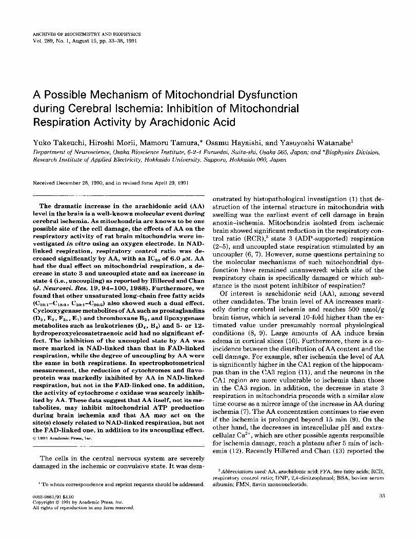
a b
RCR = state B/state 4 =@I@ of o”/@b* \
State 4 (% of contrd) =@*/@a
State 3 (% of control) =@“/@
Uncoupled state (% of control) -@II* /@I
FIG. 1. Oxygen consumption rates of mitochondrial respiratory ac- tivity in the absence (a) and in the presence (b) of arachidonic acid. To obtain state 4 respiration (@a, @b) glutamate + malate (each final concentration = 5 mM) or succinate (10 mM) were used as substrates. State 3 respiration (0) was obtained by addition of ADP (final concen- tration = 250-500 yM). Uncoupled state respiration (au) was obtained by addition of DNP (final concentration = 80 FM).
3 or uncoupled state respiration was expressed as a percentage of the control value.
Spectrophotometry. Redox state of mitochondrial cytochromes and flavoproteins was measured with a dual wavelength spectrophotometer (UV-3000, Shimadzu, Kyoto, Japan) under the same conditions as the measurement of oxygen consumption rate. Baseline was obtained in oxidized condition of state 2 (ADP + Pi, no substrate). The content of cytochrome a + a3 was determined at 605-630 nm using as extinction coefficient of 16.5/mM. cm. Cytochrome c oxidase activity was measured spectrophotometrically by the method of Wharton and Tzagoloff (17).
RESULTS
Inhibition of the Respiratory Activity by AA
The effect of AA on the mitochondrial oxygen con- sumption rate was investigated using glutamate and ma- late as substrates (NAD-linked respiration). As shown in Fig. 2, after addition of 5 or 15 PM AA, the rate of state 4 respiration increased to 1.6 or 2.5-fold from the control value, respectively. The value of the P/O ratio decreased from 3.0 to 1.9 by addition of 15 Z.LM AA. Oxygen con- sumption rates in state 3 as well as in uncoupled state respiration decreased by AA. Similar inhibition of oxygen consumption rate was obtained when AA was applied during state 3 or uncoupled state respiration (data not shown). Figure 3 shows the dose dependence of the AA effect on RCR, state 4, state 3, and uncoupled state. The concentration required for a 50% decrease (I&) of AA for RCR was 6.0 PM. The decrease in RCR value was derived from both the increase in state 4 and the decrease in state 3. The half-maximal effective dose (ED& of AA for the increase in state 4 was 22.8 PM and the IC& value of AA for state 3 inhibition was 37.6 PM. I&, value of AA
ARACHIDONIC ACID ON MITOCHONDRIAL RESPIRATORY CHAIN 35
glutamate + malate
-----% AA
Time (min)
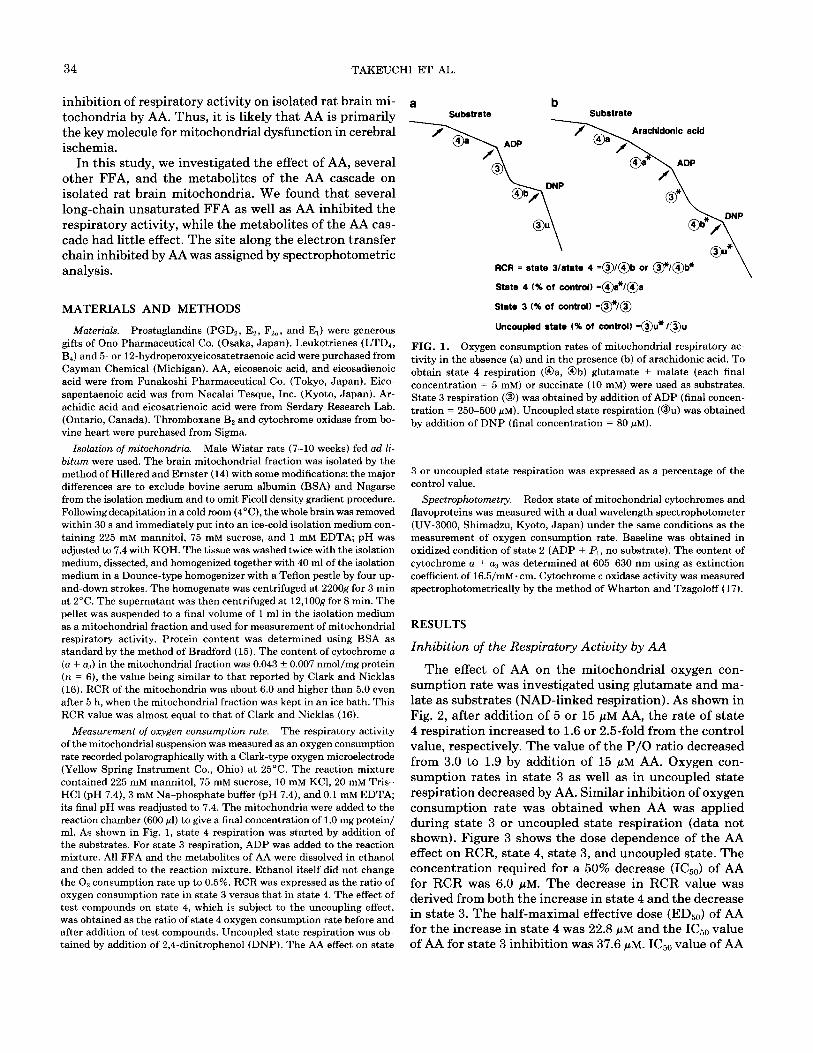
FIG. 2. Changes in oxygen consumption rate of brain mitochondria elicited by arachidonic acid (AA). Mitochondria were added at a final concentration of 1.0 mg protein/ml to 600 p 1 of the reaction mixture that contained 225 mM mannitol, 75 mM sucrose, 10 mM KCl, 20 mM Tris-HCl, 3 mM Na-phosphate, and 0.1 mM EDTA at pH 7.4. The respiration of state 4 was initiated by the addition of 5 mM glutamate + 5 mM malate as substrates at 25°C (w). Following AA addition (Q), 300pM ADP was applied to obtain state 3 respiration (V). Subsequently, DNP (80 pM) was used to start uncoupled state respiration ( I ). The concentrations of AA used were 0 ( -),5( -.-.-),and15p~(-----).
for the uncoupled state was 26.4 @M. Although BSA is well known to bind free fatty acids (FFA) (l&21), the addition of BSA around 2 min after the incubation with AA blocked the increase in state 4 by AA but did not restored the inhibition of state 3 and the uncoupled state (data not shown) as described previously (13, 22-24).
Effects of Free Fatty Acids on Mitochondrial Respiration
Palmitic acid (C16:o), stearic acid (C1&, oleic acid (C&, and AA (C&J mainly increase during cerebral
TABLE I
Changes in Brain Mitochondrial Respiration Induced by Free Fatty Acids (FFA)
ICbO (ED,) value (PM)
FFA” RCR State 4 Uncoupled state
16:0 24.7 58.9 60.4 18:0 40.9 79.6 >lOO.O 18:l 13.0 45.7 43.8 18:2 9.4 36.1 31.1 l&3 7.0 45.4 23.2 20:o 70.0 > 100.0 94.4 2O:l 7.9 40.4 40.4 20:2 8.3 26.4 31.6 20:3 5.1 16.3 15.9 20:4 6.0 22.8 26.4 20~5 4.2 15.5 14.5
Note. The IC, values of respiratory control ratio (RCR) and uncoupled state and EDbo value of state 4 were determined as described in the text. For the reaction mixture and the content of mitochondria, see the legend for Fig. 2. Glutamate (5 mM) + malate (5 mM) were used as substrates.
’ FFA are 160 (palmitic acid), 18:0 (stearic acid), 18~1 (oleic acid:& 9), 18~2 (linoleic acid:& 9, 12), 18:3 (linolenic acid:& 9,12,15), 2O:O (arachidic acid), 2O:l (eicosenoic acid:& ll), 20:2 (eicosadienoic acid: cis 11,14), 20~3 (eicosatrienoic acid:& 8,11,14), 20:4 (arachidonic acid: cis 5,8,11,14) and 20~5 (eicosapentaenoic acid:& 5,8,11,14,17). All FFA were dissolved in ethanol to be 10 IIIM as the stock solutions. In the reaction mixture, the final concentration of 10 PM FFA contained 0.1% ethanol.
ischemia and reach 500, 850, 250, and 500 nmol/g wet weight brain tissue, respectively (8,9). As shown in Table I, FFA tested exhibited a dual effect as well as AA as described previously using other tissues than the brain (24-27). Among these FFA, AA was most effective for uncoupling as well as for inhibition of the uncoupled state. The ICsO values of C&.., C18:0, and C& for RCR were ca.
0' 0 10 20 30 40 T;j
Arachidonic acid ( pM ) G
b A
6 100 100
e
$ L
60 =
E 80 2
8 60 60 5
6 40 40 5
c 0 a3 20 20 5
m z aJ 0’ I
_.
t B
10 20 30 40 p
Arachidonic acid (PM) a :
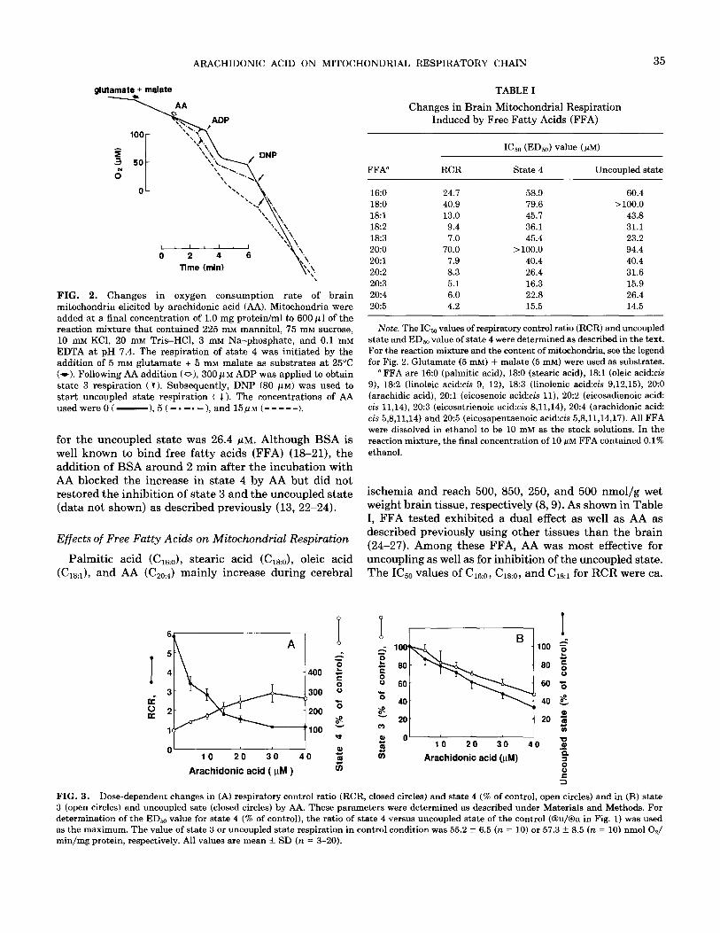
FIG. 3. Dose-dependent changes in (A) respiratory control ratio (RCR, closed circles) and state 4 (% of control, open circles) and in (B) state 3 (open circles) and uncoupled sate (closed circles) by AA. These parameters were determined as described under Materials and Methods. For determination of the EDr,,, value for state 4 (% of control), the ratio of state 4 versus uncoupled state of the control (@u/@a in Fig. 1) was used as the maximum. The value of state 3 or uncoupled state respiration in control condition was 55.2 + 6.5 (n = 10) or 57.3 + 8.5 (n = 10) nmol O,/ min/mg protein, respectively. All values are mean f SD (n = 3-20).
36 TAKEUCHI ET AL.
4.0,7.0, and 2.0 times higher than that of AA, respectively. Furthermore, the unsaturated FFA were more effective than the saturated one as reported previously using rat liver mitochondria (24). Among the Cl8 and CZO series, &a*, Ci8:. , C&:2, CZti3, and C20:5 showed similar I&,, values for RCR and the uncoupled state and also showed similar ED5,, values for state 4 to those by AA.
Effects of the Metabotites of the AA Cascade on Mitochondrial Respiration
PGD2, Ez, Fz,, and El and thromboxane B,, which are the cyclooxygenase metabolites of AA, did not change the respiratory activity of isolated mitochondria when added up to 20 PM. The metabolites of the lipoxygenase pathway, LTD, (3 PM), LTB, (20 PM), and 5 or 12-hydroperoxy- eicosatetraenoic acid (3 PM), also showed little effect (data not shown).
AA Effect in NAD-Linked and FAD-Linked Respiration
The effect of AA was investigated on the oxygen con- sumption rates in NAD-linked respiration via complex I (glutamate + malate were used as substrates) and also in FAD-linked respiration via complex II (succinate was used as substrate with rotenone). As shown in Table II, the EDs0 value of AA for state 4 was almost the same in both cases. However, the I(&, value for the uncoupled state in FAD-linked respiration was 3.0 times higher than that in the NAD-linked one.
Changes in the redox state of cytochromes elicited by AA were measured by use of a dual wavelength spectro- photometer. Figure 4 shows the absorption spectra of mi- tochondrial suspensions under various conditions. In the
TABLE II
Inhibition by AA of Brain Mitochondrial Respiration with Glutamate f Malate or Succinate as Substrates
IC,, (ED,,) value (FM)
Substrate State 4” Uncoupled state b
Glutamate + malate 19.8 26.1 Succinate 15.5 78.6
Note. The ED,, value of AA for state 4 respiration and the IC,, value for uncoupled state respiration were determined as described in the text. Concerning the reaction mixture and the content of mitochondria, see the legend for Fig. 2. For NAD-linked respiration, 5 mM glutamate and 5 mM malate were used as substrates. For FAD-linked respiration, 10 mM succinate was used as substrate, along with 10 pM rotenone.
n In the absence of AA, the value of state 4 respiration (@a in Fig. 1) in NAD-linked respiration or in FAD-linked respiration was 12.7 f 1.8 (n = 8) or 20.7 + 2.5 (n = 8) nmol O,/min/mg protein, respectively.
* In the absence of AA, the value of uncoupled state respiration in NAD-linked respiration or in FAD-linked respiration was 51.9 f 12.1 (n = 11) or 69.7 ? 9.8 (n = 11) nmol O,/min/mg protein, respectively.
I I 3 1 I I 1 I
500 550 600
Wavelength fnm)
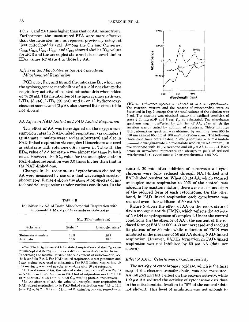
FIG. 4. Difference spectra of reduced or oxidized cytochromes. The reaction mixture and the content of mitochondria were as described in Fig. 2, except that the total volume of the solution was 3 ml. The baseline was obtained under the oxidized condition of state 2 (1 mM ADP and 3 mM P,, no substrate). The absorbance spectrum was not affected by addition of AA, after which the reaction was initiated by addition of substrate. Thirty minutes later, absorption spectrum was obtained by scanning from 500 to 650 nm against 650 nm at 100 nm/min of scan speed. The following three conditions were tested: 5 mM glutamate + 5 mM malate ( -),5 mM glutamate + 5 mM malate with 50~~ AA (-*-*-1, 10 mM succinate with 10 PM rotenone and 50 FM AA f-----l. Each arrow or arrowhead represents the absorption peak of reduced cytochrome b (r), cytochrome c ( I), or cytochrome n + a3 (0).
control, 30 min after addition of substrates all cyto- chromes were fully reduced through NAD-linked and FAD-linked respiration. When 50 /*M AA, which reduced uncoupled state respiration to 20% of the control, was added in the reaction mixture, there was no accumulation of the reduced form of each cytochrome. On the other hand, in FAD-linked respiration each cytochrome was reduced even after addition of 50 PM AA.
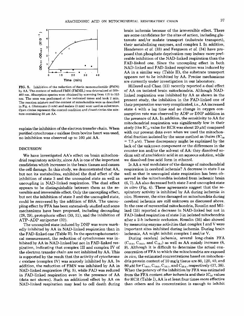
Figure 5 shows the effect of AA on the redox state of flavin mononucleotide (FMN), which reflects the activity of NADH dehydrogenase of complex I. Under the control conditions (in the absence of AA), the content of the re- duced form of FMN at 500-460 nm increased and reached its plateau after 30 min, while reduction of FMN was inhibited in the presence of 50 pM AA during NAD-linked respiration. However, FADH2 formation in FAD-linked respiration was not inhibited by 50 pM AA (data not shown).
Effect of AA on Cytochrome c Oxidase Activity
The activity of cytochrome c oxidase, which is the final step of the electron transfer chain, was also measured. AA (50 pM) had little effect on the enzyme activity, while 100 pM AA reduced the activity of cytochrome c oxidase in the mitochondrial fraction to 70% of the control (data not shown). This level of inhibition was not enough to
ARACHIDONIC ACID ON MITOCHONDRIAL RESPIRATORY CHAIN 37
0 10 20 30 40
Time (min)
FIG. 5. Inhibition of the reduction of flavin mononucleotide (FMN) by AA. The content of reduced FMN (FMNHJ was determined at 500- 460 nm. Absorption spectra were obtained by scanning from 415 to 515 nm. The scan was performed at the indicated times and took 2 min. The reaction mixture and the content of mitochondria were as described in Fig. 4. Glutamate (5 mM) and malate (5 mM) were used as substrates. Open circles represent the control condition and closed circles the mix- ture containing 50 pM AA.
explain the inhibition of the electron transfer chain. When purified cytochrome c oxidase from bovine heart was used, no inhibition was observed up to 100 PM AA.
DISCUSSION
We have investigated AA’s effect on brain mitochon- drial respiratory activity, since AA is one of the important candidates which increases in the brain tissues and causes the cell damage. In this study, we demonstrated that AA, but not its metabolites, exhibited the dual effect of the inhibition of state 3 and the uncoupled state as well as uncoupling in NAD-linked respiration. The dual effect appears to be distinguishable between them as the re- versible and irreversible effect. Only the uncoupling effect, but not the inhibition of state 3 and the uncoupled state, could be recovered by the addition of BSA. The uncou- pling effect by FFA has been extensively studied and some mechanisms have been proposed, including decoupling (28,29), protophoric effect (30,31), and the inhibition of ATP-ADP antiporter (32).
The uncoupled state induced by DNP was more mark- edly inhibited by AA in NAD-linked respiration than in the FAD-linked one (Table II). In the spectrophotometri- cal measurement, the reduction of cytochromes was in- hibited by AA in NAD-linked but not in FAD-linked res- piration, indicating that complex III and complex IV of the electron transfer chain are not inhibited by AA. This is supported by the result that the activity of cytochrome c oxidase (complex IV) was scarcely inhibited by AA. In addition, the reduction of FMN was inhibited by AA in NAD-linked respiration (Fig. 5), while FAD was reduced in FAD-linked respiration even in the presence of AA (data not shown). Such an additional effect by AA on NAD-linked respiration may lead to cell death during
brain ischemia because of the irreversible effect. There are some candidates for the sites of action, including glu- tamate and/or malate transport (substrate transport), their metabolizing enzymes, and complex I. In addition, Henderson et al. (33) and Ferguson et al. (34) have pro- posed that phosphate deprivation may induce more pref- erable inhibition of the NAD-linked respiration than the FAD-linked one. Since the uncoupling effect in both NAD-linked and FAD-linked respiration was induced by AA in a similar way (Table II), the substrate transport appears not to be inhibited by AA. Precise mechanisms are currently under investigation in our laboratory.
Hillered and Chan (13) recently reported a dual effect of AA on isolated brain mitochondria. Although NAD- linked respiration was inhibited by AA as shown in the present study, the inhibition in the FAD-linked one of their preparation was very complicated, i.e., AA increased state 4 with a lag time and no change in oxygen con- sumption rate was observed by ADP or DNP addition in the presence of AA. In addition, the sensitivity to AA for mitochondrial respiration was significantly low in their study (the IC& value for RCR was about 20 PM) compared with our present data even when we used the mitochon- drial fraction isolated by the same method as theirs (IC& = 7.5 FM). These discrepancy might be explained by the lack of the unknown component or the differences in the counter ion and/or the solvent of AA: they dissolved so- dium salt of arachidonic acid in an aqueous solution, while we dissolved free acid form in ethanol.
Is AA a real modulator of the damage of mitochondrial respiration in cerebral ischemia? A decrease in state 3 as well as that in uncoupled state respiration has been ob- served in the mitochondria isolated from ischemic brain (6,7). AA also decreased both state 3 and uncoupled state in vitro (Fig. 4). These agreements suggest that the re- spiratory activity is inhibited by AA during ischemia in vivo. However, the sites damaged on mitochondria during cerebral ischemia are still unknown as discussed above. In the case of myocardial mitochondria, Rouslin and Mil- lard (35) reported a decrease in NAD-linked but not in FAD-linked respiration of state 3 in isolated mitochondria after a 2-h ischemic occlusion, Rouslin (36) also showed by measuring enzyme activities that complex I and V were important sites inhibited during ischemia. During brain ischemia, AA might inhibit complex I and/or V.
During cerebral ischemia, several long-chain FFA (C16:0, C1s:o, and C,,,.) as well as AA mainly increase (8, 9). Although it is difficult to determine the actual con- centration of FFA to which the mitochondria are exposed in vivo, the estimated concentrations based on mitochon- dria protein content of 10 mg/g tissue are 80,120,40, and 80 PM for C16:0, C18:o, C&, and C&. , respectively (37,38). When the potency of the inhibition by FFA was estimated from the FFA content after ischemia and their IC&,, values for RCR (Table I), AA is at least four times more effective than others and its concentration is enough to inhibit
38 TAKEUCHI ET AL.
mitochondrial respiratory activity. Although other long- 15. Bradford, M. M. (1976) Anal. Biockem. 72, 248-254.
chain unsaturated fatty acids such as Cla2, (&s, and 16. yelk, J. B., and Nicklas, W. J. (1970) J. Bill. Chem. 245, 4724-
C2,..-C2,.. were effective in the same order of magnitude as AA (Table I), the contents of these FFA during isch- 17. Wharton, D. C., and Tzagoloff, A. (1967) in Methods in Enzymology
emia are at a trace level (8). In addition, the metabolites (Estahrook, R. W., and Pullman, M. E., Eds.), Vol. 10, pp. 245-250, Academic Press, New York and London.
of the AA cascade, which increase following reperfusion (39,40), had little effect on the mitochondrial respiration.
18. Goodman, D. S. (1958) J. Am. Ckem. Sot. 80,3892-3898.
Therefore, AA might be the most important FFA that 19. Reshef, L., and Shapiro, B. (1965) Biochim. Biophys. Acta 98, 73-
80. causes damage to mitochondrial respiratory activity dur- 20. Spector, A. A., John, K., and Fletcher, J. E. (1969) J. Lipid Res. 10, ing ischemia. The activation of phospholipases, phospho- 56-67.
lipase A2 and/or phospholipase C, during cerebral isch- 21. Spector, A. A., and Brenneman, D. E. (1970) Biochim. Biophys. Acta
emia seems to be an important event for the irreversible 260,433-438.
dysfunction of brain tissue. 22. Vaartjes, W. J., and Van den Bergh, S. G. (1978) Biochim. Biophys. Acta 503, 437-449.
ACKNOWLEDGMENTS 23. Matsuoka, I., and Nakamura, T. (1979) J. Biochem. (Tokyo) 86,
675-681. We thank Dr. N. Kamiike of Hotani Project, ERATO, JRDC, and 24. Borst, P., Loos, J. A., Christ, E. J., and Slater, E. C. (1962) Biochim.
Dr. S. Kawai of Naruto University of Education for valuable advice. Biophys. Acta 62, 509-518. We also thank Dr. L. D. Frye for critical reading of the manuscript. This work was supported in part by the Special Coordination Funds for
25. Pressman, B. C., and Lardy, H. A. (1956) Biochim. Biophys. Acta
Promoting Science and Technology Agency, Japan, and by grants from 2 1,458-466.
the Naito Foundation, ON0 Medical Research Foundation, Sankyo Life 26. Wojtezak, L., and Lehninger, A. L. (1961) Biochim. Biopkys. Acta
Science Foundation, and Sankyo Co., Ltd. 51,442-456. 27. Wojtezak, L. (1976) J. Bioenerg. Biomemb. 8, 293-311.
REFERENCES 28. Rottenberg, H., and Hashimoto, K. (1986) Biochemistry 25, 1747- 1755.
1. McGee-Russell, S. M., Brown, A. W., and Brierley, J. B. (1970) Brain Res. 20, 193-200.
29. Rottenberg, H. (1983) Proc. NC&. Acad. Sci. USA 80, 3313-3317.
2. Ozawa, K., Seta, K., Araki, H., and Handa, H. (1967) J. Biockem. 30. Luvisetto, S., Pietroben, D., and Azzone, G. P. (1987) Biochemistry
61, 512-514. 26, 7332-7338.
3. Schutz, H., Silverstein, P. R., Vapalahti, M., Bruce, D. A., Mela, 31. Pietroben, D., Luvisetto, S., and Azzone, G. P. (1987) Biochemistry
L., and Langfitt, T. W. (1973) Arch. Neural. 29,408-416. 26, 7339-7347.
4. Rehncrona, S., Mela, L., and Siesjo, B. K. (1979) Stroke 10, 437- 32. Andreyev, A. Yu, Bondareva, T. O., Dedukhova, V. I., Mokhova,
E. N., Skulachev, V. P., Tsofina, L. M., Volkov, N. I., and Vygodina, 446. T. V. (1989) Eur. J. Biochem. 182, 585-592.
5. Mela, L. (1979) Neural. Res. 1, 51-63. 33. Henderson, P. J. F., McGivan, J. D., and Chappell, J. B. (1969) 6. Hillered, L., SiesjB, B. K., and Arfors, K.-E. (1984) J. Cerebral Blood Biochen. J. 111,521-535.
Flow Metabol. 4, 438-446. 34. Ferguson, S. M. F., Estrada-O., S., and Lardy, H. A. (1971) J. Biol. 7. Sims, N. R., Finegan, J. M., and Blass, J. P. (1986) J. Neurochem. Chem. 246,5645-5652.
47, 506-511. 35. Rouslin, W., and Millard, R. W. (1981) Am. J. Physiol. 240, 308- 8. Bazan, N. G. (1970) Biochim. Biophys. Acta 218, l-10. 313. 9. Yoshida, S., Inoh, S., Asano, T., Sano, K., Shimasaki, H., and Ueta, 36. Rouslin, W. (1983) Am. J. Physiol. 244, 743-748.
N. (1983) J. Neurockem. 40, 1278-1286. 37. Abood, L. G. (1969) in Handbook of Neurochemistry (Lajtha, A., 10. Chan, P. H., and Fishman, R. A. (1978) Science 201,358-360. Ed.) pp. 303-326, Plenum, New York. 11. Westerberg, E., Deshpande, J. K., and Wieloch, T. (1987) J. Cerebral 38. MacIlwain, W. J., and Bachelard, H. S. (1985) in Biochemistry of
Blood Flow Metabol. 7, 189-192. the Central Nervous System, Churchill Livingstone, New York. 12. Siesjo, B. K. (1984) J. Neurosurg. 60, 883-908. 39. Gaudet, R. J., Alam, I., and Leviene, L. (1980) J. Neurockem. 35, 13. Hillered, L., and Chan, P. H. (1988) J. Neurosci. Res. 19, 94-100. 653-658.
14. Hillered, L., and Ernster, L. (1983) J. Cerebral Blood Flow Metabol. 40. Petroni, A., Bertazzo, A., Sarti, S., and Galli, C. (1989) J. Neurockem. 3, 207-214. 53, 747-752.