a pilot study on sensitivity and specificity...
TRANSCRIPT

A PILOT STUDY ON SENSITIVITY AND SPECIFICITY OF CYSTATIN C IN DETECTING
RENAL IMPAIRMENT IN HYPERTENSIVE PREGNANCIES
By
DR FAUZIAH BINTI JUMMAAT
DISSERTATION SUBMITTED IN PARTIAL FULFILLMENT OF THE REQUIREMENT FOR THE DEGREE OF MASTER OF MEDICINE
(OBSTETRICS AND GYNECOLOGY)
UNIVERSITI SAINS MALAYSIA
MAY 2011

Acknowledgements
“In the name of Allah, most Gracious, most Compassionate”.
Firstly, to my most beloved husband Dr. Azreen Syazril Bin Adnan, thousands of thanks and
appreciation for your valuable time day and night, endless support, never ending help, love,
understanding and sharing the hardships with me in completing this study. Thank you for being
my inspiration. I pray to Allah, He would give all the best thing into your life.
To our princesses Aida Farhana, Anis Fatihah and Alya Faridah, who always give me strength
and always cheer up my life. May Allah be with you all the time.
To my parents Encik Jummaat Kahman and Puan Sapiah Abdullah, may Allah bless you for your
sacrifices in replacing me to feed my children, send and pick them up from schools, shower them
with care and love, during my limited time in this Master programme. Thank you for your
continuous support and strong motivation.
In memory of my late parents in law Encik Adnan Omar and Puan Fatimah Jaman, may Allah
bless you, Al-Fatihah..
To my supervisors, Assoc. Prof. Dr. Nor Aliza Abdul Ghaffar, thank you for your full support,
encouragement and continuous motivation.
To Dr Julia Omar, thanks for your guidance and understanding.
Last but not least, to Prof. Dr. Syed Hatim Noor, thank you for the guidance and time.
Fauziah Jummaat

TABLE OF CONTENTS Page
TITLE PAGE
ACKNOWLEDGEMENTS ii
TABLE OF CONTENTS iii
LIST OF FIGURES vi
LIST OF TABLES viii
LIST OF ABBREVIATIONS ix
ABSTRACT xi
ABSTRAK xii
CHAPTER 1 INTRODUCTION 1
1.1. Chronic kidney disease in hypertensive pregnancies – Morbidities And
Mortalities 2
1.2. Current Available Measures In Detection of Chronic Kidney Disease 3
1.2.1. Urine Analysis 3
1.2.2. 24 hour urine creatinine clearance 4
1.2.3. Blood investigations 4
1.2.4. Radiological investigation 5
1.2.5. Renal biopsy 6
1.2.6. Protein Creatinine Index Ratio And Albumin Creatinine Ratio 6
1.2.7. Gold standard test for diagnosis of renal impairment 7
1.3. Pathophysiology of Chronic Kidney Disease In Hypertensive Pregnancies 7
1.4. New biomarkers in detecting renal impairment 9
1.4.1. Cystatin C 9
1.4.2. Neutrophil Gelatinase-associated Lipocalin 10
1.4.3. Kidney Injury Molecule – 1 10
1.4.4. Interleukin – 18 11
1.4.5. The need for rapid and reliable diagnostic test in detecting kidney
disease in hypertensive pregnancies 11
CHAPTER 2 HYPOTHESIS 13
CHAPTER 3 OBJECTIVES 15
CHAPTER 4 METHODOLOGY 17
4.1. Materials 18
4.1.1 Subjects 18
4.1.1.1. Inclusion Criteria 19
4.1.1.2. Exclusion Criteria 19
4.1.2 Reagents/Kits 19
4.2. Methods 21
4.2.1 Cystatin C 23
4.2.2 24 Hour Urine creatinine clearance 27
4.3. Statistical Analysis 28
CHAPTER 5 RESULTS 29
5.1 Results 30
5.2 Diagnostic Value Of Cystatin C In Detecting Renal Impairment In Hypertensive
Pregnancies 43
5.2.1 Sensitivity And Specificity, Positive Predictive Value, Negative Predictive
Value, ROC Curve and P Value Of Cystatin C in Second Trimester 43
5.2.2 Sensitivity And Specificity, Positive Predictive Value, Negative Predictive
Value, ROC Curve and P Value Of Cystatin C in Third Trimester 47
CHAPTER 6 DISCUSSION 52
CHAPTER 7 CONCLUSION, LIMITATIONS AND SUGGESTIONS 59
REFERENCES 62
APPENDICES 66
Appendix 1 Borang Keizinan Pesakit 67
Appendix 2 Patient Consent Form 73
Appendix 3 Ethical Committee Approval Letter 79
LIST OF FIGURES
Page
Figure 1.1: Uteroplacental perfusion in normal pregnancy compares to preeclampsia 8
Figure 1.2: Pathophysiology of kidney injury 9
Figure 4.1: Flow chart on the methodology of study 22
Figure 4.2: Sample cups 23
Figure 4.3: N latex Cystatin C reagent 24
Figure 4.4: Dade Behring analysis machine 24
Figure 4.5: Cystatin C process 25
Figure 4.6: Urine collector 27
Figure 4.7: Hitachi 912 analyzer 27
Figure 5.1: Age distribution 30
Figure 5.2: Distribution of patients on gestational age (weeks) 31
Figure 5.3: Distribution of patients based on CKD staging 32
Figure 5.4: Distribution of delivery based on gestational age (weeks) 33
Figure 5.5: Mode of deliveries 34
Figure 5.6: Birth weight of the baby 35
Figure 5.7: Indication for LSCS 36
Figure 5.8: Type of antihypertensive 37
Figure 5.9: Systolic blood pressure control 38
Figure 5.10: Diastolic blood pressure control 39
Figure 5.11: Creatinine clearance in hypertensive pregnancies 40
Figure 5.12: Cystatin C in hypertensive pregnancies 41
Figure 5.13: Cystatin C level and gestational age (weeks) 42
Figure 5.14: Receiver Operating Characteristics for Cystatin C in second trimester 45
Figure 5.15: Receiver Operating Characteristics for Cystatin C in third trimester 49

LIST OF TABLES
Page Table 4.1: 24 hour urine creatinine clearance and reagents 20
Table 4.2: Cystatin C and control reagents 20
Table 5.1: Age distribution in percentage 30
Table 5.2: Gestational age (weeks) in percentage 31
Table 5.3: Gestational age at delivery in percentage 33
Table 5.4: 2x2 table for sensitivity and specificity of Cystatin C in second trimester 43
Table 5.5: 2x2 table for positive predictive value of Cystatin C in second trimester 44
Table 5.6: 2x2 table for negative predictive value of Cystatin C in second trimester 44
Table 5.7: P value and area under the curve for receiver operating characteristic curve of
Cystatin C in second trimester 46
Table 5.8: 2x2 for sensitivity and specificity of Cystatin C in third trimester 47
Table 5.9: 2x2 table for positive predictive value of Cystatin C in third trimester 48
Table 5.10: 2x2 table for negative predictive value of Cystatin C in third trimester 48
Table 5.11: P value and Area under the curve for receiver operating characteristic curve of
Cystatin C in third trimester 50
LIST OF ABBREVIATIONS
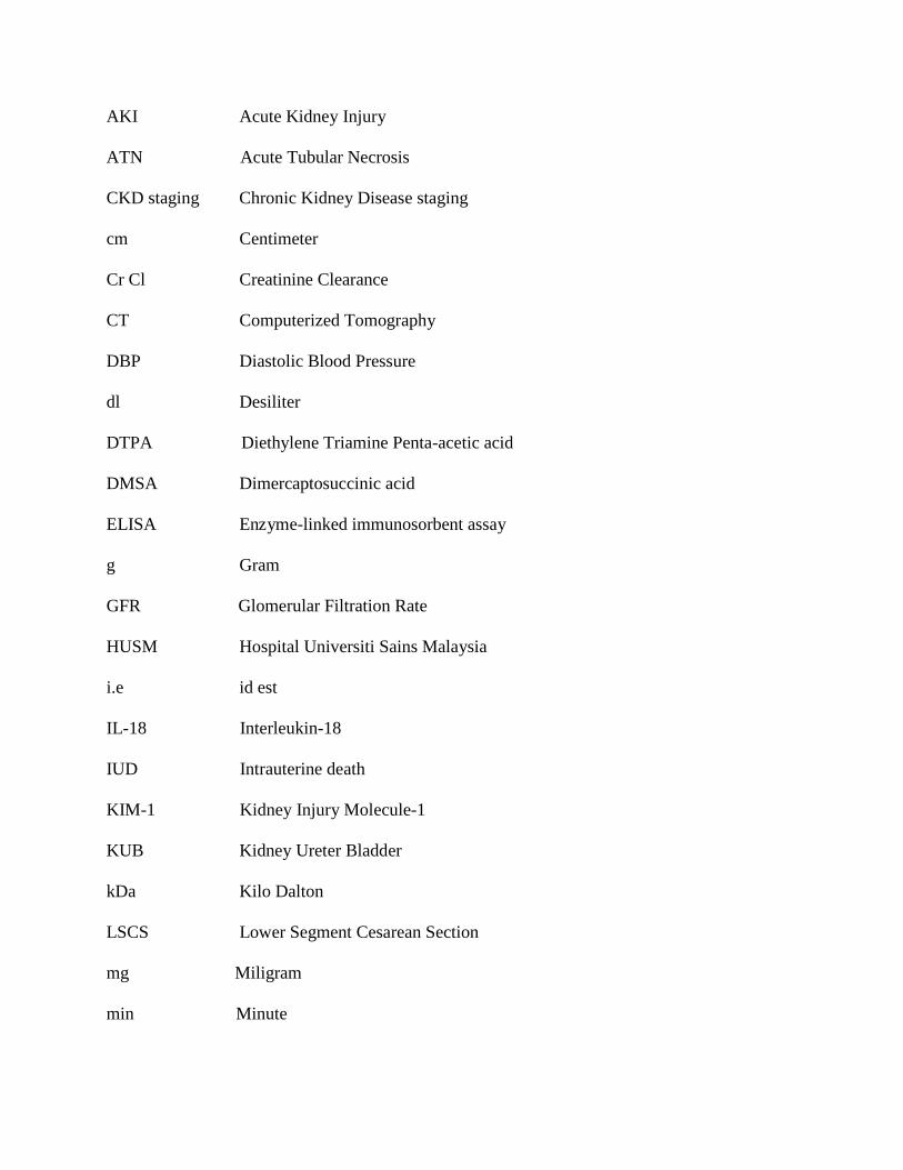
AKI Acute Kidney Injury
ATN Acute Tubular Necrosis
CKD staging Chronic Kidney Disease staging
cm Centimeter
Cr Cl Creatinine Clearance
CT Computerized Tomography
DBP Diastolic Blood Pressure
dl Desiliter
DTPA Diethylene Triamine Penta-acetic acid
DMSA Dimercaptosuccinic acid
ELISA Enzyme-linked immunosorbent assay
g Gram
GFR Glomerular Filtration Rate
HUSM Hospital Universiti Sains Malaysia
i.e id est
IL-18 Interleukin-18
IUD Intrauterine death
KIM-1 Kidney Injury Molecule-1
KUB Kidney Ureter Bladder
kDa Kilo Dalton
LSCS Lower Segment Cesarean Section
mg Miligram
min Minute

ml Mililiter
NGAL Neutrophil Gelatinase-associated Lipocalin
PENIA Particle-enhanced nephelometric immunoassay
ROC Receiver Operating Characteristics
SPSS Statistical package for social science
SD Standard Deviation
SVD Spontaneous Vaginal Delivery
µmol Micromol
ABSTRACT
Objectives : This study is to compare between Cystatin C and 24 hour urine creatinine
clearance for detection of renal impairment in hypertensive pregnancies population.
Study Methods: 64 patients enrolled in this cross sectional study and each patient was required
to collect 24 hour urine and 15 mls of blood. Blood taking was taken once in second and third
trimester. Urine collection was carried out during the first visit. Creatinine clearance below 90
mls/min is taken as renal impairment and compared to the serum Cystatin C level. The Receiver
Operating Characteristic Curve is drawn to obtain the sensitivity and specificity of Cystatin C.
Results : The results have shown that when compared to 24 hour urine creatinine clearance, in
second trimester, Cystatin C is 84.6 % sensitive and 86.7% specific for detection of renal
impairment at Cystatin C level of 0.574-0.898 (p value < 0.012) , area under curve: 0.736,
positive predictive value is 0.92 and negative predictive value is 0.76. While in the third
trimester, the sensitivity and specificity is 76.9% and 60% at the Cystatin C level of 0.657-1.00
(p value < 0.006), area under curve 0.838, the positive predictive value is 0.71 and negative
predictive value is 0.67.
Conclusion: Our study suggests that Cystatin C is a useful diagnostic kit for diagnosis of renal
impairment in hypertensive pregnancies population.
ABSTRAK

Objektif: Kegagalan fungsi buah pinggang wanita hamil yang mempunyai tekanan darah tinggi
dapat dikenalpasti dengan ujian Cystatin C. Ia dibandingkan dengan ujian air kencing ‘creatinine
clearance’ 24 jam.
Metodologi: Seramai 64 pesakit terlibat didalam kajian ini. Sampel air kencing dikumpulkan
selama 24 jam dan pengambilan darah 15 ml diperlukan. Darah pesakit diambil pada trimester
kedua dan ketiga. Manakala air kencing 24 jam diambil pada lawatan pertama. Nilai ‘Creatinine
clearance’ kurang daripada 90 ml/min menunjukkan kegagalan fungsi buah pinggang dan ini
dibandingkan dengan paras Cystatin C. Graf ‘Receiver Operating Characteristic Curve’ untuk
mendapatkan sensitiviti dan spesifisiti Cystatin C dilakarkan.
Keputusan: Keputusan dibandingkan dengan ujian air kencing ‘creatinine clearance’ 24 jam.
Trimester kedua menunjukkan Cystatin C adalah 84.6% sensitif dan 86.7% spesifik, dalam
mengesan kegagalan fungsi buah pinggang. Paras Cystatin C adalah 0.574-0.898 (nilai p<
0.012) ,‘area under curve’ adalah 0.736. Manakala ‘nilai prediktif positif’ ialah 0.92 dan ‘nilai
prediktif negatif’ ialah 0.76. Pada trimester ketiga, sensitiviti dan spesifisiti adalah 76.9% dan
60% masing-masing. Paras Cystatin C 0.657-1.000 (nilai p<0.006), ‘area under the curve’
adalah 0.838, ‘nilai prediktif positif’ ialah 0.71 dan ‘nilai prediktif negatif’ ialah 0.67.
Kesimpulan: Kajian ini menunjukkan Cystatin C berguna dalam mendiagnosa kegagalan fungsi
buah pinggang kepada wanita hamil yang mempunyai tekanan darah tinggi.


INTRODUCTION
CHAPTER 1
INTRODUCTION
1.1 Renal impairment in hypertensive pregnancies – morbidities and mortalities
Kidney is an important organ in maintaining physiology of pregnancy. Pregnancy related acute
renal failure predisposes the pregnancy to multiple medical risks, thus increased morbidity and
mortality (Brown et al., 2001). Preeclampsia and uncontrolled hypertension are recognized
factors causing renal impairment. In third world countries pregnancy causes 25% of acute kidney
injury and maternal mortality rate ranges from 9-55% (Prakash et al., 1995). Preeclampsia
increases maternal morbidity and mortality. Renal impairment is associated in hypertensive
pregnancies (Chee, 1988) . Early detection of renal impairment in hypertensive pregnancies
enable immediate referral and multidisciplinary approach. Consequently further deterioration of
kidney function is preventable. Diagnostic tools with high sensitivity and acceptable specificity
is important for renal function assessment (Hoek et al., 2003).
Preeclampsia and eclampsia constitutes 75% as the cause of acute kidney injury in pregnancy,
while sepsis 11% and followed by hemorrhage 7.2%. Fetal death was reported at 5.5 % while
maternal death at 9.1% (Silva et al., 2009). Another study conducted in India, discovered
maternal mortality due to pregnancy related acute renal failure was 24.3% (Misra et al., 2003).
Perinatal mortality rate is significantly higher with Glomerular Filtration Rate (GFR) of 70
ml/min. A consistent observation showing that serum creatinine >2.5 mg/dl (220 µmol/l) is
associated with more preterm deliveries and lower birth weights than women with a lower serum
creatinine. 73% of such women were delivered before 37 weeks and 57% had intra-uterine
growth retardation (Knutzen and Davey, 1977)
.
1.2. Current available measures in detection of chronic kidney disease
Several investigations available in diagnosing kidney impairment. Investigations are divided to
urine, blood, radiological investigation and renal biopsy. Urine analysis include urine for
microalbumin, protein and hematuria. Radiological investigations include, ultrasonography of
the kidneys, X-ray of the kidney, ureter and bladder, radionuclide imaging Diethylene Triamine
Penta-acetic acid (DTPA), Dimercaptosuccinic acid (DMSA) scan. Renal biopsy is carried out
in exclusive conditions, and general indications include assessment of causes of proteinuria and
for identifying the cause of acute glomerulonephritis. Blood investigations include serum
creatinine and urea level. 24 hour urine creatinine clearance is another method requiring patient
to collect 24 hours urine sampling and blood for serum creatinine taken simultaneously.
1.2.1. Urine Analysis
Urine analysis is performed as initial assessment in any patients presented with renal failure.
Indications include confirmation and diagnosis of proteinuria, urinary tract infection, and
establishing the cause of acute renal failure. There is no contraindication for the test and it can be
performed routinely by the bedside. The test is cheap and readily available. Urine phase contrast
is another tool available in urinalysis to detect glomerular injury. However this test can only be
performed in centers with the facility and trained staff. These investigations depend on the
quality of the sample taken, uncooperative patient is a disadvantage (Hou and Peano, 1998).
Sensitivity and specificity of the test depends upon the laboratory and quality of the sample
collected.
1.2.2. 24 hour urine creatinine clearance
24 hour endogenous creatinine clearance is employed as it is widely available in any laboratory
facilities. However the reliability of this method is diminished by patient’s accuracy in collecting
timed urine sample and tubular variability creatinine secretion. This method requires patient to
be present once to the clinic for sample collection and blood taking. Inadequate urine volume
may result in underestimation of kidney function (Johnson, 2005). Other factors that may affect
its accuracy include prolonged urine storage may cause deranged urine creatinine levels. High
temperature and acidified urine will cause more conversion of creatine to creatinine.
Multiplication of urinary bacterial colony especially contributes to the changes of urine pH.
Drugs like cimetidine inhibits tubular secretion of creatinine, therefore improves its accuracy .
Unfortunately, variability in individual response to this method has been observed. In cases of
oliguric or anuric acute kidney injury this method may not be suitable. This method is taken as
the ‘gold standard’ and under appropriate instruction to the patient will reduce the chances of
inaccuracy. Inulin clearance, even though is the most accurate method of assessing creatinine
clearance , is not applicable in day-to-day clinical practice (Brown et al., 2001) .
1.2.3. Blood investigations
There are numerous blood investigations available in assessment of renal failure. Conventionally
serum creatinine and blood urea are widely and readily available throughout the country.
Indications of these tests mainly to establish the diagnosis of renal failure. The sensitivity and
specificity of these tests depends upon the clinical situation. Serum creatinine is very useful in
determining the GFR. However certain drugs such as cimetidine, an H2 antagonists inhibits
tubular secretion of creatinine causing it to be falsely high in blood. Serum creatinine value is
reproducible for Chronic Kidney Disease Staging (CKD staging). It is particularly useful in risks
stratification of patients with renal failure in specific clinical situation. For example in pregnancy
related acute renal failure, decisions to sustain or terminate the pregnancy will depend on the
clinical staging of the renal impairment. CKD staging is classified to 5 clinical stages. Stage I is
creatinine clearance above 90 mls/min, Stage II between 60 to 89 mls/min, Stage III between 30
to 59 mls/min, Stage IV between 15 to 29 mls/min and Stage V below 15 mls/min (K/DOQI,
2004).
1.2.4. Radiological Investigation
Radiological investigations are performed to identify aetiology of the renal impairment or
supporting the diagnosis. Investigations are divided to invasive and non-invasive. Invasive
investigations include computerized tomography (CT) angiogram, magnetic resonance
angiography and intravenous pyelography. These investigations include the use of contrast media
and carries risks of contrast induced nephropathy and allergic reactions. Magnetic resonance
angiography exposes patient to nephrogenic systemic fibrosis. Non-invasive investigations
include ultrasonography and Kidney Ureter Bladder (KUB) x-ray. However, non-radioopaque
will not be visible in this investigation. Indications for CT scan include in renal vein thrombosis,
renal artery stenosis and renal infarction. CT urography is indicated in cases of obstructive
uropathy to identify the level of obstruction. Contraindications for these investigations would
include hypersensitivity to contrast media.
1.2.5. Renal Biopsy
Renal biopsy is performed under local anesthesia, via posterior approach. The procedure is
invasive and ultrasound guided. This procedure is indicated in unexplained state of acute or
chronic kidney disease with normal renal size kidneys. The histopathological finding will help to
assist the management of these patients. Information regarding the disease activity can also be
obtained from this tissue diagnosis. Contraindications for this procedure include small size
kidneys, multiple cysts, suspected renal tumor, hydronephrosis, urinary tract infection, bleeding
tendencies and uncooperative patients. Renal biopsy is technically difficult and carries
complications in pregnant ladies. Schewitz et al., (1965) describes gross hematuria in 16.7% of
77 pregnant women, 4.4 % developed perirenal hematoma and one death.
Contraindication for renal biopsy include small kidney size, bipolar length less than 9cm, single
kidney, coagulopathy, uncooperative patient and ongoing urinary tract infection.
1.2.6. Protein Creatinine Index Ratio And Albumin Creatinine Ratio
Normally, daily protein excretion does not exceed 150 mg, most filtered protein is reabsorbed.
Proteinuria is a marker of nephropathy, gross proteinuria as in nephritic range suggest overt
nephropathy. However proteinuria slightly increased in healthy pregnancy, levels above
260mg/24 hours considered abnormal. Conventionally 24 hour urine collection is used to
quantify total 24 hour urine excretion, however this method requires patient to present twice to
the clinic and to follow the instructions. It is bound to inaccuracy. Alternatively protein
creatinine ratio can be used, it has good correlation with 24 hour urine protein collection.

1.2.7 Gold Standard Test For Diagnosis Of Renal Impairment
Inulin clearance has been recognized as the gold standard for assessment of renal function. The
test which determines GFR is not feasible to be carried out in the clinical setting. Even though
the investigation has high accuracy unfortunately it is tedious and requires continuous infusion of
inulin and serial blood taking from the patients. The investigation is mainly been carried out in
experimental laboratories (Locatelli et al., 2002).
1.3. Pathophysiology of renal impairment in hypertensive pregnancies
Renal impairment in pregnancies is multifactorial and maybe preexisted or contributory to the
pregnancy. Physiological renal adaptation is a natural process and important in sustaining the
pregnancy and ensuring the maternal and fetal outcome. Failure of this natural mechanism
resulted in homeostasis imbalance and fetal or maternal loss. There are numerous other causes of
renal impairment in pregnancy but hypertension is common and early detection and appropriate
management may improve the outcome. Hypertensive disorders are major cause of maternal
mortality, accounting 20% of maternal death (Bosio et al., 1999). Most deaths occur in women
with eclampsia and severe hypertension and are due to intracerebral hemorrhage (Atlas of
Kidney Disease, 2007).
The pathophysiology of hypertension in pregnancy is postulated from preeclampsia which later
resulted in renal hypoperfusion and prerenal failure. The major mechanism is widespread
vascular endothelial dysfunction resulting in vasospasm, intravascular coagulation, hypertension,
renal, hepatic and central nervous system abnormalities (Hladunewich et al., 2007).
Abnormalities in uteroplacental circulation and release of toxic substances affecting the maternal
endothelial cells. Endothelial cell dysfunction results in increased platelet aggregation leading
to widespread systemic vasospasm, intravascular coagulation and decreased organ flow.
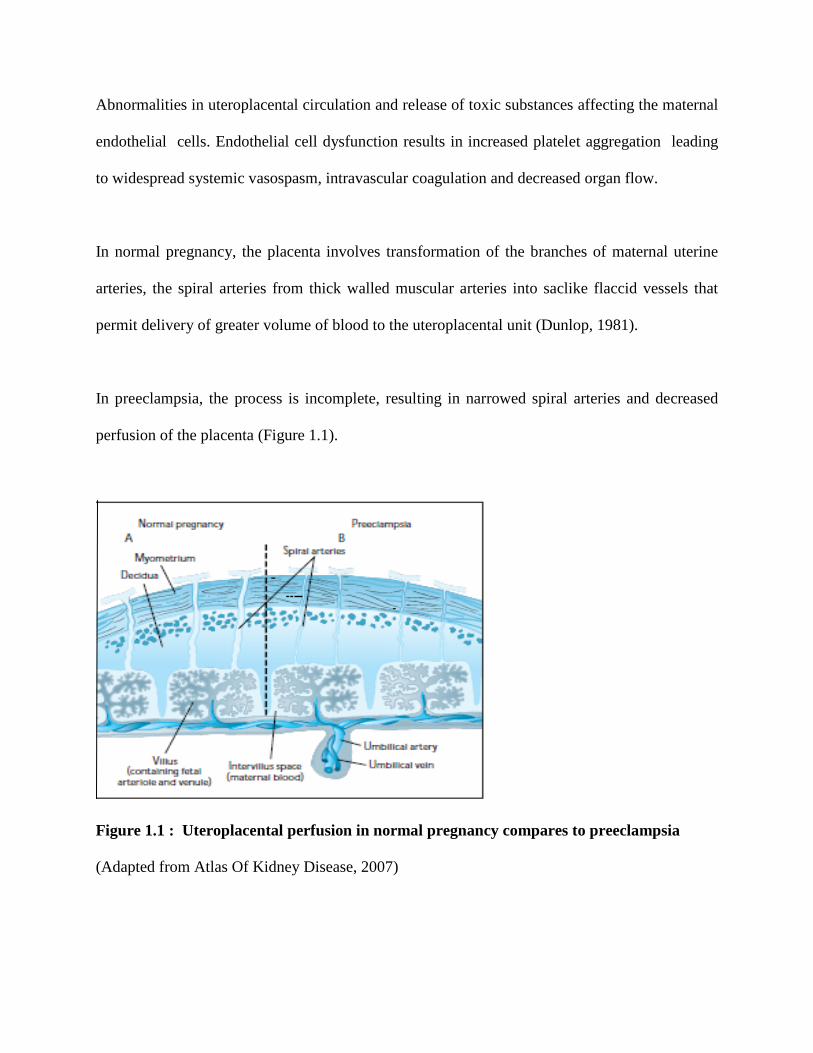
In normal pregnancy, the placenta involves transformation of the branches of maternal uterine
arteries, the spiral arteries from thick walled muscular arteries into saclike flaccid vessels that
permit delivery of greater volume of blood to the uteroplacental unit (Dunlop, 1981).
In preeclampsia, the process is incomplete, resulting in narrowed spiral arteries and decreased
perfusion of the placenta (Figure 1.1).
Figure 1.1 : Uteroplacental perfusion in normal pregnancy compares to preeclampsia
(Adapted from Atlas Of Kidney Disease, 2007)
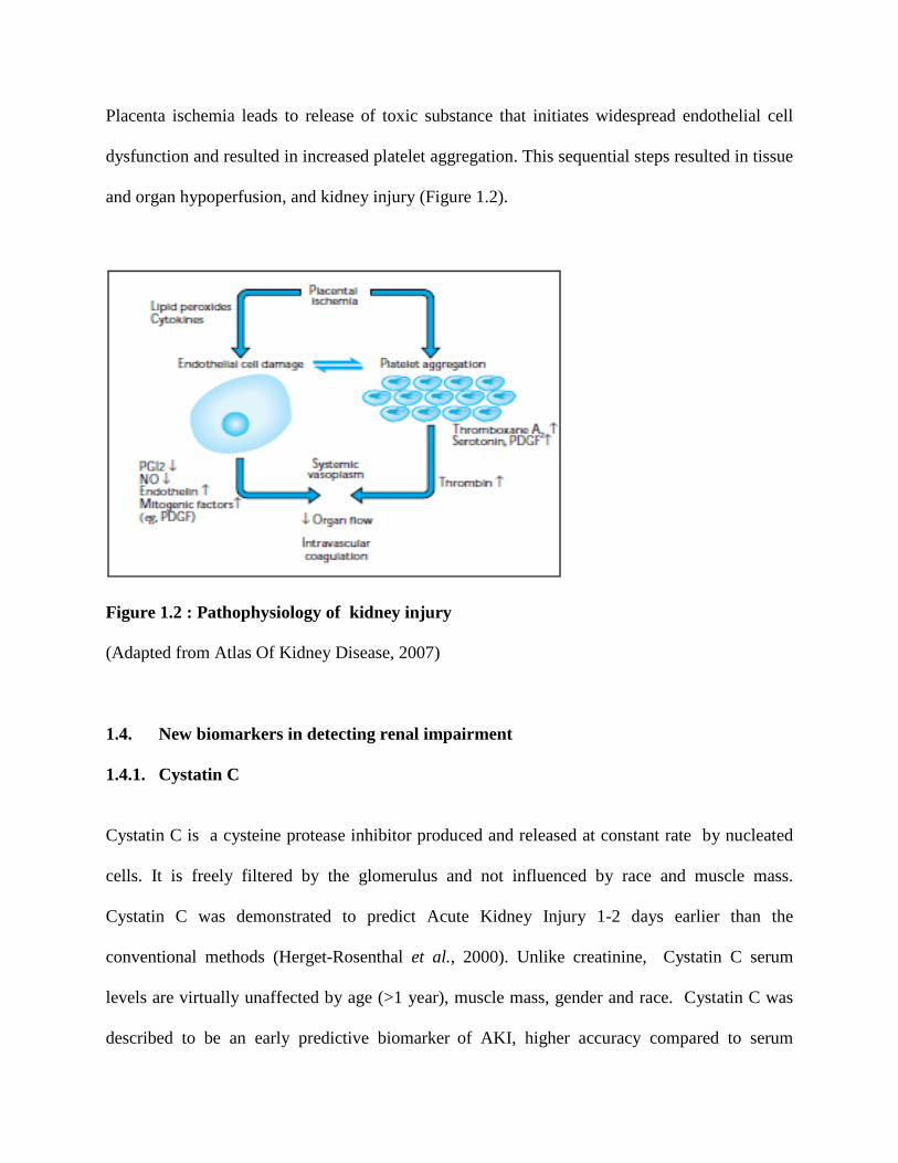
Placenta ischemia leads to release of toxic substance that initiates widespread endothelial cell
dysfunction and resulted in increased platelet aggregation. This sequential steps resulted in tissue
and organ hypoperfusion, and kidney injury (Figure 1.2).
Figure 1.2 : Pathophysiology of kidney injury
(Adapted from Atlas Of Kidney Disease, 2007)
1.4. New biomarkers in detecting renal impairment
1.4.1. Cystatin C
Cystatin C is a cysteine protease inhibitor produced and released at constant rate by nucleated
cells. It is freely filtered by the glomerulus and not influenced by race and muscle mass.
Cystatin C was demonstrated to predict Acute Kidney Injury 1-2 days earlier than the
conventional methods (Herget-Rosenthal et al., 2000). Unlike creatinine, Cystatin C serum
levels are virtually unaffected by age (>1 year), muscle mass, gender and race. Cystatin C was
described to be an early predictive biomarker of AKI, higher accuracy compared to serum
creatinine in emergency department setting (Misra et al., 2003). Patient plasma is required for
analysis via immunoassays. Urinary Cystatin C assay has also been used for earlier detection of
kidney injury and need for renal replacement therapy. The mean serum Cystatin C levels reflect
changes in the GFR in normal pregnancy population and also in the postnatal period (Strevens et
al., 2002). It has also been shown that serum Cystatin C levels are independent of age, height,
weight, or blood sugar level (Babay et al., 2005).
1.4.2. Neutrophil Gelatinase-associated Lipocalin (NGAL)
Human NGAL is a 25 kDa protein that is expressed by neutrophils and various epithelial cells,
including cells of the proximal convoluted tubule. It is upregulated after ischemic injury. Urinary
NGAL is a promising biomarker for AKI because it is upregulated within 2 hours of acute renal
cellular injury. NGAL is normally expressed in low levels in human tissues, including the
kidney, lung, stomach and colon, the expression is marked in the presence of renal tubular cells
injury. In preclinical studies, NGAL has been shown to be easily detected in blood and urine in
early AKI. It has also been used as kidney biomarkers in renal transplant patients (Liangos et al.,
2009).
1.4.3. Kidney Injury Molecule – 1 (KIM-1)
Kidney injury molecule 1 (KIM-1) is a transmembrane protein overexpressed in proximal tubule
cells of the kidney in response to ischemic or nephrotoxic injury to human and animal models.
One study was prospectively evaluated the expression of KIM-1 levels in 103 patients
undergoing cardiopulmonary bypass surgery. AKI defined as 0.3 mg/dl increase of creatinine
more common in patients with elevated KIM-1 levels compared with other biomarkers (Liangos
et al., 2009). Other studies also demonstrated urinary KIM-1 able to differentiate between pre-
renal and renal causes of kidney injury. The sensitivity of specificity of KIM-1 are 99% sensitive
and 95% specific, however the results obtained is not in pregnant ladies with hypertension.
1.4.4. Interleukin – 18 (IL-18)
IL-18 is a proinflammatory cytokine activated within the proximal tubule and excreted in urine
after ischemic kidney injury. One study was demonstrated to compare urinary IL-18
concentration in patients with diagnosis of Acute Tubular Necrosis (ATN) with healthy
controls. Patients with ATN had markedly increased IL-18 and elevated ratios of urinary IL-18
to creatinine compared with other group (Parikh et al., 2006) .
1.4.5. The need for rapid and reliable diagnostic test in detecting kidney disease in
hypertensive pregnancies
Rapid diagnosis of acute kidney injury in pregnancy will prevent potential adverse outcomes to
the mother and fetus. Earlier intervention can be initiated to reverse the potential ongoing insult.
In one study, described mortality between 20-30% in pregnant ladies with acute kidney injury
(Hou and Peano, 1998). It is important of earlier initiation of dialysis for better control of
azotemia and improving the overall outcomes (Gammill and Jeyabalan, 2005). However the role
of prophylactic dialysis in pregnant ladies with acute kidney injury is debatable.

HYPOTHESIS
CHAPTER 2
HYPOTHESIS
We hypothesized that Cystatin C is a reliable test for detection of renal impairment in
hypertensive pregnancies that carries acceptable sensitivity and specificity.

OBJECTIVES
CHAPTER 3
OBJECTIVES
Objectives :
1. To determine sensitivity and specificity of Cystatin C in comparison to 24 hour urine
Creatinine clearance in hypertensive pregnancies.
2. To compare Cystatin C level in hypertensive pregnant lady between 2nd and 3rd
trimester.

METHODOLOGY
CHAPTER 4
METHODOLOGY
This section describes the materials and general methods used in this work. Specific methods are
described in the respective chapters.
4.1. Materials
4.1.1. Subjects
This study was approved by the Research Ethics Committee (Human) on 21st
and funded by short term grant (304/PPSP/6139008) Hospital Universiti Sains Malaysia
(HUSM). This pilot prospective cross sectional study was conducted in HUSM between January
2009 and January 2010. Flow chart of the study is shown in Figure 4.1. All pregnant patients
beginning with 2
November 2010
nd
trimester at the age of 16 years old to 55 years old and hypertensives
including gestational hypertension, chronic hypertension, chronic hypertension with
superimposed preeclampsia, preeclampsia and unclassified hypertension were included. Verbally
informed consent was obtained prior to the involvement of the patients into the study followed
by written consent. Recruitment was done on patients that meet the inclusion and exclusion
criteria and consented to participate.
Patients included in this cross-sectional study whom had satisfied the inclusion criteria, were
informed regarding their involvement in the study and explained regarding the need to collect 5
mls of blood sample exclusive of other routine blood investigations. They were also required to
provide 24 hour urine collection sample for the analysis. Blood investigations was taken in serial
during the 2nd and 3rd
trimester. 64 patients were selected and enrolled in this cross sectional
study.
The inclusion and exclusion criteria for the patients were as follows:
4.1.1.1 Inclusion criteria:
Consented for the study
Patients beginning from 2nd
gestational hypertension, chronic hypertension, chronic hypertension with superimposed
trimester of pregnancy whom are hypertensives including
preeclampsia, preeclampsia and unclassified hypertension.
Age : 16-55 years
4.1.1.2 Exclusion criteria:
Hyper/Hypothyroidism
Smokers
Receiving corticosteroids
Suffering from any forms of malignancy
4.1.2. Reagents/Kits
The reagents were used for the analysis of the blood sample as listed in the product inlet. The
laboratory analysis was performed and prepared as recommended in the product inlet. The 24
hour urine creatinine clearance and the reagents are listed in Table 4.1. The Cystatin C reagents
are as listed in Table 4.2.
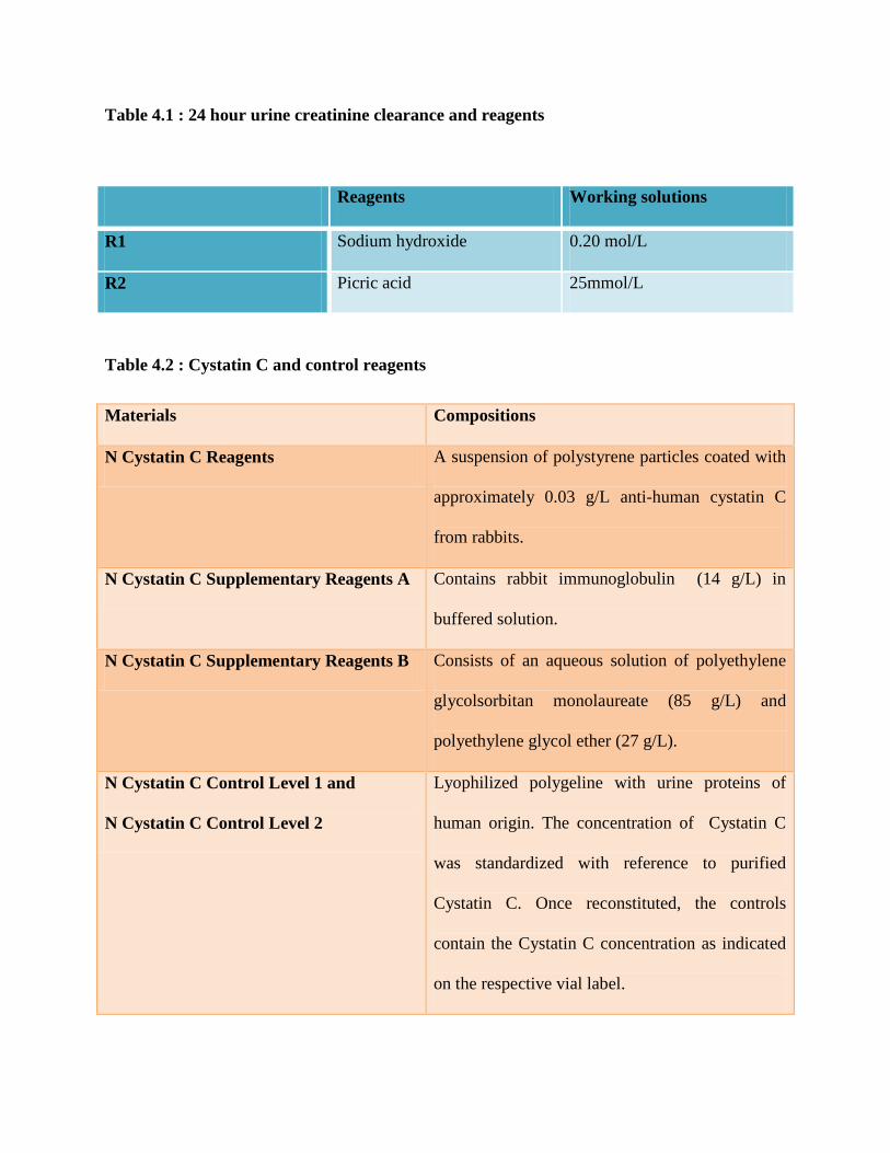
Table 4.1 : 24 hour urine creatinine clearance and reagents
Reagents Working solutions
R1 Sodium hydroxide 0.20 mol/L
R2 Picric acid 25mmol/L
Table 4.2 : Cystatin C and control reagents
Materials Compositions
N Cystatin C Reagents A suspension of polystyrene particles coated with
approximately 0.03 g/L anti-human cystatin C
from rabbits.
N Cystatin C Supplementary Reagents A Contains rabbit immunoglobulin (14 g/L) in
buffered solution.
N Cystatin C Supplementary Reagents B Consists of an aqueous solution of polyethylene
glycolsorbitan monolaureate (85 g/L) and
polyethylene glycol ether (27 g/L).
N Cystatin C Control Level 1 and
N Cystatin C Control Level 2
Lyophilized polygeline with urine proteins of
human origin. The concentration of Cystatin C
was standardized with reference to purified
Cystatin C. Once reconstituted, the controls
contain the Cystatin C concentration as indicated
on the respective vial label.
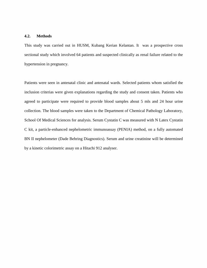
4.2. Methods
This study was carried out in HUSM, Kubang Kerian Kelantan. It was a prospective cross
sectional study which involved 64 patients and suspected clinically as renal failure related to the
hypertension in pregnancy.
Patients were seen in antenatal clinic and antenatal wards. Selected patients whom satisfied the
inclusion criterias were given explanations regarding the study and consent taken. Patients who
agreed to participate were required to provide blood samples about 5 mls and 24 hour urine
collection. The blood samples were taken to the Department of Chemical Pathology Laboratory,
School Of Medical Sciences for analysis. Serum Cystatin C was measured with N Latex Cystatin
C kit, a particle-enhanced nephelometric immunoassay (PENIA) method, on a fully automated
BN II nephelometer (Dade Behring Diagnostics). Serum and urine creatinine will be determined
by a kinetic colorimetric assay on a Hitachi 912 analyser.
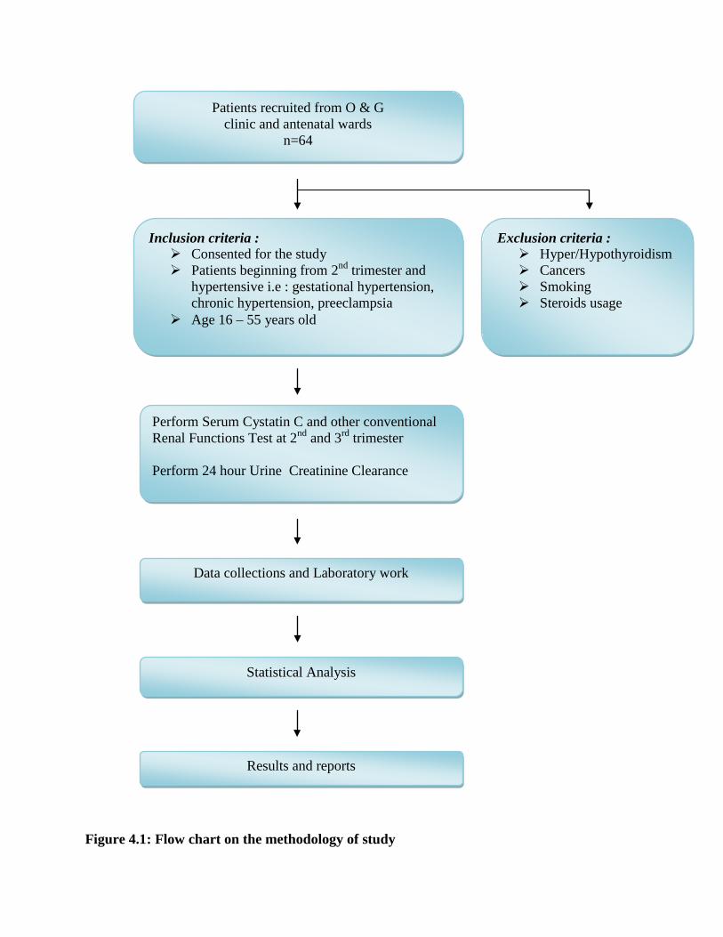
Figure 4.1: Flow chart on the methodology of study
Perform Serum Cystatin C and other conventional Renal Functions Test at 2nd and 3rd trimester Perform 24 hour Urine Creatinine Clearance
Data collections and Laboratory work
Results and reports
Patients recruited from O & G clinic and antenatal wards
n=64
Exclusion criteria : Hyper/Hypothyroidism Cancers Smoking Steroids usage
Inclusion criteria : Consented for the study Patients beginning from 2nd trimester and
hypertensive i.e : gestational hypertension, chronic hypertension, preeclampsia
Age 16 – 55 years old
Statistical Analysis

4.2.1.Cystatin C
Patients who were selected for the study were requested for blood sample. About 5mls of blood
were taken during antenatal clinic or inpatients. They were required to give the blood each in 2nd
and 3rd
The serum was manually transferred to the sample cups which is small in size.
trimester. The blood was sent to the Chemical Pathology Laboratory, HUSM. In the
laboratory, the blood was run in the Eppendorf Centrifuge 5810 machine to obtain the serum and
supernatant.
Figure 4.2 : Sample cups
The serum were kept in the freezer in temperature below -17°C before being analyzed .
The serum sample collection was equilibrated in room temperature with reagent N Latex
Cystatin C and automatically diluted 1:100 with N Diluent.

Figure 4.3 : N latex Cystatin C reagent
Analyzing process started by using Dade–Behring Nephelometer Systems and results were
automatically evaluated .
Figure 4.4 : Dade Behring analysis machine

Manually transferred
Figure 4.5 : Cystatin C process
CENTRIFUGE MACHINE : EPPENDORF CENTRIFUGE 5810
BLOOD SAMPLE FROM PATIENT
SERUM
SAMPLE CUP
CYSTATIN C ( keep in freezer : -17° C as it runs by batches )
Sample + Reagent ( equilibrate in room temperature ) Reagent : N Latex Cystatin C
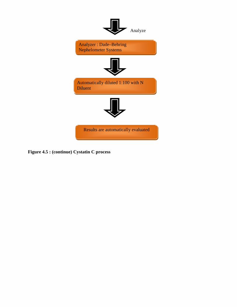
Analyze
Figure 4.5 : (continue) Cystatin C process
Analyzer : Dade–Behring Nephelometer Systems
Automatically diluted 1:100 with N Diluent
Results are automatically evaluated