a peptide derived from a single-modified viroid-rna can be used as an “in vivo” nucleolar marker
TRANSCRIPT

A
sdmvur©
K
rtamicckc(octmc1e(
0d
Journal of Virological Methods 144 (2007) 169–171
Short communication
A peptide derived from a single-modified viroid-RNAcan be used as an “in vivo” nucleolar marker
G. Gomez, V. Pallas ∗Instituto de Biologıa Molecular y Celular de Plantas, Consejo Superior de Investigaciones Cientıficas-UPV,
Av. de los Naranjos s/n, 46022 Valencia, Spain
Received 8 January 2007; received in revised form 4 April 2007; accepted 25 April 2007Available online 14 June 2007
bstract
Viroids are small, single-stranded, circular, non-coding pathogenic RNAs. Hop stunt viroid (HSVd) is characterized by possesses rod-liketructure and replicate in the host nuclei. Green fluorescent protein (GFP) fusions with transit sequences or entire proteins can be used foreliberate labelling of particular cell compartments. Different GFP-fusions have been obtained to selectively illuminate different organelles andembranes in many cell types. However, as far as we know, examples for established efficient markers for nucleoli are scarce. In this work, a
iroid-RNA was made translatable by inserting an ATG at position 1 and fused to the GFP. The results showed that the resultant fusion can besed as an efficient “in vivo” nucleolar marker in “real time” cellular observations. Thus, this construct can be a very useful tool to study processeselated with nucleolus functions.
2007 Elsevier B.V. All rights reserved.
Nucle
npttbtfasdttmmd
eywords: Fluorescent probes; Nucleolar targeting; Hop stunt viroid; Peptide;
Organelle-specific fluorescent probes encoding green fluo-escent protein (GFP) within cells have engendered experimentshat could not be conceived a few years ago. One of the chiefdvantages of GFP as a fluorescent probe is its lack of require-ent for an exogenous cofactor. GFP can be expressed within
ntact tissues and processes monitored without the disturbanceaused by the introduction of reagents. Among the many appli-ations that GFP permits are those that allow an in-depthnowledge into the understanding of both the organization ofellular activities and compartmentalization within plant cellsHanson and Kohler, 2001). GFP-fusions with transit sequencesr entire proteins can be used for deliberate labelling of particularell compartments. Different GFP-fusions have been obtainedo selectively illuminate different organelles and membranes in
any cell types such as endoplasmic reticulum, Golgi, mito-hondria, nucleus, endosomes and lysosomes (Grebenok et al.,
997; Hanson and Kohler, 2001). However, as far as we know,xamples for established efficient markers for nucleoli are scarceLorkovic et al., 2004).∗ Corresponding author. Fax: +34 963877859.E-mail address: [email protected] (V. Pallas).
it
amaw
166-0934/$ – see front matter © 2007 Elsevier B.V. All rights reserved.oi:10.1016/j.jviromet.2007.04.009
olus
The nucleolus is the most obvious structure in the eukaryoticucleus (Sato et al., 2005). It is known to be a ribosome-roducing apparatus where ribosomal (r) DNA is transcribed andhe primary rRNA transcripts are processed to produce three ofhe four rRNA species. Therefore, the nucleolus can functionallye interpreted as a loop of rDNA associated with its transcripts,heir processed products and other functional proteins requiredor rDNA transcription, transcript processing and pre-ribosomessembly (Sato et al., 2005). Although there has been exten-ive work on the plant nucleolar ultrastructure, it has been veryifficult to allocate functions to specific nucleolar ultrastruc-ures until recently. One of the reasons for this difficulty is thathe nucleolar structures have been examined mainly by electron
icroscopy using conventional double staining. Another reasonay be that the nucleolus easily changes its structure and size
uring the cell cycle or under abnormal conditions. This makest difficult to characterize the specific functions associated withhe fundamental structure in the nucleolus (Sato et al., 2005).
Viroids are plant subviral pathogens whose small genomes
re constituted by a single-stranded and covalently closed RNAolecule that does not encode for any protein. Viroid speciesre clustered into the families Pospiviroidae and Avsunviroidae,hose members have evolved to replicate in their host nuclei or
1 rolog
cv(P
atb
twmoawu3swc8mefioGfbnsc
3tmwaba
Sa
wGicsss
thaGgpa24nioHsoittran
p1
FGai
70 G. Gomez, V. Pallas / Journal of Vi
hloroplasts, respectively (Flores and Pallas, 2006). Hop stuntiroid (HSVd) is a typical Pospiviroidae that possesses a small294–300 nt) circular RNA with rod-like structure (Gomez andallas, 2004, 2006).
In this work, it is described the use of a peptide derived fromsingle-modified viroid-RNA fused to GFP that translocates to
he nucleolus 36–48 h after its transient expression in Nicotianaenthamiana plants as an efficient “in vivo” nucleolar marker.
In silico analysis of the HSVd-RNA sequence revealed that,his RNA could render a complete open reading frame if an ATGere provided. Thus, a modified coding ATG-HSVd cDNA frag-ent (300 nt) was amplified by PCR starting from the cDNA
f HSVd isolate PR1A (Y09347: Kofalvi et al., 1997; Amari etl., 2001) using the VP/atg-HSVd (5′-ctctctccatgggcccctctgg-3′,ith a 5′ extra sequence of 11 nt including a Nco restriction site,nderlined) and the VP/end-HSVd (5′-gctagcgagaggatccgcggc-′, with a 5′ extra sequence of 6 nt including a NheI restrictionite, underlined) primers. The amplified ATG-HSVd fragmentas NcoI-NheI digested, ligated to GFP NheI/ZBglII cDNA and
loned in a previously NcoI/BglII digested binary plasmid pMog00 under the control of the Cauliflower mosaic virus 35S pro-oter and the nopaline synthase terminator (t-Nos) (Knoester
t al., 1998). The resultant vector HSVd:GFP, contains a modi-ed HSVd cDNA (with an ATG insertion at the position “290”f the viroid cDNA) transcriptionally fused to the 5′ end of theFP cDNA. The correct transcriptional fusion of the HSVd-GFP
ragments was checked by DNA sequencing and by Westernlot assays of the transiently expressed chimeric protein (dataot shown). A binary pMog 800 plasmid carrying the con-truction 35S:GFP:t-Nos was used as a free-GFP expressionontrol.
The binary plasmids carrying the 35S-HSVd-GFP-tNos or5S:GFP:t-Nos cassettes were transfected into Agrobacteriumumefaciens strain C58C1 containing the virulence helper plas-
id pCH32 (Hamilton et al., 1996). The N. benthamiana plants
ere agroinfiltrated as previously described (Vlot et al., 2001)nd maintained in environmentally controlled growing cham-ers (28 ◦C, 14 h of light). The GFP expression in plants wasnalyzed at 36, 48, 72 and 96 h after agroinfiltration with a TCS
Roni
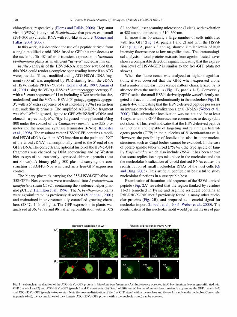
ig. 1. Subnuclear localization of the ATG-HSVd-GFP protein in Nicotiana benthamFP (panels 1 and 2) and ATG-HSVd-GFP (panels 3 and 4) constructs. (B) Detail of
nd ATG-HSVd-GFP (panels 4–6) proteins. Note the uneven distribution of the free Gn panels (4–6), the accumulation of the chimeric ATG-HSVd-GFP protein within th
ical Methods 144 (2007) 169–171
L confocal laser scanning microscope (Leica), with excitationt 488 nm and emission at 510–560 nm.
In more than 50 assays, a large number of cells infiltratedith free GFP (Fig. 1A, panels 1 and 2) and with the HSVd-FP (Fig. 1A, panels 3 and 4), showed similar levels of high
ntensity fluorescence at low magnifications. The immunologi-al analysis of total proteins extracts from agroinfiltrated leaveshows a comparable detection signal, indicating that the expres-ion level of HSVd-GFP is similar to the free-GFP (data nothown).
When the fluorescence was analyzed at higher magnifica-ions, it was observed that the GFP, when expressed alone,ad a uniform nuclear fluorescence pattern characterized by itsbsence from the nucleolus (Fig. 1B, panels 1–3). Conversely,FP fused to the small HSVd-derived peptide was efficiently tar-eted and accumulated predominantly in the nucleolus (Fig. 1B,anels 4–6) indicating that the HSVd-derived peptide possessesn intrinsic nucleolar localization signal (NoLS) (Weber et al.,000). This subnuclear localization was maintained for at leastdays, when the GFP-fluorescence commences to decay (data
ot shown). This result indicates that the HSVd-derived peptides functional and capable of targeting and retaining a heterol-gous protein (GFP) in the nucleolus of N. benthamiana cells.owever, the possibility of localization also in other nucleus
tructures such as Cajal bodies cannot be excluded. In the casef potato spindle tuber viroid (PSTVd), the type specie of fam-ly Pospiviroidae which also include HSVd, it has been shownhat some replication steps take place in the nucleolus and thathe nucleololar localization of viroid-derived RNAs causes theedistribution of small nucleololar RNAs of the host cells (Qind Ding, 2003). This artificial peptide can be useful to studyucleololar functions in a susceptible host.
Examination of the amino acid sequence of the HSVd-derivedeptide (Fig. 2A) revealed that the region flanked by residues1–31 (enriched in lysine and arginine residues) contains an
/K-R/K-X-R/K motif previously found in many other nucle-lar proteins (Fig. 2B), and proposed as a crucial signal forucleolar import (Libault et al., 2005; Weber et al., 2000). Thedentification of this nucleolar motif would permit the use of par-iana. (A) Fluorescence observed in N. benthamiana leaves agroinfiltrated withdifferent N. benthamiana nucleus transiently expressing the GFP (panels 1–3)FP signal within the nucleus and the exclusion from the nucleolus. Conversely,
e nucleolus (nuc) can be observed.

G. Gomez, V. Pallas / Journal of Virolog
Fig. 2. HSVd-derived peptide contains a nucleolar localization signal (NoLS).(A) Detailed cDNA and the deduced sequence description of the HSVd-derivedpeptide. The region enriched in lysine and arginine residues contains the R/K-R/K-X-R/K motif (in bold), previously found in many other nucleolar proteins,ili
tin
ffn
A
losaVc
R
A
F
G
G
G
H
H
K
K
L
L
Q
S
V
s boxed. (B) Nucleololar localization signals. Regions necessary for nucleolarocalization in several known cellular and viral proteins (Weber et al., 2000) arendicated and aligned. The conserved R/K-R/K-X-R/K motif is boxed.
ial HSVd-RNA sequences, to avoid the potential risk of viroidnfection, in the case that this signal-peptide were used to studyucleolar functions in susceptible hosts.
In conclusion, this brief report shows that the peptide derivedrom the single-modified HSVd, can be used as molecular probeor selective and simple efficient staining or targeting of plantucleolus.
cknowledgements
We are indebted to Dr. M.D. Gomez-Jimenez for excel-ent technical assistance in the observation and photographyf GFP expression via a confocal microscope. This work was
upported by grant BIO2005-07331 from the Spanish grantinggency DGICYT and by grant GV05-238 from the Generalitatalenciana (region government). G. Gomez is the recipient of aontract from the CSIC.W
ical Methods 144 (2007) 169–171 171
eferences
mari, K., Gomez, G., Myrta, A., Di Terlizzi, B., Pallas, V., 2001. The molecularcharacterization of 16 new sequence variants of Hop stunt viroid reveals theexistence of invariable regions and a conserved hammerhead-like structureon the viroid molecule. J. Gen. Virol. 82, 953–962.
lores, R., Pallas, V., 2006. Viroids. In: Khan, J.A., Dijkstra, J. (Eds.), Handbookof Plant Virology. The Haworth Press Inc., New York, USA, pp. 93–104.
omez, G., Pallas, V., 2004. A long-distance translocatable phloem protein fromcucumber forms a ribonucleoprotein complex in vivo with Hop stunt viroidRNA. J. Virol. 78, 10104–10110.
omez, G., Pallas, V., 2006. Hop stunt viroid is processed and translocated intransgenic Nicotiana benthamiana plants. Mol. Plant Pathol. 7, 511–517.
rebenok, R.J., Pierson, E., Lambert, G.M., Gong, F.C., Afonso, C.L.,Haldeman-Cahill, R., Carrington, J.C., Galbraith, D.W., 1997. Green-fluorescent protein fusions for efficient characterization of nuclear targeting.Plant J. 11, 573–586.
amilton, C.M., Frary, A., Lewis, C., Tanksley, S.D., 1996. Stable transfer ofintact high molecular weight DNA into plant chromosomes. Proc. Natl.Acad. Sci. U.S.A. 93, 9975–9979.
anson, M.R., Kohler, R.H., 2001. GFP imaging: methodology and applicationto investigate cellular compartmentation in plants. J. Exp. Bot. 356, 529–539.
noester, M., van Loon, L.C., van den Heuvel, J., Hennig, J., Bol, J.F., Linthorst,H., 1998. Ethylene-insensitive tobacco lacks nonhost resistance against soil-bornefungi. Proc. Natl. Acad. Sci. U.S.A. 95, 1933–1937.
ofalvi, S.A., Marcos, J.F., Canizares, M.C., Pallas, V., Candresse, T., 1997.Hop stunt viroid (HSVd) sequence variants from Prunus species: evidencefor recombination between HSVd isolates. J. Gen. Virol. 78, 3177–3186.
ibault, M., Tessadori, F., Germann, S., Snijder, B., Fransz, P., Gaudin, V., 2005.The Arabidopsis LHP1 protein is a component of euchromatin. Planta 222,910–925.
orkovic, Z.J., Hilscher, J., Barta, A., 2004. Use of fluorescent proteins tagsto study nuclear organization of the splicesomal machinery in transientlytransformed living plant cells. Mol. Biol. Cell 15, 3233–3243.
i, Y., Ding, B., 2003. Differential subnuclear localization of RNA strands ofopposite polarity derived from an autonomously replicating viroid. PlantCell 15, 2566–2577.
ato, S., Yano, H., Makimoto, Y., Kaneta, T., Sato, Y., 2005. Nucleolonema asa fundamental substructure of the nucleolus. J. Plant Res. 118, 71–81.
lot, C.A., Neeleman, L., Linthorst, H.J., Bol, J.F., 2001. Role of the 3′-untranslated regions of Alfalfa mosaic virus RNAs in the formation of atransiently expressed replicase in plants and in the assembly of virions. J.
Virol. 75, 6440–6449.eber, J.D., Kuo, M.L., Bothner, B., Di Giammarino, E.L., Kriwacki, R.W.,Roussel, M.F., Sherr, C.J., 2000. Cooperative signals governing ARF–mdm2interaction and nucleolar localization of the complex. Mol. Cell Biol. 20,2517–2528.