a new class of bifunctional major histocompatibility class...
TRANSCRIPT

1
Title:
A new class of bifunctional major histocompatibility class I antibody fusion molecules to
redirect CD8 T cells
Authors:
Martina Schmittnaegel1,4, Eike Hoffmann1, Sabine Imhof-Jung1, Cornelia Fischer1, Georg Drabner1,
Guy Georges1, Christian Klein2, Hendrik Knoetgen3
Authors' Affiliations: ROCHE Pharma Research and Early Development:
1 Large Molecule Research, ROCHE Innovation Center Munich, Germany;
2 Discovery Oncology, ROCHE Innovation Center Zurich, Switzerland;
3 Therapeutic Modalities, ROCHE Innovation Center Basel, Switzerland;
4 Current Affiliation: Swiss Federal Institute of Technology Lausanne (EPFL), The Swiss Institute for
Experimental Cancer Research (ISREC), Lausanne, Switzerland
Corresponding Author: Hendrik Knoetgen, ROCHE Pharma Research and Early Development:
Therapeutic Modalities, ROCHE Innovation Center Basel, F. Hoffmann-La Roche Ltd,
Grenzacherstrasse 124, 4070 Basel, Switzerland Phone: +41 616821959,
mailto:[email protected]
Disclosure of Potential Conflicts of Interest: All authors are or have been employees of the F.
Hoffmann-La Roche AG, Switzerland2,3 or Roche Diagnostics GmbH, Germany1. H. Knoetgen, S.
Imhof-Jung and M. Schmittnaegel have ownership interest (including patents).
Running Title: Bifunctional MHC I antibody fusions
Key Words: major histocompatibility class I, antibody fusion, CMV-pp65-specific CD8+ T cells,
targeted T-cell recruiter, MHCI restricted T-cell activation, viral mimicry on cancer cells
on July 15, 2018. © 2016 American Association for Cancer Research. mct.aacrjournals.org Downloaded from
Author manuscripts have been peer reviewed and accepted for publication but have not yet been edited. Author Manuscript Published OnlineFirst on June 28, 2016; DOI: 10.1158/1535-7163.MCT-16-0207

2
ABSTRACT
Bifunctional antibody fusion proteins engaging effector T cells for targeted elimination of tumor cells
via CD3 binding have shown efficacy in both preclinical and clinical studies. Different from such a
polyclonal T cell recruitment, an alternative concept is to engage only antigen-specific T cell subsets.
Recruitment of specific subsets of T cells may be as potent but potentially lead to fewer side effects.
Tumor-targeted peptide-MHC class I complexes (pMHCI-IgGs) bearing known antigenic peptides
complexed with MHC class I molecules mark tumor cells as antigenic and utilize the physiological way
to interact with and activate T cell receptors. If for example virus-specific CD8+ T cells are addressed,
the associated strong antigenicity and tight immune surveillance of the effector cells could lead to
efficacious anti-tumor treatment in various tissues. However, peptide-MHC class I fusions are difficult
to express recombinantly, especially when fused to entire antibody molecules. Consequently, current
formats are largely limited to small antibody fragment fusions expressed in bacteria followed by
refolding or chemical conjugation. Here we describe a new molecular format bearing a single pMHCI
complex per IgG fusion molecule characterized by enhanced stability and expression yields. This
molecular format can be expressed in a full immunoglobulin format and can be designed as mono- or
bivalent antibody binders.
on July 15, 2018. © 2016 American Association for Cancer Research. mct.aacrjournals.org Downloaded from
Author manuscripts have been peer reviewed and accepted for publication but have not yet been edited. Author Manuscript Published OnlineFirst on June 28, 2016; DOI: 10.1158/1535-7163.MCT-16-0207

3
INTRODUCTION
T lymphocytes are key mediators for the surveillance and elimination of virus-infected and abnormal
cells. The activation and redirection of a T cell immune response against tumor cells has recently
received a lot of attention for the treatment of advanced tumors (1-4). Poor antigenicity of tumor cells,
an immunosuppressive tumor environment and adaptive immune resistance by up-regulation of
checkpoint inhibitors (5) are the major hurdles for such an approach and limit the elimination of cancer
cells by T lymphocytes. Poor antigenicity caused by the lack of foreign antigens and the low
expression of major histocompatibility complex molecules has been successfully overcome by
bypassing the T cell receptor / peptide:major histocompatibility interaction. In these approaches, T
cells are recruited independent of their T cell receptor specificity via binding to the CD3 complex or via
expression of chimeric antigen receptors (CARs) on T cells directly targeting a tumor-specific surface
marker (1, 2, 4). T cell activation via CD3, irrespective of specificity or T cell subtype, by bispecific T
cell engager (BiTE) molecules still needs to prove efficacy in solid tumors, whereas they perform well
in the therapy of hematopoietic tumors. In the case of blinatumomab it requires, however, co-
treatment to manage the side effects caused by the strong cytokine release induced by this treatment
(6). CAR T cell administration is recently being tested in small clinical trials only and constitutes a
complex process requiring ex vivo manipulation of the patients' T cells, complicating a widespread
application. Another therapeutic option is to unleash an existing anti-tumor T cell response with
checkpoint immunomodulators like PD1 and CTLA-4 antagonists (7). Cancers with an elevated
somatic mutation rate seem to respond best to these immunotherapies (8). A high mutational load
leads to an increased number of neoantigens, thus increasing the potential antigenicity of tumor cells
which otherwise express predominantly self-antigens and are poorly antigenic. Selective mimicking of
a viral infection in cancer cells could represent an elegant way of conferring antigenicity to tumor cells.
Chronic infections such as cytomegalovirus (CMV) or Epstein-Barr virus (EBV) with a high prevalence
in the human population allow the recruitment of a naturally occurring and continuously resupplied
endogenous cytotoxic T cell subset (9, 10). These infections have a substantial influence especially
on CD8+ T cells, promoting progressive proliferation and memory formation, resulting in high numbers
of CMV-specific T cells against certain epitopes such as the pp65-derived peptide (NLVPMVATV) (9,
10). These specific T cell populations can constitute up to 10% of CD8+ T cells in the peripheral blood
on July 15, 2018. © 2016 American Association for Cancer Research. mct.aacrjournals.org Downloaded from
Author manuscripts have been peer reviewed and accepted for publication but have not yet been edited. Author Manuscript Published OnlineFirst on June 28, 2016; DOI: 10.1158/1535-7163.MCT-16-0207
4
of healthy virus carriers and up to 20% of CD8+ T cells in the peripheral blood of elderly individuals
(11).
MHC class I molecules are cell-surface antigen-presenting proteins, complexing and displaying
peptide fragments derived from predominantly intracellular proteins, self-antigens but also virus-
derived proteins, for the recognition by CD8+ T cells. MHCI-complexes consist of three non-covalently
linked molecules, an α chain, encoded by the highly polymorphic human leukocyte antigen (HLA)
class I genes, β2-microglobulin and the antigenic peptide. The α chain consists of three domains, α1,
α2, and α3. The first two domains, α1 and α2, shape a groove which binds the antigenic peptide. The
third domain α3 forms an interface with β2-microglobulin thus stabilizing the molecular complex. In
addition, the α-chain contains a transmembrane domain anchoring the complex to the cell surface
(12). We and others have previously shown that antibody mediated targeting of peptide-MHC class I
complexes can decorate tumor cells with antigenic peptides, thus rendering them similar to virally
infected cells and triggering elimination of these cells by CD8+ cytotoxic T cells (13-15). We found in
the previous study that human PBMCs with low frequencies of CMV-specific CD8+ T cells (<1%) were
still sufficient to eliminate targeted tumor cells effectively (15). Viral antigens are recognized as non-
self and can trigger highly specific T cells in large numbers especially for immunodominant peptides in
chronic infections such as CMV and EBV (10, 11, 16). The extensive polymorphism of HLA molecules
limits the use of a single peptide-MHC class I complex at best to 35% to 40% of all patients for HLA-
A*0201 ((17), http://www.allelefrequencies.net). The delivery of peptide:MHC class I complexes also
overcomes the problem of downregulation of MHC complexes which is frequently observed in tumors
(18). The concept of antibody-mediated delivery of pMHC class I complexes has been proposed both
for vaccination (19) and antigen specific CD8+ T cell recruitment to eliminate cancer cells (13, 20-22).
Here, we provide a deeper exploration of the recombinant protein fusion format, that we previously
described for the successful expression of peptide-MHC class I complexes with complete
immunoglobulins in a standard mammalian expression system. The format consists of two
heterodimeric antibody heavy chains, one of which is fused to the peptide-MHC class I complex.
Engineering of the antibody constant region enabled the heterodimerization of the heavy chains and
diminished side product formation during protein expression and purification, thus allowing the
expression of monovalent and bivalent, monospecific or bispecific peptide-MHC-class I fused
on July 15, 2018. © 2016 American Association for Cancer Research. mct.aacrjournals.org Downloaded from
Author manuscripts have been peer reviewed and accepted for publication but have not yet been edited. Author Manuscript Published OnlineFirst on June 28, 2016; DOI: 10.1158/1535-7163.MCT-16-0207
5
antibodies (pMHCI-mono- or bivalent IgG). A single peptide-MHC class I complex per IgG antibody
prevents TCR crosslinking in the absence of target cells and consequently reduces the risk of
unspecific T cell activation. Disulfide bridge engineering of the peptide-MHCI-complex increases the
thermal stability and results in a higher purification yield of the molecule by reducing aggregation. The
full IgG format provides improved pharmacokinetic properties and enables bivalent and more affine
antigen binding.
Material and Methods:
Cell lines
Recombinant fusion proteins were expressed in suspension-adapted human embryonic kidney cells,
HEK293 (Freestyle, obtained in 2007 from Invitrogen), cultured in shake flasks in F17 medium
(Invitrogen). Murine fibroblast cell line NIH 3T3 I24-M6 (cultured in DMEM) recombinantly expresses
high levels of human insulin-like growth factor 1 receptor (IGF-1R) (generated in 1999, (23)). Human
melanoma cell lines UCLA-SO-M14 (obtained in 1999 from ATCC, grown in RPMI1640, (24)) and
WM-266-4 (obtained in 2012 from ATCC, CRL-1676, grown in Eagle`s MEM with Earle`s BSS, 1.0
mM Sodium pyruvate, 0.1 mM NEAA) express high levels of melanoma-associated chondroitin sulfate
proteoglycan (MCSP). WM-266-4 are HLA-A*0201 positive. All media for adherent cells were
supplemented with 10% fetal bovine serum and L-glutamine (2 mM). Cell lines were verified as
pathogen-free and identity was verified at the Leibniz-Institute DSMZ, Germany, by DNA profiling
using 8 different and highly polymorphic short tandem repeat (STR) loci directly before use.
Cloning, transient transfection, protein production and analysis
Recombinant pMHCI–IgG fusions were cloned into standard mammalian expression vectors
(pcDNA3-derived; Life Technologies) carrying a CMV-promoter with Intron A, BGH poly A site (bovine
growth hormone polyadenylation signal), pUC Ori and Ampicillin resistance. Monoclonal antibodies,
R1507 directed against IGFI-R (25) and M4-3-ML2 directed against MCSP (26, 27), respectively,
mediated tumor cell-specific targeting, non-binding control antibody is DP47 (28) a germline sequence
on July 15, 2018. © 2016 American Association for Cancer Research. mct.aacrjournals.org Downloaded from
Author manuscripts have been peer reviewed and accepted for publication but have not yet been edited. Author Manuscript Published OnlineFirst on June 28, 2016; DOI: 10.1158/1535-7163.MCT-16-0207

6
antibody with modified CDR3 region. Protein sequences are provided in the supplementary material
(Supplementary Protein Sequences). HEK293 cells were transiently transfected with 293fectin™
transfection reagent (Life Technologies). Cells were incubated and harvested after 7 days. The
harvest was centrifuged and filtered through a 0.22 μm sterile filter (Merck Millipore). Fusion proteins
were purified from supernatants by protein A affinity chromatography with HiTrap MabSelect SuRe
columns (GE Healthcare). Elution of antibodies was achieved at pH 3.2 with 50 mM sodium citrate.
Preparative size-exclusion chromatography was performed with HiLoad 16/60 Superdex 200 pg
columns (GE Healthcare). Purified proteins were buffered in 20 mM Histidine and 140 mM sodium
chloride at pH 6.0. The protein concentration of purified antibodies was determined by measuring the
OD at 280 nm, using a molar extinction coefficient calculated according to (29). Eluted fusion proteins
were concentrated with Amicon Ultra - 15 with a molecular cut off of 30000 (Merck Millipore). Purified
proteins were analyzed by SDS-PAGE, size exclusion chromatography and electrospray ionization
mass spectrometry. Reducing and non-reducing SDS (Sodium dodecyl sulfate) polyacrylamide gel
electrophoresis was done on NuPAGE 4-12% Bis-Tris Gels (LifeTechnologies). Simply Blue Safe
Stain (LifeTechnologies) and Precision Plus Protein Standard Dual Color (BIO RAD) was performed
to analyze the purity of the protein preparations.
Electrospray ionization - mass spectrometry (ESI-MS)
Protein aliquots (100 µg) were deglycosylated by adding 1.0 µl N-Glycanase F (Roche, 14 U/µl) and
sodium phosphate buffer (0.1 M, pH 7.1) to obtain a final sample volume of 230 µl. The mixture was
incubated at 37 °C for 18 hrs. Afterwards to half of the sample 60 µl 0.5 M tris(2-
carboxyethyl)phosphine in 4 M guanidinium · HCl and 50 µL 8 M guanidinium · HCl were added for
reduction and denaturing. The mixture was incubated at 37 °C for 30 min. Reduced and unreduced
samples were desalted by size exclusion chromatography (Sepharose G-25, isocratic, 40%
acetonitrile with 2% formic acid). ESI mass spectra were recorded on a Q-TOF (maXis, Bruker)
instrument equipped with a nano ESI source (TriVersa NanoMate, Advion).
Western Blot
on July 15, 2018. © 2016 American Association for Cancer Research. mct.aacrjournals.org Downloaded from
Author manuscripts have been peer reviewed and accepted for publication but have not yet been edited. Author Manuscript Published OnlineFirst on June 28, 2016; DOI: 10.1158/1535-7163.MCT-16-0207
7
Blotting was performed with Trans-Blot SD semi-dry Transfer Cell (BioRad) on a Trans-blot Pure
Nitrocellulose membrane (0.45 µm) (BioRad). Blocking of the membrane was performed with 1x
Western Blocking Reagent (Roche). Staining was performed with peroxidase conjugated polyclonal
Rabbit anti-human κ-chain (DAKO, P0129, 1:4000-1:3000) and polyclonal Rabbit Anti-Human
IgG/HRP (DAKO, P0214, 1:4000-1:3000). Luminescence was detected with Lumi-Imager F1 (Roche)
with Lumi-Light Plus Western Blotting substrate.
Differential Light Scattering
Thermal stability was measured on a DynaPro Plate Reader 1 (Wyatt Technology).
Flow cytometry
Cells were stained directly with fluorochrome-conjugated monoclonal antibodies at the respective
concentrations given in the manufacturer’s protocols. For binding analysis of pMHCI-IgGs, tumor cells
were stained with pMHCI-IgG constructs in concentrations of 0.005 to 25 nM followed by secondary
anti-human IgG(H+L) PE (Jackson ImmunoResearch, concentration 1:50) or anti-human HLA-A2
FITC (Becton Dickinson, clone BB7.2, concentration 1 µg/ml). Compensation was conducted using
single-stained antibody-capturing beads (CompBeads, BD Biosciences). Flow cytometric analyses
were performed using a FACSCanto II (BD Biosciences). Data were analyzed using FlowJo (TreeStar)
or FACSDiva Software (BD Biosciences).
PBMC preparations and Expansion of CMV-pp65-specific CD8+ T cells
Peripheral blood mononuclear cells (PBMC) were isolated by Ficoll gradient centrifugation from
heparinized blood of healthy volunteers. PBMC were peptide-pulsed (25 µg/ml), washed, and
expanded. Media was supplemented with IL-2 (20 U/ml, Roche), IL-7, and IL-15 (both 25 ng/ml,
Peprotech). Cells were re-stimulated every two weeks with freshly prepared or thawed autologous
peptide-pulsed PBMC. These PBMC were irradiated and washed before use. Interleukins as
on July 15, 2018. © 2016 American Association for Cancer Research. mct.aacrjournals.org Downloaded from
Author manuscripts have been peer reviewed and accepted for publication but have not yet been edited. Author Manuscript Published OnlineFirst on June 28, 2016; DOI: 10.1158/1535-7163.MCT-16-0207
8
described above were added. Cells were expanded every 2 to 3 days with fresh medium
supplemented with interleukins. Effector to target cell ratio was calculated for total expansion culture
containing about 50% expanded CMV-pp65-specific CD8+ T cells and the remaining irradiated
PBMCs. PBMCs were washed with PBS/2%FCS and stained with anti-CD8 PE-Cy7 (BD), Dextramer
CMV APC (Immudex), anti-CD25 PE (BioLegend) or anti-CD69 PerCP/Cy5.5 (BioLegend) on ice for
45 min, washed with PBS/2%FCS and analyzed at FACS Canto II.
Cytotoxicity assays
Cytotoxicity was measured in a real-time cell analyzer xCELLigence (Roche). Adherent tumor cell
lines were incubated for 24 hours in the xCELLigence 96 well plates in the respective tumor cell
media. Cell culture medium was removed, cells were washed with AIM-V (Thermo Fisher) and test
molecules and effector cells were added in the respective concentrations in AIM-V medium. Readout
was performed over 7 to 48 hours in triplicates. For measurement of lactate dehydrogenase (LDH)
release, supernatants of cytotoxicity assays were analyzed with the Cytotoxicity Detection Kit (Roche)
according to the manufacturer's protocol. Absorption was detected at 490 nm using Tecan Sunrise
Reader (Tecan). Spontaneous release is defined as cell index from target cells and effector cells only,
specific release from cell index of each specimen. Specific Lysis in percent [%] is calculated as ((cell
index spontaneous release – cell index specimen) / (cell index spontaneous release))*100. Maximum
release was determined through addition of 1% Triton X-100 (Sigma-Aldrich) to target cells.
Stimulation of CMV-specific CD8+ T cells with single- and double pMHCI-bivalent IgGs
In vitro-expanded T cells containing 50% of CMV-pp65-peptide specific CD8+ T cells were incubated
with CMV-single pMHCI-bivalent IgG and double pMHCI-bivalent IgG containing either one or two
pMHCI-complexes, respectively, at concentrations of 50 nM and 500 nM. After 16 hours, cells were
stained with anti-CD8 PE-Cy7 (BD), HLA-A*0201-CMV-pp65 (NLVPMVATV)-specific Dextramer
(Immudex), anti-CD25 PE (BioLegend) or anti-CD69 PerCP Cy5.5 (BioLegend) to measure
downregulation of TCR and activation of T cells in FACS Canto II.
on July 15, 2018. © 2016 American Association for Cancer Research. mct.aacrjournals.org Downloaded from
Author manuscripts have been peer reviewed and accepted for publication but have not yet been edited. Author Manuscript Published OnlineFirst on June 28, 2016; DOI: 10.1158/1535-7163.MCT-16-0207

9
T cell INF-γ induction assay
WM-266-4 melanoma cells (MCSP and HLA-A*0201 positive) were incubated in serum-free AIM-V
medium (Gibco). To load endogenous MHC class I complexes with peptides the cells were incubated
for 1.5 hours with 25 nM of CMV-pp65 peptide (NLVPMVATV). Incubation with CMV-pMHCI-bivalent
IgG was done at 25 nM. Cells were subsequently washed. Treated WM-266-4 cells were then co-
cultured with human donor-derived PBMCs containing 1.6% CMV-pp65-specific CD8+ T cells isolated
from peripheral blood via Ficoll centrifugation. The amount of CMV-pp65-specific T cells was
measured before in FACS by Dextramer (Immudex) staining. The medium was supplemented with 5
µg/ml Brefeldin A (BioLegend). The target to effector cell ratio was 1:10 (30,000 WM-266-4 to 300,000
PBMCs). After 6 hours of co-incubation cells were stained for the intracellular expression of IFN-γ by
FACS. Detection antibodies: HIT8a (Mouse IgG1,κ, APC anti-human CD8a antibody, BioLegend) and
4S.B3 (Mouse IgG1,κ, PE anti-human INF-γ antibody, BioLegend).
RESULTS
Design of novel complex antibody format consisting of a peptide-MHCI-complex fused to a
complete human antibody (pMHCI-IgG)
The recombinant pMHCI complex consists of the nonameric antigenic peptide, β-2-microglobulin and
a truncated HLA-A*0201 heavy chain lacking the transmembrane and intracellular domains. We
chose the CMV-pp65 derived peptide (NLVPMVATV) to recruit a large number of CD8+ T cells from
human donors (15). Glycine-Serine linker L1 ‘GCGGS-(G4S)2’ connects the peptide with β-2-
microglobulin, linker L2 ‘(G4S)4’ links β-2-microglobulin and the truncated HLA α-chain, and linker L3
‘GS’ connects the HLA α-chain with the antibody moiety. To stabilize the peptide in the groove of the
MHC class I complex an artificial disulfide bridge was inserted between linker L1 (Cys in position 2 of
linker 1, underlined in linker 1 sequence) and the HLA domain, position 227 (30) of the HLA-A*0201
(Y227C) (31, 32). The pMHCI complex was fused in several formats to IgG variants (Fig.1). In all
cases it was fused to the N-terminus of the antibody, either to the variable domain of the antibody
heavy or light chain or to the N-terminus of the Fc region (Fig.1, see Supplementary Protein
on July 15, 2018. © 2016 American Association for Cancer Research. mct.aacrjournals.org Downloaded from
Author manuscripts have been peer reviewed and accepted for publication but have not yet been edited. Author Manuscript Published OnlineFirst on June 28, 2016; DOI: 10.1158/1535-7163.MCT-16-0207

10
Sequences). It was linked to the variable domain of the antibody light chain (Fig.1, Format 1, 8, 9), to
the variable domain of the antibody heavy chain (Fig.1 Format 6, 7, 10) or to the Fc-domain of the
antibody (Fig.1 Format 2, 3, 4, 5, 11). Formats either carried one or two pMHCI complexes (Fig.1
Format 1, 2, 3, 5, 6, 7 or 4, 8, 9, 10, 11, respectively). The heavy chains of the antibody were either
homodimers with identical antibody heavy chains (Fig.1 Format 4, 8, 9, 10, 11) or heterodimers with
two different antibody heavy chains (Fig.1 Format 1, 2, 3, 5, 6, 7). Heterodimerization of the heavy
chains was enforced by the knob-into-hole mutations (33) stabilized with an artificial disulfide bridge
(34). Two formats were cloned without variable antibody domains (Fig.1 Format 4 and 5). Format 11
carried two single chain antibody variable domains (scFv), fused to the C-terminus of the constant
region. The IgG isotype was in all cases human IgG1, mutated to eliminate all Fcγ receptor-mediated
effector functions. To this end, the CH2 domain of the Fc portion carried amino acid exchanges of
Leucines L234 and L235 to Alanin (‘L234A, L235A’) and Prolin P329 to Glycine (‘P329G’) which
abrogates binding to Fcγ-receptors FcγRI, FcγRII und FcγRIIIA, and complement associated proteins
(C1q) (35). Protein sequences of all constructs are given in the supplementary material.
pMHCI-IgGs can be expressed in mammalian cell lines as heterodimeric formats carrying a
single pMHCI complex
Expression was performed in HEK293 cells by transient transfection. Full IgG antibodies bearing two
recombinant pMHCI complexes (homodimers) did not yield any detectable amounts of protein in
Western blots when fused to the N-terminus of the light chains (Fig. 1 and 2, Format/Lane 9), and
very low amounts were observed when fused to the heavy chains (Fig. 1 and 2, Format/Lane 10).
Similarly, fusion molecules lacking the peptide (Fig. 1 and 2, Format/Lane 8) as well as pMHCI-Fc
fusions with C-terminal scFv antibody domains (Fig. 1 and 2, Format/Lane 11) could not be expressed.
In contrast, pMHCI-Fc fusion molecules lacking the antibody Fab regions were expressed
successfully (Fig. 1 and 2, Format/Lane 4 and 5) but they were not further pursued because they do
not allow specific targeting of the pMHCI fusions. Asymmetric formats carrying a single pMHCI-
complex per IgG antibody molecule are more complex to construct because they necessarily consist
of two different antibody heavy chains, but resulted in improved expression yields compared to all
other formats. The knob-into-hole technology allowed the expression of heterodimeric IgGs with two
on July 15, 2018. © 2016 American Association for Cancer Research. mct.aacrjournals.org Downloaded from
Author manuscripts have been peer reviewed and accepted for publication but have not yet been edited. Author Manuscript Published OnlineFirst on June 28, 2016; DOI: 10.1158/1535-7163.MCT-16-0207

11
different heavy chains (Fig. 1A). Thus, all constructs bearing a single pMHCI-complex per IgG
antibody could be expressed at levels that were easily detectable in the supernatant of transiently
transfected HEK293 cells (Fig. 1 and 2, Format/Lane 1, 2, 3, 5, 6, 7). Weaker expression was found
for fusions to the N-terminus of the light chain (Fig. 1 and 2, Format/Lane 1) and in format 2 in which
the pMHCI was fused to one of the Fc regions at the hinge region lacking the VH-CH1 domain (Fig. 1
and 2, Format/Lane 2). The pMHCI complex can be fused either to the ‘knob’ or to the ‘hole’ modified
antibody heavy chain. We compared both options for the fusions to the hinge region (Fig. 1 and 2,
Format/Lane 2, 3). The expression level and the amount of side products were different. When the
pMHCI complex was fused to the ‘knob’-modified antibody heavy chain (Fig. 1 and 2, Format/Lane 2)
the expression level was lower and ‘hole’-‘hole’ dimers lacking the pMHCI complex turned out to be a
significant side product. When the pMHCI complex was fused to the ‘hole’ antibody heavy chain
instead (Fig. 1 and 2, Format/Lane 3) expression levels were higher and no ‘hole’-‘hole’ side products
were observed. ‘Knob’-‘knob’ side products lacking the pMHCI complex were only observed at low
frequencies (below 5%, data not shown). Thus we fused the pMHCI complex to the ‘hole’ modified
heavy chain to prevent unwanted ‘hole’-‘hole’ side products and to achieve better expression levels.
Based on the observations described above we focused our activities on the constructs in which the
pMHCI complex was fused to the N-terminus of the variable domain of the heavy chain (Fig. 1 and 2,
Format/Lane 6 and 7). These formats not only showed the highest expression levels but also allowed
mono- and bivalent antibody binding. These preferred formats were named single pMHCI-monovalent
IgG (Fig. 1, Format 6), and single pMHCI-bivalent IgG (Fig. 1, Format 7), respectively. The single
pMHCI-monovalent IgG construct consisted of a pMHCI complex fused to the antibody heavy chain
(pMHCI-IgG HC) combined with a Fc-only chain comprising the hinge, CH2 and CH3 domains
(truncated IgG HC), and the antibody light chain (IgG LC). In the single pMHCI-bivalent IgG construct,
the pMHCI-IgG HC (in the hole format) was combined with a normal, full-length heavy chain in the
knob format (IgG HC) and the light chain (IgG LC). The expression of the preferred formats was
further improved by balancing the expression vector ratio during transfection. With the standard ratio
of all expression plasmids at a molar 1:1:1 ratio (IgG LC, pMHCI-IgG HC and IgG HC or truncated IgG
HC), the expression of the unfused heavy chain was much stronger than the pMHCI-fused heavy
chain (Fig. 2 Lane 6 compare the middle band of the truncated IgG heavy chain at 29 kDa versus the
upper band of the pMHCI-fused IgG heavy chain at 98 kDa and Lane 7 compare the middle band of
on July 15, 2018. © 2016 American Association for Cancer Research. mct.aacrjournals.org Downloaded from
Author manuscripts have been peer reviewed and accepted for publication but have not yet been edited. Author Manuscript Published OnlineFirst on June 28, 2016; DOI: 10.1158/1535-7163.MCT-16-0207
12
the IgG heavy chain at 52 kDa versus the upper band of the pMHCI-fused IgG heavy chain at 98 kDa).
However, an expression vector ratio of 1:3:1 (IgG LC : pMHCI-IgG HC : IgG HC or truncated IgG HC)
turned out to be optimal.
Modification of pMHCI-IgGs to improve stability and expression yield
Expression was scaled up to the 3L culture volume. Transient transfection in HEK293 suspension
cells normally yields about 100 mg/L for normal human IgG1 antibodies after purification (typical
range 40 to 200 mg/L). After Protein A affinity purification and size exclusion chromatography (SEC)
expression levels of single pMHCI-mono- and bivalent IgG were in the range of 1 to 10 mg/L. A large
proportion of the product formed aggregates which were removed by the SEC leading to a loss of 25
to 60% of the product (Fig. 3B).
We analyzed the thermal stability of the single pMHCI-mono- and bivalent IgG by dynamic light
scattering analysis (DLS) and found a biphasic denaturing pattern for the fusion proteins at 44°C and
70°C (Fig. 3A), respectively. In order to increase the stability of the pMHCI complex, we introduced an
artificial disulfide bridge between the linker L1 (position 2 of L1) and the HLA α2-domain (Y227C)
which was described before (31, 32). This stabilization increased the first denaturing temperature to
50°C while the second denaturing point remained unchanged (Fig. 3A). As a second consequence
the aggregate content after Protein A affinity purification was reduced from the previously 25 to 60%
to below 10% (Fig. 3C). The reduced amount of aggregates increased the yields to 15 to 30 mg/L
after purification. Gel electrophoresis (SDS-PAGE) and analytical size exclusion chromatography
(SEC) confirmed the expected band sizes and intensities and a level of aggregates below 5% (Fig. 4).
After successful stabilization of pMHCI-IgG by inserting the additional disulfide bridge between linker
L1 and the MHCI complex we again addressed the expression of the double pMHCI-bivalent IgG
fusions with two pMHCI complexes (Fig. 1 Format 10). Without disulfide stabilization, format 10 yields
3.3 mg per L product after Protein A purification. SEC analysis revealed that the product solely
consisted of aggregates and no monomeric protein could be recovered. Upon disulfide stabilization of
the pMHCI-complex, a slightly better yield of 4.9 mg/L was achieved after Protein A purification of
which only 60% was aggregated. Eventually, 1.7 mg/L monomeric product could be purified after SEC
(data not shown), still being about 10-20 fold lower than the single pMHCI-IgGs. This double pMHCI-
on July 15, 2018. © 2016 American Association for Cancer Research. mct.aacrjournals.org Downloaded from
Author manuscripts have been peer reviewed and accepted for publication but have not yet been edited. Author Manuscript Published OnlineFirst on June 28, 2016; DOI: 10.1158/1535-7163.MCT-16-0207
13
bivalent IgG was used subsequently for comparative test but was not further pursued due to its
inferiority to the single pMHCI-bivalent IgG in terms of yields and unspecific T cell activation (see
below).
With the optimized transfection protocol and protein engineering both the single pMHCI-monovalent
and bivalent IgG contained the different chains in the correct ratios. The intensities in the SDS-PAGE
indicated the correct molar ratios of all chains (Fig. 4A right, SDS-PAGE under reducing conditions).
In the mass spectrometry we found series of peaks corresponding to the expected mass and the
absence of unwanted side products (Fig.4C, D).
Functional characterization
pMHCI-IgG binding and pMHCI delivery to tumor cells
Binding properties of single pMHCI-mono- and bivalent IgG were tested using flow cytometry analysis
in comparison to the parental, unfused antibody. Single pMHCI-bivalent IgG (Fig.1 Format 7) showed
unimpaired binding to the antigen-positive tumor cells at all tested concentrations from 0.005 nM to 25
nM (Fig. 5). Similarly, monovalent pMHCI-IgG (Fig.1 Format 6) bound to the target cells to the same
extent as bivalent pMHCI-IgG at high concentrations (5 and 25 nM) but with a slight loss of binding at
lower concentrations (Fig. 5) as expected for a monovalent antibody. Antibody binding was
independent from peptide fusion (EBV or CMV), and was observed for two different parental
antibodies (anti-MCSP and anti-IGF1-R) on all tested cell lines (3 for each target). No unspecific
binding of pMHCI-IgGs was detected. pMHCI-IgGs delivered properly folded HLA-A*0201 complexes
to the cell surface of target cells, verified by detection with an antibody (36) recognizing a
conformational epitope of correctly folded HLA-A2 (Fig. 5B).
pMHCI-IgG mediated tumor cell lysis in vitro
The functional activity of pMHCI-IgGs to specifically activate antigen specific human CD8+ T cells was
tested by xCELLigence analysis with two adherent human tumor cell lines, WM-266-4 and UCLA-SO-
M14, and human CD8+ T cells (Fig. 6 and (15)) derived from chronically CMV-infected donors. CD8+ T
on July 15, 2018. © 2016 American Association for Cancer Research. mct.aacrjournals.org Downloaded from
Author manuscripts have been peer reviewed and accepted for publication but have not yet been edited. Author Manuscript Published OnlineFirst on June 28, 2016; DOI: 10.1158/1535-7163.MCT-16-0207

14
cells directed against the immunodominant pp65 CMV peptide (‘NLVPMVATV’) were expanded in
vitro as previously described (15). These in vitro expanded CD8+ T cells consisted of about 50%
CMV-pp65 specific CD8+ (15). The CD8+ T cell mediated target cell killing was potent, achieving more
than 50% of target cell killing at sub-nanomolar concentrations. The mediated cytotoxicity was peptide
and antibody dependent. When the CMV-derived peptide in the pMHCI-IgG construct was replaced
by an EBV-derived peptide, or when a non-binding antibody was used, no lysis of the target cells was
observed (Fig. 6A, B). Different effector to target cell ratio (E:T) were tested and showed successful
tumor cell killing at low E:T ratios (Fig.6C).
pMHCI-IgGs induce similar tumor cell lysis compared to peptide loading
Tumor cell lysis mediated by pMHCI-IgGs was subsequently compared to peptide-loaded HLA-A2
positive tumor cells (Fig. 6D). To this end, HLA-A*0201 expressing tumor cells were either loaded with
CMV peptide (pp65 ‘NLVPMVATV’) or decorated with the CMV-single pMHCI-bivalent IgG antibody
fusion containing the same peptide. The specific killing through peptide-specific CD8+ T cells was
similar for both peptide-loaded and pMHCI-IgG-targeted cells and was in the sub-nanomolar range.
By measuring IFN-γ induction at a concentration of 10 µM of peptide or 1 nM, 5 nM, or 25 nM of
pMHCI-IgGs, we investigated the proportion of activated CMV-specific T cells from freshly isolated
human PBMCs after exposure to either peptide-loaded or pMHCI-IgG-decorated tumor cells (Fig. 6E).
In both cases, the identical proportion of T cells was induced to express IFN-γ, demonstrating that the
pMHCI-IgG complexes can activate T cells to the same extent as peptide-loaded MHCI-complexes.
We observed an unspecific activation with the non-binding control construct at the highest
concentration tested (Fig. 6E).
Unspecific activation of T cells in the absence of target cells – formats with one vs two pMHCI
The single pMHCI-mono- and bivalent IgG fusions (Fig.1) carried only a single pMHCI complex per
IgG antibody. Consequently, the crosslinking of T cell receptors (TCR) in the absence of target cells
was unlikely as these fusion proteins can only interact with a single TCR per molecule. After binding
of the fusion proteins on the cell surface of a target cell, however, they allow a multivalent binding to
on July 15, 2018. © 2016 American Association for Cancer Research. mct.aacrjournals.org Downloaded from
Author manuscripts have been peer reviewed and accepted for publication but have not yet been edited. Author Manuscript Published OnlineFirst on June 28, 2016; DOI: 10.1158/1535-7163.MCT-16-0207

15
specific T cells and thus can efficiently cross-link TCRs and activate T cells. For comparison we
tested the double pMHCI-bivalent IgG format (Fig.1, Format 10) which carried two pMHCI complexes
per IgG antibody, one fused to each variable domain of the antibody heavy chain. Each of these
fusion antibodies could potentially link two TCR on T cells in the absence of target cells. We
measured the activation of T cells as downregulation of the TCR, induction of CD69 and CD25. All
experiments showed that at concentrations of up to 50 nM, only the double pMHCI-bivalent IgG
format with two pMHCI complexes activated T cells in the absence of target cells. At higher
concentration of 500 nM, both formats with a single or double pMHCI activated T cells but the double
pMHCI-bivalent IgG format with two pMHCI complexes per IgG antibody to a larger extent (Fig. 6F).
DISCUSSION
Recombinant pMHCI complexes are notoriously instable and their production is not robust. First
attempts to express recombinant MHC class I molecules started as fusion proteins of β2-
microglobulin and the α chain lacking the transmembrane domain (37, 38) followed by fusions with an
antigenic peptide shortly thereafter (39). Fusions of the β2-microglobulin to the α chain have been
designed both as N- and C-terminal fusion molecules (31, 37, 39, 40), and standard Glycine-Serine
based linkers have been used. Fusions of all three components, the antigenic peptide, β2-
microglobulin and the soluble α chain lacking the transmembrane domain have been named ‘single
chain trimers’ and the fusion significantly increased the complex stability (31, 40). Displacement of the
antigenic peptide is reduced at least 1000-fold when the peptide is covalently connected to the
complex by a linker peptide (40). Still, the recombinant expression of peptide-MHC class I fusion
molecules to full IgG antibodies remained a technical challenge because of low expression levels, a
high degree of aggregate formation and low thermal stability of the fusion protein. Some of the
hurdles could be lowered by using smaller and less complex antibody fragments instead of full
antibodies such as single chain Fv fragments (scFv). These simpler formats could be expressed as
non-functional proteins in bacterial expression systems, requiring in vitro refolding to obtain
functionally active molecules (20). However, these constructs were lacking FcRn binding properties
and as a consequence were rapidly cleared and had short in vivo half-lives. An alternative was to
generate the pMHCI complex and the antibody separately and fuse both of them by chemical
on July 15, 2018. © 2016 American Association for Cancer Research. mct.aacrjournals.org Downloaded from
Author manuscripts have been peer reviewed and accepted for publication but have not yet been edited. Author Manuscript Published OnlineFirst on June 28, 2016; DOI: 10.1158/1535-7163.MCT-16-0207

16
conjugation or by biotin-streptavidin coupling (14, 22, 41-45). Chemical conjugation is technically
laborious especially at a larger scale and site-specific conjugation is not fully developed yet. Random
conjugation generates heterogeneous molecules which may show batch-dependent functionality. The
fusion of pMHCI complexes to full length IgG antibodies is very attractive because of the superior
pharmacokinetics and the bivalent binding properties of full monoclonal IgG antibodies. Our first
attempts to express pMHCI-IgGs as a simple fusion of the recombinant pMHCI complex to the N-
terminus of either the light or the heavy chain of the antibody in standard mammalian expression
systems failed to produce the expected fusion protein in reasonable quantity and quality. We tested
different monoclonal antibodies and antigenic peptides and even omitted the peptide. None of these
modifications could improve the results indicating that a format-inherent feature impaired expression
in mammalian cells. However, when we analyzed transfected cells after fixation with fluorescently
labeled monoclonal antibodies either directed against the human IgG1 or the MHC complex, a strong
vesicular staining inside the cells indicated that the proteins were indeed made by the cells but that
they were trapped in the vesicular compartment without being secreted into the supernatant (data not
shown). Next, we reduced the number of pMHCI complexes per fusion protein by adding the pMHCI
complex to only one of the two antibody heavy chains. Now significant amounts of the intact fusion
protein were secreted by transiently transfected cells although the yield was still much lower than for
normal unmodified monoclonal IgG1 antibodies. Expression of the antibody heavy chain fused to the
pMHCI was much reduced compared to the unfused heavy chain. Increasing the relative amount of
expression vector encoding the pMHCI-fused IgG heavy chain during transfection helped to overcome
the problem to some extent. It was also important to fuse the pMHCI to the IgG heavy chain carrying
the ‘hole’ mutation. The unfused IgG heavy chain was always expressed at higher levels than the
pMHCI-fused chain. Importantly, the normal IgG heavy chain formed much fewer unwanted side
products in the ‘knob’ version because unwanted ‘knob’-‘knob’ homodimers are much less favored
than ‘hole’-‘hole’ homodimers.
With artificial TCR-ligand systems it was found that elongation of the pMHC ectodomain greatly
reduces TCR triggering without affecting TCR-pMHC ligation (46). Interfaces between T cells and
target cells expressing elongated pMHC showed an increased intermembrane separation distance
and less TCR activation. Therefore, we decided to minimize the distance between the antibody
binding interface and the pMHCI : TCR interface. This distance was shorter for the constructs in which
on July 15, 2018. © 2016 American Association for Cancer Research. mct.aacrjournals.org Downloaded from
Author manuscripts have been peer reviewed and accepted for publication but have not yet been edited. Author Manuscript Published OnlineFirst on June 28, 2016; DOI: 10.1158/1535-7163.MCT-16-0207

17
the pMHCI was fused to the N-terminus of the variable domain of the monoclonal antibody (Fig.1,
Format 1,6,7,8,9,10, see black double-headed arrow in Format 1) compared to constructs in which
the pMHCI was fused to the hinge region of the antibody (Fig.1, Format 2 and 3, see black double-
headed arrow in Format 2). In molecular models the peptide groove of the pMHCI complex was about
6 to 7 nm separated from the binding paratope of the antibody Fab arm if the pMHCI was fused to the
variable domain of the antibody. This distance was larger and about 9 to 13 nm for the constructs in
which the pMHCI was fused to the hinge region of the antibody.
The thermal stability of the pMHCI-IgG fusion protein was biphasic. At less than 40°C one part of the
fusion protein began to denature while a second denaturing point was reached at 65°C. Since MHC
complexes are known to be less stable than antibodies (47) we assumed that the biphasic nature of
the thermal stability reflects the melting points of the two subunits of the fusion protein, the less stable
pMHCI complex and the more stable antibody. Indeed, the introduction of an artificial disulfide bond
between the linker of the MHC peptide and HLA heavy chain (31, 32) increased the first melting point
by roughly 10°C to about 50°C confirming that i) the pMHCI complex had the lower thermal stability
compared to the antibody and ii) that both the pMHCI complex and the antibody structurally denature
independently of each other. The disulfide stabilization also reduced the degree of aggregation and
thereby increased the final expression yield of fusion protein.
We addressed potential potency and safety aspects inherent to the molecular format. An important
question concerning the potency was, if all T cells specific for the native peptide-MHC class I complex
can also recognize the recombinant complex in the pMHCI-IgG format. The antigenic peptide in the
native complex is not covalently linked to the groove and has a free C-terminus. In the pMHCI-IgG
format the antigenic peptide is fused at the C-terminus to a Gly-Ser-linker. This modification may
change the conformation of the peptide and the MHC class I complex and thereby limiting the number
of T cells which can recognize the recombinant complex compared to the unfused native complex.
Furthermore, the MHC class I complex is displayed differently on the cell surface of the target cell.
The native MHC class I complex is close to the membrane whereas the pMHCI-IgG is bound to the
extracellular domain of another membrane protein. First, the in vitro analysis of pMHCI-IgG-coated
versus peptide-loaded target cells revealed that pMHCI-IgGs are equally effective at inducing T cell-
mediated target cell lysis in a concentration-dependent manner compared to peptide-loaded target
on July 15, 2018. © 2016 American Association for Cancer Research. mct.aacrjournals.org Downloaded from
Author manuscripts have been peer reviewed and accepted for publication but have not yet been edited. Author Manuscript Published OnlineFirst on June 28, 2016; DOI: 10.1158/1535-7163.MCT-16-0207

18
cells. Second, peptide-loaded target cells activated as many T cells in human donor derived PBMCs
as pMHCI-IgG loaded target cells shown by IFN-γ staining. Thus, the modification of the antigenic
peptide and the fusion of the MHC to the IgG antibody did not appear to limit the number of T cells
which can be activated. The recombinant pMHCI complex seems to sufficiently mimic the native
peptide displaying MHC class I complex, at least for the HLA allotype and the antigenic peptide used
here. The binding to the peptide-MHC class I complex is dependent on the nature of the peptide, the
HLA allotype and human donor specific T cells and therefore needs to be confirmed for all constructs
separately.
A potential safety issue is the activation of T cells in the absence of target cells due to the unwanted
TCR cross-link. In solution, a single pMHCI-IgG fusion molecule carrying only one pMHCI-complex
can only interact with a single TCR on the CD8+ T cell. Therefore, TCRs cannot be cross-linked and
the risk of unspecific activation of T cells is reduced for the single pMHCI-IgG format. The double
pMHCI-IgG format can cross-link TCR in solution without binding to target cells. Consistently the
double pMHCI-IgG triggered unspecific activation of T cells at concentrations at which the single
pMHCI-IgG was not activating T cells in the absence of target cells. Therefore, the single pMHCI-IgG
is also the preferred format in terms of safety.
Only at high concentrations we observed an unspecific activation of T cells either for the non-binding
control or in the absence of target cells (Fig. 6E at 25 nM, Fig. 6F at 500 nM). These concentrations
are about 1000-fold higher than the in vitro potency of the preferred format (15). We believe that
unspecific protein absorption under the experimental setting used here may explain this activation
showing that the experimental settings need to be carefully designed and the protein quality is
essential for these potent T cell activating compounds.
The optimized pMHCI-IgG fusion formats proposed here overcome several technical hurdles such as
low expression levels, protein aggregation and instability. The pMHCI-IgGs allow the fusion of MHC
class I complexes to full IgG monoclonal antibodies and confer monoclonal antibody-like
characteristics such as bivalent (avidity) binding and superior pharmacokinetic properties (see also
(15)) to the fusion molecules. They also offer the possibility to have monovalent antibody binding,
which can be important if the cell surface target tends to internalize after bivalent antibody binding.
The pMHCI-IgG fusion proteins allow the delivery of functional peptide-MHC class I complexes to
on July 15, 2018. © 2016 American Association for Cancer Research. mct.aacrjournals.org Downloaded from
Author manuscripts have been peer reviewed and accepted for publication but have not yet been edited. Author Manuscript Published OnlineFirst on June 28, 2016; DOI: 10.1158/1535-7163.MCT-16-0207
19
tumor cells, trigger a potent and highly peptide- and antibody-specific activation of a specific CD8+ T
cell population and lead to a powerful tumor cell elimination (see also (15)). Our pMHCI-fused IgG
antibodies can activate CD8+ T cells to eliminate tumor cells at sub-nanomolar concentrations and at
low effector to target cell ratios similar to peptide-loaded tumor cells, but are independent of the
expression of MHC class I by the tumor cells.
The data presented here demonstrate the feasibility of expressing pMHCI-IgG fusion proteins to
redirect peptide-specific CD8+ T cells for the elimination of tumor cells. We believe that especially for
tumor types with a low frequency of neo-antigens in which check point immunomodulation therapies
e.g. with PD-L1 or PD-1 blocking antibodies are less efficacious, targeted pMHCI-IgGs can deliver
strong neo/viral-antigens to the tumor cell surface and can act independently of MHC class I
expression by tumor cells. Therefore pMHCI-IgGs will help to complement cancer immunotherapy in
terms of the redirection of endogenous antigen-specific T cells to non-immunogenic tumors. They can
be designed to employ naturally occurring epitopes from widespread viral infections like CMV and
EBV but also for vaccination induced epitopes as for influenza or even allogeneic MHCs. Compared
to CD3-based T cell recruiting molecules, our pMHCI-IgGs activate only a subpopulation of virus-
peptide specific CD8+ T cells thereby potentially improving clinical/therapeutic safety.
In summary, in our preferred format a single pMHCI-complex per full IgG antibody is fused to the N-
terminus of the variable domain of the antibody heavy chain. This does not only lead to improved
expression yield of the pMHCI-IgG fusion but also reduces the risk of unwanted TCR crosslinking in
the absence of target cells. In addition, pMHCI-IgGs were found to be stable in mouse serum and
after repeated freeze-thaw cycles (data not shown, (15)). The expression levels are in the range as
for monoclonal antibodies. Although it is difficult to predict the expression levels and yields at a large,
technical scale, these proteins are of sufficient stability and quality that a technical process can
successfully be developed. The expected therapeutic dose will be lower as for normal antibody
therapies and is expected to be in the range of CD3-based T cell recruiters. We believe that further
investigation is warranted to develop this concept towards clinical application.
Acknowledgements
on July 15, 2018. © 2016 American Association for Cancer Research. mct.aacrjournals.org Downloaded from
Author manuscripts have been peer reviewed and accepted for publication but have not yet been edited. Author Manuscript Published OnlineFirst on June 28, 2016; DOI: 10.1158/1535-7163.MCT-16-0207
20
We thank Sylke Poehling, Pablo Umana, William Pao, Christian Rommel and John C. Reed for
supporting the program, Georg Tiefenthaler and Shantanu Karkare for critical reading of the
manuscript.
References
1. Heiss MM, Murawa P, Koralewski P, Kutarska E, Kolesnik OO, Ivanchenko VV, et al. The trifunctional antibody catumaxomab for the treatment of malignant ascites due to epithelial cancer: Results of a prospective randomized phase II/III trial. Int J Cancer. 2010;127:2209-21. 2. Bargou R, Leo E, Zugmaier G, Klinger M, Goebeler M, Knop S, et al. Tumor regression in cancer patients by very low doses of a T cell-engaging antibody. Science. 2008;321:974-7. 3. Klebanoff CA, Yamamoto TN, Restifo NP. Immunotherapy: Treatment of aggressive lymphomas with anti-CD19 CAR T cells. Nat Rev Clin Oncol. 2014;11:685-6. 4. Rosenberg SA. Decade in review-cancer immunotherapy: entering the mainstream of cancer treatment. Nat Rev Clin Oncol. 2014;11:630-2. 5. Ribas A. Adaptive Immune Resistance: How Cancer Protects from Immune Attack. Cancer Discov. 2015;5:915-9. 6. Topp MS, Gökbuget N, Stein AS, Zugmaier G, O'Brien S, Bargou RC, et al. Safety and activity of blinatumomab for adult patients with relapsed or refractory B-precursor acute lymphoblastic leukaemia: a multicentre, single-arm, phase 2 study. Lancet Oncol. 2015;16:57-66. 7. Hodi FS, O'Day SJ, McDermott DF, Weber RW, Sosman JA, Haanen JB, et al. Improved survival with ipilimumab in patients with metastatic melanoma. N Engl J Med. 2010;363:711-23. 8. Snyder A, Makarov V, Merghoub T, Yuan J, Zaretsky JM, Desrichard A, et al. Genetic basis for clinical response to CTLA-4 blockade in melanoma. N Engl J Med. 2014;371:2189-99. 9. Snyder CM. Buffered memory: a hypothesis for the maintenance of functional, virus-specific CD8(+) T cells during cytomegalovirus infection. Immunol Res. 2011;51:195-204. 10. Hislop AD, Annels NE, Gudgeon NH, Leese AM, Rickinson AB. Epitope-specific evolution of human CD8(+) T cell responses from primary to persistent phases of Epstein-Barr virus infection. J Exp Med. 2002;195:893-905. 11. Khan N, Shariff N, Cobbold M, Bruton R, Ainsworth JA, Sinclair AJ, et al. Cytomegalovirus seropositivity drives the CD8 T cell repertoire toward greater clonality in healthy elderly individuals. J Immunol. 2002;169:1984-92. 12. Bjorkman PJ, Saper MA, Samraoui B, Bennett WS, Strominger JL, Wiley DC. Structure of the human class I histocompatibility antigen, HLA-A2. Nature. 1987;329:506-12. 13. Lev A, Noy R, Oved K, Novak H, Segal D, Walden P, et al. Tumor-specific Ab-mediated targeting of MHC-peptide complexes induces regression of human tumor xenografts in vivo. Proc Natl Acad Sci U S A. 2004;101:9051-6. 14. Cesson V, Stirnemann K, Robert B, Luescher I, Filleron T, Corradin G, et al. Active antiviral T-lymphocyte response can be redirected against tumor cells by antitumor antibody x MHC/viral peptide conjugates. Clin Cancer Res. 2006;12:7422-30.
on July 15, 2018. © 2016 American Association for Cancer Research. mct.aacrjournals.org Downloaded from
Author manuscripts have been peer reviewed and accepted for publication but have not yet been edited. Author Manuscript Published OnlineFirst on June 28, 2016; DOI: 10.1158/1535-7163.MCT-16-0207

21
15. Schmittnaegel M, Levitsky V, Hoffmann E, Georges G, Mundigl O, Klein C, et al. Committing Cytomegalovirus-Specific CD8 T Cells to Eliminate Tumor Cells by Bifunctional Major Histocompatibility Class I Antibody Fusion Molecules. Cancer Immunol Res. 2015;3:764-76. 16. Wills MR, Carmichael AJ, Mynard K, Jin X, Weekes MP, Plachter B, et al. The human cytotoxic T-lymphocyte (CTL) response to cytomegalovirus is dominated by structural protein pp65: frequency, specificity, and T-cell receptor usage of pp65-specific CTL. J Virol. 1996;70:7569-79. 17. Solberg OD, Mack SJ, Lancaster AK, Single RM, Tsai Y, Sanchez-Mazas A, et al. Balancing selection and heterogeneity across the classical human leukocyte antigen loci: a meta-analytic review of 497 population studies. Hum Immunol. 2008;69:443-64. 18. Aptsiauri N, Carretero R, Garcia-Lora A, Real LM, Cabrera T, Garrido F. Regressing and progressing metastatic lesions: resistance to immunotherapy is predetermined by irreversible HLA class I antigen alterations. Cancer Immunol Immunother. 2008;57:1727-33. 19. Kim S, Li L, McMurtrey CP, Hildebrand WH, Weidanz JA, Gillanders WE, et al. Single-chain HLA-A2 MHC trimers that incorporate an immundominant peptide elicit protective T cell immunity against lethal West Nile virus infection. J Immunol. 2010;184:4423-30. 20. Lev A, Novak H, Segal D, Reiter Y. Recruitment of CTL activity by tumor-specific antibody-mediated targeting of single-chain class I MHC-peptide complexes. J Immunol. 2002;169:2988-96. 21. Novak H, Noy R, Oved K, Segal D, Wels WS, Reiter Y. Selective antibody-mediated targeting of class I MHC to EGFR-expressing tumor cells induces potent antitumor CTL activity in vitro and in vivo. Int J Cancer. 2007;120:329-36. 22. King BC, Hamblin AD, Savage PM, Douglas LR, Hansen TH, French RR, et al. Antibody-peptide-MHC fusion conjugates target non-cognate T cells to kill tumour cells. Cancer Immunol Immunother. 2013;62:1093-105. 23. Ligensa T, Krauss S, Demuth D, Schumacher R, Camonis J, Jaques G, et al. A PDZ domain protein interacts with the C-terminal tail of the insulin-like growth factor-1 receptor but not with the insulin receptor. J Biol Chem. 2001;276:33419-27. 24. Chee DO, Boddie AW, Roth JA, Holmes EC, Morton DL. Production of melanoma-associated antigen(s) by a defined malignant melanoma cell strain grown in chemically defined medium. Cancer Res. 1976;36:1503-9. 25. Croasdale R, Wartha K, Schanzer JM, Kuenkele KP, Ries C, Mayer K, et al. Development of tetravalent IgG1 dual targeting IGF-1R-EGFR antibodies with potent tumor inhibition. Arch Biochem Biophys. 2012;526:206-18. 26. Burns WR, Zhao Y, Frankel TL, Hinrichs CS, Zheng Z, Xu H, et al. A high molecular weight melanoma-associated antigen-specific chimeric antigen receptor redirects lymphocytes to target human melanomas. Cancer Res. 2010;70:3027-33. 27. Moessner E. M4-3-ML2, a novel glycoengineered humanized IgG1 antibody, targeting a membrane-proximal epitope of MCSP/CSPG4 exhibits potent ADCC induction in vitro and in vivo anti-tumoral efficacy in disseminated melanoma models. Cancer Res. 2012;Volume 72:Issue 8, Supplement 1. 28. Tomlinson IM, Walter G, Marks JD, Llewelyn MB, Winter G. The repertoire of human germline VH sequences reveals about fifty groups of VH segments with different hypervariable loops. J Mol Biol. 1992;227:776-98. 29. Pace CN, Vajdos F, Fee L, Grimsley G, Gray T. How to measure and predict the molar absorption coefficient of a protein. Protein Sci. 1995;4:2411-23. 30. Kabat EA. Sequences of Proteins of Immunological Interest: Bethesda; 1991. 31. Yu YY, Netuschil N, Lybarger L, Connolly JM, Hansen TH. Cutting edge: single-chain trimers of MHC class I molecules form stable structures that potently stimulate antigen-specific T cells and B cells. J Immunol. 2002;168:3145-9. 32. Truscott SM, Lybarger L, Martinko JM, Mitaksov VE, Kranz DM, Connolly JM, et al. Disulfide bond engineering to trap peptides in the MHC class I binding groove. J Immunol. 2007;178:6280-9.
on July 15, 2018. © 2016 American Association for Cancer Research. mct.aacrjournals.org Downloaded from
Author manuscripts have been peer reviewed and accepted for publication but have not yet been edited. Author Manuscript Published OnlineFirst on June 28, 2016; DOI: 10.1158/1535-7163.MCT-16-0207

22
33. Merchant AM, Zhu Z, Yuan JQ, Goddard A, Adams CW, Presta LG, et al. An efficient route to human bispecific IgG. Nat Biotechnol. 1998;16:677-81. 34. Ridgway JB, Presta LG, Carter P. 'Knobs-into-holes' engineering of antibody CH3 domains for heavy chain heterodimerization. Protein Eng. 1996;9:617-21. 35. Baehner M. JS, Kubbies M., Moessner E., Schlothauer T., inventor Genentech, assignee. Antibody Fc Variants patent US 20120251531. 2012. 36. Parham P, Brodsky FM. Partial purification and some properties of BB7.2. A cytotoxic monoclonal antibody with specificity for HLA-A2 and a variant of HLA-A28. Hum Immunol. 1981;3:277-99. 37. Mottez E, Jaulin C, Godeau F, Choppin J, Levy JP, Kourilsky P. A single-chain murine class I major transplantation antigen. Eur J Immunol. 1991;21:467-71. 38. Godeau F, Luescher IF, Ojcius DM, Saucier C, Mottez E, Cabanie L, et al. Purification and ligand binding of a soluble class I major histocompatibility complex molecule consisting of the first three domains of H-2Kd fused to beta 2-microglobulin expressed in the baculovirus-insect cell system. J Biol Chem. 1992;267:24223-9. 39. Mottez E, Langlade-Demoyen P, Gournier H, Martinon F, Maryanski J, Kourilsky P, et al. Cells expressing a major histocompatibility complex class I molecule with a single covalently bound peptide are highly immunogenic. J Exp Med. 1995;181:493-502. 40. Lybarger L, Yu YY, Miley MJ, Fremont DH, Myers N, Primeau T, et al. Enhanced immune presentation of a single-chain major histocompatibility complex class I molecule engineered to optimize linkage of a C-terminally extended peptide. J Biol Chem. 2003;278:27105-11. 41. Ogg GS, Dunbar PR, Cerundolo V, McMichael AJ, Lemoine NR, Savage P. Sensitization of tumour cells to lysis by virus-specific CTL using antibody-targeted MHC class I/peptide complexes. Br J Cancer. 2000;82:1058-62. 42. Robert B, Guillaume P, Luescher I, Romero P, Mach JP. Antibody-conjugated MHC class I tetramers can target tumor cells for specific lysis by T lymphocytes. Eur J Immunol. 2000;30:3165-70. 43. Donda A, Cesson V, Mach JP, Corradin G, Primus FJ, Robert B. In vivo targeting of an anti-tumor antibody coupled to antigenic MHC class I complexes induces specific growth inhibition and regression of established syngeneic tumor grafts. Cancer Immun. 2003;3:11. 44. Robert B, Guillaume P, Luescher I, Doucey MA, Cerottini JC, Romero P, et al. Redirecting anti-viral CTL against cancer cells by surface targeting of monomeric MHC class I-viral peptide conjugated to antibody fragments. Cancer Immun. 2001;1:2. 45. Mous R, Savage P, Remmerswaal EB, van Lier RA, Eldering E, van Oers MH. Redirection of CMV-specific CTL towards B-CLL via CD20-targeted HLA/CMV complexes. Leukemia. 2006;20:1096-102. 46. Choudhuri K, Wiseman D, Brown MH, Gould K, van der Merwe PA. T-cell receptor triggering is critically dependent on the dimensions of its peptide-MHC ligand. Nature. 2005;436:578-82. 47. Yu Z, Theoret MR, Touloukian CE, Surman DR, Garman SC, Feigenbaum L, et al. Poor immunogenicity of a self/tumor antigen derives from peptide-MHC-I instability and is independent of tolerance. J Clin Invest. 2004;114:551-9.
Figure Legends:
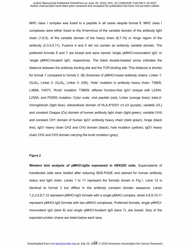
Figure 1
Schematic illustration of peptide-MHC class I-fused IgG antibodies (pMHCI-IgG). (A)
Formats either carried one or two pMHC class I complexes (1,2,3,5,6,7 or 4,8,9,10,11). The
on July 15, 2018. © 2016 American Association for Cancer Research. mct.aacrjournals.org Downloaded from
Author manuscripts have been peer reviewed and accepted for publication but have not yet been edited. Author Manuscript Published OnlineFirst on June 28, 2016; DOI: 10.1158/1535-7163.MCT-16-0207
23
MHC class I complex was fused to a peptide in all cases despite format 8. MHC class I
complexes were either fused to the N-terminus of the variable domain of the antibody light
chain (1,8,9), of the variable domain of the heavy chain (6,7,10) or hinge region of the
antibody (2,3,4,5,11). Fusions 4 and 5 did not contain an antibody variable domain. The
preferred formats 6 and 7 are boxed and were named ‘single pMHCI-monovalent IgG’ or
‘single pMHCI-bivalent IgG’, respectively. The black double-headed arrow indicates the
distance between the antibody binding site and the TCR binding site. This distance is shorter
for format 1 compared to format 2. (B) Schemes of pMHCI-fused antibody chains. Linker 1:
(G4S)3, Linker 2: (G4S)4, Linker 3: (GS), ‘Hole’ mutation in antibody heavy chain: T366S,
L368A, Y407V, ‘Knob’ mutation: T366W, effector function-free IgG1 isotype with L234A,
L235A, and P329G mutation. Color code: viral peptide (red), Linker (orange lines), beta-2-
microglobulin (light blue), extracellular domain of HLA-A*0201 α1-α3 (purple), variable (VL)
and constant Ckappa (Cκ) domain of human antibody light chain (light green), variable (VH)
and constant CH1 domain of human IgG1 antibody heavy chain (dark green), hinge (black
line), IgG1 heavy chain CH2 and CH3 domain (black), hole mutation (yellow); IgG1 heavy
chain CH2 and CH3 domain carrying the knob mutation (grey).
Figure 2
Western blot analysis of pMHCI-IgGs expressed in HEK293 cells. Supernatants of
transfected cells were blotted after reducing SDS-PAGE and stained for human antibody
heavy and light chain. Lanes 1 to 11 represent the formats shown in Fig.1. Lane 12 is
identical to format 3 but differs in the antibody constant domain sequence. Lanes
1,2,3,5,6,7,12 represent pMHCI-IgG formats with a single pMHCI complex, lanes 4,8,9,10,11
represent pMHCI-IgG formats with two pMHCI complexes. Preferred formats, single pMHCI-
monovalent IgG (lane 6) and single pMHCI-bivalent IgG (lane 7), are boxed. Size of the
expected protein chains are listed below each lane.
on July 15, 2018. © 2016 American Association for Cancer Research. mct.aacrjournals.org Downloaded from
Author manuscripts have been peer reviewed and accepted for publication but have not yet been edited. Author Manuscript Published OnlineFirst on June 28, 2016; DOI: 10.1158/1535-7163.MCT-16-0207
24
Figure 3
Analysis of thermal stability and aggregation properties before and after disulfide
stabilization of the pMHCI-complex. (A) Dynamic light scattering analysis of thermal
stability of single pMHCI-bivalent IgG before (purple) and after (blue) disulfide stabilization.
Reference IgG antibody (Herceptin) without pMHCI-fusion (green) was included as control.
(B, C) Analytical size exclusion chromatography of single pMHCI-bivalent IgG after Protein A
chromatography purification before (B) and after disulfide stabilization (C).
Figure 4
Protein purity and integrity after Protein A affinity and size exclusion chromatography.
(A) SDS gel electrophoreses under non-reducing (left) and reducing (right) conditions with
single pMHCI-monovalent IgG and single pMHCI-bivalent IgG after Protein A affinity
chromatography and size exclusion chromatography. Non-reducing gel shows the expected
size of the 145 kDa single pMHCI-monovalent IgG and 193 kDa of the single pMHCI-bivalent
IgG. Reducing gel shows the pMHCI-IgG HC fusion (95 kDa) and the antibody light chain
(25 kDa) for both constructs. Additionally the single pMHCI-monovalent IgG consists of a
truncated antibody heavy chain (26 kDa) lacking the variable domain and the single pMHCI-
bivalent IgG consists of the antibody heavy chain (50 kDa). (B) Analytical size exclusion
chromatography of the single pMHCI-monovalent (left) and bivalent IgG (right) after Protein
A purification. Percentages of aggregates and monomeric product are indicated in the
schemes. (C) Mass spectroscopy (ESI-MS) of single pMHCI-monovalent IgG (left) and
single pMHCI-bivalent IgG (right). Main peak under deglycosylated, non-reducing conditions
is the expected product consisting of the antibody light chain, the truncated antibody IgG
knob and the pMHCI-fused antibody IgG hole heavy chain (left) and two antibody light
chains, the antibody IgG knob and the pMHCI-fused antibody IgG hole heavy chain (right).
on July 15, 2018. © 2016 American Association for Cancer Research. mct.aacrjournals.org Downloaded from
Author manuscripts have been peer reviewed and accepted for publication but have not yet been edited. Author Manuscript Published OnlineFirst on June 28, 2016; DOI: 10.1158/1535-7163.MCT-16-0207

25
Minor peaks are PNGaseF, which was used for deglycosylation and adducts of
phophate/sulfate, or one or two hexoses. (D) Mass spectroscopy (ESI-MS) of single pMHCI-
monovalent IgG (left) and single pMHCI-bivalent IgG (right) under deglycosylated and
reducing conditions. All peptide chains have the expected mass. Antibody heavy chains are
lacking the C-terminal Lysine. Other modifications are indicated.
Figure 5
Flow cytometric analysis of single pMHCI-IgG binding and pMHCI delivery to hIGF-1R+
NIH3T3 and MCSP+ UCLA-SO-M14 cells target cells at different concentrations. (A)
Binding of anti-IGF-1R normal bivalent control antibody without pMHCI-fusion (column 1),
single CMV pMHCI-monovalent anti-IGF-1R IgG (column 2), single CMV pMHCI-bivalent
anti-IGF-1R IgG (column 3), single EBV pMHCI-monovalent anti-IGF-1R IgG (column 4),
and non-binding single CMV pMHCI-bivalent DP47 IgG (column 5). Antibody/fusion protein
concentration is indicated on the left. (B) HLA-A2 complexes detected on the cell surface of
MCSP+ HLA-A2- UCLA-SO-M14 cells after binding of single CMV pMHCI-anti-MCSP IgG in
concentrations of 25 nM to 0.5 nM. grey: Isotype control and secondary antibody, black: test
antibody or fusion protein and secondary antibody.
Figure 6
Functional analysis of single and double pMHCI-mono- and bivalent IgGs. Induction of
specific tumor cell lysis by MCSP-targeted pMHCI-mono- and bivalent IgGs (A, B). MCSP+
tumor cells (A: WM-266-4 cells, B: UCLA-SO-M14 cells) incubated with single CMV pMHCI-
bivalent anti-MCSP IgG (black), single CMV pMHCI-monovalent anti-MCSP IgG (light grey),
single EBV pMHCI-monovalent anti-MCSP IgG control (dark grey) or non-binding single
on July 15, 2018. © 2016 American Association for Cancer Research. mct.aacrjournals.org Downloaded from
Author manuscripts have been peer reviewed and accepted for publication but have not yet been edited. Author Manuscript Published OnlineFirst on June 28, 2016; DOI: 10.1158/1535-7163.MCT-16-0207

26
CMV pMHCI-bivalent IgG control (white with grey border) in the indicated concentrations. In
vitro expanded CMV-pp65-specific CD8+ T cells were added in an effector to target cell ratio
of 3:1. Cell lysis was measured after 7 hours in the xCELLigence system. (C) Lysis of tumor
cells by IGF-1R-targeted single CMV pMHCI-bivalent anti-IGF-1R IgG at different effector to
target cell ratios. IGF1R+ expressing NIH 3T3 cells (I24M6) were incubated at the indicated
concentrations. In vitro expanded CMV-pp65-specific CD8+ T cells were added in an effector
to target cell ratio of 3:1, 2:1 and 1:1. Cell lysis was measured after 17 hours in the
xCELLigence system. (D) Comparison of tumor cell lysis with peptide-loaded versus single
CMV pMHCI-bivalent anti-MCSP IgG-decorated tumor cells. MCSP+ and HLA-A*0201+
tumor cells (WM266-4) were either loaded with CMV peptide (pp65 495-503) or decorated
with single CMV pMHCI-bivalent anti-MCSP IgG in the indicated concentrations. CMV-pp65-
specific CD8+ T cells were added in an effector to target cell ratio of 3:1. Readout of lysis
was performed by measurement of LDH release after 24 hours. (E) Activation of T cells in
CMV-positive human donor PBMCs is measured as the frequency of IFN-γ expressing cells
in response to the exposure to target cells. WM266-4 target cells are either incubated with
PBMCs (black), or loaded with 10 µM CMV-derived pp65 peptide (dark grey) or incubated
with single CMV pMHCI-bivalent IgG (grey), or single pMHCI-bivalent non-binding IgG
control (light grey ) at 1 nM, 5 nM or 25 nM. (F) Flow cytometric analysis of unspecific in vitro
activation of primary human CMV-specific T cells with single and double CMV pMHCI-
bivalent IgGs. PBMCs from a human HLA-A*0201+ and chronically CMV-infected donor with
about 3 % of CMV-pp65-specific CD8+ T cells were incubated with double CMV pMHCI-
bivalent IgGs or single CMV pMHCI-bivalent IgGs. After 16 hours incubation, downregulation
of CMV pp65-specific T cell receptor and upregulation of activation markers CD69 and CD25
were measured and calculated in percent of all CMV pp65-specific CD8+ T cells. All
constructs were applied at 50 and 500 nM. All error bars show standard deviation of
replicates (n=3). Statistical significance was determined using students t-test for three
independent experiments.
on July 15, 2018. © 2016 American Association for Cancer Research. mct.aacrjournals.org Downloaded from
Author manuscripts have been peer reviewed and accepted for publication but have not yet been edited. Author Manuscript Published OnlineFirst on June 28, 2016; DOI: 10.1158/1535-7163.MCT-16-0207
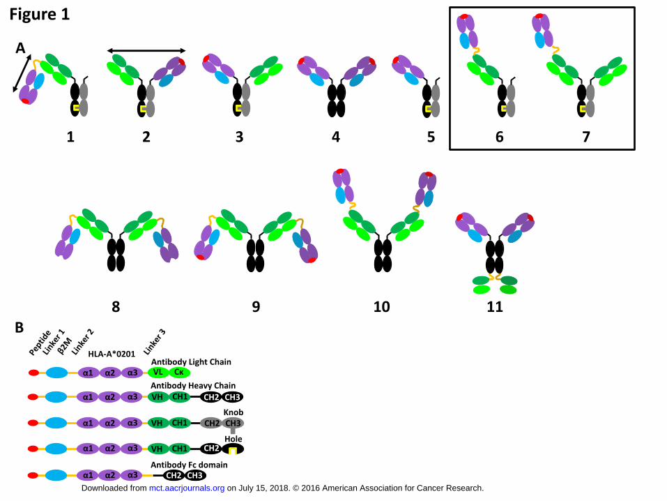
Figure 1
1 2 3 4 5 6 7
8 9 10 11
HLA-A*0201 Antibody Light Chain
α1 α2 α3 Cκ VL
Antibody Heavy Chain CH1 VH CH2 CH3
Knob
Hole
A
B
α1 α2 α3
α1 α2 α3
α1 α2 α3
α1 α2 α3 CH2 CH3
CH1 VH
CH1 VH
CH2 CH3
CH2
Antibody Fc domain
on July 15, 2018. © 2016 American Association for Cancer Research. mct.aacrjournals.org Downloaded from
Author manuscripts have been peer reviewed and accepted for publication but have not yet been edited. Author Manuscript Published OnlineFirst on June 28, 2016; DOI: 10.1158/1535-7163.MCT-16-0207

Figure 2
8 7 6 4 3 2 1 10 9 11 5 12 Marker
30
40
50
60
80
100 120
220
20
98
29
26
98
52
26
75
52
26
75
29
98
26
102 75
52
26
75
59
52
29
70
52
69
52
75
52
26
Lane
Expected Size [kDa]
Size
[kDa]
on July 15, 2018. © 2016 American Association for Cancer Research. mct.aacrjournals.org Downloaded from
Author manuscripts have been peer reviewed and accepted for publication but have not yet been edited. Author Manuscript Published OnlineFirst on June 28, 2016; DOI: 10.1158/1535-7163.MCT-16-0207
Figure 3
on July 15, 2018. © 2016 American Association for Cancer Research. mct.aacrjournals.org Downloaded from
Author manuscripts have been peer reviewed and accepted for publication but have not yet been edited. Author Manuscript Published OnlineFirst on June 28, 2016; DOI: 10.1158/1535-7163.MCT-16-0207
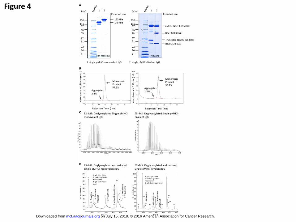
Figure 4
on July 15, 2018. © 2016 American Association for Cancer Research. mct.aacrjournals.org Downloaded from
Author manuscripts have been peer reviewed and accepted for publication but have not yet been edited. Author Manuscript Published OnlineFirst on June 28, 2016; DOI: 10.1158/1535-7163.MCT-16-0207
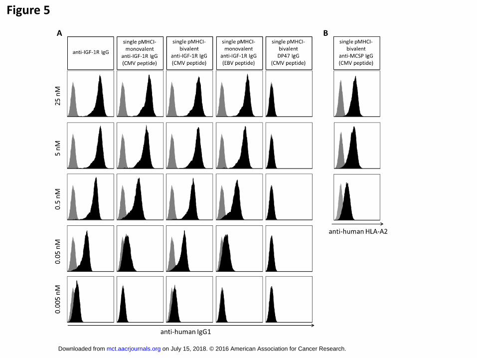
Figure 5
on July 15, 2018. © 2016 American Association for Cancer Research. mct.aacrjournals.org Downloaded from
Author manuscripts have been peer reviewed and accepted for publication but have not yet been edited. Author Manuscript Published OnlineFirst on June 28, 2016; DOI: 10.1158/1535-7163.MCT-16-0207

Figure 6
on July 15, 2018. © 2016 American Association for Cancer Research. mct.aacrjournals.org Downloaded from
Author manuscripts have been peer reviewed and accepted for publication but have not yet been edited. Author Manuscript Published OnlineFirst on June 28, 2016; DOI: 10.1158/1535-7163.MCT-16-0207
Published OnlineFirst June 28, 2016.Mol Cancer Ther Martina Schmittnaegel, Eike Hoffmann, Sabine Imhof-Jung, et al. antibody fusion molecules to redirect CD8 T cellsA new class of bifunctional major histocompatibility class I
Updated version
10.1158/1535-7163.MCT-16-0207doi:
Access the most recent version of this article at:
Material
Supplementary
http://mct.aacrjournals.org/content/suppl/2016/06/28/1535-7163.MCT-16-0207.DC1
Access the most recent supplemental material at:
Manuscript
Authoredited. Author manuscripts have been peer reviewed and accepted for publication but have not yet been
E-mail alerts related to this article or journal.Sign up to receive free email-alerts
Subscriptions
Reprints and
To order reprints of this article or to subscribe to the journal, contact the AACR Publications
Permissions
Rightslink site. Click on "Request Permissions" which will take you to the Copyright Clearance Center's (CCC)
.http://mct.aacrjournals.org/content/early/2016/06/28/1535-7163.MCT-16-0207To request permission to re-use all or part of this article, use this link
on July 15, 2018. © 2016 American Association for Cancer Research. mct.aacrjournals.org Downloaded from
Author manuscripts have been peer reviewed and accepted for publication but have not yet been edited. Author Manuscript Published OnlineFirst on June 28, 2016; DOI: 10.1158/1535-7163.MCT-16-0207