a method for anchoring round shaped cells for atomic force microscope imaging
TRANSCRIPT
Biophysical Journal Volume 68 May 1995 1678-1680
A Method for Anchoring Round Shaped Cells for Atomic ForceMicroscope Imaging
S. Kasas and A. IkaiTokyo Institute of Technology, Faculty of Biosciences, 4259 Nagatsuta, Midoriku, Yokohama 227, Japan
ABSTRACT More and more researchers are interested in imaging living (Henderson, 1994) or fixed cells in their naturalenvironment using the atomic force microscope (AFM). However, the AFM tip interacts strongly with the sample, and its z rangefreedom is limited to a few micrometers. This means that the cells to be imaged have to be strongly attached to the substrate,and imaging is restricted to cells having a flattened shape. Here we propose a simple and inexpensive solution to overcomethese limitations. The method we propose is trapping living round shaped cells in a Millipore filter with a pore size comparableto the dimensions of the cell. The highest part of some of the blocked cells protrude through the holes of the filter and can thisway be easily observed using the AFM without detachment.
INTRODUCTION
The atomic force microscope (AFM) developed by Binnigand co-workers (Binnig et al., 1986) belongs to the new,rapidly growing family of scanning probe microscopes. TheAFM consists of a tip at the end of a cantilever that is placedin contact with the sample (Wickramasinghe, 1989). Piezo-electric crystals scan the sample under the tip, and the minutedeformations of the lever are detected and recorded by acomputer to reproduce the sample's topography. The AFMallows the observation of conducting and non-conductingsamples with high lateral and vertical resolutions, in vacuum,air, and liquids (Manne et al., 1990). Additionally the mi-croscope can probe some physical properties such as frictionand softness on a nanometric scale. All these possibilitiesmake this instrument very useful in several biologicalapplications, and up to now a very large number of biologi-cal samples have already been observed with the AFM(Radmacher et al., 1992). The observation of living cellsusing this instrument is a very exciting, but for the momentquite difficult research field. The difficulty is due to the lim-ited capability of the microscope to image soft, high, andrough samples in addition to the main difficulty in fixing thesample to the substrate in liquid, which is the key point forobtaining good resolution images. Soft samples are deformedby the tip and rough ones cannot be imaged, because thevertical motion of the tip is limited to 5-15 ,um, dependingon the type of the instrument. Additionally there are stronginteractions between the tip and the sample that limit thelateral resolution of the microscope by moving the looselyattached particles or even cells right and left during the scan.Nevertheless, the new insights this microscope can giveabout the morphology, dynamics (Fritz et al., 1994), and
Receivedfor publication 18 November 1994 and in finalform 2 February1995.Address reprint requests to Dr. Sandor Kasas, c/o Professor A. Ikai, Facultyof Biosciences, 4259 Nagatsuta, Midoriku, Yokohama 227, Japan. Tel.:81-459-2457-39; Fax: 81-459-2458-05.© 1995 by the Biophysical Society0006-3495/95/05/1678/03 $2.00
mechanical properties (Shroff et al., 1994) of living cells areso exciting that they largely motivate efforts to overcomethese limitations. One of these challenges is to fix biologicalsamples to the substrate under physiological conditions. Theproblem can sometimes be solved by choosing flat and welladhering cell types (Kasas et al., 1993). Stable scanning overspherical-shaped cells is much more difficult to achieve. Avery elegant solution, although technically difficult to set up,was developed by Horber et al. (1992). It consists of trappinga single cell at the mouth of a micropipette and imaging theprotruding part by the AFM. This technique has several ad-vantages. Any cell type can be observed, and there is noheight problem any more, because the microscope is directlyengaged on the cell. Another advantage of this method is thatit keeps the cell membrane stretched, which significantlyincreases the resolution of the AFM images. Additionally,electrophysiological experiments can be carried on simul-taneously. However, the disadvantage is the heavy instru-mentation needed in this method.The solution proposed here is a combination of the pre-
ceding two techniques. The cells to be observed are grownin suspension in the presence of a Millipore filter with a poresize slightly smaller than the diameter of the cells. Whilebeing cultured, some cells are trapped in the holes and a partof their cell body protrudes outside. This part of the mem-brane is thus accessible to AFM imaging. A similar trappingcan be achieved by passing the cell suspension through thefilter placed in a modified syringe. In spite of the fact thatthis method allows the observation of just a small part of thecell surface this part is stretched and a reasonable resolutioncan be obtained. A very similar sample trapping techniquewas independently developed by Holstein et al. (1994) toimmobilize and dissect fixed Hydra vulgaris polyps.
MATERIALS AND METHODSCommercially available Saccharomyces cerviceae (dried form) were grownovernight in tap water in 20% sucrose in the presence of 5 ,um pore sizemillipore filters in a shaking bath at 30°C. The filters were removed fromthe liquid, stuck on a coverglass using double-faced adhesive tape, and fixedagain on a small metallic disc for AFM observation.
1 678
Immobilization of Cells for AFM
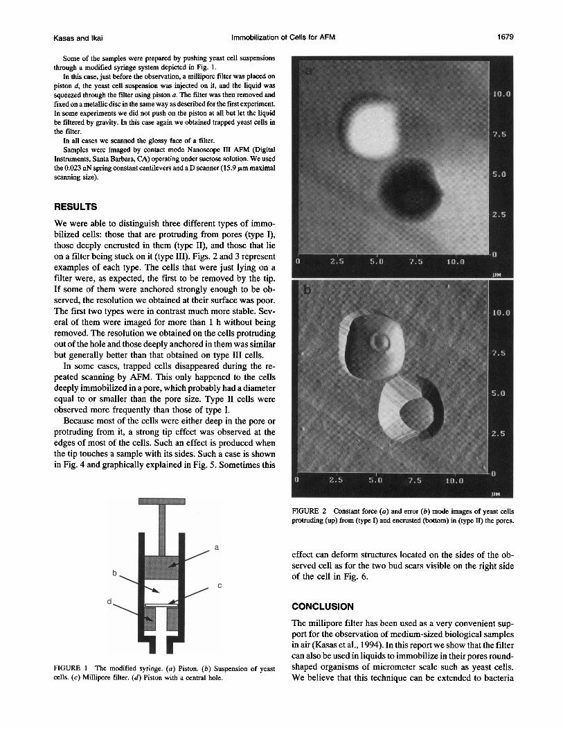
Some of the samples were prepared by pushing yeast cell suspensionsthrough a modified syringe system depicted in Fig. 1.
In this case, just before the observation, a millipore filter was placed onpiston d, the yeast cell suspension was injected on it, and the liquid wassqueezed through the filter using piston a. The filter was then removed andfixed on a metallic disc in the same way as described for the first experiment.In some experiments we did not push on the piston at all but let the liquidbe filtered by gravity. In this case again we obtained trapped yeast cells inthe filter.
In all cases we scanned the glossy face of a filter.Samples were imaged by contact mode Nanoscope III AEM (Digital
Instruments, Santa Barbara, CA) operating under sucrose solution. We usedthe 0.023 nN spring constant cantilevers and a D scanner (15.9 gm maximal l,.scanning size).
RESULTS
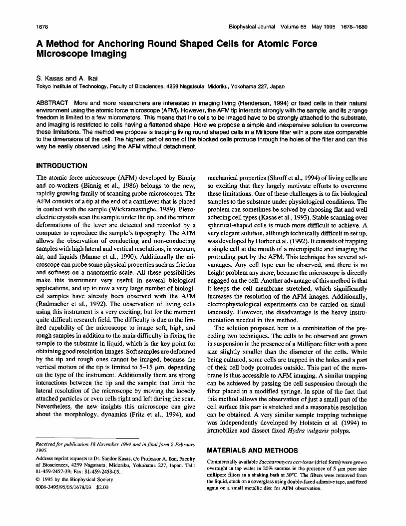
We were able to distinguish three different types of immo-bilized cells: those that are protruding from pores (type I),those deeply encrusted in them (type II), and those that lieon a filter being stuck on it (type III). Figs. 2 and 3 representexamples of each type. The cells that were just lying on afilter were, as expected, the first to be removed by the tip.If some of them were anchored strongly enough to be ob-served, the resolution we obtained at their surface was poor.The first two types were in contrast much more stable. Sev-eral of them were imaged for more than 1 h without beingremoved. The resolution we obtained on the cells protrudingout of the hole and those deeply anchored in them was similarbut generally better than that obtained on type III cells.
In some cases, trapped cells disappeared during the re-peated scanning by AFM. This only happened to the cellsdeeply immobilized in a pore, which probably had a diameterequal to or smaller than the pore size. Type II cells wereobserved more frequently than those of type I.
Because most of the cells were either deep in the pore orprotruding from it, a strong tip effect was observed at theedges of most of the cells. Such an effect is produced whenthe tip touches a sample with its sides. Such a case is shown .'....in Fig. 4 and graphically explained in Fig. 5. Sometimes this K _ A
FIGURE 2 Constant force (a) and error (b) mode images of yeast cellsprotruding (up) from (type I) and encrusted (bottom) in (type II) the pores.
aeffect can deform structures located on the sides of the ob-
bserved cell as for the two bud scars visible on the right side
b _ of the cell in Fig. 6.
d * ]-_ 9 CONCLUSION
The millipore filter has been used as a very convenient sup-port for the observation of medium-sized biological samplesin air (Kasas et al., 1994). In this report we show that the filtercan also be used in liquids to immobilize in their pores round-
FIGURE 1 The modified syringe. (a) Piston. (b) Suspension of yeast shaped organisms of micrometer scale such as yeast cells.cells. (c) Millipore filter. (d) Piston with a central hole. We believe that this technique can be extended to bacteria
Kasas and Ikai 1 679
1680 Biophysical Journal Volume 68 May 1995
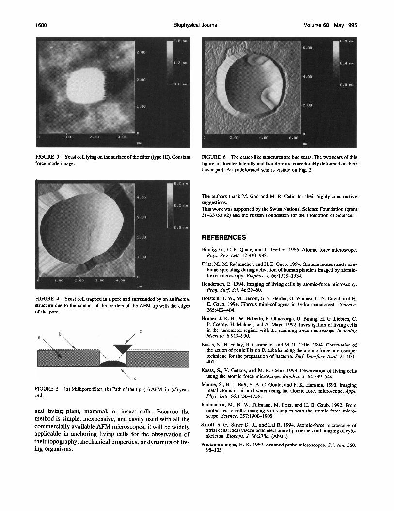
FIGURE 3 Yeast cell lying on the surface of the filter (type III). Constantforce mode image.
FIGURE 4 Yeast cell trapped mn a pore and surrounded by an artifactualstructure due to the contact of the borders of the AFM tip with the edgesof the pore.
b
| D _ 1 1 1 ^ 1 S
FIGURE 5 (a) Millipore filter. (b) Path of the tip. (c) AFM tip. (d) yeastcell.
and living plant, mammal, or insect cells. Because themethod is simple, inexpensive, and easily used with all thecommercially available AFM microscopes, it will be widelyapplicable in anchoring living cells for the observation oftheir topography, mechanical properties, or dynamics of liv-ing organisms.
ll
FIGURE 6 The crater-like structures are bud scars. The two scars of thisfigure are located laterally and therefore are considerably deformed on theirlower part. An undeformed scar is visible on Fig. 2.
The authors thank M. Gad and M. R. Celio for their highly constructivesuggestions.This work was supported by the Swiss National Science Foundation (grant31-33753.92) and the Nissan Foundation for the Promotion of Science.
REFERENCES
Binnig, G., C. F. Quate, and C. Gerber. 1986. Atomic force microscope.Phys. Rev. Lett. 12:930-933.
Fritz, M., M. Radmacher, and H. E. Gaub. 1994. Granula motion and mem-brane spreading during activation of human platelets imaged by atomic-force microscopy. Biophys. J. 66:1328-1334.
Henderson, E. 1994. Imaging of living cells by atomic-force microscopy.Prog. Surf. Sci. 46:39-60.
Holstein, T. W., M. Benoit, G. v. Herder, G. Wanner, C. N. David, and H.E. Gaub. 1994. Fibrous mini-collagens in hydra nematocysts. Science.265:402-404.
Horber, J. K. H., W. Haberle, F. Ohnesorge, G. Binnig, H. G. Liebich, C.P. Csemy, H. Mahnel, and A. Mayr. 1992. Investigation of living cellsin the nanonWeter regime with the scanning force microscope. ScanningMicrosc. 6:909-930.
Kasas, S., B. Fellay, R. Cargnello, and M. R. Celio. 1994. Observation ofthe action of penicillin on B. subtilis using the atomic force microscope:technique for the preparation of bacteria. Surf Interface Anal. 21:400-401.
Kasas, S., V. Gotzos, and M. R. Celio. 1993. Observation of living cellsusing the atomic force microscope. Biophys. J. 64:539-544.
Manne, S., H.-J. Butt, S. A. C. Gould, and P. K. Hansma. 1990. Imagingmetal atoms in air and water using the atomic force microscope. Appl.Phys. Lett. 56:1758-1759.
Radmacher, M., R. W. Tillmann, M. Fritz, and H. E. Gaub. 1992. Frommolecules to cells: imaging soft samples with the atomic force micro-scope. Science. 257:1900-1905.
Shroff, S. G., Saner D. R., and Lal R. 1994. Atomic-force microscopy ofatrial cells: local viscoelastic mechanical-properties and imaging of cyto-skeleton. Biophys. J. 66:278a. (Abstr.)
Wickramasinghe, H. K. 1989. Scanned-probe microscopes. Sci. Am. 260:98-105.