a guide to overnight orthokeratology - swisslens · corneal astigmatism of as much as 0.80d. this...
TRANSCRIPT
A Guide to Overnight
ORTHOKERATOLOGY
Polymer Technology,a Bausch & Lomb company Second Edition, 2004North America

All rights reserved. No part of this book may be reproduced or transmitted in anyform by any means, electronic or mechanical, including photocopying, recording,or by any information storage and retrieval system, without express writtenpermission from Polymer Technology, a Bausch & Lomb company, except for theinclusion of brief quotations in a review.
Copyright © 2002, 2004 by Polymer Technology, a Bausch & Lomb company
A Guide to Overnight ORTHOKERATOLOGY 3
TABLE OF CONTENTS
Foreword . . . . . . . . . . . . . . . . . . . . . . . . . . . . . . . . . . . . . . . . . . . . 5I. Background . . . . . . . . . . . . . . . . . . . . . . . . . . . . . . . . . . . . . . . . . . 5
• About Myopia . . . . . . . . . . . . . . . . . . . . . . . . . . . . . . . . . . . . . . . . 5• History . . . . . . . . . . . . . . . . . . . . . . . . . . . . . . . . . . . . . . . . . . . . . 6• Ortho-k Designs, Then . . . . . . . . . . . . . . . . . . . . . . . . . . . . . . . . . . 8
II. Modern Orthokeratology . . . . . . . . . . . . . . . . . . . . . . . . . . . . . . 12• Theory/Mechanisms . . . . . . . . . . . . . . . . . . . . . . . . . . . . . . . . . . 12• Ortho-k Designs, Now . . . . . . . . . . . . . . . . . . . . . . . . . . . . . . . . . 15• Daily Wear Orthokeratology Versus Overnight Orthokeratology . . . . 16• Is Ortho-k Myopia Control?. . . . . . . . . . . . . . . . . . . . . . . . . . . . . . 18
III. The Modern Ortho-k Fitting Process . . . . . . . . . . . . . . . . . . . . 19• Alternate Ways to Correct Nearsightedness . . . . . . . . . . . . . . . . . . 22• Patient Interview and Selection. . . . . . . . . . . . . . . . . . . . . . . . . . . 22• Pre-Fitting Examination . . . . . . . . . . . . . . . . . . . . . . . . . . . . . . . . 23• Instruments Required/Suggested . . . . . . . . . . . . . . . . . . . . . . . . . 24• The General Fitting Process . . . . . . . . . . . . . . . . . . . . . . . . . . . . . 25• Shaping Lens Adaptation and Follow-Up . . . . . . . . . . . . . . . . . . . . 25• The Importance of Using High Dk Materials . . . . . . . . . . . . . . . . . 28• Treatment Considerations . . . . . . . . . . . . . . . . . . . . . . . . . . . . . . 29• Importance and Use of Topography . . . . . . . . . . . . . . . . . . . . . . . . 31
IV. Practice Management. . . . . . . . . . . . . . . . . . . . . . . . . . . . . . . . . 34• Fees and Costs . . . . . . . . . . . . . . . . . . . . . . . . . . . . . . . . . . . . . . 34• How to Incorporate Ortho-k into the Practice . . . . . . . . . . . . . . . . . 36• Ortho-k Lens Care and Handling . . . . . . . . . . . . . . . . . . . . . . . . . . 36• Sample Fitting and Care Agreement . . . . . . . . . . . . . . . . . . . . . . . 40
V. Glossary . . . . . . . . . . . . . . . . . . . . . . . . . . . . . . . . . . . . . . . . . . . . 42VI. Frequently Asked Questions (FAQs) . . . . . . . . . . . . . . . . . . . . . 49
VII. Suggested Reading . . . . . . . . . . . . . . . . . . . . . . . . . . . . . . . . . . . 54VIII. Useful Ortho-k Web sites . . . . . . . . . . . . . . . . . . . . . . . . . . . . . . 55
IX. Bibliography. . . . . . . . . . . . . . . . . . . . . . . . . . . . . . . . . . . . . . . . . 56
Background I.5
FOREWORD
This publication is an educational tool intended to provide an overview for the use of specialized contact lenses for overnight orthokeratology(corneal reshaping) treatment for the temporary reduction of myopia.
Presented is a general foundation behind the history and principles of theorthokeratology process. Training for fitting specific ortho-k designs canbe obtained from individual ortho-k lens designers and/or manufacturinglaboratories.
I . BACKGROUND
IN THIS CHAPTER…
• This section discusses the worldwide incidence of myopia and its potentialimpact on ocular health and vision.
• A brief history of orthokeratology is presented as it began in the 1960sright up to the present-day evolution.
• Early ortho-k lens designs are reviewed with regard to their advantagesover the earlier systems and their shortcomings.
About Myopia
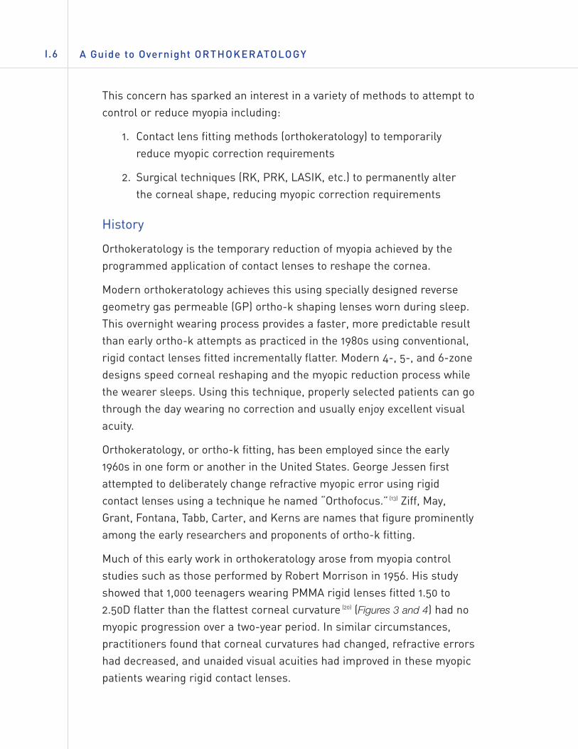
Myopia (nearsightedness) has been labeled as one of the most commonocular disorders affecting human beings—and is increasing worldwide.The increased incidence is occurring worldwide ascountries industrialize and education levels rise.The percentage varies from country to countryfrom as little as 25% in the United States to asmuch as 90% in some parts of China (16) (Figure 1).This has caused concern among somegovernments and their health agencies with regardto the ability of citizens to function in times ofemergency and natural disasters.
Figure 1. Epidemiology of myopia in some countries
This concern has sparked an interest in a variety of methods to attempt tocontrol or reduce myopia including:
1. Contact lens fitting methods (orthokeratology) to temporarilyreduce myopic correction requirements
2. Surgical techniques (RK, PRK, LASIK, etc.) to permanently alterthe corneal shape, reducing myopic correction requirements
History
Orthokeratology is the temporary reduction of myopia achieved by theprogrammed application of contact lenses to reshape the cornea.
Modern orthokeratology achieves this using specially designed reversegeometry gas permeable (GP) ortho-k shaping lenses worn during sleep.This overnight wearing process provides a faster, more predictable resultthan early ortho-k attempts as practiced in the 1980s using conventional,rigid contact lenses fitted incrementally flatter. Modern 4-, 5-, and 6-zonedesigns speed corneal reshaping and the myopic reduction process whilethe wearer sleeps. Using this technique, properly selected patients can gothrough the day wearing no correction and usually enjoy excellent visualacuity.
Orthokeratology, or ortho-k fitting, has been employed since the early1960s in one form or another in the United States. George Jessen firstattempted to deliberately change refractive myopic error using rigidcontact lenses using a technique he named “Orthofocus.” (13) Ziff, May,Grant, Fontana, Tabb, Carter, and Kerns are names that figure prominentlyamong the early researchers and proponents of ortho-k fitting.
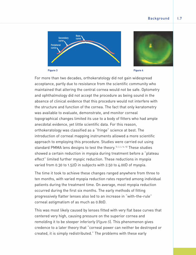
Much of this early work in orthokeratology arose from myopia controlstudies such as those performed by Robert Morrison in 1956. His studyshowed that 1,000 teenagers wearing PMMA rigid lenses fitted 1.50 to2.50D flatter than the flattest corneal curvature (20) (Figures 3 and 4) had nomyopic progression over a two-year period. In similar circumstances,practitioners found that corneal curvatures had changed, refractive errorshad decreased, and unaided visual acuities had improved in these myopicpatients wearing rigid contact lenses.
A Guide to Overnight ORTHOKERATOLOGYI.6
For more than two decades, orthokeratology did not gain widespreadacceptance, partly due to resistance from the scientific community whomaintained that altering the central cornea would not be safe. Optometryand ophthalmology did not accept the procedure as being sound in theabsence of clinical evidence that this procedure would not interfere withthe structure and function of the cornea. The fact that only keratometrywas available to evaluate, demonstrate, and monitor cornealtopographical changes limited its use to a body of fitters who had ampleanecdotal evidence, yet little scientific data. For this reason,orthokeratology was classified as a “fringe” science at best. Theintroduction of corneal mapping instruments allowed a more scientificapproach to employing this procedure. Studies were carried out usingstandard PMMA lens designs to test the theory. (2, 3, 4, 14, 15) These studiesshowed a certain reduction in myopia during treatment before a “plateaueffect” limited further myopic reduction. These reductions in myopiavaried from 0.30 to 1.52D in subjects with 2.50 to 4.00D of myopia.
The time it took to achieve these changes ranged anywhere from three toten months, with varied myopia reduction rates reported among individualpatients during the treatment time. On average, most myopia reductionoccurred during the first six months. The early methods of fittingprogressively flatter lenses also led to an increase in “with-the-rule”corneal astigmatism of as much as 0.80D.
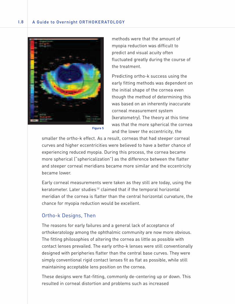
This was most likely caused by lenses fitted with very flat base curves thatcentered very high, causing pressure on the superior cornea andremolding it to be steeper inferiorly (Figure 5). This phenomenon givescredence to a later theory that “corneal power can neither be destroyed orcreated, it is simply redistributed.” The problems with these early
Background I.7
Figure 3 Figure 4
A Guide to Overnight ORTHOKERATOLOGYI.8
methods were that the amount ofmyopia reduction was difficult topredict and visual acuity oftenfluctuated greatly during the course ofthe treatment.
Predicting ortho-k success using theearly fitting methods was dependent onthe initial shape of the cornea eventhough the method of determining thiswas based on an inherently inaccuratecorneal measurement system(keratometry). The theory at this timewas that the more spherical the corneaand the lower the eccentricity, the
smaller the ortho-k effect. As a result, corneas that had steeper cornealcurves and higher eccentricities were believed to have a better chance ofexperiencing reduced myopia. During this process, the cornea becamemore spherical (“sphericalization”) as the difference between the flatterand steeper corneal meridians became more similar and the eccentricitybecame lower.
Early corneal measurements were taken as they still are today, using thekeratometer. Later studies (3) claimed that if the temporal horizontalmeridian of the cornea is flatter than the central horizontal curvature, thechance for myopia reduction would be excellent.
Ortho-k Designs, Then
The reasons for early failures and a general lack of acceptance oforthokeratology among the ophthalmic community are now more obvious.The fitting philosophies of altering the cornea as little as possible withcontact lenses prevailed. The early ortho-k lenses were still conventionallydesigned with peripheries flatter than the central base curves. They weresimply conventional rigid contact lenses fit as flat as possible, while stillmaintaining acceptable lens position on the cornea.
These designs were flat-fitting, commonly de-centering up or down. Thisresulted in corneal distortion and problems such as increased
Figure 5
astigmatism. Another factor in early failures wasdue to the use of PMMA lenses. They causedcorneal edema, thereby exacerbating cornealdistortion. However, despite the acknowledgedphysiological disadvantages of PMMA contactlenses, no other significant lasting effects werenoted as a result of ortho-k lens wear.
The procedure involved making very smallincremental lens design changes. The process was very slow, costly, and tedious for fitter andpatient alike.
Myopia reduction did not last very long whenlenses were worn occasionally on a daily wearbasis. The lack of high-permeability GP materialsdid not allow for safe overnight wear of a retainerlens to maintain corneal shape.
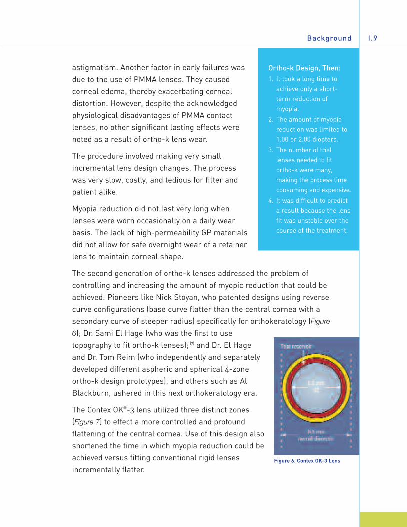
The second generation of ortho-k lenses addressed the problem ofcontrolling and increasing the amount of myopic reduction that could beachieved. Pioneers like Nick Stoyan, who patented designs using reversecurve configurations (base curve flatter than the central cornea with asecondary curve of steeper radius) specifically for orthokeratology (Figure
6); Dr. Sami El Hage (who was the first to usetopography to fit ortho-k lenses); (7) and Dr. El Hageand Dr. Tom Reim (who independently and separatelydeveloped different aspheric and spherical 4-zoneortho-k design prototypes), and others such as AlBlackburn, ushered in this next orthokeratology era.
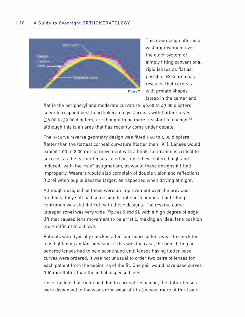
The Contex OK®-3 lens utilized three distinct zones(Figure 7) to effect a more controlled and profoundflattening of the central cornea. Use of this design alsoshortened the time in which myopia reduction could beachieved versus fitting conventional rigid lensesincrementally flatter.
Background I.9
Figure 6. Contex OK-3 Lens
Ortho-k Design, Then:1. It took a long time to
achieve only a short-term reduction ofmyopia.
2. The amount of myopiareduction was limited to1.00 or 2.00 diopters.
3. The number of triallenses needed to fitortho-k were many,making the process timeconsuming and expensive.
4. It was difficult to predicta result because the lensfit was unstable over thecourse of the treatment.
A Guide to Overnight ORTHOKERATOLOGYI.10
This new design offered avast improvement overthe older system ofsimply fitting conventionalrigid lenses as flat aspossible. Research hasrevealed that corneaswith prolate shapes(steep in the center and
flat in the periphery) and moderate curvature (46.00 to 49.00 diopters)seem to respond best to orthokeratology. Corneas with flatter curves(36.00 to 39.00 diopters) are thought to be more resistant to change, (9)
although this is an area that has recently come under debate.
The 3-curve reverse geometry design was fitted 1.50 to 4.00 dioptersflatter than the flattest corneal curvature (flatter than “K”). Lenses wouldexhibit 1.00 to 2.00 mm of movement with a blink. Centration is critical tosuccess, as the earlier lenses failed because they centered high andinduced “with-the-rule” astigmatism, as would these designs if fittedimproperly. Wearers would also complain of double vision and reflections(flare) when pupils became larger, as happened when driving at night.
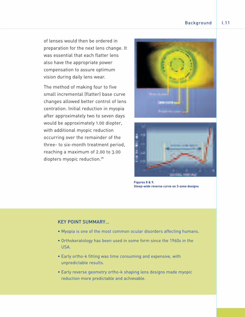
Although designs like these were an improvement over the previousmethods, they still had some significant shortcomings. Controllingcentration was still difficult with these designs. The reverse curve(steeper zone) was very wide (Figures 8 and 9), with a high degree of edgelift that caused lens movement to be erratic, making an ideal lens positionmore difficult to achieve.
Patients were typically checked after four hours of lens wear to check forlens tightening and/or adhesion. If this was the case, the tight-fitting oradhered lenses had to be discontinued until lenses having flatter basecurves were ordered. It was not unusual to order two pairs of lenses foreach patient from the beginning of the fit. One pair would have base curves0.10 mm flatter than the initial dispensed lens.
Once the lens had tightened due to corneal reshaping, the flatter lenseswere dispensed to the wearer for wear of 1 to 3 weeks more. A third pair
Figure 7
Background I.11
of lenses would then be ordered inpreparation for the next lens change. Itwas essential that each flatter lensalso have the appropriate powercompensation to assure optimumvision during daily lens wear.
The method of making four to fivesmall incremental (flatter) base curvechanges allowed better control of lenscentration. Initial reduction in myopiaafter approximately two to seven dayswould be approximately 1.00 diopter,with additional myopic reductionoccurring over the remainder of thethree- to six-month treatment period,reaching a maximum of 2.00 to 3.00diopters myopic reduction.(8)
Figures 8 & 9. Steep-wide reverse curve on 3-zone designs
KEY POINT SUMMARY…
• Myopia is one of the most common ocular disorders affecting humans.
• Orthokeratology has been used in some form since the 1960s in theUSA.
• Early ortho-k fitting was time consuming and expensive, withunpredictable results.
• Early reverse geometry ortho-k shaping lens designs made myopicreduction more predictable and achievable.
A Guide to Overnight ORTHOKERATOLOGYII.12
I I . MODERN ORTHOKERATOLOGY
IN THIS CHAPTER…
• Modern ortho-k designs are discussed and compared to theirpredecessors.
• Theories for how ortho-k treatment effects myopic reduction arepresented.
• This chapter discusses the advantages of overnight (accelerated)orthokeratology versus previous methods.
Theory/Mechanisms
The new orthokeratology designs have allowed the reshaping process totake place rather quickly. This accelerated form of ortho-k (also known as
AOK) offers some immediate change after one-nightwear of the shaping lens, with the remainder usuallyoccurring over a treatment period of about 10–30 days.
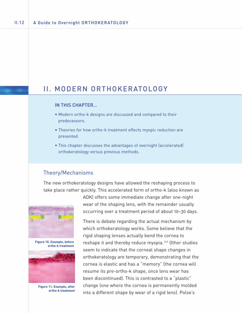
There is debate regarding the actual mechanism bywhich orthokeratology works. Some believe that therigid shaping lenses actually bend the cornea toreshape it and thereby reduce myopia. (23) Other studiesseem to indicate that the corneal shape changes inorthokeratology are temporary, demonstrating that thecornea is elastic and has a “memory” (the cornea willresume its pre-ortho-k shape, once lens wear hasbeen discontinued). This is contrasted to a “plastic”change (one where the cornea is permanently moldedinto a different shape by wear of a rigid lens). Polse’s
Figure 10. Example, beforeortho-k treatment
Figure 11. Example, afterortho-k treatment
Modern Orthokeratology II.13
result in this study showed that whatever the mechanism, the changes tothe shape of the cornea and resultant myopic reduction were temporaryand reversed themselves once rigid contact lens wear was discontinued.The question remained whether this was due to a bending of the cornealsurface or by another mechanism.
Newer studies suggest that ortho-k shaping lens fitting using reversegeometry GP designs may compress corneal tissue (in some fashion)rather than changing refractive error by bending the cornea. Thehypothesis is that a thin layer of tear film exists between the back of theortho-k shaping lens and the central cornea. These tear film “shear” forcesact hydraulically to force a compression and possibly subsequentredistribution of very anterior epithelial cells under the shaper from thecenter toward the periphery (29) (Figures 10 & 11). This seems to refute theold theory that ortho-k lens wear changes myopic correction by bendingthe cornea. It also may explain why these patients can wear shaping lenseswhose base curves are flatter than “K” and stillexperience no central corneal staining or irritationwith well-fitted ortho-k shaping lenses.
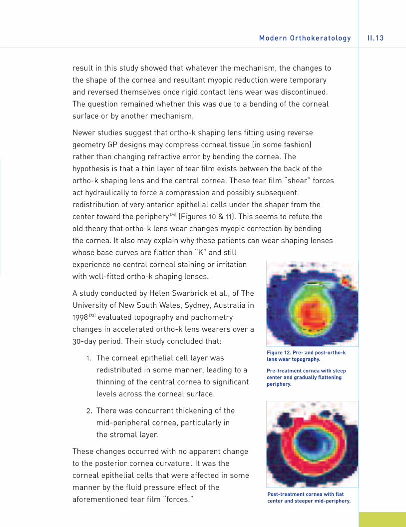
A study conducted by Helen Swarbrick et al., of TheUniversity of New South Wales, Sydney, Australia in1998 (32) evaluated topography and pachometrychanges in accelerated ortho-k lens wearers over a30-day period. Their study concluded that:
1. The corneal epithelial cell layer wasredistributed in some manner, leading to athinning of the central cornea to significantlevels across the corneal surface.
2. There was concurrent thickening of the mid-peripheral cornea, particularly in the stromal layer.
These changes occurred with no apparent changeto the posterior cornea curvature . It was thecorneal epithelial cells that were affected in somemanner by the fluid pressure effect of theaforementioned tear film “forces.”
Figure 12. Pre- and post-ortho-klens wear topography.
Pre-treatment cornea with steepcenter and gradually flatteningperiphery.
Post-treatment cornea with flatcenter and steeper mid-periphery.
A Guide to Overnight ORTHOKERATOLOGYII.14
In theory, these tear film forces cause a compression that results in aredistribution of the epithelial cells (and possibly some stromal) towardthe corneal periphery. (1, 32) This process of compression and redistributionis thought to produce a reduction in corneal sagittal height, which results
in a change (flattening) in cornealcurvature of the eye (Figures 12 and 13).This corneal shape change results inthe refocusing of the light rays on theretina (macula) of the eye, reducing oreliminating the need for myopiccorrection.
It must be remembered that thereduction of myopia, whether donepermanently by means of removingtissue by use of a laser (LASIK and
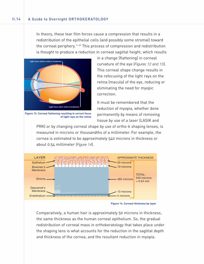
PRK) or by changing corneal shape by use of ortho-k shaping lenses, ismeasured in microns or thousandths of a millimeter. For example, thecornea is estimated to be approximately 540 microns in thickness orabout 0.54 millimeter (Figure 14).
Comparatively, a human hair is approximately 50 microns in thickness,the same thickness as the human corneal epithelium. So, the gradualredistribution of corneal mass in orthokeratology that takes place underthe shaping lens is what accounts for the reduction in the sagittal depthand thickness of the cornea, and the resultant reduction in myopia.
Figure 14. Corneal thickness by layer
Figure 13. Corneal flattening resulting in correct focusof light rays on the retina
Modern Orthokeratology II.15
Ortho-k Designs, Now
Depending on the fitting philosophy of thedesign being used, an initial base curve ischosen that is 0.30 mm to 1.40 mm flatterthan the flattest corneal curvature (flat“K”). This optical zone width may varyfrom 6.0 mm to 8.0 mm. Commonly a posterior optical zone diameter of6.0 to 6.5 mm is most often used.
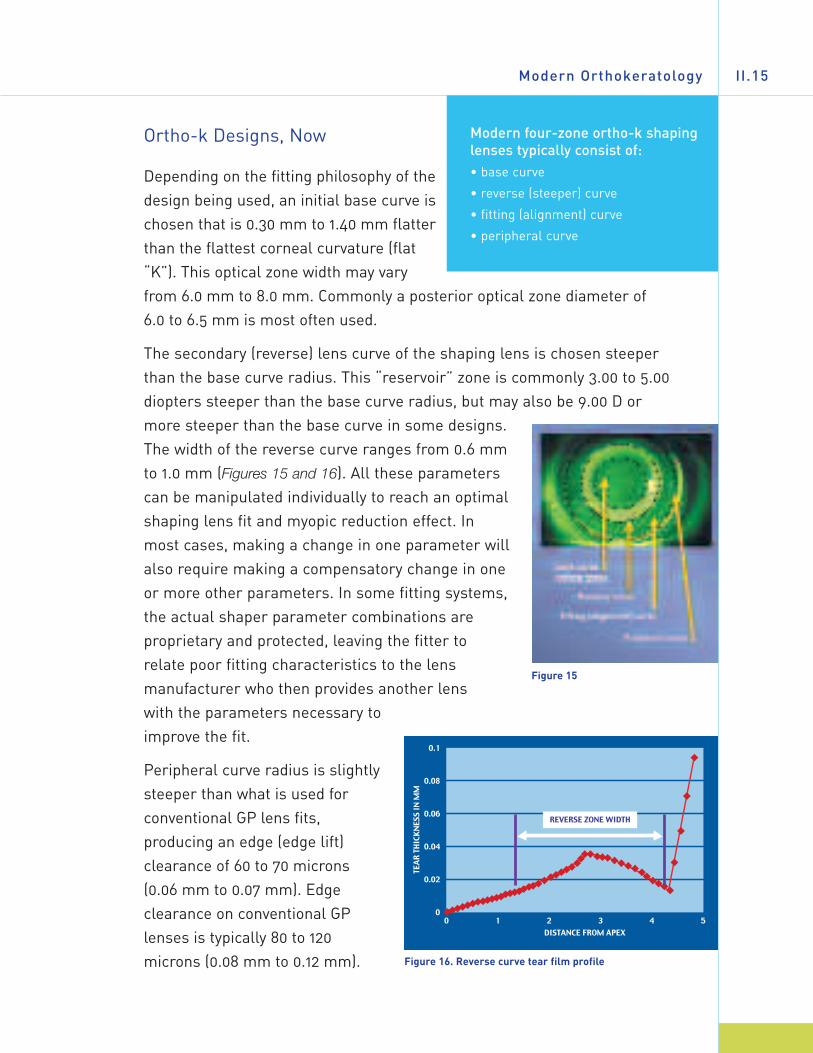
The secondary (reverse) lens curve of the shaping lens is chosen steeperthan the base curve radius. This “reservoir” zone is commonly 3.00 to 5.00diopters steeper than the base curve radius, but may also be 9.00 D ormore steeper than the base curve in some designs.The width of the reverse curve ranges from 0.6 mmto 1.0 mm (Figures 15 and 16). All these parameterscan be manipulated individually to reach an optimalshaping lens fit and myopic reduction effect. Inmost cases, making a change in one parameter willalso require making a compensatory change in oneor more other parameters. In some fitting systems,the actual shaper parameter combinations areproprietary and protected, leaving the fitter torelate poor fitting characteristics to the lensmanufacturer who then provides another lens with the parameters necessary toimprove the fit.
Peripheral curve radius is slightlysteeper than what is used forconventional GP lens fits,producing an edge (edge lift)clearance of 60 to 70 microns(0.06 mm to 0.07 mm). Edgeclearance on conventional GPlenses is typically 80 to 120microns (0.08 mm to 0.12 mm).
Figure 15
Figure 16. Reverse curve tear film profile
Modern four-zone ortho-k shapinglenses typically consist of: • base curve
• reverse (steeper) curve
• fitting (alignment) curve
• peripheral curve
A Guide to Overnight ORTHOKERATOLOGYII.16
Modern three-zone ortho-k shaping lens designs were made availableby Roger Tabb, Jim Day (Fargo™), Donald Harris, and Nick Stoyan (ContexOK®). Modern four-zone designs were introduced by Sami El Hage(CKR), Tom Reim (Dreimlens®/DreamLens™), Euclid (Emerald™), DonNoack and John Mountford (BE Retainer™), Rinehart-Reeves, ParagonCRT®, and others.
Daily Wear Orthokeratology Versus Overnight Orthokeratology
The limitations imposed by the poor physiologic effects of PMMA lenswear dictated that early ortho-k lens wear be limited to daily lens wear.The same is true today of ortho-k shaping lenses that are made of gaspermeable plastics of medium and low permeabilities. Like PMMA, thesematerials are not optimal for safe overnight ortho-k wear, since the newgeneration of ortho-k lens designs are typically larger and thicker thanconventional gas permeable lenses.
Being limited to daily wear made the early ortho-k process more difficultfor the patient to endure in terms of comfort and consistent vision ascompared to wearing conventional rigid lenses for vision correction alone.Added to this was the cost of this procedure in terms of the number oflenses required (eight pairs or more) and the treatment period (nine totwelve months), with no way to accurately predict the visual result.
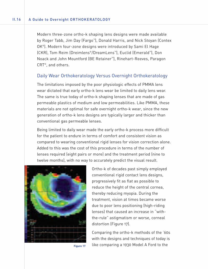
Ortho-k of decades past simply employedconventional rigid contact lens designs,progressively fit as flat as possible toreduce the height of the central cornea,thereby reducing myopia. During thetreatment, vision at times became worsedue to poor lens positioning (high-ridinglenses) that caused an increase in “with-the-rule” astigmatism or worse, cornealdistortion (Figure 17).
Comparing the ortho-k methods of the ’60swith the designs and techniques of today islike comparing a 1930 Model A Ford to theFigure 17
Modern Orthokeratology II.17
latest model Mercedes Benz. This brings up the issue of what to call thismodality. In its beginnings in the 1960s, George Jessen called it“Orthofocus.” It soon came to be known simply as “orthokeratology.”Looking at the PDR Medical Dictionary definition, orthokeratology isdefined as: “A method of molding the cornea with contact lenses toimprove unaided vision.” And in fact, that’s exactly what it is. The processinvolves the programmed application of a contact “shaping” lens for thepurpose of systematically and predictably reshaping the corneal surfaceto temporarily reduce the need for myopic correction.
Today, other names have been coined for the more modern processutilizing four and five zone reverse geometry designs, such as“accelerated orthokeratology” (AOK), “Ortho-K,” “AdvancedOrthokeratology,” Corneal Refractive Therapy (CRT ®), etc.
The advent of new high-permeability GP materials (ISO/Fatt Dk of 85 ormore) has allowed overnight wear of these ortho-k shaping lensesinstead of during the day.* This provides easy and fast lens adaptation forthe patient.
New innovative four, five, and six curve reverse geometry designs inlarge diameters have not only allowed for better control of position of theshaping lens, but have also provided ortho-k fitters with a scientific andmore accurate means to control and predict myopic reduction. Thesemodern ortho-k shaping lenses allow for rapid myopia reduction as well.What took nine to twelve months to achieve in the ’60s now will occurusually within 30 days. Approximately 70 to 80% of the patients treatedwith modern ortho-k shaping lenses achieve their desired myopiareduction with only one pair of shapers, as compared to the old processthat often took eight or more pairs of conventional rigid contact lenses toachieve myopia reduction. The first approvals for overnightorthokeratology in the U.S. were obtained by Paragon Vision Sciences(CRT®) and Euclid Systems Inc. (Emerald Lens).
* Overnight orthokeratology should only be performed using GP lens materials and designsapproved for overnight use.
A Guide to Overnight ORTHOKERATOLOGYII.18
Is Ortho-k Myopia Control?
Orthokeratology treatment effects a rapid reduction in myopia. Thisreduction is temporary in nature and is maintained by regular nightlywear of the shaping lens as prescribed.
Since orthokeratology treatment results in this myopic reduction, it islogical that questions have arisen regarding the effect this process mighthave on myopic progression. Among these question are:
• Would the systematic reshaping of the cornea to reduce myopiaalso slow its progression?
• If ortho-k treatment is discontinued, what (if any) is the effecton myopic progression? Will there be a “rebound” effect thatresults in an increased amount and rate of progression ofmyopia or will it slow?
There are no definitive answers or scientific evidence to address thesequestions at this point. The role that orthokeratology may play in affectingmyopic progression is currently under investigation.
KEY POINT SUMMARY…
• Overnight (accelerated) orthokeratology allows for treatment resultswithin approximately 30 days.
• Reverse geometry designs utilize tear film forces to effect cornealshape changes.
• Modern four and five zone designs allow better control of treatmentprocess.
• Patients easily accept overnight ortho-k because adaptation is rapidwith minimal awareness of the shaping lens.
• The role that orthokeratology may play in myopia control is currentlyunder investigation.
The Modern Ortho-k Fitting Process III.19
I I I . THE MODERN ORTHO-K FITTING PROCESS
IN THIS CHAPTER…
• Modern orthokeratology involves the combination of reverse geometrydesigns, high permeability GP materials, and corneal topography tounderstand the relationship of the shaping lenses to the cornea.
• This chapter provides guidance to practitioners to help select andinterview potential ortho-k wearers.
• The pre-fitting screening examination is outlined with tips on how toevaluate patients as good candidates.
• A list of essential equipment to fit, evaluate, and manage ortho-kpatients in the practice is provided.
• An overview of the general fitting process is provided.
• Emphasized is the importance of using high Dk GP materials forovernight lens wear of orthokeratology shaping lenses.
• Possible complications are presented with a discussion on how to usecorneal topography to assess fit, follow progress, and avoidunfavorable results.
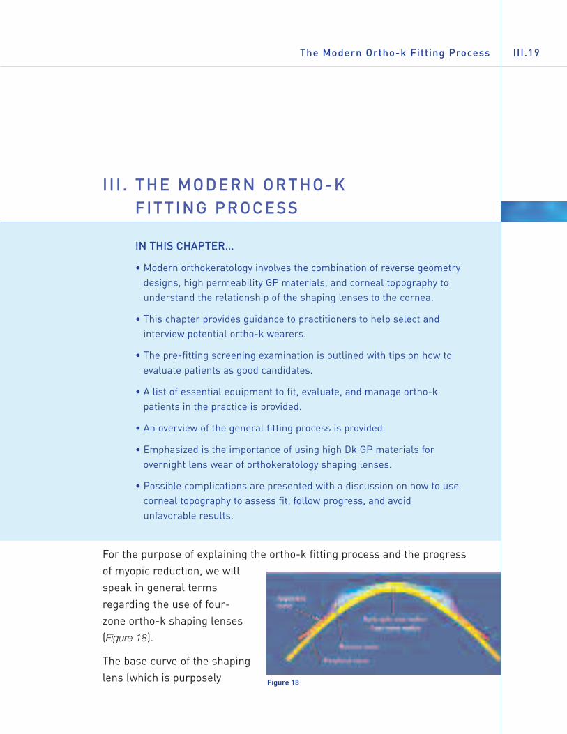
For the purpose of explaining the ortho-k fitting process and the progressof myopic reduction, we willspeak in general termsregarding the use of four-zone ortho-k shaping lenses(Figure 18).
The base curve of the shapinglens (which is purposely Figure 18
chosen to be flatter than the flattest central apical radius) appliespressure on the thin layer of tear film that lies between the back of theortho-k shaper and the corneal surface. Even though the use offluorescein to observe the lens fit gives the appearance of apical touch, atear film layer of less than 10 microns exists.
Theoretically, these “tear film forces” cause the apical epithelial cells tocompress and possibly “migrate” toward the periphery. This cornealreshaping process creates a decrease in corneal sagittal height andcauses the cornea to become more spherical and flatter, therebyreducing or eliminating the need for myopic correction.
As epithelial mass is shifted toward the periphery, the steeper secondarycurve (reverse curve) forms a tear reservoir where excess tear and thedisplaced corneal cells may form.
The mid-peripheral or fitting curve (also known as alignment curve) isactually the curve that allows the shaping lens to center and positionproperly on the eye. This curve is calculated to be in alignment (parallel)with the mid-periphery of the cornea.
The purpose of the peripheral curve on an ortho-k shaping lens is thesame as on any GP lens, to allow for tear circulation under the shaper,allow for its easy removal, and provide a means to remove debris fromunder the shaper.
Generally, an ortho-k fitter will calculate the parameters of the trialshaping lens with a design-specific nomogram or computer program. Insome cases, “K”s and refraction are given to the laboratory to calculatethe shaping lens parameters. The trial shaping lens or the shaper to be
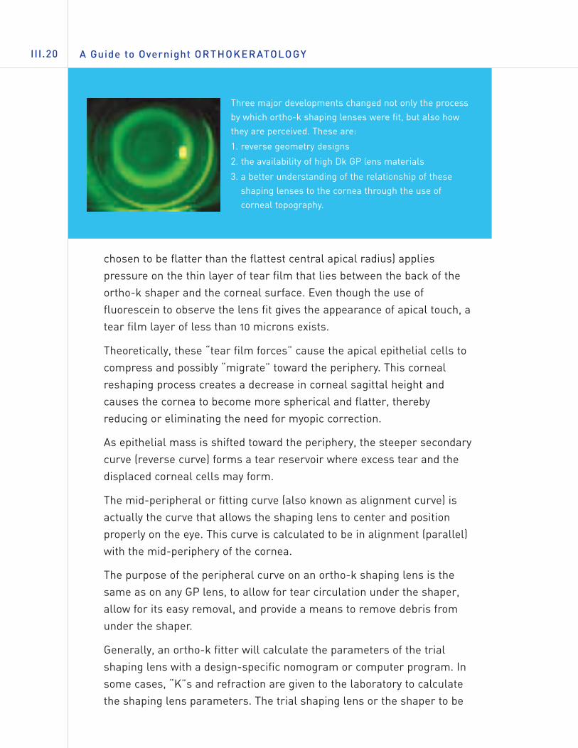
Three major developments changed not only the processby which ortho-k shaping lenses were fit, but also howthey are perceived. These are:
1. reverse geometry designs
2. the availability of high Dk GP lens materials
3. a better understanding of the relationship of theseshaping lenses to the cornea through the use ofcorneal topography.
A Guide to Overnight ORTHOKERATOLOGYIII.20
The Modern Ortho-k Fitting Process III.21
dispensed is then placed on the patient’s eye and allowed to settle beforeit is evaluated to ensure that it does not tighten excessively. There havebeen cases of “corneal responders” who will display corneal changes inas little as 30 minutes. (14) A slight reduction in myopia will usually be seenin as little as four to six hours. (31) This allows the fitter to decide whetherthe patient will respond successfully to the treatment.
Once it is decided to allow the patient to wear the ortho-k shaping lensesovernight, the patient is instructed to return to the office as early aspossible (within a few hours of awakening) in the morning. Prior towearing the shaping lenses overnight, the patient must be instructed howto insert, remove, and care for the shapers. Under no circumstancesshould patients be allowed to leave the office and wear the shapinglenses overnight if they cannot confidently remove them. Patients shouldbe instructed in shaper removal using the eyelids (blink/scissor method).At the practitioner’s discretion, the patient may also be supplied with andinstructed in the use of the silicone rubber (DMV) lens remover.
Probably the most critical step in the fitting process is to examine thepatient early the first morning after the first night of ortho-k shaping lenswear. It is important for the fitter to assess position of the shaping lensesand the location of the flattened zone on the cornea for centration. This isaccomplished by observing the shapers on the eyes and evaluatingcorneal topography after removal of the shaping lenses. Unaided visualacuity is also checked at this time. De-centered shaping lenses will notproduce the desired myopia reduction and may even cause cornealdistortion. Shapers must also be checked for adherence and cornealintegrity must be determined.
A significant degree of myopic reduction (1.00 to2.00 diopters) often occurs after the first night ofortho-k shaper lens wear. If the initial shaperappears to have tightened, a shaping lens with alower sagittal height should be fitted.
The patient should not be allowed to wear a tight-fitting shaper in orderto avoid metabolic and corneal distortion problems. Some fitters order asecond shaper that has a lower sagittal height at the same time that theinitial shaper is ordered to facilitate this change easily.
A significant degree ofmyopic reduction (as much as 2.00 diopters)often occurs after the firstnight of lens wear.
A Guide to Overnight ORTHOKERATOLOGYIII.22
As mentioned, a large part of the ortho-k effect occurs in the first sevendays of shaping lens wear, with maximum results usually achieved in onemonth. (19, 22, 30) In some cases, myopia reduction may take up to three months.
The initial goal in ortho-k shaping lens fitting is to achieve the desiredamount of myopia reduction. Having reached that point, the fitter’s goal isthen to try to reduce overnight shaper wear to a frequency that will stillmaintain the patient’s cornea in the desired shape and visual acuity level.
One benefit to the ortho-k treatment is that the process is reversible.Studies have shown that regression will usually occur in a period ofapproximately 30–90 days, (12, 25, 26) with most wearers showing completereversal in less time. While this factor represents an advantage oversurgical refractive correction, it is a disadvantage at the same time. Formost, nightly wear of the shaping lens will be required to maintain myopicreduction. For others, lens wear may be required only every second orthird night. The last shaping lens worn that produced the optimum cornealshape change is typically used for nightly wear to maintain reduction.
Alternative Ways to Correct Nearsightedness
Myopia (nearsightedness) can be corrected by any method that reduces thefocusing power of the eye. The most common methods of correction utilizeeyeglasses or regular standard daily, extended, or continuous wear contactlenses. These represent a means of correcting myopia only during the timethat the eyeglasses or regular contact lenses are worn, with no lastingeffect on the myopia. Other methods of correcting myopia involve varioussurgical procedures such as LASIK.
Patient Interview and Selection
Patient selection criteria will depend on the approach and philosophy of the fitter. Patient selection criteria are presented here using a “broad-range” approach.
The Modern Ortho-k Fitting Process III.23
Orthokeratology Candidate Profile
• Age: juvenile to adult myopes
• Spherical refractive error: -1.00 D to -5.00 D spherical power correction
• Cylindrical refractive error: - 1.50 D or less “with-the-rule” corneal astigmatism - 0.75 D or less “against-the-rule” astigmatism
• Recreational and sports activities where periods withoutwearing visual correction are beneficial
• Those whose vocation requires unaided visual acuity for certainperiods, such as police, firemen, military, or occupations whererefractive surgery may be a cause for exclusion (deep-seadivers, high altitude pilots, etc.)
• Free of corneal dystrophies (e.g. keratoconus), ocular diseases,or any condition that may preclude the patient from wearing anytype of GP lens
• Motivated to undergo full or partial myopia reduction and willingto return to the office for two to three months of active treatmentand every six months for passive treatment
• Committed to the initial and ongoing cost of ortho-k treatment(See section on fees and costs)
• Practitioners should consult fitting information provided byspecific design/fitting systems.
Pre-Fitting Examination
This should include:• Refraction (with dilation)
• Baseline topography (keratometry optional, but topography is a must)
• Tear film analysis
a. Schirmer test (quantitative)
b. Tear Break-Up Time or TBUT (qualitative)
• Biomicroscopy
A Guide to Overnight ORTHOKERATOLOGYIII.24
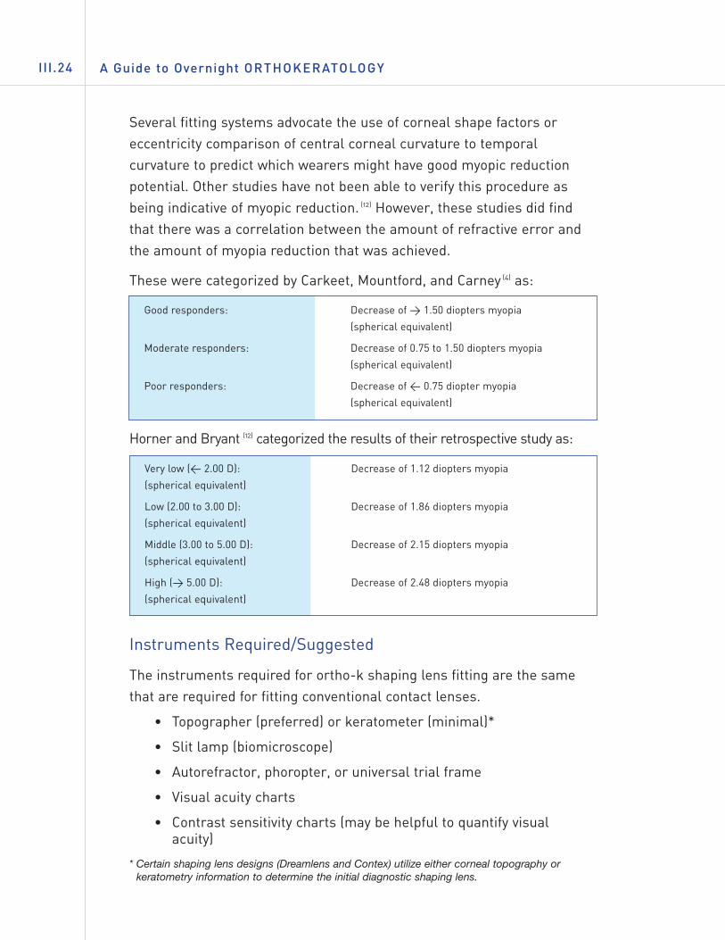
Several fitting systems advocate the use of corneal shape factors oreccentricity comparison of central corneal curvature to temporalcurvature to predict which wearers might have good myopic reductionpotential. Other studies have not been able to verify this procedure asbeing indicative of myopic reduction. (12) However, these studies did findthat there was a correlation between the amount of refractive error andthe amount of myopia reduction that was achieved.
These were categorized by Carkeet, Mountford, and Carney (4) as:
Horner and Bryant (12) categorized the results of their retrospective study as:
Instruments Required/Suggested
The instruments required for ortho-k shaping lens fitting are the samethat are required for fitting conventional contact lenses.
• Topographer (preferred) or keratometer (minimal)*
• Slit lamp (biomicroscope)
• Autorefractor, phoropter, or universal trial frame
• Visual acuity charts
• Contrast sensitivity charts (may be helpful to quantify visualacuity)
* Certain shaping lens designs (Dreamlens and Contex) utilize either corneal topography orkeratometry information to determine the initial diagnostic shaping lens.
Very low (< 2.00 D): Decrease of 1.12 diopters myopia
(spherical equivalent)
Low (2.00 to 3.00 D): Decrease of 1.86 diopters myopia
(spherical equivalent)
Middle (3.00 to 5.00 D): Decrease of 2.15 diopters myopia
(spherical equivalent)
High (> 5.00 D): Decrease of 2.48 diopters myopia
(spherical equivalent)
Good responders: Decrease of > 1.50 diopters myopia
(spherical equivalent)
Moderate responders: Decrease of 0.75 to 1.50 diopters myopia
(spherical equivalent)
Poor responders: Decrease of < 0.75 diopter myopia
(spherical equivalent)
The Modern Ortho-k Fitting Process III.25
The General Fitting Process
Initial Fitting:Some ortho-k fitting systems advocate use of diagnostic trial shapinglenses for initial parameter selection. Diagnostic fitting not only yieldsvaluable clinical information, but also provides important information onpatient response and potential for successful ortho-k shaping lensadaptation. However, equally successful ortho-k fitting systems requirethat the initial shaper be ordered from keratometry readings andrefraction. In either case, baseline topography, evaluation of response towear of the shaping lens, and post-wear topography will serve asvaluable information that will be used to guide the course of treatment.
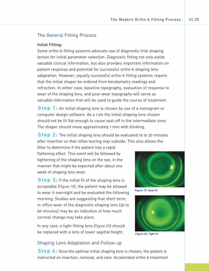
Step 1: An initial shaping lens is chosen by use of a nomogram orcomputer design software. As a rule the initial shaping lens chosenshould not be fit flat enough to cause seal-off in the intermediate zone.The shaper should move approximately 1 mm with blinking.
Step 2: The initial shaping lens should be evaluated 10 to 30 minutesafter insertion so that reflex tearing may subside. This also allows thefitter to determine if the patient has a rapidflattening effect. This event will be followed bytightening of the shaping lens on the eye, in themanner that might be expected after about oneweek of shaping lens wear.
Step 3: If the initial fit of the shaping lens isacceptable (Figure 19), the patient may be allowedto wear it overnight and be evaluated the followingmorning. Studies are suggesting that short term,in-office wear of the diagnostic shaping lens (30 to60 minutes) may be an indication of how muchcorneal change may take place.
In any case, a tight-fitting lens (Figure 20) shouldbe replaced with a lens of lower sagittal height.
Shaping Lens Adaptation and Follow-up
Step 4: Once the optimal initial shaping lens is chosen, the patient isinstructed on insertion, removal, and care. Accelerated ortho-k treatment
Figure 19. Ideal fit
Figure 20. Tight fit
A Guide to Overnight ORTHOKERATOLOGYIII.26
allows patients to wear their shaping lenses overnight immediately. For thisreason, these patients should be seen in the office (wearing their shapers)the morning after the first night of wear. Shaping lenses are checked for:
• Centration: Centration of the shaping lens is critical to the ortho-keffect. De-centered shapers do not produce the desired myopiareduction. The result will not only be poor visual acuity, but mayalso cause localized corneal distortion.
• Movement: Shaping lenses should move approximately 1 mm withblinking. Shapers that are adhered during wear should bereplaced with shaping lenses of lower sagittal height.
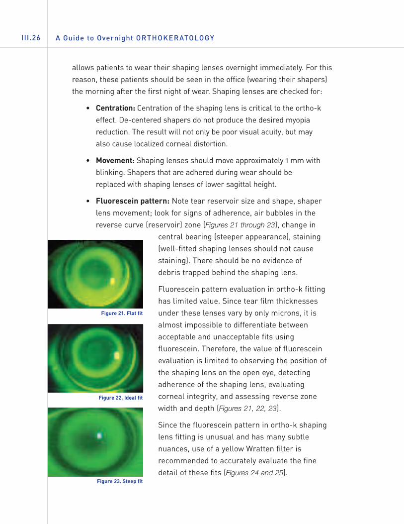
• Fluorescein pattern: Note tear reservoir size and shape, shaperlens movement; look for signs of adherence, air bubbles in thereverse curve (reservoir) zone (Figures 21 through 23), change in
central bearing (steeper appearance), staining(well-fitted shaping lenses should not causestaining). There should be no evidence ofdebris trapped behind the shaping lens.
Fluorescein pattern evaluation in ortho-k fittinghas limited value. Since tear film thicknessesunder these lenses vary by only microns, it isalmost impossible to differentiate betweenacceptable and unacceptable fits usingfluorescein. Therefore, the value of fluoresceinevaluation is limited to observing the position ofthe shaping lens on the open eye, detectingadherence of the shaping lens, evaluatingcorneal integrity, and assessing reverse zonewidth and depth (Figures 21, 22, 23).
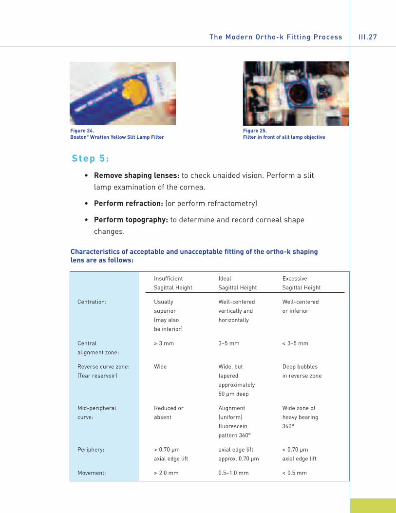
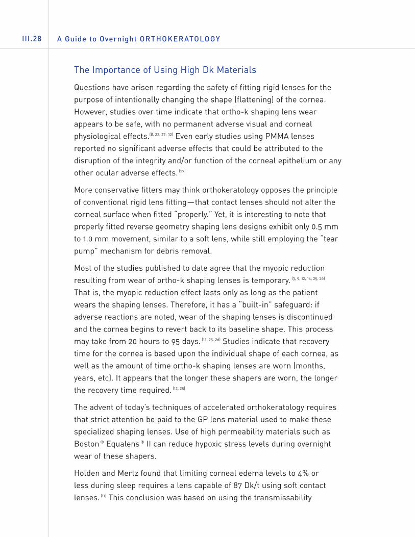
Since the fluorescein pattern in ortho-k shapinglens fitting is unusual and has many subtlenuances, use of a yellow Wratten filter isrecommended to accurately evaluate the finedetail of these fits (Figures 24 and 25).
Figure 21. Flat fit
Figure 22. Ideal fit
Figure 23. Steep fit
The Modern Ortho-k Fitting Process III.27
Step 5:
• Remove shaping lenses: to check unaided vision. Perform a slitlamp examination of the cornea.
• Perform refraction: (or perform refractometry)
• Perform topography: to determine and record corneal shapechanges.
Characteristics of acceptable and unacceptable fitting of the ortho-k shapinglens are as follows:
Insufficient Ideal Excessive
Sagittal Height Sagittal Height Sagittal Height
Centration: Usually Well-centered Well-centered
superior vertically and or inferior
(may also horizontally
be inferior)
Central > 3 mm 3–5 mm < 3–5 mm
alignment zone:
Reverse curve zone: Wide Wide, but Deep bubbles
(Tear reservoir) tapered in reverse zone
approximately
50 µm deep
Mid-peripheral Reduced or Alignment Wide zone of
curve: absent (uniform) heavy bearing
fluorescein 360°
pattern 360°
Periphery: > 0.70 µm axial edge lift < 0.70 µm
axial edge lift approx. 0.70 µm axial edge lift
Movement: > 2.0 mm 0.5–1.0 mm < 0.5 mm
Figure 25. Filter in front of slit lamp objective
Figure 24. Boston® Wratten Yellow Slit Lamp Filter
The Importance of Using High Dk Materials
Questions have arisen regarding the safety of fitting rigid lenses for thepurpose of intentionally changing the shape (flattening) of the cornea.However, studies over time indicate that ortho-k shaping lens wearappears to be safe, with no permanent adverse visual and cornealphysiological effects.(8, 23, 27, 32) Even early studies using PMMA lensesreported no significant adverse effects that could be attributed to thedisruption of the integrity and/or function of the corneal epithelium or anyother ocular adverse effects. (27)
More conservative fitters may think orthokeratology opposes the principleof conventional rigid lens fitting—that contact lenses should not alter thecorneal surface when fitted “properly.” Yet, it is interesting to note thatproperly fitted reverse geometry shaping lens designs exhibit only 0.5 mmto 1.0 mm movement, similar to a soft lens, while still employing the “tearpump” mechanism for debris removal.
Most of the studies published to date agree that the myopic reductionresulting from wear of ortho-k shaping lenses is temporary. (3, 9, 12, 14, 25, 26)
That is, the myopic reduction effect lasts only as long as the patientwears the shaping lenses. Therefore, it has a “built-in” safeguard: ifadverse reactions are noted, wear of the shaping lenses is discontinuedand the cornea begins to revert back to its baseline shape. This processmay take from 20 hours to 95 days. (12, 25, 26) Studies indicate that recoverytime for the cornea is based upon the individual shape of each cornea, aswell as the amount of time ortho-k shaping lenses are worn (months,years, etc). It appears that the longer these shapers are worn, the longerthe recovery time required. (12, 25)
The advent of today’s techniques of accelerated orthokeratology requiresthat strict attention be paid to the GP lens material used to make thesespecialized shaping lenses. Use of high permeability materials such asBoston ® Equalens ® II can reduce hypoxic stress levels during overnightwear of these shapers.
Holden and Mertz found that limiting corneal edema levels to 4% or less during sleep requires a lens capable of 87 Dk/t using soft contactlenses. (11) This conclusion was based on using the transmissability
A Guide to Overnight ORTHOKERATOLOGYIII.28
formula x10-9(cm2/sec)(mlO2/ml x mmHg). It should also be taken intoconsideration that GPs cover a smaller area of the cornea and the Dkratings are generally higher than with conventional soft contact lenses.
There is another reason why use of high Dk materials is critical. Ortho-kshaping lenses generally have center thicknesses which are greater thanconventional GP lenses,(19) to avoid flexure and produce the desired,controlled corneal flattening.
Ortho-k Treatment Considerations
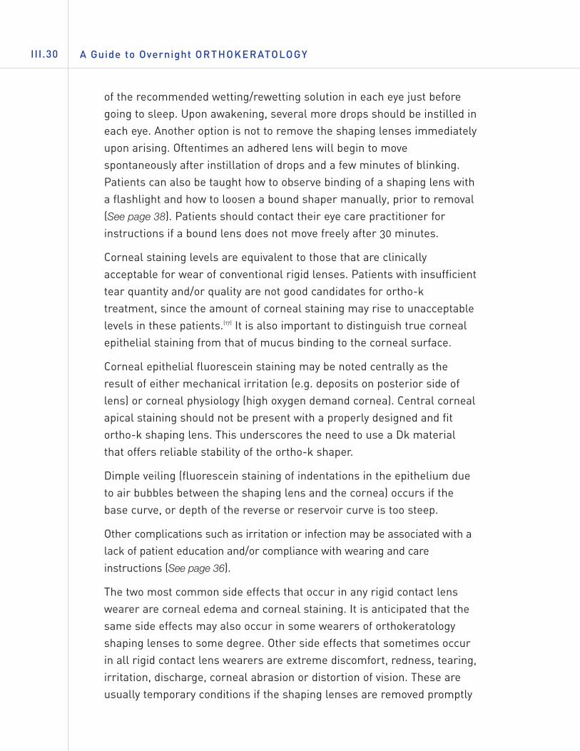
One of the most critical fitting criteria is centration of the ortho-k shapinglens (See “The Importance and Use of Topography”). Without goodcentration (both vertical and lateral), visual acuity will not be optimal andmay be unacceptable to the wearer because the“treatment” zone will not be centered over thepupil and optical axis of the eye. This is whyobservation of the fit of the shaping lens,movement, visual acuity, and corneal topographyare so important and why the fitter must see thepatient within several hours of awakening in themorning after the first night of overnight wear ofthe shaping lens. Patients must not be allowed tocontinue to wear shapers that are misfit in any way.
Modern orthokeratology studies have shown thatthe complication rate associated with this modalityis within the confines of acceptable clinicalsequelae as seen in fitting conventional GP lenses. (23, 32) Adaptation may even be easier andfaster since the lenses are of large diameter andworn only at night during sleep, so issues of lidsensation are minimized.
Adherence (binding) of the shaping lenses isalways a possibility when a large diameter shapinglens is worn overnight (Figures 26 through 28).Fitters often advise wearers to instill several drops
The Modern Ortho-k Fitting Process III.29
Figure 26. Slit lamp white light view of adhered lens
Figure 27. Adhered lens viewed withfluorescein
Figure 28. Epithelial indentation(fluorescein staining) following lensremoval
A Guide to Overnight ORTHOKERATOLOGYIII.30
of the recommended wetting/rewetting solution in each eye just beforegoing to sleep. Upon awakening, several more drops should be instilled ineach eye. Another option is not to remove the shaping lenses immediatelyupon arising. Oftentimes an adhered lens will begin to movespontaneously after instillation of drops and a few minutes of blinking.Patients can also be taught how to observe binding of a shaping lens witha flashlight and how to loosen a bound shaper manually, prior to removal(See page 38). Patients should contact their eye care practitioner forinstructions if a bound lens does not move freely after 30 minutes.
Corneal staining levels are equivalent to those that are clinicallyacceptable for wear of conventional rigid lenses. Patients with insufficienttear quantity and/or quality are not good candidates for ortho-ktreatment, since the amount of corneal staining may rise to unacceptablelevels in these patients.(17) It is also important to distinguish true cornealepithelial staining from that of mucus binding to the corneal surface.
Corneal epithelial fluorescein staining may be noted centrally as theresult of either mechanical irritation (e.g. deposits on posterior side oflens) or corneal physiology (high oxygen demand cornea). Central cornealapical staining should not be present with a properly designed and fitortho-k shaping lens. This underscores the need to use a Dk materialthat offers reliable stability of the ortho-k shaper.
Dimple veiling (fluorescein staining of indentations in the epithelium dueto air bubbles between the shaping lens and the cornea) occurs if thebase curve, or depth of the reverse or reservoir curve is too steep.
Other complications such as irritation or infection may be associated with alack of patient education and/or compliance with wearing and careinstructions (See page 36).
The two most common side effects that occur in any rigid contact lenswearer are corneal edema and corneal staining. It is anticipated that thesame side effects may also occur in some wearers of orthokeratologyshaping lenses to some degree. Other side effects that sometimes occurin all rigid contact lens wearers are extreme discomfort, redness, tearing,irritation, discharge, corneal abrasion or distortion of vision. These areusually temporary conditions if the shaping lenses are removed promptly
and professional care is obtained. The wearer should be advised toremove the shaping lenses and not to re-insert them if any of thesesymptoms are present and to contact the eye care practitionerimmediately.
In rare instances, there may occur permanent corneal scarring,decreased vision, and infections of the eye, corneal ulcer, iritis, orneovascularization. The occurrence of these side effects should beminimized or completely eliminated if a proper schedule of shaping lenscare and professional follow-up is exercised.
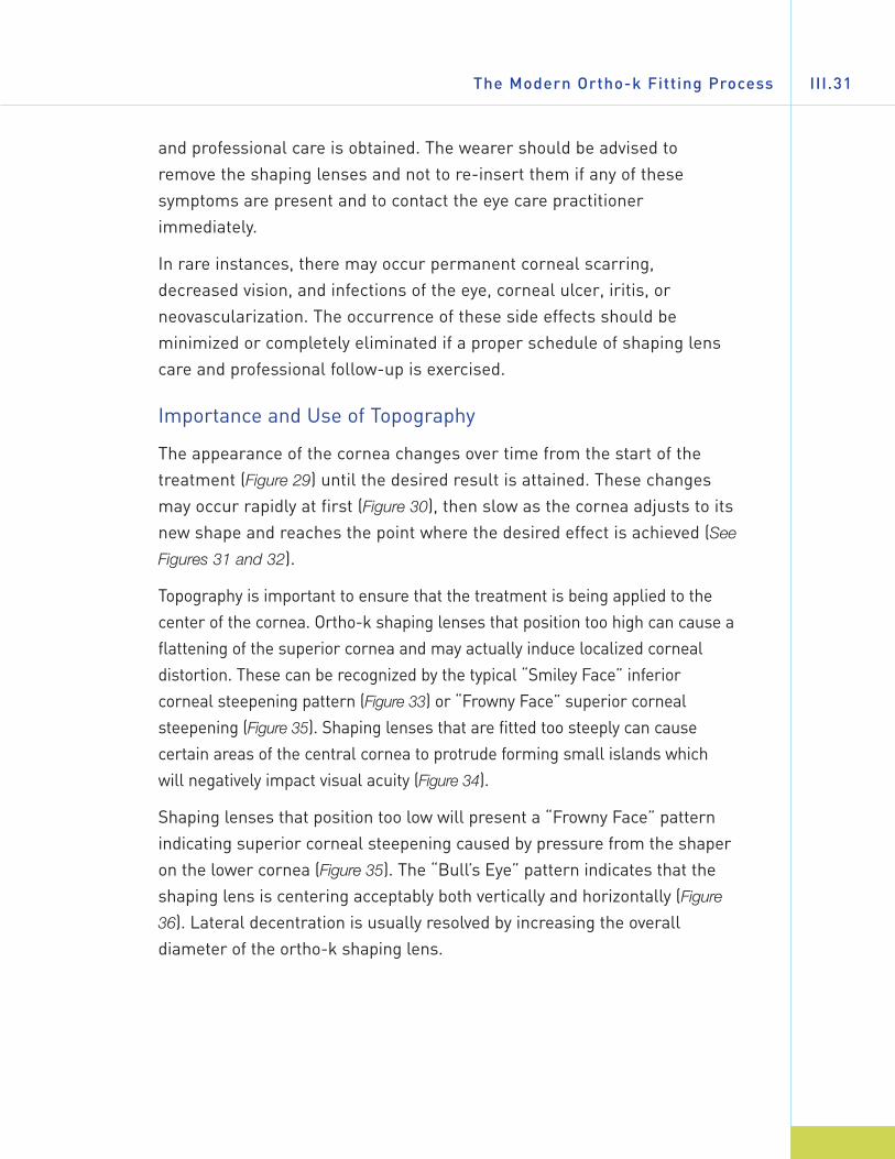
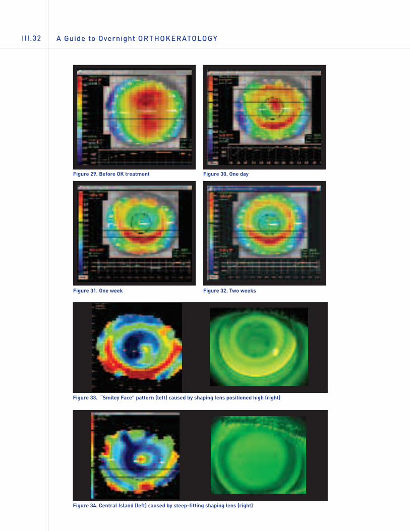
Importance and Use of Topography
The appearance of the cornea changes over time from the start of thetreatment (Figure 29) until the desired result is attained. These changesmay occur rapidly at first (Figure 30), then slow as the cornea adjusts to itsnew shape and reaches the point where the desired effect is achieved (See
Figures 31 and 32).
Topography is important to ensure that the treatment is being applied to thecenter of the cornea. Ortho-k shaping lenses that position too high can cause aflattening of the superior cornea and may actually induce localized cornealdistortion. These can be recognized by the typical “Smiley Face” inferiorcorneal steepening pattern (Figure 33) or “Frowny Face” superior cornealsteepening (Figure 35). Shaping lenses that are fitted too steeply can causecertain areas of the central cornea to protrude forming small islands whichwill negatively impact visual acuity (Figure 34).
Shaping lenses that position too low will present a “Frowny Face” patternindicating superior corneal steepening caused by pressure from the shaperon the lower cornea (Figure 35). The “Bull’s Eye” pattern indicates that theshaping lens is centering acceptably both vertically and horizontally (Figure
36). Lateral decentration is usually resolved by increasing the overalldiameter of the ortho-k shaping lens.
The Modern Ortho-k Fitting Process III.31
A Guide to Overnight ORTHOKERATOLOGYIII.32
Figure 29. Before OK treatment Figure 30. One day
Figure 31. One week Figure 32. Two weeks
Figure 33. “Smiley Face” pattern (left) caused by shaping lens positioned high (right)
Figure 34. Central Island (left) caused by steep-fitting shaping lens (right)

The Modern Ortho-k Fitting Process III.33
Figure 36. Bull’s Eye pattern indicating perfect centration of the shaping lens
Figure 35. “Frowny Face” pattern (left) caused by shaping lens that is positioning low (right)
KEY POINT SUMMARY…
• The combination of modern reverse geometry designs, high Dk GP materials,and corneal topographers make orthokeratology fitting fast, safe, and morepredictable.
• One of the most critical visits for both the patient and the practitioner is thefirst morning after the shaping lenses are worn at night.
• An advantage of orthokeratology is that it is reversible.
• Both objective and subjective patient information must be considered whenpresenting options and evaluating a potential wearer for ortho-k shapinglenses.
• Pre-fitting examination does not vary much from those routine exams done inthe office.
• It has been suggested that patients who may expect the best chance forsuccess with ortho-k treatment are those candidates who have steep corneaswith high eccentricity, although this is not always the case.
A Guide to Overnight ORTHOKERATOLOGYIV.34
IV. PRACTICE MANAGEMENT
IN THIS CHAPTER…
• This section offers examples of fee structures for ortho-k shapinglenses and services.
• Follow-up programs are discussed for continued wear of ortho-kshaping lenses.
• Care and handling of these special designs is reviewed.
• Important points on incorporating orthokeratology into the practice arepresented.
• Examples of pre-treatment permission forms are presented.
• Helping the patient to decide if ortho-k treatment is the right visioncorrection modality.
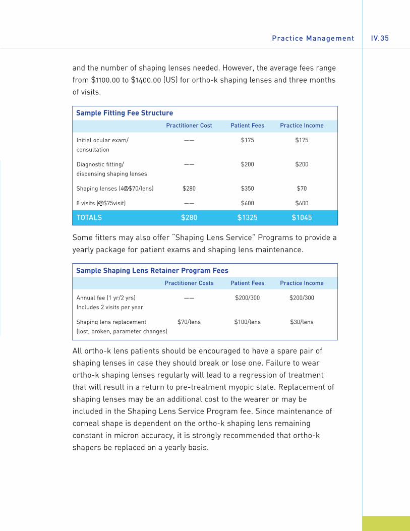
Fees and Costs
Fitting of ortho-k shaping lenses involves higher costs for the fitter, bothin terms of materials (shaping lenses) and increased time for fitting andfollow-up. Therefore, fees for ortho-k treatment are significantly higherthan for conventional GP lens fitting. That said, ortho-k fitting competesto some extent with refractive surgery and other vision correctionmodalities, such as conventional contact lenses and eyeglasses. As thenumber of laser surgical centers and LASIK procedures increases, sodoes the competition for the same adult myopic patients. Fees for ortho-kfitting vary from place to place and may be comprehensive (all-inclusive) orbased on the duration of the treatment. Generally, all-inclusive fees arebetween $750 and $2,500, depending on the length of treatment required
Practice Management IV.35
and the number of shaping lenses needed. However, the average fees rangefrom $1100.00 to $1400.00 (US) for ortho-k shaping lenses and three monthsof visits.
Sample Fitting Fee Structure
Practitioner Cost Patient Fees Practice Income
Initial ocular exam/ —— $175 $175
consultation
Diagnostic fitting/ —— $200 $200
dispensing shaping lenses
Shaping lenses (4@$70/lens) $280 $350 $70
8 visits (@$75visit) —— $600 $600
TOTALS $280 $1325 $1045
Some fitters may also offer “Shaping Lens Service” Programs to provide ayearly package for patient exams and shaping lens maintenance.
Sample Shaping Lens Retainer Program Fees
Practitioner Costs Patient Fees Practice Income
Annual fee (1 yr/2 yrs) —— $200/300 $200/300
Includes 2 visits per year
Shaping lens replacement $70/lens $100/lens $30/lens
(lost, broken, parameter changes)
All ortho-k lens patients should be encouraged to have a spare pair ofshaping lenses in case they should break or lose one. Failure to wearortho-k shaping lenses regularly will lead to a regression of treatmentthat will result in a return to pre-treatment myopic state. Replacement ofshaping lenses may be an additional cost to the wearer or may beincluded in the Shaping Lens Service Program fee. Since maintenance ofcorneal shape is dependent on the ortho-k shaping lens remainingconstant in micron accuracy, it is strongly recommended that ortho-kshapers be replaced on a yearly basis.
A Guide to Overnight ORTHOKERATOLOGYIV.36
How to Incorporate Ortho-k into the Practice
The introduction and incorporation of orthokeratology to the practicerequires a change in mindset for not only the practitioner, but also for theoffice staff. The following steps are recommended prior to beginning tooffer orthokeratology:
1. Fitter research into the various ortho-k shaping lens designoptions.
2. Upgrade or refresh GP fitting skills as needed.
3. Education and training for the fitter in using the design chosenby attending courses, seminars and home study.
4. Training for the office staff on discussing the ortho-k option withpatients, handling patient questions, and instructing ortho-kwearers on lens care, insertion, removal, etc. of the ortho-kshaping lenses.
5. Establishing office fees and policies for ortho-k treatment. Thisshould include development of ortho-k information sheets (FAQs),ortho-k shaping lens wear and care instructions, and Patient Fittingand Care Agreements for patients or parents of minors to read andsign (See pages 40 and 41).
Ortho-k Shaping Lens Care and Handling
In order to minimize the potential for wearing complications such as eyeirritations or serious infections, patients must be thoroughly trained in theproper way to wear and care for their ortho-k shaping lenses, and usinggood hygienic methods whenever they handle their ortho-k shapers.
Preparing the Lens for WearingCleanliness is a very important aspect of proper care of the shapinglenses. Hands should be clean and free of any foreign substanceswhenever the shaping lenses are handled.
• Always wash, rinse, and dry hands thoroughly before handlingthe shaping lenses.
• Avoid soaps containing cold cream, lotions, or oily cosmeticsprior to handling shapers. These substances can adhere to thesurface of the shaping lens and be difficult to remove.
• Handle shaping lenses with the fingertips, avoiding use offingernails that can scratch or chip them.
• Always start with the same shaping lens first to avoid mix-ups.
• Remove the shaping lens from its storage case and examine it.Be sure it is clean, moist, and free of any nicks or cracks.
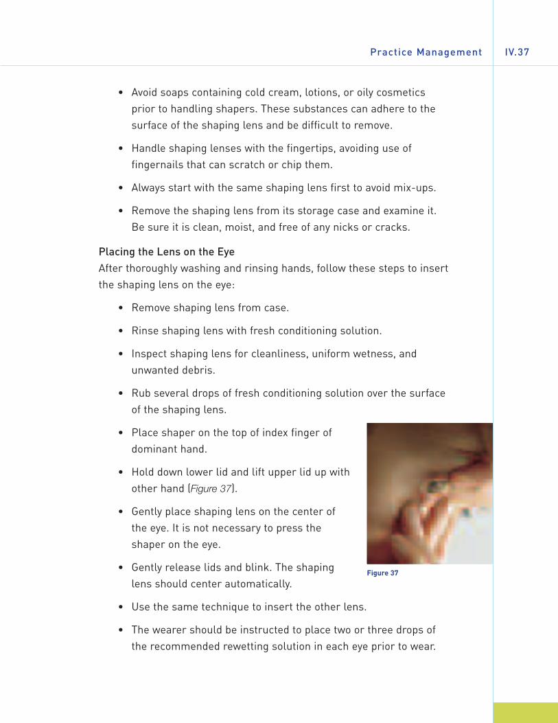
Placing the Lens on the EyeAfter thoroughly washing and rinsing hands, follow these steps to insertthe shaping lens on the eye:
• Remove shaping lens from case.
• Rinse shaping lens with fresh conditioning solution.
• Inspect shaping lens for cleanliness, uniform wetness, andunwanted debris.
• Rub several drops of fresh conditioning solution over the surfaceof the shaping lens.
• Place shaper on the top of index finger ofdominant hand.
• Hold down lower lid and lift upper lid up withother hand (Figure 37).
• Gently place shaping lens on the center ofthe eye. It is not necessary to press theshaper on the eye.
• Gently release lids and blink. The shapinglens should center automatically.
• Use the same technique to insert the other lens.
• The wearer should be instructed to place two or three drops ofthe recommended rewetting solution in each eye prior to wear.
Practice Management IV.37
Figure 37

A Guide to Overnight ORTHOKERATOLOGYIV.38
Removing the Shaping LensesBefore attempting to remove a shaping lens, it is very important that thewearer verify that it is moving. If the shaping lens is not moving, instill 5drops of the recommended rewetting solution. Oftentimes an adheredlens will begin to move spontaneously after instillation of drops and a fewminutes of blinking. Wait until the shaper begins to move freely with theblink before attempting to remove it.
From the office, the practitioner should instruct the patient as follows:While looking upwards, a finger is placed at the lower eyelid margin at theedge of the shaper to gently but firmly apply pressure. Looking downward,the process is repeated using the fingertip placed on the upper eyelid atthe shaper edge. The patient should then look straight ahead and blinkseveral times.
Once the shaping lens begins to move, it can beremoved using one of the following methods: Theshaping lens may be removed manually by using the“blink” or “scissor” method customary with standardGP lenses. Removing large diameter ortho-k shapinglenses may require use of a soft silicone rubberremoval device (Figure 38).
Cleaning and Storing the Shaping LensesThe shaping lenses should be rubbed gently for 20seconds on each side with the recommendedcleaner, followed by a thorough rinse in therecommended solution. Care must be taken not topress or squeeze the shapers excessively duringhandling. Ortho-k shaping lenses are susceptible todistortion and breakage.
The cleaned shaping lenses should be placed in the proper well of thecase and covered completely with the storage (conditioning) solution.Maintaining the proper orthokeratology effect depends on the patientwearing the prescribed shaping lens on the correct eye. Laboratoriesmanufacturing ortho-k lenses produce them in different colors betweenright and left to help the patient avoid a mixup.
Figure 38
Practice Management IV.39
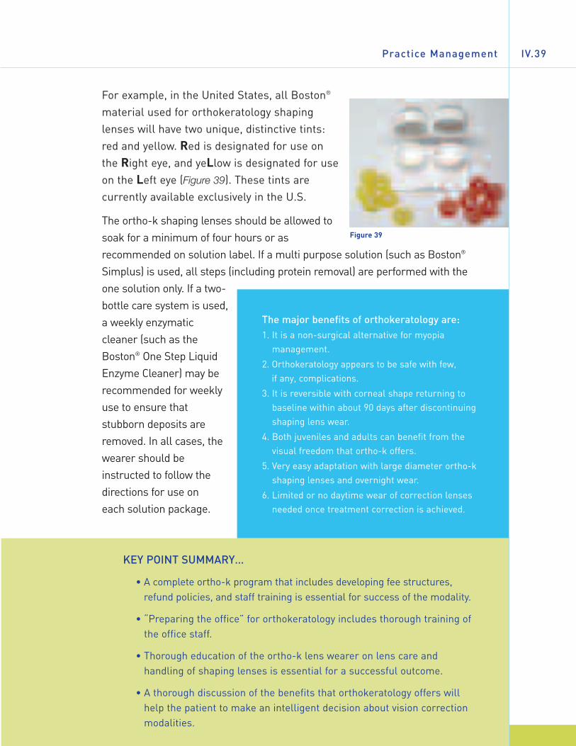
For example, in the United States, all Boston®
material used for orthokeratology shapinglenses will have two unique, distinctive tints:red and yellow. Red is designated for use onthe Right eye, and yeLlow is designated for useon the Left eye (Figure 39). These tints arecurrently available exclusively in the U.S.
The ortho-k shaping lenses should be allowed tosoak for a minimum of four hours or asrecommended on solution label. If a multi purpose solution (such as Boston®
Simplus) is used, all steps (including protein removal) are performed with theone solution only. If a two-bottle care system is used,a weekly enzymaticcleaner (such as theBoston® One Step LiquidEnzyme Cleaner) may berecommended for weeklyuse to ensure thatstubborn deposits areremoved. In all cases, thewearer should beinstructed to follow thedirections for use on each solution package.
Figure 39
The major benefits of orthokeratology are:1. It is a non-surgical alternative for myopia
management.
2. Orthokeratology appears to be safe with few, if any, complications.
3. It is reversible with corneal shape returning tobaseline within about 90 days after discontinuingshaping lens wear.
4. Both juveniles and adults can benefit from thevisual freedom that ortho-k offers.
5. Very easy adaptation with large diameter ortho-kshaping lenses and overnight wear.
6. Limited or no daytime wear of correction lensesneeded once treatment correction is achieved.
KEY POINT SUMMARY…
• A complete ortho-k program that includes developing fee structures,refund policies, and staff training is essential for success of the modality.
• “Preparing the office” for orthokeratology includes thorough training ofthe office staff.
• Thorough education of the ortho-k lens wearer on lens care andhandling of shaping lenses is essential for a successful outcome.
• A thorough discussion of the benefits that orthokeratology offers willhelp the patient to make an intelligent decision about vision correctionmodalities.
A Guide to Overnight ORTHOKERATOLOGYIV.40
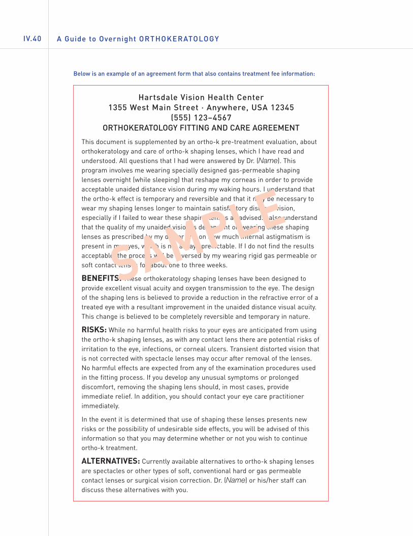
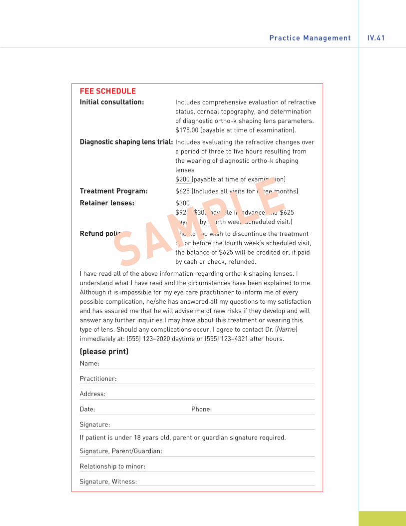
Below is an example of an agreement form that also contains treatment fee information:
Hartsdale Vision Health Center1355 West Main Street · Anywhere, USA 12345
(555) 123–4567ORTHOKERATOLOGY FITTING AND CARE AGREEMENT
This document is supplemented by an ortho-k pre-treatment evaluation, aboutorthokeratology and care of ortho-k shaping lenses, which I have read andunderstood. All questions that I had were answered by Dr. (Name). Thisprogram involves me wearing specially designed gas-permeable shapinglenses overnight (while sleeping) that reshape my corneas in order to provideacceptable unaided distance vision during my waking hours. I understand thatthe ortho-k effect is temporary and reversible and that it may be necessary towear my shaping lenses longer to maintain satisfactory distance vision,especially if I failed to wear these shaping lenses as advised. I also understandthat the quality of my unaided vision is dependent on wearing these shapinglenses as prescribed by my doctor and on how much internal astigmatism ispresent in my eyes, which is not always predictable. If I do not find the resultsacceptable, the process will be reversed by my wearing rigid gas permeable orsoft contact lenses for about one to three weeks.
BENEFITS: These orthokeratology shaping lenses have been designed toprovide excellent visual acuity and oxygen transmission to the eye. The designof the shaping lens is believed to provide a reduction in the refractive error of atreated eye with a resultant improvement in the unaided distance visual acuity.This change is believed to be completely reversible and temporary in nature.
RISKS: While no harmful health risks to your eyes are anticipated from usingthe ortho-k shaping lenses, as with any contact lens there are potential risks ofirritation to the eye, infections, or corneal ulcers. Transient distorted vision thatis not corrected with spectacle lenses may occur after removal of the lenses.No harmful effects are expected from any of the examination procedures usedin the fitting process. If you develop any unusual symptoms or prolongeddiscomfort, removing the shaping lens should, in most cases, provideimmediate relief. In addition, you should contact your eye care practitionerimmediately.
In the event it is determined that use of shaping these lenses presents newrisks or the possibility of undesirable side effects, you will be advised of thisinformation so that you may determine whether or not you wish to continueortho-k treatment.
ALTERNATIVES: Currently available alternatives to ortho-k shaping lensesare spectacles or other types of soft, conventional hard or gas permeablecontact lenses or surgical vision correction. Dr. (Name) or his/her staff candiscuss these alternatives with you.
SAMPLE
Practice Management IV.41
FEE SCHEDULEInitial consultation: Includes comprehensive evaluation of refractive
status, corneal topography, and determinationof diagnostic ortho-k shaping lens parameters. $175.00 (payable at time of examination).
Diagnostic shaping lens trial: Includes evaluating the refractive changes overa period of three to five hours resulting fromthe wearing of diagnostic ortho-k shapinglenses $200 (payable at time of examination)
Treatment Program: $625 (Includes all visits for three months)
Retainer lenses: $300 $925 ($300 payable in advance and $625payable by fourth week scheduled visit.)
Refund policy: Should you wish to discontinue the treatmenton or before the fourth week’s scheduled visit,the balance of $625 will be credited or, if paidby cash or check, refunded.
I have read all of the above information regarding ortho-k shaping lenses. Iunderstand what I have read and the circumstances have been explained to me.Although it is impossible for my eye care practitioner to inform me of everypossible complication, he/she has answered all my questions to my satisfactionand has assured me that he will advise me of new risks if they develop and willanswer any further inquiries I may have about this treatment or wearing thistype of lens. Should any complications occur, I agree to contact Dr. (Name)immediately at: (555) 123–2020 daytime or (555) 123–4321 after hours.
(please print)Name:
Practitioner:
Address:
Date: Phone:
Signature:
If patient is under 18 years old, parent or guardian signature required.
Signature, Parent/Guardian:
Relationship to minor:
Signature, Witness:
SAMPLE
A Guide to Overnight ORTHOKERATOLOGYV.42
V. GLOSSARY
Ablate/Ablation The process of using a laser light to vaporize corneal epithelial
cells in a specific area (treatment zone) during a LASIK or PRK
refractive surgical treatment.
Accelerated orthokeratology The use of specially designed 4, 5, and 6 zone lens designs that
employ base curves calculated to be flatter than the flat
corneal curvature, reverse zones of steeper radii, and
alignment zones that provide improved lens positioning and the
positive-negative pressures between lens and cornea
necessary to effect desired myopic reduction quickly and
systematically, while worn during sleep.
“Against-the-rule” astigmatism The flattest (longest) radius of corneal curvature lies vertically
at 90° and the steepest (shortest) curve of the cornea is
horizontal at 180°.
Alignment Curve (AC) Based on specific design, this curve may also be called Anchor
Curve, Landing Zone, or Fitting Curve. Area of posterior lens
surface that aligns lens with mid-periphery of cornea to control
lens position and centration during lens wear.
Anterior optical zone (AOZ) Front, central lens curve that (combined with the base curve
and refractive index) determines lens power.
Apical/Apex The extreme top or tip of a curve (e.g. corneal apex: tip of the
cornea).
Apical bearing Posterior lens curvature lightly touches the corneal apex.
Apical touch Posterior lens curvature rests on corneal apex.
Axial edge lift Vertical distance from the lens edge to an extension of the base
curve of a lens.
Aspheric Not spherical. A posterior or anterior lens surface design
which progressively flattens at a given rate (eccentricity) as the
curve progresses toward the periphery.
Astigmatism Refractive defect where the refractive components of the eye
have differing powers in different meridians. The result is that
light rays will focus at more than one point inside the eye.
Glossary V.43
Back Optic Zone diameter Chord length measurement across back optic zone.
(BOZD)/Optic Zone diameter In ortho-k, determines size of central corneal
(OZD) treatment area.
Back Optic Zone radius The major central posterior curve of the lens that fits over
(BOZR)/Base curve (BC)/ the apex of the cornea.
Posterior optical zone (POZ)
Biomicroscopy/slit lamp exam Use of high power microscope and light source to examine the
anterior segments of the eye using various amounts of
magnification.
Bowman’s Membrane Second layer of the cornea located between the epithelium and
the stroma, approximately 10 microns in thickness.
“Bull’s Eye” topography Corneal topography map that shows acceptable centration and
central corneal flattening with an ortho-k shaping lens fit.
“Central Island” topography Topography maps that show areas of steepness in the central
cornea caused by an ortho-k shaping lens that is fitted too
steeply, causing a squeezing of the cornea and central
steepening.
Contrast sensitivity test Contrast sensitivity tests plot the lowest contrast level at which
a person can detect an object of a given size to detect
variations in the quality of the image on the retina.
Cornea Transparent, small dome-shaped anterior portion of the eye
through which light rays pass into the eye.
Corneal astigmatism Condition where the toroidal (oval) shape of the cornea is the
reason that light rays are caused to focus at various points
within the eye.
Corneal staining Healthy corneal epithelial cells normally will not take up
fluorescein dye. Damaged or dead cells will allow fluorescein
dye to enter. The extent and location of this staining is an
indication of corneal irritation (mechanical or chemical) and/or
insufficient oxygen (hypoxia).
Cylindrical refractive Requiring a lens whose correcting power is located in one
error/power specific meridian.
Descemet’s Membrane Thin elastic fourth layer of the cornea that adds to the flexibility
of the cornea, approximately 10 microns in thickness.
Dk Refers to the inherent permeability of a lens material to allow
the passage of gases through it. D = diffusion coefficient, k =
degree of material solubility.
Dk/L or Dk/t Refers to the amount of oxygen (gases) that pass through a
lens material of a specified thickness (“D” and “k” references
are same as above. “L” or “t” refers to specific lens average
thickness).
A Guide to Overnight ORTHOKERATOLOGYV.44
Dimple veiling Fluorescein staining of indentations made in the corneal
epithelium by trapped air bubbles under the lens.
Diopter Unit of measurement of light ray convergence or divergence
equal to the reciprocal focal length of a lens in meters (e.g. a
2.00 diopter lens brings light rays into convergence at 0.5
meter).
Eccentricity The rate of flattening of an aspheric curve measured as an “e”
value.
Edema (corneal) Condition where the corneal water content increases causing a
loss of clarity.
Edge lift (clearance) Distance between lens edge and the corneal surface. Created
by the width and radii of the peripheral curves. Allows for tear
circulation under the lens and aids in lens removal.
Endothelium Fifth (innermost) layer of the cornea, responsible for corneal
metabolism and maintaining the water content of the cornea.
Approximately 5 microns in thickness.
Epithelial flap Use of a microkeratome instrument to incise and lift an
epithelial layer of the central cornea prior to applying laser
ablation.
Epithelium The outermost layer of the cornea comprised of approximately
five layers of cells that is approximately 50 microns in
thickness.
Fitting curve (FC) Depending on specific ortho-k design, this may also be called
Alignment curve or Reverse curve.
Flat meridian/Flat “K” Meridian of the cornea having the longer radius of curvature.
Fluorescein pattern The appearance of the tear film distribution and thickness
between the posterior of a rigid lens and the anterior corneal
curvature, as viewed with fluorescent dye that stains the tear
film.
Fluoro-silicone acrylate (F–S/A) An oxygen permeable rigid contact lens material developed in
the 1980s that combines fluorine for lens stability, wettability,
and added oxygen transmission; with silicone for oxygen
permeability; and methyl methacrylate for stability,
machinability, durability, and optical quality.
“Frowny Face” topography Topography map that shows a steepened area of the superior
cornea caused by an ortho-k lens that centers too low on the
cornea causing inferior flattening.
GP/RGP Gas permeable (rigid) contact lens.
High Dk Having permeability ratings 31 to 60 (ISO/Fatt).
Hyper Dk Having permeability ratings greater than 100 (ISO/Fatt).
Glossary V.45
Hyperopia Farsightedness; condition where the eye is underpowered
causing light rays to focus at a point behind the retina.
Correction is supplied using “plus” or “positive” powered
optical lenses.
Hypoxic stress Refers to corneal oxygen uptake rates on a given eye with a
lens of a given permeability or the lack thereof.
ISO/Fatt International Standard Organization (ISO) method for
measuring and stating oxygen permeability and other values
for all contact lens materials.
Keratoconus Degenerative corneal disease characterized by thinning and
cone-shaped protrusion of the central cornea.
Keratometry (K-readings) Use of a keratometer to measure central apical cornea
curvature zone of 3 to 4 millimeters in diameter.
LASEK Laser Assisted Subepithelial Keratectomy is a refractive
surgery procedure where a thin flap of epithelium only is
created using an alcohol solution to soften the epithelium and
a thin surgical blade to lift it. This process is said to be
beneficial for corneas too thin to have LASIK or PRK. The
underlying cornea is then treated with a laser to reshape the
central cornea. The flap is then replaced to heal.
LASIK Laser Assisted In-Situ Keratomileusis; same as LASEK except
a thicker hinged flap consisting of the epithelium and some
stroma is created using a microkeratome instrument. The
central cornea is then treated by laser as in LASEK and PRK
and the flap replaced to heal.
Low permeability GP materials having Dk ratings of less than 15 (ISO/Fatt).
Macular degeneration Degeneration of the macular portion of the retina which leads
to permanent loss of central vision.
Medium Dk Having Dk ratings between 15 and 30 (ISO/Fatt).
Micron One-thousandth of a millimeter. One average human cell is 10
microns in height. One human hair is approximately 50 microns
thick.
Myopia (nearsightedness) Nearsightedness; condition where the eye is over powered
causing light rays to focus at a point in front of the retina.
Correction is supplied using “minus” or “negative” powered
optical lenses.
Myopic progression Process by which myopia in an individual gradually increases
over a period of time, requiring the need for increased myopic
prescription changes.
Nomogram Calculation system (charts, tables, computer programs) used
to calculate appropriate diagnostic or final lens design.
Oblate shape A curve whose central curve is nearly flat (similar to the long
curvature of an egg).
Ophthalmoscopy Direct or indirect examination of the internal structures of the
eye, especially the optic nerve, macula, and retina.
Optical zone (POZ)/Back Central portion of the posterior lens, not including the
Optical Zone radius (BOZR)/ peripheral curves, that the wearer sees through.
Base curve (BC)
Orthokeratology, ortho-k Use of gas permeable (rigid) contact lenses to gradually and
systematically alter the shape of the cornea to cause a
temporary and reversible reduction in myopia.
Ortho-k shaping lens, lens A specially designed gas permeable (rigid) contact lens
shaper, ortho-k shaper, used in ortho-k treatment to change the shape of the
retainer, etc. cornea to reduce sagittal height, refocus light rays onto the
retina, and temporarily reduce the myopic requirement of the
eye.
Overall diameter (OAD) Chord length of a lens across the widest point of lens.
or Total Lens Diameter
Over-refraction Refraction performed over an existing correction (contact lens
or ortho-k treatment) to gauge extent of under- or over-
correction.
Pachometry Measurement of corneal thickness.
Peripheral curve(s) (PC) Curve(s) of flatter radius added to the posterior surface of the
rigid lens edge to approximate the flattening of the peripheral
cornea. Essential for lens comfort, tear circulation, debris
removal, and to facilitate lens removal.
Permeability See “Dk.”
Phoropter Refraction device containing a large variety of spherical plus
and minus powered lenses, cylinder powers, prisms, etc. used
to determine the eye’s best optical correction.
Photorefractive keratectomy Use of eximer laser light to “sculpt” a prescribed area
(PRK) of the central cornea, creating a flatter zone and reducing
myopia.
Polymethylmethacrylate Plastic very similar to Plexiglas™ used in contact
(PMMA) lens manufacture from the 1930s as the original contact lens
material. Totally impermeable to gases.
Prolate shape A curve having a steeper (higher) center than its periphery
(similar to the curve at the tip of an egg).
A Guide to Overnight ORTHOKERATOLOGYV.46
Glossary V.47
Punctate staining Appearance of the cornea when stained with fluorescein dye
where scattered to diffuse areas of “pinpoint” staining occur.
This usually indicates corneal epithelial cell damage or
destruction.
Radial Keratotomy (RK) Radial incision made in the periphery of the cornea that
causes central corneal flattening with resultant reduction in
myopia.
Refraction Subjective test used to determine the refractive error of the
eye in order to prescribe the best correcting lens powers.
Refractive error Internal optical defect where light rays are not focused on the
retina of the eye causing blurred vision.
Refractive surgery Use of incisions, implants, or laser light to alter the shape of
the corneal surface to affect vision.
Retainer lens Ortho-k shaping lens worn nightly as prescribed to maintain
(See shaping lens) the shape of the cornea and myopic reduction. Similar to a
retainer worn for teeth.
Reverse Curve Also known as Reservoir Curve, Fitting Curve, depending on
specific ortho-k design. Secondary curve with steeper radius of
curvature than preceding (Back Optic Zone Radius/BOZR)
curve. Area of shaping lens where corneal mass will
accumulate during ortho-k treatment.
Reverse geometry design Designs that use secondary curves that are steeper than the
preceding curve.
Sagittal depth The distance between the crest of an arc and a straight
baseline. Area under the arc of a curve depends on the
steepness or flatness of the curve radius.
Schirmer tear test Screening test that uses sterile filter paper strips to determine
tear production volume for a given time period.
Seal-off A contact lens whose edges have sealed against the cornea
360° preventing tear exchange and debris removal.
Shaping lens See “Ortho-k shaping lens.”
Silicone acrylate (S/A ) Oxygen permeable plastic developed in the 1970s for contact
lenses (GP) that combines silicone for oxygen transmissibility
and methacrylate for optical quality, lens machinability, and
stability.
“Smiley Face” topography Corneal topography map that shows an area of inferior
steepening caused by an ortho-k shaping lens that rides too
high, causing superior corneal flattening.
Spherical equivalent Average power of a sphero-cylindrical lens equal to the sum of
the spherical power plus half of the cylindrical power.
A Guide to Overnight ORTHOKERATOLOGYV.48
Spherical refractive error/power Requiring a lens whose correcting power is equal in all
meridians.
Steep meridian/Steep “K” Meridian of the cornea having the shorter radius of curvature.
Stroma Middle (third) layer of the cornea composed of layered collagen
fibrils and cells, approximately 460 microns thick. Comprises
90% of corneal thickness.
Super-Dk Having permeability ratings between 61 and 100 (ISO/Fatt).
Tear Breakup Time (TBUT) Time interval between a blink and the appearance of a dry spot
in the tear film. Indication of the quality of the tear film.
Tear pump mechanism Dynamic mechanism created by lens movement with each
blink which causes an exchange of tears under the lens to
remove de-oxygenated tears and cellular debris.
Topography Corneal modeling instrument that produces color-coded maps
of the corneal surface using a variety of views.
Visual acuity Measure of the eye’s ability to distinguish objects, details, and
shape assessed by the smallest identifiable object that can be
seen at a specific distance (usually 20 feet for distance and 16
inches for near).
“With-the-rule” astigmatism The flattest (longest) radius of corneal curvature lies
horizontally at 180° and the steepest (shortest) curve of the
cornea is vertical at 90°.
Wratten (yellow) filter A photographic yellow filter of a specific density that allows the
visualization of greater detail gradations in the fluorescein
stained-tear film.
Frequently Asked Questions VI.49
VI. FREQUENTLY ASKED QUESTIONS (FAQS)
IN THIS CHAPTER…
• The most often asked questions about orthokeratology by patients topractitioners.
• Questions that are frequently asked about orthokeratology from fitters to the laboratory.
The FAQ (Frequently Asked Questions) section may be applicable to fitter,manufacturer, or consumer, depending on their knowledge oforthokeratology.
General
Q: How do ortho-k lenses work?
A: Ortho-k shaping lenses are designed to progressively reshape the central surface ofthe cornea systematically, similar to the effect of lasers in reversing nearsightedness.However, unlike laser surgery, the effect of ortho-k treatment is temporary andreversible.
Q: Is everyone a candidate for ortho-k?
A: No! While orthokeratology can help most individuals (including those with certaintypes and amounts of astigmatism and moderately high myopia) improve their vision, theprocedure is often most effective for those prescriptions falling within a specific range.Additional factors may include individual corneal rigidity and shape factors as well as anability to wear contact lenses. A thorough consultation and examination using advancedcomputerized diagnostic instrumentation can easily determine if orthokeratology is rightfor an individual.
VI.50 A Guide to Overnight ORTHOKERATOLOGY
Q: Who is not a candidate for ortho-k?
A: While ortho-k can be performed on practically anyone with healthy eyes who caninsert a contact lens and follow lens care directions, it is not recommended for peoplewith prescriptions above 5.00 diopters of myopia or above 1.50 diopters of astigmatism.Also, people with pupils that are larger than normal (> 6 mm in normal light) and thosehaving irregular corneal astigmatism or any corneal disorder such as keratoconus arenot candidates.
Q: How safe is ortho-k?
A: Ortho-k is believed to be safe when appropriately fitted and managed properly. Manypeople have been able to eliminate their dependence upon their glasses and standardcontacts with no adverse effects. Unlike surgical procedures like radial keratotomy (RK),photorefractive keratectomy (PRK), laser assisted in-situ keratomileusis (LASIK), andlaser assisted subepithelial keratectomy (LASEK), the corneal tissue is not incised orvaporized by a laser. As with all contact lenses, proper lens care and handling must beperformed to maintain eye health. The state-of-the-art, high-permeability GP materialsnow available provide adequate amounts of oxygen to the tissues of the eye.
Q: Is ortho-k permanent?
A: After treatment, maximum results are achieved and retainer shaping lenses are wornto stabilize and maintain the new corneal shape. Failure to wear the shaping lens on anongoing basis will result in a return to the pre-existing prescription. Retainer shapinglenses will likely be prescribed for overnight wear.
Q: What are the risks of wearing lenses overnight?
A: The complications of wearing contact lenses include corneal ingrowth of vessels,ulcers, and abrasions.The risks associated with wearing contact lenses overnight arehigher than wearing contact lenses only while awake. Contact lenses cannot become lostbehind the eye (this is anatomically impossible) and it is rare for ortho-k shaping lensesto become decentered from the cornea.
Q: How long does it take for the process to stabilize and provide functional vision for thewhole day?
A: It generally requires four to seven consecutive nights of wearing the shaping lensesto achieve the desired result. It may take somewhat longer for those with higher degreesof nearsightedness.

Frequently Asked Questions VI.51
Q: Are ortho-k lenses uncomfortable to wear?
A: Overnight wearing of the shaping lenses is surprisingly comfortable. Most patientsare unaware of their presence within a very short time after insertion. And because theortho-k shapers are made in large diameters and worn during sleep, the normaladaptation process is very short.
Q: Will I still have to wear glasses or contacts?
A: Once the desired myopic reduction is obtained, the final shaping lenses act asretainers to maintain that level. Regular contact lenses are not needed. Glasses may beneeded for reading or other part-time use. During the initial treatment period, if unaidedvision does not last a full day, the patient will be given soft disposable contact lenses towear to maintain normal distance vision.
Q: Once the treatment phase is completed, how frequently will I need to wear theovernight retainer lenses?
A: Most people will need to wear the shaping lenses six to seven consecutive nights inorder to enjoy good, unaided vision during the entire day. Patients with lesser degrees ofmyopia (nearsightedness) may find that wearing them every other night is satisfactory.However, this will be determined on an individual basis by the eye care practitioner.
Q: How much myopia can a person realistically expect to be able to reduce usingorthokeratology?
A: Generally, 5.00 diopters of myopia is the upper limit for myopia reduction. However,work is underway on designs that will hopefully correct higher amounts of myopia.Lower amounts of myopia are easier and faster to reduce.
Q: How about ortho-k for astigmatism?
A: The amount of astigmatism reduction achievable will depend on the amount and thetype. 1.50 diopters of “with-the-rule” corneal astigmatism and 0.75 diopter of “against-the-rule” corneal astigmatism are considered the upper limits for astigmatismreduction. Ortho-k will not have an effect on residual (internal) astigmatism.
Q: How long does it take to be able to see well without glasses?
A: Rapid visual improvement normally occurs in the first few days. Stabilization thenfollows over the next few weeks and months. Once the wearer’s eyes are stabilized,improved eyesight is maintained by wearing shaping lenses as recommended to maintainvision at the desired level.
A Guide to Overnight ORTHOKERATOLOGYVI.52
Q: If the patient decides to return to wearing glasses, can vision be restored to its pre-treatment level?
A: The wearer’s eyes will return to their pre-treatment nearsightedness after about twoweeks. Depending on how long ortho-k lenses have been worn, this process may be aslong as 30–90 days. In order to provide good vision during this transitional period,patients are refitted with gas permeable lenses or disposable soft lenses while thecorneas recover their pre-treatment shape.
Q: If the patient becomes less nearsighted or presbyopic in later years how will that behandled?
A: Unlike laser surgery that cannot be reversed, corneas can usually be remodeled toaccommodate the change in prescription by changing the design of the maintenancelenses to correct for near vision using a technique called “monovision.”
Q: How much does ortho-k cost?
A: The actual cost will depend on the complexity of the case. Each doctor sets feesaccordingly. Fees can range from $750 to $2,000 depending on length of treatment andthe number of shaping lenses needed. The doctor will be able to provide an estimate foreach patient.
For the Fitter/Lab:
Q: What is the current prediction for the number of orthokeratology fits that will be done worldwide?
A: The use of orthokeratology will vary from market to market. Current estimates in theUS market suggest that there could be as many as 50,000 to 100,000 ortho-k fitsannually. Much of this will depend on the acceptance of the professional community,potential candidates, and promotion from GP lens manufacturers and the media.
Q: Is it necessary to use topography to fit today’s ortho-k lenses or can I usekeratometry as I always have to fit GP lenses?
A: Use of topography is highly recommended in all phases of the fitting and follow-up. Some fitting systems base initial shaping lens selection on topography data,while others recommend use of manual keratometry. Virtually all ortho-k fitting systemsrecommend the use of topographical data to establish a baseline reference as part of thefit evaluation, to identify and help solve fitting problems and to monitor the progress ofmyopic reduction.
Frequently Asked Questions VI.53
Q: What are the benefits of using four and five zone designs over the earlier three zonedesigns?
A: These modern four and five zone shaping lens designs use wider alignment (mid-peripheral) zones to achieve easier, more stable lens centration. Corneal shape changescan be made in larger increments; myopia reduction occurs more rapidly, requiring onlyone pair of shaping lenses in a high percentage of cases. This makes material costs toboth fitter and patient lower.
Q: If ortho-k shaping lenses need modification or blending, can I do this in my office tosave time?
A: While in-office lens modification has long been a skill valued by lens fitters, modernortho-k shaping lens designs are complex and precise. Any hand modification made bythe fitter to improve the lens fit will probably be impossible to duplicate if a lens is lost,broken, or requires other parameter changes.
Q: How important is dispensing and the next-day visit in evaluating the overnight ortho-kshaping lens fit?
A: Each patient will respond differently to orthokeratology treatment. These shapinglenses generally tighten with wear, which may occur in as little as an hour. It isimportant to evaluate the shaping lens after allowing it to settle-down in the office.Centration is the key to a successful ortho-k treatment; therefore, shaping lenses mustbe checked early the first morning for centration, binding, and adhesion. Shaping lensesare then removed to evaluate topography and to check unaided visual acuity.
Q: If an ortho-k shaping lens is lost, broken, or not able to be worn, how can I providesuitable vision to the patient until the replacement lens arrives?
A: Soft disposable lenses can be prescribed until the proper replacement shaping lensis received. If the cornea begins to revert back to higher myopia, stronger disposablelenses can be given. The wearer should be checked once again the day after a newshaping lens is worn overnight.
KEY POINT SUMMARY…
• The success of orthokeratology in the practice and in the laboratory isdependent on being able to address and adequately answer anyquestions that arise before, during, or after orthokeratology treatment.
A Guide to Overnight ORTHOKERATOLOGYVII.54
VI I . SUGGESTED READING
• Orthokeratology Handbook, Winkler_TD, Kame_RT, ButterworthHeinemann, January 1995
• Orthokeratology: Principles and Practice, Mountford_J, Ruston_D,Dave_T, Butterworth Heinemann, March 2004, ISBN 0750640073
• Myopia and the Control of Eye Growth: Symposium No. 155, Back_G,John Wiley & Son, Ltd. February 1991, ISBN 0471926922
• Clinical Management of Myopia, Grosvenor_TP, Goss_DA,Hofstetter_HW, Butterworth Heinemann, October 1998, ISBN0750670606
• Controlled Kerato-reformation (CKR): An Alternative to RefractiveSurgery, El Hage_S, Practical Optometry, Canadian Edition, December1999, 10:6
VII I . USEFUL ORTHO-K WEB SITES
• British Orthokeratology Societywww.boks.org.uk
• All About Visionwww.allaboutvision.com
• The Ortho-k Networkwww.ortho-k.net
• National Eye Research Foundation (NERF) www.nerf.org

Bibliography IX.55
IX. BIBLIOGRAPHY
1. Alharbi_A, Swarbrick_HA, “The Effects of Overnight Orthokeratology Lens Wear on Corneal
Thickness,” Investigative Ophthalmology and Visual Science, 44:6, June 2003
2. Binder_P, May_C, Grant_S, “An Evaluation of Orthokeratology,” American Journal of
Ophthalmology, 87:729, 1980
3. Brand_RJ, Polse_KA, Schwalbe_JS, “The Berkeley Orthokeratology Study, Part I: General
Conduct of the Study,” American Journal of Optometry and Physiologic Optics, 60:175, 1983
4. Carkeet_NL, Mountford_JA, Carney_LG,“Predicting Success with Orthokeratology Lens Wear:
A Retrospective Analysis of Ocular Characteristics,” Optometry and Vision Science, Vol. 72,
No. 12, 892–898
5. Coons_LJ, “Orthokeratology Part II: Evaluating the Tabb Method,” Journal of the American
Optometric Association, 55:409, 1984
6. Dave_T, Ruston_D, “Current Trends in Modern Orthokeratology,” Ophthalmic and Physiology
Optics, Vol. 18, No. 2, 1998
7. El Hage_S, “Photokeratoscopy and Controlled Keratoreformation,” paper presented at the
International Symposium of Ophthalmological Optics, Tokyo, Japan, May 1978
8. Grant_SC, “Orthokeratology: A Safe and Effective Treatment for a Disabling Problem,” Survey
of Ophthalmology, Vol. 24, No. 5, March–April 1980, 291–297
9. Harris_DH, Stoyan_N, “A New Approach to Orthokeratology,” Contact Lens Spectrum, April
1992, 37–39
10. Holden_BA, Mertz_GW, McNally_JJ, “Corneal Swelling Response to Contact Lens Worn Under
Extended Wear Conditions,” Investigative Ophthalmology and Vision Science, 1983,
24:218–226
11. Holden_BA, Mertz_GW, “Critical Oxygen Levels to Avoid Corneal Edema for Daily and
Extended Wear Contact Lenses,” Investigative Ophthalmology and Vision Science, 1984,
25:1161–1167
12. Horner_DG, Bryant_MK, “Take Another Look at Today’s Ortho-k,” Review of Optometry, June
1994
13. Jessen_G, “Orthofocus Techniques,” Contacto, 6:200, 1962
14. Kame_RT, “Flatten the Cornea the Nonsurgical Way,” Review of Optometry, April 1995
A Guide to Overnight ORTHOKERATOLOGYIX.56
15. Kerns_R. “Research in Orthokeratology II: Experimental Design Protocol Method,” JAOA,
47:1275, 1976
16. Lin_LK, Shih_YF, Lee_YC, Hung_PT, Hou_PK, “Changes in Ocular Refraction and Its
Components Among Medical Students: A 5-Year Longitudinal Study,” Optometry and Vision
Science, 73, 1996, 495–498
17. Lu_Fan, Jiang_Jun, Qu_Jia, Jin_Wangqing, Mao_Xinjie, She_Yi, “Clinical Study of the
Orthokeratology for Young Myopic Adolescents,” presented at the Myopia 2001 Conference,
Boston MA USA, July 2001
18. Lund_ME, Reim_TR, Wu_RLT, “Myopia Progression in Orthokeratology,” paper presented at VII
International Conference on Myopia, Taipei, Taiwan, 1999
19. Marsden_HJ, “Common Sense Orthokeratology,” Contact Lens Spectrum, Oct. 1995, 24–31
20. Morrison_RJ, “Contact Lenses and the Progression of Myopia,” Optometry Weekly, 1956, 47,
1487–1488
21. Mountford_J, “An Analysis of the Changes in Corneal Shape and Refractive Error Induced by
Accelerated Orthokeratology,” International Contact Lens Clinics, 1997, 24: 128–143
22. Mountford_J, Orthokeratology in Contact Lenses, Butterworth–Heinemann, Oxford UK, 1997,
653–692
23. Nichols_JJ, Marsich_MM, Nguyen_M, Barr_JT, Bullimore_MA, “Overnight Orthokeratology,”
Optometry and Vision Science, Vol. 77, No. 5, May 2000
24. Perrigin_J, Perrigin_D, Quintero_S, Grosvenor_T, “Silicone Acrylate Contact Lenses for
Myopia Control: Three-Year Results,” Optometry and Vision Science, 1990, 67:764–769
25. Polse_KA, Brand_RJ, Vastine_DW, Schwalbe_JS, “Corneal Change Accompanying
Orthokeratology: Plastic or Elastic?,” Archives of Ophthalmology, Vol. 101, 1983
26. Polse_KA, Brand_RJ, Schwalbe_JS, Vastine_DW, Keener_RJ, “The Berkeley Orthokeratology
Study, Part II: Efficacy and Duration,” American Journal of Optometry and Physiological
Optics, Vol. 60, No. 3, 1983, 187–198
27. Polse_KA, Brand_RJ, Keener_RJ, Schwalbe_JS, Vastine_DW, “The Berkeley Orthokeratology
Study, Part III: Safety,” American Journal of Optometry and Physiological Optics, Vol. 60, No.
4, 1983, 321–326
28. Ramkuhar_S, Swarbrick_H, “Corneal Response to Short-Term Orthokeratology Lens Wear,”
Optometry and Vision Science, 80:3, March 2003
29. Reim_TR, “Overflowing With Ortho-k,” Optometric Management, December 1998, 36–39
30. Schachet_JL, “Optimal Lens Choices for Orthokeratology,” unknown publication
31. Stone_J, “Possible Influence of Contact Lenses on Myopia,” British Journal of Optometry,
1976, 31, 89–114
32. Swarbrick_HA, Wong_G, O’Leary_DJ, “Corneal Response to Orthokeratology,” Optometry and
Vision Science, 75:11, November 1998