a computational study on dna bases interactions with dinuclear
TRANSCRIPT

This article was published in an Elsevier journal. The attached copyis furnished to the author for non-commercial research and
education use, including for instruction at the author’s institution,sharing with colleagues and providing to institution administration.
Other uses, including reproduction and distribution, or selling orlicensing copies, or posting to personal, institutional or third party
websites are prohibited.
In most cases authors are permitted to post their version of thearticle (e.g. in Word or Tex form) to their personal website orinstitutional repository. Authors requiring further information
regarding Elsevier’s archiving and manuscript policies areencouraged to visit:
http://www.elsevier.com/copyright

Author's personal copy
A computational study on DNA bases interactions with dinucleartetraacetato-diaqua-dirhodium(II,II) complex
Jaroslav V. Burda a,*, Jiande Gu b
a Department of Chemical Physics and Optics, Faculty of Mathematics and Physics, Charles University, Ke Karlovu 3, 121 16 Prague 2, Czech Republicb Center for Drug Discovery and Design and State Key Laboratory of Drug Research, Shanghai Institute of Materia Medica,
Shanghai Institutes for Biological Sciences, Chinese Academy of Sciences, 294 Taiyuan Road, Shanghai 200031, PR China
Received 3 April 2007; received in revised form 22 June 2007; accepted 26 June 2007Available online 7 July 2007
Abstract
In our study, we have determined the thermodynamic behavior for the replacement reaction of one and two acetyl-ligands from thediaqua-tetrakis(l-acetylato)dirhodium(II,II) complex by purine DNA bases. The complexes were optimized at the density functional the-ory (DFT) level with the B3LYP functional. Stuttgart–Dresden pseudopotentials were used for the description of the Rh atoms. Most ofthe replacement reactions are mildly exothermic, DG is up to 12 kcal/mol for the first acetyl-ligand and up to 8 kcal/mol for the secondligand replacement. For all explored complexes, stabilization and bonding energies were computed together with selected electronic prop-erties. Adenine base coordinates to the dirhodium complex slightly more firmly than guanine. In head-to-tail conformation the two gua-nines are better stabilized (by about 8 kcal/mol) than in head-to-head arrangement due to minimization of sterical repulsion of bothbases. We have shown that the bonding energy of axial water ligands is very small (up to 13 kcal/mol), resembling more H-bonds thandative coordination. Despite the larger stabilization energies of adenine-containing complexes, the thermodynamic parameters of thestudied replacement reactions are more favorable in case of guanine complexes. Higher exothermicity is connected with easier deprot-onization of guanine N1-site in comparison with N6-site of adenine in accord with experimental data.� 2007 Elsevier Inc. All rights reserved.
Keywords: Quantum chemical calculations; Dirhodium complexes; Reaction thermodynamics; DNA interactions
1. Introduction
Since the early 60s when anticancer activity of cisplatinbecame known [1], many other transition metal complexeswere probed as antitumor drugs, too. Especially due to thevery high toxicity of cisplatin it is worth to study other sub-stances in parallel to the development of cisplatin ana-logues. It is known that metallocene complexes of the Ti,Mo or V atoms can be used for this purpose, see, e.g.,the review of Kuo et al. [2] where basic properties andactivities are summarized. Some other recent works onthe topic of metallocenes can be found in studies [3–7].
Titanocene dichloride has recently successfully passed thefirst phase of clinical tests. Also, the activity of Ru(III)complexes has been thoroughly studied [8–11]. The firstphase of clinical trials has been recently passed by trans-Cl4(Me2SO)(Im)Ru(III) (Im = imidazole) (cf. Formula 1).
Also the Ru(II) compounds – the so-called piano-stoolcomplexes – have been recognized as very potent antican-cer agents [12–15]. Dirhodium(II) complexes have been dis-covered as possible candidates for anticancer treatment,too. Their antitumor properties were noticed already inthe 90s, as can be noticed in several studies [16–19]. Anexcellent review on rhodium anticancer activity was writtenby Katsaros and Anagnostopoulou [20], where not onlyRh(II) but also other rhodium complexes were addressed.In this work, considerations on the length of carboxylligands are presented as well as possible interactions with
0162-0134/$ - see front matter � 2007 Elsevier Inc. All rights reserved.
doi:10.1016/j.jinorgbio.2007.06.041
* Corresponding author. Fax: +420 221 911 249.E-mail address: [email protected] (J.V. Burda).
www.elsevier.com/locate/jinorgbio
Available online at www.sciencedirect.com
Journal of Inorganic Biochemistry 102 (2008) 53–62
JOURNAL OFInorganicBiochemistry

Author's personal copy
some amino acids in proteins and peptides. Since the verybeginning of the anticancer investigations of the dirhodi-um(II) compounds, the research group of Chifotides andDunbar has been very active. They have performed manyphysico-chemical characterizations, especially an NMRstructural exploration of metal adducts with diguanine ord(GpG) sequences [21,22] and a mass-spectroscopy studyon binding properties [23]. In the latter study, the kineticaspects of the metal addition were also examined. Theobtained results were compared with the properties of theplatinum complexes (cisplatin and carboplatin). The paperalso suggests a probable molecular mechanism for theDNA base interactions with tetracarboxylate complexesRh2(l-O2CR)4. Moreover, measurements of variable oligo-meric sequences where two adenine, adenine–guanine, andtwo guanine bases interact with a dirhodium complex weredone, too. Comparable rates for the replacement of theacetyl group were observed in the case of coordination ofthe same bases (AA and GG). Slightly slower reaction rateswere found for the formation of mixed AG and GAadducts. Their recent publications [24,25] present somedeeper insights into the reaction mechanism and the roleof various ligands in the formation of complexes active inantitumor processes.
In our study, the energy relations of the acetyl ligand(s)replacement with purine DNA base(s) are explored.Although adenine has not been used in experimental mea-surements, we have used it in model interactions of paddle-wheel dirhodium complex with poly/oligo-nucleotides,which were reported, e.g., in Refs. [23,25].
Since these dinuclear complexes have relatively compli-cated electronic structures, only the gas phase calculationswere performed at this first stage. The influence of solva-tion effects and an exploration of possible reaction mecha-nisms (with a kinetic description of the reaction) are underinvestigation.
2. Computational details
The optimization of the selected structures was per-formed using the DFT technique with the B3PW91 func-tional and 6-31G(d) basis set. For the description of theRh atoms, the Stuttgart–Dresden pseudopotentials wereused [26]. The appropriateness of this description wastested with respect to all-electron calculations using the 3-21G(f) (the exponent of the f functions was taken fromour optimization at the coupled clusters (CCSD) level onthe atomic ground state a = 0.975) and well-temperedHuzinaga’s basis set [27,28] on the smallest tetraacetylato-dirhodium complex, comparing the eigenvectors and eigen-values of the chosen basis sets. A correct electronicdescription is represented by the highest occupied molecu-lar orbital (HOMO) composed from a sigma antibondingcombination of Rh atomic orbitals (AOs) and the lowestunoccupied molecular orbital (LUMO) composed from adelta antibonding combination of Rh AOs (cf. the discus-sion of MOs below). Also a reasonable agreement of the
dinuclear distance of 2.386 A with the experimental valuewas obtained. The final energy analyses (reaction, stabiliza-tion, and bonding energies) were determined at theB3LYP/6-31++G(d,p) level of DFT. Stabilization energiesand bonding energies (BE) were estimated in the frame-work of the basis set superposition error (BSSE) correc-tions with the inclusion of deformation energies [29]:
DEStab ¼ �ðEcomplex �X
EfragmentÞ � DEdeform ð1Þ
where Efragment energies were evaluated in fixed geometryfrom the complex optimized structure with basis functionslocalized on ‘ghost’ atoms. DEdeform (fragment) was evalu-ated for each ligand (base, acetic acid, and water mole-cules): DEdeformðfragmentÞ ¼ EOpt � EFr as a differencebetween fully optimized geometry and the fixed structuretaken from the complex (evaluated without additionalfunctions of the ghost atoms). The final DEdeform energyis a sum over all fragments deformation energies in thecomplex. The determination of Gibbs free energies is basedon the microcanonical statistical ensemble. Individualcontributions to partition functions were obtained fromcombination of the (single point) energies at B3LYP/6-31++G(d,p) level and frequency analysis performed atthe B3PW91/6-31G(d) level. At this level, the calculationsalso served as a test that the obtained structures have theproper character of minimum (all the frequencies of normalvibration modes have positive values).
Two types of stabilization energies were considered,namely energies with and without ligand repulsion correc-tions [30]. In the calculation of DEStex, all ligands are con-sidered as a single fragment in Eq. (1) in the form of a fixedshell (optimized position taken from complexes geometry)where only the two rhodium cations are missing. Thesetwo cations (with fixed Rh–Rh distance) are consideredas the second fragment. In the evaluation of DEStab ener-gies (without these corrections) every ligand is consideredseparately. It means that 6 separate calculations for eachligands + one calculation of dirhodium kernel were neces-sary for evaluation of the DEStab energy (besides the totalenergy of the whole complex) while only two calculationsare required for the determination of the DEStex energy.
BE’s were calculated according to the analogous formulawithout the deformation energy corrections and the parti-tioning of the complex for the calculation of the Efragment
energies is taken according to the examined bond(s).Electronic properties (partial charges, dipole moments,
MOs, and the natural population analysis (NPA)) and elec-trostatic potentials were determined at the level used inenergy analyses. For determination of the electronic struc-tures, the Gaussian 98 program package was used; theatomic population from NPA was obtained using the pro-gram NBO v.5.0 from Wisconsin university [31].
The starting point for the substitution reactions is theelectroneutral diaqua-tetrakis(l-acetylato)dirhodium(II,II)complex [Rh2(OAc)4w2 (where OAc = CH3COO� and wmeans aqua-ligands in axial positions)] in the singlet elec-
54 J.V. Burda, J. Gu / Journal of Inorganic Biochemistry 102 (2008) 53–62

Author's personal copy
tronic ground state. Then, one of the acetyl groups wasreplaced by N1-deprotonated 9-ethylguanine or 9-ethylad-enine deprotonated in N6 position. The standard [32] num-bering of the DNA-bases atoms is used in this study. The
deprotonation is expected to occur during metal addition[21]. Some more details on the experiments where deproto-nation of N1 site of guanine was observed can also befound in Ref. [33]. In the second reaction step, the replace-ment of the adjacent acetyl ligand by another deprotonatedbase (either 9EtG or 9EtA) was simulated. In this waythree head-to-head (HH) complexes [Rh2(OAc)2(B1B2)w2
(Bi = 9EtG or 9EtA)] and one head-to-tail (HT) complex[Rh2(OAc)2(9EtG)2w2] were examined. Head-to-headarrangement means that both guanine bases are attachedwith same position (e.g., N7) to the first metal atom andwith the other (O6) to second metal atom as it can be seein Scheme 1.
In case of HT orientation, O6 of one guanine and N7site of another coordinate to one metal atom. For the esti-mation of the base–base repulsion, the classical electro-static interaction was estimated according to Coulomblaw where atomic partial charges qi were taken from theNPA analysis.
3. Structural parameters of the complexes
All optimized structures of dirhodium complexes can befound in Fig. 1 and the most important geometry charac-teristics are collected in Table 1. The intermetallic distancesreflect the number and types of the bases. While the tetra-acetate complex exhibits a relatively short Rh–Rh bondlength (about 2.39 A), this distance is 2.43 A in the complexwith guanine and 2.44 A in the complex with adenine. Sim-ilarly in the two-base complexes, the diguanine structure
Rh Rh
OO
O O
OH2
H2O
O6N7
O6
N
N
N
N7
NH2
Rh Rh
OO
O O
OH2
H2O
O6N7
O6
N
N
N
N7
NH2
N
NH
RuCl Cl
Cl Cl
S
O CH3
CH3
Formula 1
Scheme 1. HH and HT arrangement of diguanine complexes.
Fig. 1. Optimized structures with the orientation of dipole moments (violet arrows) of dirhodium complexes with 9Et-guanine(s) and 9Et-adenine(s): (a)isolated adenine, (b) isolated guanine, (c) tetraacetato-diaqua-dirhodium complex, (d) adenine adduct with dirhodium complex, (e) guanine adduct withdirhodium complex, (f) diadenine complex with dirhodium, (g) mixed adenine–guanine complex, (h) HH–diguanine complex, (i) HT–diguanine-diacetato-diaqua-dirhodium complex. (For interpretation of the references to colour in this figure legend, the reader is referred to the web version of this article.)
J.V. Burda, J. Gu / Journal of Inorganic Biochemistry 102 (2008) 53–62 55

Author's personal copy
displays Rh–Rh bond length ca. 2.48 A (in both HH andHT orientations), the distance of 2.49 A can be observedfor the mixed AG complex, and 2.50 A for the diadeninecomplex. The larger dinuclear bond length correspondsto the longer distance between N7 and X6 sites in DNApurine bases in comparison with the distance in theacetyl group.
While the Rh–N bond is relatively insensitive to itsneighborhood (1.99 A for Rh–N6 and 2.01–2.02 A forRh–N7), the Rh–O distances exhibit a much larger vari-ability. Even in the symmetrical tetraacetylatodirhodiumcomplex, the specific orientation of water molecules causesa differentiation of the Rh–O(Ac) bonds by about 0.015 A.The longer Rh–O bonds occur in the presence ofO(Ac)� � �H(w) interactions despite the fact that these inter-actions are relatively very weak (the O� � �H distance is morethan 2.1 A). Due to the different base interaction sites, thedifference increases up to 0.05 A for Rh–O of acetyl ligandsin trans-position to N6–adenine coordination. This can beexplained by a strong trans effect of the partially deproto-nated N6 amino group. The Rh–O6 distance in guaninecomplexes varies within 0.01 A – the shortest dative bondis in the HH diguanine isomer (2.018 A), while the longestbond (2.027 A) is in the HT isomer. An interesting situa-tion occurs in the case of the Rh–O(w) distances. They rep-resent the longest bond distances and exhibit the widestrange of fluctuations (from 2.27 to 2.50 A). These factspoint to a very weak electron pair donation from the wateroxygen to the metal cation. The largest difference in Rh–O(aqua) is caused by the different bonding moiety of theN7 and O6 bonding sites in the HH diguanine complex.While at the O6 sites, the H-bondings strengthen the Rh–O dative bond, in the N7 domain the Rh–O coordinationis weakened by the additional H8� � �O(w) interactions,which take away part of the electron density from theRh–O(w) area.
In the case of complexes with two bases in HH orienta-tion, some distortion of the molecular structure occurred.
Here, the axis of the Rh–Rh bond displays deviation fromthe planes of DNA bases or acetyl-ligands. In the lowerpart of Table 1, the X–Rh–Rh–Y dihedral angles were col-lected where X and Y are either oxygens in OAc ligands orN7 and O6/N6 atoms in DNA bases. Such a distortiondecreases the base–base repulsion and improves the finalstabilization of the complex. Without such a distortion,the distances between the pairs of atoms of the same kind(e.g., O6� � �O6, with the same charge either negative orpositive) are shorter than when some shift between basesoccurs.
In the complexes with guanine, relatively strong H-bonds are formed between the water molecule (closer tothe O6 site) and oxygen atom of guanine. Such an H-bondweakens the Rh–O6 coordination. This can be demon-strated, e.g., by areas of lower values of electrostatic poten-tial displayed in the Fig. 2 and in Table 2.
4. Energy relations and thermodynamics
Energy analyses of the studied dirhodium complexes aresummarized in Table 2. Many local minima were exploredand the following analyses were performed for the lowestlying conformers. The first row contains the stabilizationenergies DEStab and, similarly to square platinum com-plexes [34], the stabilization energy increases with the num-ber of DNA bases in the Rh2 moiety by about 10–15 kcal/mol. This reflects the higher affinity of the dirhodiumkernel to nitrogen atoms in correspondence with thehard–soft–acid–base (HSAB) principle [35]. The estima-tion of the corrected stabilization DEStex (where theligand� � �ligand interaction is subtracted from the DEStab
energy) gives substantially higher metal–ligand coordina-tion energies. This is due to the fact that the electrostaticrepulsion of ligands (each of them carries �1 negativecharge) is not included. On the contrary to the DEStab ener-gies, the DEStex values decrease with the number of coordi-nated DNA bases. An explanation follows from the fact
Table 1Selected bond distances (in A) involving in the co-ordinations of Rh(II) cations and torsion angles (in degrees) which demonstrate the distortions of dibasecomplexes
(OAc)4 (OAc)3A (OAc)3G (OAc)2A2 (OAc)2AGc (OAc)2G2 (OAc)2G2_HT
Rh–Rh 2.386 2.435 2.429 2.496 2.488 2.481 2.483Rh–O6/Rh–N6 1.992 2.023 1.992 2.022g/1.991a 2.018 2.027Rh–N7 2.012 2.015 2.013 2.019g/2.013a 2.017 2.014Rh–O(OAc) 6–end 2.041 2.056b 2.045 2.100 2.049g/2.098a 2.05 2.054Rh–O(OAc) 7-end 2.056a 2.055b 2.047 2.069 2.064g/2.060a 2.06 2.054
Rh–O(w6) 6-end 2.347 2.325 2.318 2.296 2.292 2.272 2.389Rh–O(w7) 7-end 2.347 2.422 2.422 2.490 2.493 2.502 2.389
O(OAc)–Rh–Rh–O(OAc) 0.0 0.0 0.0 9.4 9.0 8.2 0.0O(OAc)–Rh–Rh–O(OAc) 0.0 0.0 0.0 9.2 8.3 10.5 0.0N7–Rh–Rh–X6 0.0 0.0 0.0 10.0 10.2a 11.2 0.0N7–Rh–Rh–X6 0.0 0.0 0.0 10.3 11.8g 10.5 0.0
a Rh–O distances influenced by H-bonding from water in axial position.b Rh–O in cis-positions to adenine, values of OAc in trans-positions are 2.092 and 2.067 A for 6-end and 7-end, respectively.c Numbers labeled with ‘‘g’’ and ‘‘a’’ mean value for guanine and adenine, respectively, and in lines Rh–O (OAc) it signs value for adjacent acetato-
ligand to the given base.
56 J.V. Burda, J. Gu / Journal of Inorganic Biochemistry 102 (2008) 53–62

Author's personal copy
that smaller acetyl ligands have their negative charge con-centrated only on the oxygen atoms (close to rhodium cat-ions), which causes higher interligand repulsion. In the case
of larger DNA bases, the negative charge is (at least par-tially) smeared over several electronegative heteroatoms,which are more distant from the dirhodium kernel. Thus,
Fig. 2. Maps of electrostatic potentials for dirhodium complexes. The scaling (Vmin and Vmax) for the depicted structures can be found in Table 2.(a) Rh2OAc4w2; (b) Rh2OAc3Aw2; (c) Rh2OAc3Gw2; (d) Rh2OAc2A2w2; (e) Rh2OAc2AGw2; (f) Rh2OAc2G2w2; (HH) (g) Rh2OAc2G2w2; (HT) (h) anionA�; (i) electroneutral 9EtAH; (j) anion G�; (k) electroneutral 9EtGH. Dark blue areas corresponds to the most positive values, red color corresponds tothe surface with negative potential.
Table 2Stabilization and bonding energies of dirhodium complexes (in kcal/mol), extremal values of electrostatic potentials and dipole moments l (in D)
(OAc)4 (OAc)3A (OAc)3G (OAc)2A2 (OAc)2AG (OAc)2G2 (OAc)2G2_HT
DEStab 1917.3 1930.1 1922.1 1941.8 1933.6 1923.8 1931.0DEStex 2349.9 2334.3 2330.2 2323.1 2321.7 2317.4 2324.1BE(B1) 191.8 186.4 182.7 187.5 177.2 183.4BE(B2) 184.2 178.1 177.8 183.4
BE(OAc) 171.4a 173.0a
BE(OAc1) 179.5 168.7a 172.6a 165.6 168.2 169.5 170.6BE(OAc2) 176.1 170.7a 173.8a 166.2 168.5 170.2 170.6
BE(w6) 7.5 8.8 10.5 10.3 12.1 13.3 11.7BE(w7) 7.5 8.5 8.7 9.1 9.5 9.1 11.7
Vmin �38.1 �44.2 �42.6 �43.3 �45.3 �45.5 �39.3Vmax 44.2 45.9 43.0 43.6 39.6 35.9 37.3l 3.44 6.40 5.22 9.35 8.66 9.04 0.92
Vmin = �53.6/�42.7 and Vmax = 54.3/44.6 kcal/mol for isolated guanine/adenine can be compared with similar calculations from Ref. [45]. Dipolemoments of isolated 9Eth-adenine/guanine are 2.67/7.28 D.
a BE(OAc) and BE(OAc1) means cis-acetyl ligands towards DNA base, BE(OAc2) is trans-acetyl ligand.
J.V. Burda, J. Gu / Journal of Inorganic Biochemistry 102 (2008) 53–62 57

Author's personal copy
the electrostatic repulsion is substantially (by up to 30 kcal/mol) reduced. Interestingly, the total ligand repulsionenergy (DEStab � EStex) is practically the same in the bothHH and HT diguanine systems. Nevertheless the base-base repulsion energy is smaller in HT orientation. Thebase� � �base repulsion energies were determined using theclassical Coulomb-law formula employing NBO partialcharges. In this way, it can be estimated that guanine basesin HH arrangement repel each other by about 40 kcal/molwhile the repulsion of only 23 kcal/mol was obtainedfor the HT conformation. A partial compensation of thelarger base� � �base repulsion in HH arrangement correlateswith the distortion of the ligands in all the three HH com-plexes explored, as discussed in the section on complexstructures. This can be compared with the HH and HTarrangements in cisplatin complexes, where DEStab for theHT conformer is by about 2.5 kcal/mol higher than forthe HH one [36].
Table 2 further contains the coordination energies ofindividual ligands. Notice that in the tetraacetylatodirhodi-um complex, the four acetyl groups are not equivalentlycoordinated. A small difference (about 3 kcal/mol) iscaused by asymmetrically bonded water molecules inaccord with the geometry parameters discussed above.Seemingly stronger Rh–O(OAc) dative bonds occur in thecase where H-bonds between H(axial aqua) and O(OAc)are formed. The explanation can be seen in the fact thatthe actual reduction of the Rh–O bond is compensatedby the formation of relatively strong H-bonds betweenpolarized O(OAc) and axial water. This results in a largertotal BE value for those acetyl-ligands that are involvedin H-bonding. In complexes with a single adenine, H-bondsbetween water and two acetyl-ligands are preferred leadingto an increase in net Rh–O(OAc) coordination energies by2 kcal/mol (in comparison with the third acetyl ligand,which is not involved in H-bonding). Practically no energyweakening caused by the trans-effect is observable in single-base complexes. It can be estimated that the reduction inthe coordination energy of the visibly longer Rh–O(OAc)distance in trans position to the N6 site of adenine is com-pensated by H-bond interaction in analogy with bondingrelations in the tetraacetate complex. The lower coordina-tion energies of acetyl ligands in the adenine complexesare in comparison with the guanine complexes is due tothe stronger donation competition with two nitrogen atoms(N6, N7) in adenine than with O6 and N7 sites of guanine.Also, the monotonic decrease in the acetyl-ligand bindingenergy with number of bases is linked to the higher affinityof both DNA bases to the dinuclear Rh kernel.
The most important characteristics are the binding ener-gies of the DNA bases. In all studied complexes, adeninedative bonds are slightly stronger (up to 5 kcal/mol perbase) in comparison with guanine bonds. This is a com-pletely different picture than in the case of the cisplatininteraction with bases. However, it is in good accord withPearson’s HSAB principle, since the interaction of (softer)transition metals like rhodium with nitrogen atoms should
be stronger than with the relatively hard oxygen atoms.Contrary to cisplatin or the recently reported ‘‘piano-stool’’ Ru(II) complexes [13,15,37,38], where only N7 coor-dination can occur, here two different sites (N7 and X6) areinvolved in the metal–base interaction. Thus not only thedifferent polarizability or dipole moment can be used forthe characterization of the differences between metal–basebondings [34,39–42]. The stronger interaction of adeninecan be explained by the stronger Rh–N6 dative bond incomparison with the Rh–O6 bond in guanine. When theHH and HT diguanine conformers are considered, mark-edly higher guanine coordination energies follow fromthe fact that in the HT conformation the two N7 sitesare not localized on the same rhodium atom and thereforethe competition between these two N7 sites vanishes. Thispoints to some kind of saturation of the electron donationto the vacant orbitals of the transition metal. We also sawthis effect in some other calculations, which involved plat-inum [34,40], ruthenium [43], and copper [44] complexes.Also, the lower coordination energy of adenine in the diad-enine complex in comparison with the mixed AG complexcan be explained on the same basis. It can be guessed thatHT orientation in the diadenine complex would not lead toan analogous increase in stability like in diguanine com-plexes due to the approximately same local softness of bothN6 and N7 atoms and thus the donation to the metal atomshould not increase.
The axial water coordination to the dirhodium(II) com-plex is very weak and resembles much more the H-bondingbehavior than a dative bond. This is further supported bythe fact that the lone electron pairs of water are not properlyoriented towards the metal atoms due to H-bond interac-tions to the oxygen atoms of adjacent acetyl-ligands or O6site of guanine. Notice that no H-bonds involve adenine.From the NBO analysis (cf. discussion of partial chargesbelow), the less negative water oxygen is in the X6 region.This corresponds to the stronger donation of the oxygenlone electron pair to metal and correlates also with shorterRh–O(w6) distances in the X6 region. In the case of guanine,the stronger interaction of water with the dirhodium kernelin the O6 vicinity (above 10 kcal/mol) is connected withstronger H-bonds in this region. Since the N6 positions donot interact with the axial water molecules, a smaller differ-ence between both axial ligands exists in the diadenine case(as well as in the symmetrical HT–diguanine conformation).
Finally, reaction energies (DG) for the acetyl ligandreplacement were determined. Two possible types of reac-tants (and products) were considered: (i) the interactionof electroneutral 9-ethylguanine/adenine, which led to neu-tral acetic acid and (ii) the interaction of 9-ethylguanine/adenine anions, where the N1/N6 position was deproto-nated. In this case, an acetyl anion was the reaction prod-uct. The obtained reaction energies are collected in Table 3.From this table, it can be noticed that while the replace-ment of neutral guanine is a less demanding process, theN6 deprotonated form is connected with a higher energyrelease in the adenine case. The preference for a deproto-
58 J.V. Burda, J. Gu / Journal of Inorganic Biochemistry 102 (2008) 53–62
Author's personal copy
nated base correlates with the corresponding pK constants.When only the N7 bonding site was employed, the expectedpKa(N1) would be about 8.5 similarly to cisplatin d(GpG)crosslinks. In the case of l-dirhodium complexes, pKa(N1–guanine) is about 5.7 in the given environment [21]. LargerpKa value can be expected for the dissociation of the N6–Hbond of adenine. The formation of the N6 deprotonizedadenine is substantially more demanding. Therefore, whena less stable deprotonated adenine form is considered, amore exothermic reaction course is obtained.
In the first reaction step, when one of the four acetylligands is replaced, the thermodynamic potential for guan-ine (regular N1-protonated form, which is present in realDNA chains) is by about 7 kcal/mol lower (a more exo-thermic course) than for adenine. Despite that the exactmechanism for the replacement reaction is not known, itcan be assumed that the first interaction site will concernthe N7 position of a base and in this case a stronger affinityto guanine can be expected in accord with analogous inter-actions with other metal complexes like cisplatin.
In the second step, only a minor DG preference wasobtained in the second adenine replacement in the case ofthe dirhodium–monoguanine complex in comparison withthe dirhodium–monoadenine one. For the second guaninereplacement, it holds that the creation of a mixed complexis preferred to a diguanine complex. However, this assumesthat the dirhodium–monoadenine complex already exists.In the case when the HT diguanine structure is formed, alarger energy release can be noticed. This point to a largerdonation competition of the N7 guanine site in the HHorientation.
5. Electronic properties
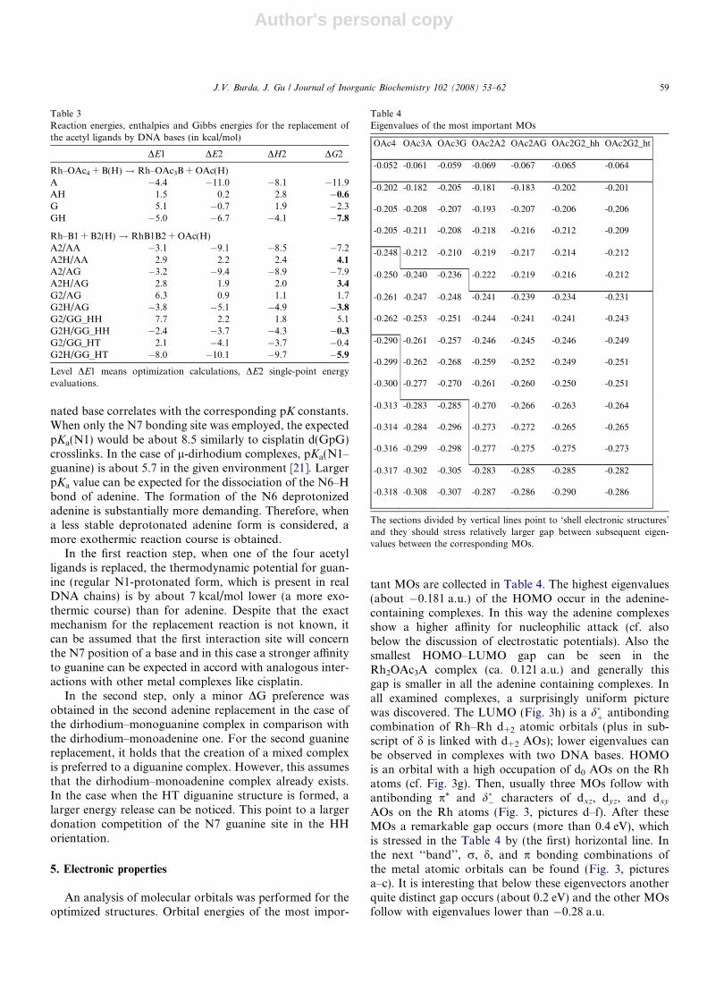
An analysis of molecular orbitals was performed for theoptimized structures. Orbital energies of the most impor-
tant MOs are collected in Table 4. The highest eigenvalues(about �0.181 a.u.) of the HOMO occur in the adenine-containing complexes. In this way the adenine complexesshow a higher affinity for nucleophilic attack (cf. alsobelow the discussion of electrostatic potentials). Also thesmallest HOMO–LUMO gap can be seen in theRh2OAc3A complex (ca. 0.121 a.u.) and generally thisgap is smaller in all the adenine containing complexes. Inall examined complexes, a surprisingly uniform picturewas discovered. The LUMO (Fig. 3h) is a d�þ antibondingcombination of Rh–Rh d+2 atomic orbitals (plus in sub-script of d is linked with d+2 AOs); lower eigenvalues canbe observed in complexes with two DNA bases. HOMOis an orbital with a high occupation of d0 AOs on the Rhatoms (cf. Fig. 3g). Then, usually three MOs follow withantibonding p* and d�� characters of dxz, dyz, and dxy
AOs on the Rh atoms (Fig. 3, pictures d–f). After theseMOs a remarkable gap occurs (more than 0.4 eV), whichis stressed in the Table 4 by (the first) horizontal line. Inthe next ‘‘band’’, r, d, and p bonding combinations ofthe metal atomic orbitals can be found (Fig. 3, picturesa–c). It is interesting that below these eigenvectors anotherquite distinct gap occurs (about 0.2 eV) and the other MOsfollow with eigenvalues lower than �0.28 a.u.
Table 3Reaction energies, enthalpies and Gibbs energies for the replacement ofthe acetyl ligands by DNA bases (in kcal/mol)
DE1 DE2 DH2 DG2
Rh–OAc4 + B(H) ! Rh–OAc3B + OAc(H)A �4.4 �11.0 �8.1 �11.9AH 1.5 0.2 2.8 �0.6
G 5.1 �0.7 1.9 �2.3GH �5.0 �6.7 �4.1 �7.8
Rh–B1 + B2(H) ! RhB1B2 + OAc(H)A2/AA �3.1 �9.1 �8.5 �7.2A2H/AA 2.9 2.2 2.4 4.1
A2/AG �3.2 �9.4 �8.9 �7.9A2H/AG 2.8 1.9 2.0 3.4
G2/AG 6.3 0.9 1.1 1.7G2H/AG �3.8 �5.1 �4.9 �3.8
G2/GG_HH 7.7 2.2 1.8 5.1G2H/GG_HH �2.4 �3.7 �4.3 �0.3
G2/GG_HT 2.1 �4.1 �3.7 �0.4G2H/GG_HT �8.0 �10.1 �9.7 �5.9
Level DE1 means optimization calculations, DE2 single-point energyevaluations.
Table 4Eigenvalues of the most important MOs
OAc4 OAc3A OAc3G OAc2A2 OAc2AG OAc2G2_hh OAc2G2_ht
-0.052 -0.061 -0.059 -0.069 -0.067 -0.065 -0.064
-0.202 -0.182 -0.205 -0.181 -0.183 -0.202 -0.201
-0.205 -0.208 -0.207 -0.193 -0.207 -0.206 -0.206
-0.205 -0.211 -0.208 -0.218 -0.216 -0.212 -0.209
-0.248 -0.212 -0.210 -0.219 -0.217 -0.214 -0.212
-0.250 -0.240 -0.236 -0.222 -0.219 -0.216 -0.212
-0.261 -0.247 -0.248 -0.241 -0.239 -0.234 -0.231
-0.262 -0.253 -0.251 -0.244 -0.241 -0.241 -0.243
-0.290 -0.261 -0.257 -0.246 -0.245 -0.246 -0.249
-0.299 -0.262 -0.268 -0.259 -0.252 -0.249 -0.251
-0.300 -0.277 -0.270 -0.261 -0.260 -0.250 -0.251
-0.313 -0.283 -0.285 -0.270 -0.266 -0.263 -0.264
-0.314 -0.284 -0.296 -0.273 -0.272 -0.265 -0.265
-0.316 -0.299 -0.298 -0.277 -0.275 -0.275 -0.273
-0.317 -0.302 -0.305 -0.283 -0.285 -0.285 -0.282
-0.318 -0.308 -0.307 -0.287 -0.286 -0.290 -0.286
The sections divided by vertical lines point to ‘shell electronic structures’and they should stress relatively larger gap between subsequent eigen-values between the corresponding MOs.
J.V. Burda, J. Gu / Journal of Inorganic Biochemistry 102 (2008) 53–62 59

Author's personal copy
The NPA partial charges of the DFT optimized struc-tures were examined and the determined charges of chosenatoms are summarized in Table 5. In the tetraacetate com-plex, a symmetrical electron density distribution occursbetween both Rh atoms. In single-base and HH complexes,a difference between the Rh atoms is enforced by the differ-ent bonding character of the N7 and X6 coordination sitesof the bases. The larger positive partial charge of the Rhatom is linked with a smaller donation from the ligands.In Table 5 it can be observed that the highest d(Rh) is con-nected with O6 guanine coordination (d = 0.97 in the digua-nine complex). Simultaneously, the lowest partial chargeoccurs on the neighbouring Rh atom of the same complex,which is coordinated to the N7 site (d = 0.67). The differentcharge population of both Rh atoms corresponds with thecoefficients of the natural bond orbital (NBO) of the Rh–Rh bond. This orbital contains a higher contribution ofthe Rh atom, which is coordinated to the N7 atom(s). Thelargest Rh partial charge disproportion (58%Rh(N7) vs.42%Rh(O6)) was found in the HH diguanine complex.
The consequences of the different base interaction sites(N7 and X6) can be also noticed in the electron densitylocalized on the water molecules. A small positive chargeprevails on these molecules in all examined complexes even
without any DNA base. This charge transfer can be alsoobserved in the maps of electrostatic potentials where posi-tive blue areas dominate in the Rh-aqua ligands regions(cf. Fig. 2). The difference of the electron density on bothwater molecules is about 0.06e in the diguanine complex.As to partial charges on atoms of the bases, the pronouncedpolarization effects are remarkable under base coordinationto the dirhodium complex. The most pronounced deviationsfrom the isolated base were found in the partial charges ofthe C8 (up to 0.08e), H8, N1, and N3 sites (0.06e) in guanineas well as in adenine complexes. These pronounced polar-izations speak out about substantial changes in electrondensities due to the formation of Rh2 adducts.
Analogous changes are also visible from the electrostaticmaps depicted in Fig. 2. The values of minima and maximaof the individual potentials on the isodensity surface(q = 0.001 e/A3) are compiled in the lower section of Table2. It can be seen that always the N7 moiety is connectedwith a relatively large positive potential (H8 and H(w))and the N1 proximity with a negative potential. This corre-sponds to the inhomogeneity of charge distribution on theRh atoms discussed above. The orientations of negativeand positive areas are confirmed by the dipole momentssummarized in the last line of Table 2 and Fig. 1. The
Fig. 3. Molecular orbitals of the Rh2OAc2G2w2 (HH) complex: (a) 144th and (b) 145th MOs with r-bonding character of the Rh–Rh atoms; (c) 146thMO with d-bonding character; (d) 147th and (e) 148th MOs represent p-antibonding character; (f) 149th MO has d and (g) HOMO (152nd MO) hasr-antibonding character; (h) LUMO with d-antibonding character on the Rh–Rh subpart. (For interpretation of the references to colour in this figurelegend, the reader is referred to the web version of this article.)
60 J.V. Burda, J. Gu / Journal of Inorganic Biochemistry 102 (2008) 53–62

Author's personal copy
dipole moment occurring in the diadenine complex isslightly larger than in the diguanine complex. However,the orientation and size of the dipole moment is influencedby the orientation of the axial water molecules, which canpartially mask or modify the effects of the electron redistri-bution of the bases. The dipole moments of the isolatedneutral bases are included for the demonstration of theextent of the electron density redistribution.
6. Conclusions
In this study, the diaqua-tetrakis(l-acetylato)dirhodi-um(II,II) complexes were explored. The replacement ofthe acetyl-ligand by a DNA base was simulated in twosteps. Both head-to-head and head-to-tail diguanine com-plexes were considered. Only HH diadenine and mixed ade-nine–guanine complexes were examined.
The optimization of the complexes was performed at theDFT level employing the B3PW91 functional and 6-31G*
basis set. For the Rh description, Stuttgart–DresdenMWB-28 pseudopotentials were used. The complexes weredescribed in singlet electronic ground states. The energyand electron density analyses were determined at theB3LYP/6-31++G(d,p) level.
Higher stabilization as well as bonding energies wasfound for the adenine coordination in comparison withguanine (by about 5 kcal/mol per base). Since adenine inter-acts through two nitrogen atoms (N6 and N7) and guaninethrough O6 and N7 sites, this result matches Pearson’sHSAB principle and is also supported by the NPA analysis.
However, despite the larger stabilization energies of ade-nine complexes, the thermodynamic description of thereplacement reaction favors neutral guanine. The moreexothermic reaction course (by about 7 kcal/mol) is con-nected with an easier proton transfer from the N1 positionof neutral guanine to acetic acid anion in comparison withthe N6 site of adenine. This is also in good accord with theexperimentally found decrease of pKa(N1–guanine). Whilethe value of 9.5 was determined for isolated guanine,
pKa = 5.7 was estimated for the dirhodium–guanine com-plex in study [21].
It was found that the axial aqua-ligands are bonded veryweakly, up to 13 kcal/mol. Such an amount of energy cor-responds to an H-bond rather than a dative bond especiallyin highly polarized complexes like these.
According to the higher-lying HOMO and a smallerHOMO–LUMO gap of the adenine complexes, we canexpect their higher reactivity.
Electrostatic potential maps were drawn for the com-plexes showing a large negative potential in the N1–X6area and a positive potential in the moiety of the N7 coor-dination for all the complexes with DNA base(s).
Acknowledgements
The study was supported by NSF-MSMT Grant No.1P05ME784, GA-AV IAA400550701, and MSM0021620835. The authors thank the Meta-Centers in Pra-gue (Charles University and Czech Technical University),Brno (Masaryk University), Pilsen (University of WestBohemia), and Mississippi Center for SupercomputingResearch for their generous support by providing thecomputational resources.
References
[1] B. Rosenberg, L. Van Camp, J.L. Trosko, V.H. Mansour, Nature 222(1969) 385–391.
[2] L.Y. Kuo, A.H. Liu, T.J. Marks, in: A.S.H. Sigel (Ed.), Met. IonsBiol. Syst., Marcel Dekker, Inc., New York, Basel, Hong Kong, 1995,pp. 53–85.
[3] F. Caruso, M. Rossi, Mini-Rev. Med. Chem. 4 (2004) 49–60.[4] J.B. Waern, C.T. Dillon, M.M. Harding, J. Med. Chem. 48 (2005)
2093–2099.[5] R. Meyer, S. Brink, C.E.J. van Rensburg, G.K. Joone, H. Gorls, S.
Lotz, J. Organomet. Chem. 690 (2005) 117–125.[6] B.M. zu Berstenhorst, G. Erker, G. Kehr, J.C. Wasilke, J. Muller, H.
Redlich, J. Pyplo-Schnieders, Eur. J. Inorg. Chem. (2005) 92–99.[7] R.K. Narla, C.L. Chen, Y.H. Dong, F.M. Uckun, Clin. Cancer Res. 7
(2001) 2124–2133.
Table 5NPA partial charges on selected atoms of the dirhodium complexes (in e)
OAc4 OAc3A OAc3G OAc2A2 OAc2AG OAc2G2_HH OAc2G2_HT
Rh 0.884 Rh6 0.843 Rh6 0.931 Rh6 0.801 Rh6 0.886 Rh6 0.969 Rh 0.828Rh 0.884 Rh7 0.795 Rh7 0.781 Rh7 0.697 Rh7 0.687 Rh7 0.671 Rh 0.828O �0.674 Oac6i �0.662 Oac6i �0.640 Oac6 �0.685 Oac6 �0.684 Oac6 �0.638 Oac6 �0.683O �0.677 Oac7i �0.686 Oac7i �0.691 Oac7 �0.712 Oac7 �0.708 Oac7 �0.706 Oac60 �0.683O �0.653 Oac6o �0.648 Oaco6 �0.667 Oac60 �0.692 Oac60 �0.633 Oac70 �0.711 Oac7 �0.652O �0.652 Oac7o �0.661 Oaco7 �0.659 Oac70 �0.705 Oac70 �0.712 Oac60 �0.640 Oac70 �0.652O �0.674 Oac6t �0.693 Oac7t �0.697 N6 �0.719 O6 �0.690 O6 �0.679 O6 �0.710O �0.677 Oac7t �0.700 Oac6t �0.644 N7 �0.442 N7 �0.438 N7 �0.438 N7 �0.441O �0.653 N6 �0.732 O6 �0.695 N6 0 �0.722 N6 0 �0.719 O6’ �0.681 O6’ �0.710O �0.652 N7 �0.446 N7 �0.445 N7 0 �0.439 N7 0 �0.443 N7’ �0.443 N7’ �0.441Ow �0.954 Ow6 �0.952 Ow6 �0.954 Ow6 �0.947 Ow6 �0.951 Ow6 �0.942 O �0.983Ow �0.954 Ow7 �0.974 Ow7 �0.977 Ow7 �0.988 Ow7 �0.989 Ow7 �0.990 O �0.983H 0.520 Hw6 0.525 Hw6 0.533 Hw6 0.529 Hw6 0.536 Hw6 0.530 H 0.535H 0.519 Hw6 0.524 Hw6 0.515 Hw6 0.529 Hw6 0.521 Hw6 0.529 H 0.535H 0.519 Hw7 0.526 Hw7 0.524 Hw7 0.523 Hw7 0.524 Hw7 0.523 H 0.522H 0.520 Hw7 0.523 Hw7 0.525 Hw7 0.524 Hw7 0.524 Hw7 0.523 H 0.522
J.V. Burda, J. Gu / Journal of Inorganic Biochemistry 102 (2008) 53–62 61

Author's personal copy
[8] A. Anagnostopoulou, E. Moldrheim, N. Katsaros, E. Sletten, J. Biol.Inorg. Chem. 1999 (1999) 199–208.
[9] M. Eriksson, M. Leijon, C. Hiort, B. Norden, A. Graeslund,Biochemistry 33 (1994) 5031–5040.
[10] A. Kueng, T. Pieper, R. Wissiack, E. Rosenberg, B.K. Keppler,J. Biol. Inorg. Chem. 6 (2001) 292–299.
[11] G. Sava, A. Bergarno, Int. J. Oncol. 17 (2000) 353–365.[12] R. Aird, J. Cummings, A. Ritchie, M. Muir, R. Morris, H. Chen, P.
Sadler, D. Jodrell, Br. J. Cancer 86 (2002) 1652–1657.[13] H. Chen, J.A. Parkinson, S. Parsons, R.A. Coxal, R.O. Gould, P.
Sadler, J. Am. Chem. Soc. 124 (2002) 3064–3082.[14] R.E. Morris, R. Aird, P.D. Murdoch, H. Chen, J. Cummings, N.D.
Hughes, S. Parson, A. Parkin, G. Boyd, P. Sadler, D. Jodrell, J. Med.Chem. 44 (2001) 3616–3621.
[15] O. Novakova, H. Chen, O. Vrana, A. Rodger, P.J. Sadler, V. Brabec,Biochemistry 42 (2003) 11544–11554.
[16] H.T. Chifotides, K.R. Dunbar, J.H. Matonic, N. Katsaros, Inorg.Chem. 31 (1992) 4628–4634.
[17] R. Cini, G. Giorgi, L. Pasquini, Inorg. Chim. Acta 196 (1992) 7–18.[18] J.R. Rubin, T.P. Haromy, M. Sundaralingam, Acta Crystallogr. Sect.
C-Cryst. Struct. Commun. 47 (1991) 1712–1714.[19] D.M.L. Goodgame, C.A. Omahoney, C.J. Page, D.J. Williams,
Inorg. Chim. Acta 175 (1990) 141–147.[20] N. Katsaros, A. Anagnostopoulou, Crit. Rev. Oncol. Hematol. 42
(2002) 297–308.[21] H.T. Chifotides, K.M. Koshlap, L.M. Perez, K.R. Dunbar, J. Am.
Chem. Soc. 125 (2003) 10703–10713.[22] H.T. Chifotides, K.M. Koshlap, L.M. Perez, K.R. Dunbar, J. Am.
Chem. Soc. 125 (2003) 10714–10724.[23] H.T. Chifotides, J.M. Koomen, M.J. Kang, S.E. Tichy, K.R. Dunbar,
D.H. Russell, Inorg. Chem. 43 (2004) 6177–6187.[24] H.T. Chifotides, P.K.L. Fu, K.R. Dunbar, C. Turro, Inorg. Chem. 43
(2004) 1175–1183.[25] H.T. Chifotides, K.R. Dunbar, Acc. Chem. Res. 38 (2005) 146–156.
[26] D. Andrae, U. Haussermann, M. Dolg, H. Stoll, H. Preuss, Theor.Chim. Acta 77 (1990) 123–141.
[27] S. Huzinaga, B. Miguel, Chem. Phys. Lett. 175 (1990) 289.[28] S. Huzinaga, M. Klobukowski, Chem. Phys. Lett. 212 (1993) 260–
264.[29] S.F. Boys, F. Bernardi, Mol. Phys. 19 (1970) 553–566.[30] J.V. Burda, M. Pavelka, M. Simanek, J. Mol. Struct. THEOCHEM
683 (2004) 183–193.[31] F. Weinhold, University of Wisconsin, Madison, Wisconsin 53706,
Wisconsin (2001).[32] W. Saenger, Principles of Nucleic Acid Structure, Springer-Verlag,
New York, 1983, pp. 556.[33] K.R. Dunbar, J.H. Matonic, V.P. Saharan, C.A. Crawford, G.
Christou, J. Am. Chem. Soc. 116 (1994) 2201–2202.[34] J.V. Burda, J. Leszczynski, Inorg. Chem. 42 (2003) 7162–7172.[35] R.G. Parr, R.G. Pearson, J. Am. Chem. Soc. 105 (1983) 7512.[36] M. Pavelka, J.V. Burda, Chem. Phys. 312 (2005) 193–204.[37] J.-G. Liu, B.-H. Ye, Q.-L. Zhang, X.-H. Zou, Q.-X. Zhen, X. Tian,
L.-N. Ji, J. Biol. Inorg. Chem. 5 (2000) 119–128.[38] F. Wang, H.M. Chen, S. Parsons, L.D.H. Oswald, J.E. Davidson,
P.J. Sadler, Chem. Eur. J. 9 (2003) 5810–5820.[39] M.H. Baik, R.A. Friesner, S.J. Lippard, J. Am. Chem. Soc. 125
(2003) 14082–14092.[40] M. Zeizinger, J.V. Burda, J. Leszczynski, Phys. Chem. Chem. Phys. 6
(2004) 3585–3590.[41] P. Carloni, M. Sprik, W. Andreoni, J. Phys. Chem. B 104 (2000) 823–
835.[42] K. Spiegel, U. Rothlisberger, P. Carloni, J. Phys. Chem. B 108 (2004)
2699–2707.[43] Z. Futera, J. Klenko, J.E. Sponer, J. Sponer, J.V. Burda, J. Phys.
Chem, submitted for publication.[44] M. Pavelka, J.V. Burda, J. Mol. Model. 13 (2006) 367–379.[45] J.S. Murray, Z. Peralta-Inga, P. Politzer, K. Ekanayake, P. Lebreton,
Int. J. Quant. Chem. 83 (2001) 245–254.
62 J.V. Burda, J. Gu / Journal of Inorganic Biochemistry 102 (2008) 53–62