a common rejection module (crm) for acute rejection across multiple organs
TRANSCRIPT
Article
The Rockefeller University Press $30.00J. Exp. Med. 2013 Vol. 210 No. 11 2205-2221www.jem.org/cgi/doi/10.1084/jem.20122709
2205
Current immune suppression regimen in organ transplantation has been very successful and has extended 1-yr graft survival rates. However, 5-yr graft survival rates have not improved (Lechler et al., 2005). Furthermore, current im-mune suppression may be responsible for the increased risk of various cancers after transplan-tation (Vajdic et al., 2006), suggesting novel, more targeted therapeutics are needed in trans-plantation. Increased transcriptional profiling of transplant biopsies has provided useful insights into allograft injury mechanisms such as acute rejection (AR) and chronic rejection. These in-sights have led to a hypothesis that there is a common rejection mechanism in all trans-planted solid organs (Morgun et al., 2006; Wang et al., 2008; Snyder et al., 2011). Identifying
such a common rejection mechanism could fa-cilitate novel diagnostics and therapeutics with-out requiring details about tissue-specific injury. Given the escalating costs of drug discovery, and the relatively greater impact of these costs on smaller disease markets such as organ trans-plantation, we believe that it is important to find common injury pathways across multiple solid organ transplants.
The NCBI Gene Expression Omnibus (GEO) contains more than 100 human microarray data-sets from heart, kidney, liver, and lung allografts
CORRESPONDENCE Atul J. Butte: [email protected] OR Minnie M. Sarwal: [email protected]
Abbreviations used: AR, acute rejection; AUC, area under the curve; CADI, chronic allograft damage index; CRM, common rejection module; FDR, false discovery rate; JT, Jonckheere-Terpstra; STA, stable.
A common rejection module (CRM) for acute rejection across multiple organs identifies novel therapeutics for organ transplantation
Purvesh Khatri,1,4,5 Silke Roedder,1,6 Naoyuki Kimura,2,3 Katrien De Vusser,7 Alexander A. Morgan,1 Yongquan Gong,2,3 Michael P. Fischbein,3 Robert C. Robbins,2,3 Maarten Naesens,6,7 Atul J. Butte,1 and Minnie M. Sarwal1,6
1Department of Pediatrics; 2Stanford Cardiovascular Institute; 3Department of Cardiothoracic Surgery; 4Stanford Center for Biomedical Informatics Research, Department of Medicine; and 5Institute for Immunity, Transplant, and Infection; Stanford University, Stanford, CA 94305
6California Pacific Medical Center Research Institute, San Francisco, CA 941077Department of Nephrology and Renal Transplantation, University Hospitals Leuven, B-3000 Leuven, Belgium
Using meta-analysis of eight independent transplant datasets (236 graft biopsy samples) from four organs, we identified a common rejection module (CRM) consisting of 11 genes that were significantly overexpressed in acute rejection (AR) across all trans-planted organs. The CRM genes could diagnose AR with high specificity and sensitivity in three additional independent cohorts (794 samples). In another two independent cohorts (151 renal transplant biopsies), the CRM genes correlated with the extent of graft injury and predicted future injury to a graft using protocol biopsies. Inferred drug mechanisms from the literature suggested that two FDA-approved drugs (atorvastatin and dasatinib), approved for nontransplant indications, could regulate specific CRM genes and reduce the number of graft-infiltrating cells during AR. We treated mice with HLA-mismatched mouse cardiac transplant with atorvastatin and dasatinib and showed reduction of the CRM genes, significant reduction of graft-infiltrating cells, and extended graft survival. We further validated the beneficial effect of atorvastatin on graft survival by retrospec-tive analysis of electronic medical records of a single-center cohort of 2,515 renal transplant patients followed for up to 22 yr. In conclusion, we identified a CRM in transplantation that provides new opportunities for diagnosis, drug repositioning, and rational drug design.
© 2013 Khatri et al. This article is distributed under the terms of an Attribution– Noncommercial–Share Alike–No Mirror Sites license for the first six months after the publication date (see http://www.rupress.org/terms). After six months it is available under a Creative Commons License (Attribution–Noncommercial– Share Alike 3.0 Unported license, as described at http://creativecommons.org/ licenses/by-nc-sa/3.0/).
The
Journ
al o
f Exp
erim
enta
l M
edic
ine
2206 Common rejection module in all transplanted organs | Khatri et al.
We applied two meta-analysis approaches to the normal-ized data as described in Materials and methods. In brief, the first approach combines effect sizes from each dataset into a meta-effect size to estimate the amount of change in expres-sion across all datasets. For each gene in each dataset, an effect size was computed using Hedges’ adjusted g. If multiple probes mapped to a gene, the effect size for each gene was summa-rized using the fixed effect inverse-variance model. We com-bined study-specific effect sizes to obtain the pooled effect size and its standard error using the random effects inverse-variance technique. We computed z-statistics as a ratio of the pooled effect size to its standard error for each gene and com-pared the result with a standard normal distribution to obtain a nominal p-value. P-values were corrected for multiple hy-potheses testing using false discovery rate (FDR; Storey, 2002). We identified 180 genes that were measured in all datasets and were overexpressed in AR with P < 0.01 (FDR ≤ 20%; Table S1 B).
We used a second nonparametric meta-analysis that com-bines p-values from individual experiments to identify genes with a large effect size in all datasets. In brief, we calculated a t-statistic for each gene in each study. After computing one-tail p-values for each gene, they were corrected for multiple hypotheses using FDR. Next, we used Fisher’s sum of logs method (Fisher, 1932), which sums the logarithm of cor-rected p-values across all datasets for each gene and compares the sum against a 2 distribution with 2k degrees of freedom, where k is the number of datasets used in the analysis. This method identified 1,772 overexpressed genes at FDR < 20% (Table S1 C).
102 genes were identified as significantly overexpressed by both methods (Table S1 D). This group contains genes that (a) had overall large effect size across all datasets and (b) were significant across all datasets. Although our selection criteria may have left out genes with varying expression in AR, the method allowed us to develop robust overlapping transcrip-tional signals in AR across all transplanted organs.
Using BioGPS, we defined a gene as preferentially ex-pressed in a tissue if its expression in a given tissue was at least three times its median expression across all 84 tissues in BioGPS (Su et al., 2004). We found that the 102 genes are highly expressed in one or more blood cell type that partici-pates in the immune response (Table S1 E), suggesting that our meta-analysis removed tissue-specific bias and identified the relevant pathogenic transcriptional signal of activated in-filtrating cells in the graft in AR, rather than being affected by various confounding factors such as organ-specific expres-sion bias, treatment protocols by different groups, or different microarray platforms.
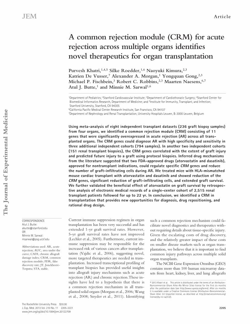
Network analysis of the 102 genes using Ingenuity Path-way Analysis (IPA) revealed that 96 of our genes are part of a network involved in cellular movement and immune cell trafficking (Fig. 1). These genes include major histocompati-bility complex class I and II molecules, interferon regulatory factors, granzymes, chemokines, interleukins, transcription factors, and the T cell receptors. All have direct relationships
that are derived from samples from tissue biopsies or blood. The conditions studied include acute and chronic rejec-tion, tolerance, and drug toxicity. However, the presence of mostly unknown biological and technical confounding fac-tors (e.g., cohort selection, treatment protocol, and microarray technology) in each individual study presents a challenge of integrating these datasets in a meaningful way, which con-sequently limits the usefulness of the publicly available data. We developed a computational framework for integrating expression data from multiple experiments. We used this frame-work to integrate transcriptional data across four different transplanted organs undergoing histologically confirmed AR to identify common rejection mechanism across all trans-planted organs.
We found a common transcriptional response in AR, con-sisting of 11 genes overexpressed during allograft rejection regardless of tissue source, of which, 6 genes are direct or indi-rect targets of immunosuppressive drugs and of drugs other-wise used in immune and inflammatory diseases. We selected two FDA-approved drugs (dasatinib and atorvastatin), which reduce expression of LCK (Lee et al., 2010) and CXCL9 (Ferreira et al., 2010) and CXCL10 (Grip and Janciauskiene, 2009), respectively, three genes within the common rejection module (CRM), for further experiments in an experimental model of rodent acute cardiac rejection. Our goal was to deter-mine whether these drugs could extend graft survival by im-proving AR as measured by a reduction of graft-infiltrating cells and extension of graft survival in an experimental model of graft rejection and to validate any drug benefit observed in human transplant studies, providing support that targeting the CRM genes is a novel approach to repositioning available FDA-approved drugs and identifying new drug targets for all solid organ transplant recipients.
RESULTSMeta-analysis of solid organ transplant datasets recapitulates known mechanisms of ARWe downloaded raw data for eight gene expression studies from organ biopsy specimens from kidney, lung, heart, and liver transplant patients, with and without diagnosis of AR (Table S1 A). To reduce the clinical complexity in defining AR and stable (STA) phenotypes, we used the phenotypes as defined in the corresponding original publications. Pheno-type definitions and sample composition for each dataset are described in Materials and methods. Notably, none of the studies had any antibody-mediated rejection samples or did not report this information. We filtered each dataset to in-clude only biopsies from patients with AR and patients who were in STA condition. This filtering reduced the number of samples available for analysis to 236 from 392 samples in eight datasets. After reannotating the probes, each dataset was sepa-rately normalized using gcRMA (Irizarry et al., 2003a). Data-sets GSE2596 and GSE4470 were not normalized because raw data were not available, and the downloaded data were already normalized.
JEM Vol. 210, No. 11
Article
2207
(b) organ-specific bias caused by unequal number of data-sets (and samples) used in the meta-analysis, we performed leave-one-organ-out meta-analysis. We excluded all datasets from individual organs, one organ at a time, and performed meta-analysis on the remaining datasets from three organs. We hypothesized that the minimal set of genes that are sig-nificantly overexpressed, irrespective of the set of organs analyzed, would constitute a CRM in solid organ trans-plant rejection.
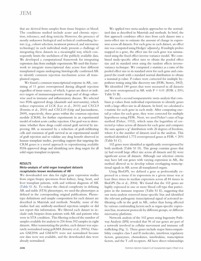
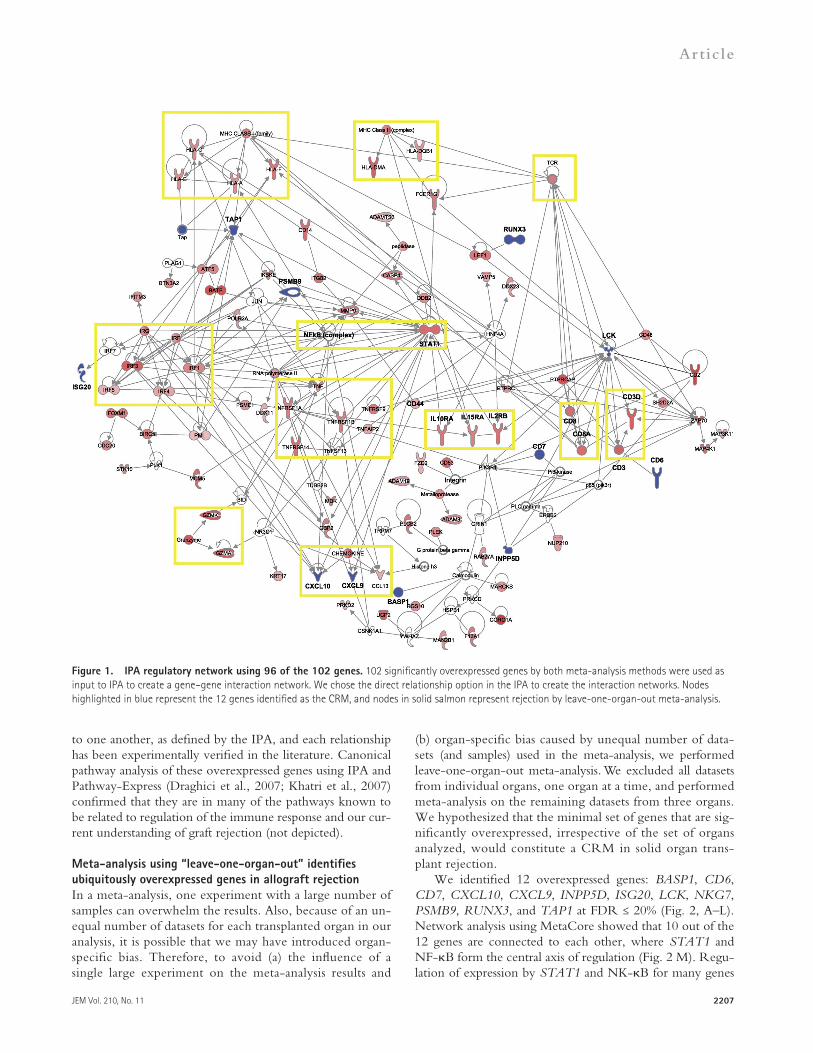
We identified 12 overexpressed genes: BASP1, CD6, CD7, CXCL10, CXCL9, INPP5D, ISG20, LCK, NKG7, PSMB9, RUNX3, and TAP1 at FDR ≤ 20% (Fig. 2, A–L). Network analysis using MetaCore showed that 10 out of the 12 genes are connected to each other, where STAT1 and NF-B form the central axis of regulation (Fig. 2 M). Regu-lation of expression by STAT1 and NK-B for many genes
to one another, as defined by the IPA, and each relationship has been experimentally verified in the literature. Canonical pathway analysis of these overexpressed genes using IPA and Pathway-Express (Draghici et al., 2007; Khatri et al., 2007) confirmed that they are in many of the pathways known to be related to regulation of the immune response and our cur-rent understanding of graft rejection (not depicted).
Meta-analysis using “leave-one-organ-out” identifies ubiquitously overexpressed genes in allograft rejectionIn a meta-analysis, one experiment with a large number of samples can overwhelm the results. Also, because of an un-equal number of datasets for each transplanted organ in our analysis, it is possible that we may have introduced organ-specific bias. Therefore, to avoid (a) the influence of a single large experiment on the meta-analysis results and
Figure 1. IPA regulatory network using 96 of the 102 genes. 102 significantly overexpressed genes by both meta-analysis methods were used as input to IPA to create a gene–gene interaction network. We chose the direct relationship option in the IPA to create the interaction networks. Nodes highlighted in blue represent the 12 genes identified as the CRM, and nodes in solid salmon represent rejection by leave-one-organ-out meta-analysis.
2208 Common rejection module in all transplanted organs | Khatri et al.
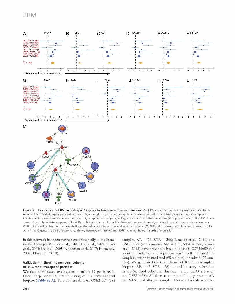
samples, AR = 76, STA = 206; Einecke et al., 2010) and GSE36059 (411 samples, AR = 122, STA = 289; Reeve et al., 2013) have previously been published. GSE36059 also identified whether the rejection was T cell mediated (35 samples), antibody mediated (65 samples), or mixed (22 sam-ples). We generated the third dataset of 101 renal transplant biopsies (AR = 43, STA = 58) in our laboratory, referred to as the Stanford cohort in this manuscript (GEO accession no. GSE50058). All datasets contained biopsy-proven AR and STA renal allograft samples. Meta-analysis showed that
in this network has been verified experimentally in the litera-ture (Chatterjee-Kishore et al., 1998; Der et al., 1998; Sharif et al., 2004; Shi et al., 2005; Robertson et al., 2007; Kuznetsov, 2009; Ellis et al., 2010).
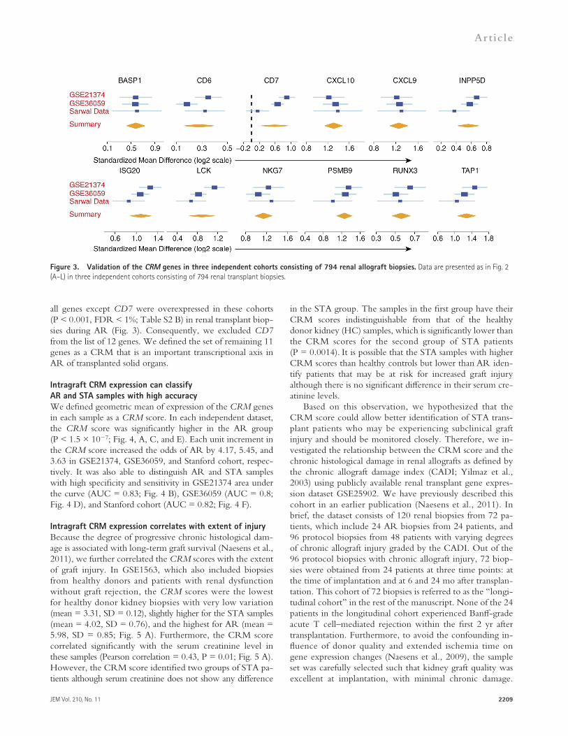
Validation in three independent cohorts of 794 renal transplant patientsWe further validated overexpression of the 12 genes set in three independent cohorts consisting of 794 renal allograft biopsies (Table S2 A). Two of these datasets, GSE21374 (282
Figure 2. Discovery of a CRM consisting of 12 genes by leave-one-organ-out analysis. (A–L) 12 genes were significantly overexpressed during AR in all transplanted organs analyzed in this study, although they may not be significantly overexpressed in individual datasets. The x axes represent standardized mean difference between AR and STA, computed as Hedges’ g, in log2 scale. The size of the blue rectangles is proportional to the SEM differ-ence in the study. Whiskers represent the 95% confidence interval. The yellow diamonds represent overall, combined mean difference for a given gene. Width of the yellow diamonds represents the 95% confidence interval of overall mean difference. (M) Network analysis using MetaCore showed that 10 out of the 12 genes are part of a single regulatory network, with NF-B and STAT1 forming the central axis of regulation.
JEM Vol. 210, No. 11
Article
2209
in the STA group. The samples in the first group have their CRM scores indistinguishable from that of the healthy donor kidney (HC) samples, which is significantly lower than the CRM scores for the second group of STA patients (P = 0.0014). It is possible that the STA samples with higher CRM scores than healthy controls but lower than AR iden-tify patients that may be at risk for increased graft injury although there is no significant difference in their serum cre-atinine levels.
Based on this observation, we hypothesized that the CRM score could allow better identification of STA trans-plant patients who may be experiencing subclinical graft injury and should be monitored closely. Therefore, we in-vestigated the relationship between the CRM score and the chronic histological damage in renal allografts as defined by the chronic allograft damage index (CADI; Yilmaz et al., 2003) using publicly available renal transplant gene expres-sion dataset GSE25902. We have previously described this cohort in an earlier publication (Naesens et al., 2011). In brief, the dataset consists of 120 renal biopsies from 72 pa-tients, which include 24 AR biopsies from 24 patients, and 96 protocol biopsies from 48 patients with varying degrees of chronic allograft injury graded by the CADI. Out of the 96 protocol biopsies with chronic allograft injury, 72 biop-sies were obtained from 24 patients at three time points: at the time of implantation and at 6 and 24 mo after transplan-tation. This cohort of 72 biopsies is referred to as the “longi-tudinal cohort” in the rest of the manuscript. None of the 24 patients in the longitudinal cohort experienced Banff-grade acute T cell–mediated rejection within the first 2 yr after transplantation. Furthermore, to avoid the confounding in-fluence of donor quality and extended ischemia time on gene expression changes (Naesens et al., 2009), the sample set was carefully selected such that kidney graft quality was excellent at implantation, with minimal chronic damage.
all genes except CD7 were overexpressed in these cohorts (P < 0.001, FDR < 1%; Table S2 B) in renal transplant biop-sies during AR (Fig. 3). Consequently, we excluded CD7 from the list of 12 genes. We defined the set of remaining 11 genes as a CRM that is an important transcriptional axis in AR of transplanted solid organs.
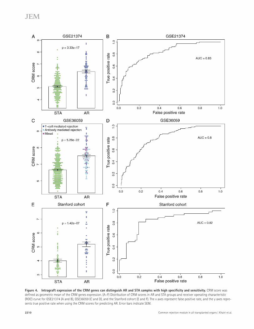
Intragraft CRM expression can classify AR and STA samples with high accuracyWe defined geometric mean of expression of the CRM genes in each sample as a CRM score. In each independent dataset, the CRM score was significantly higher in the AR group (P < 1.5 × 10⁷; Fig. 4, A, C, and E). Each unit increment in the CRM score increased the odds of AR by 4.17, 5.45, and 3.63 in GSE21374, GSE36059, and Stanford cohort, respec-tively. It was also able to distinguish AR and STA samples with high specificity and sensitivity in GSE21374 area under the curve (AUC = 0.83; Fig. 4 B), GSE36059 (AUC = 0.8; Fig. 4 D), and Stanford cohort (AUC = 0.82; Fig. 4 F).
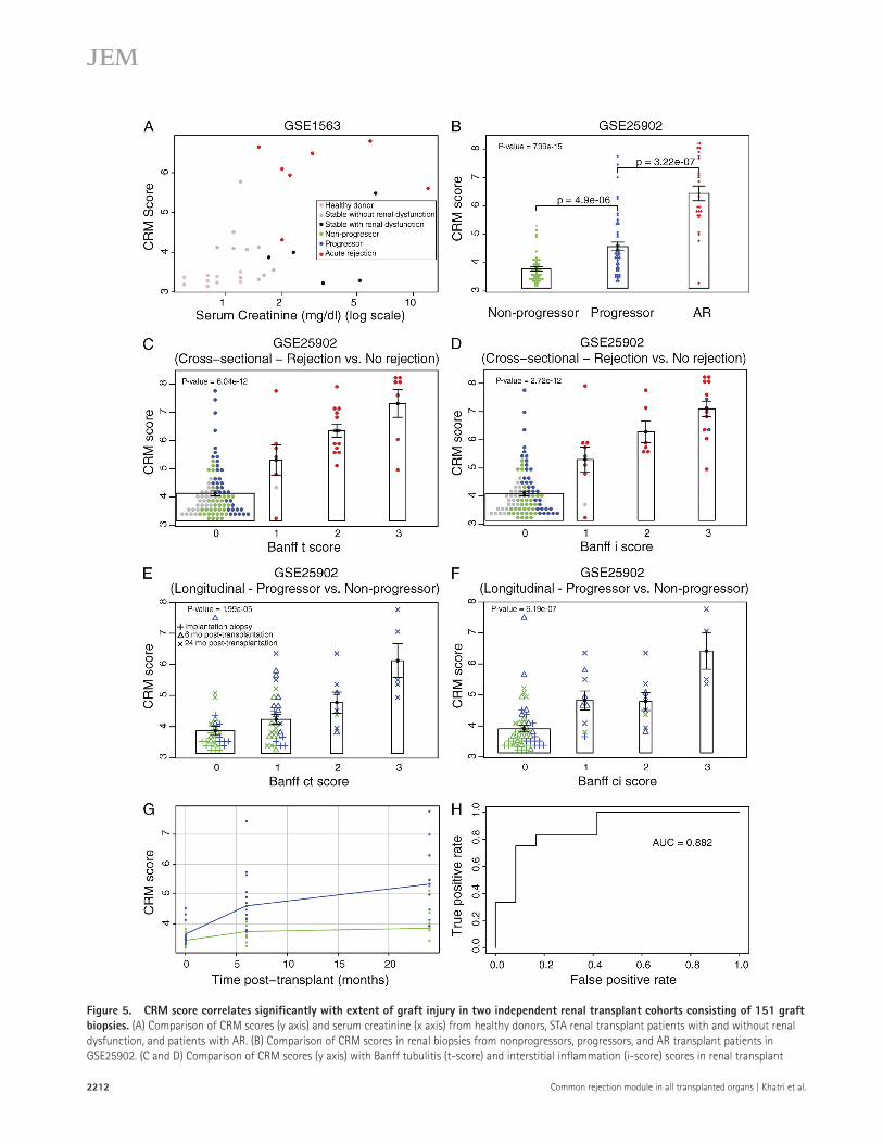
Intragraft CRM expression correlates with extent of injuryBecause the degree of progressive chronic histological dam-age is associated with long-term graft survival (Naesens et al., 2011), we further correlated the CRM scores with the extent of graft injury. In GSE1563, which also included biopsies from healthy donors and patients with renal dysfunction without graft rejection, the CRM scores were the lowest for healthy donor kidney biopsies with very low variation (mean = 3.31, SD = 0.12), slightly higher for the STA samples (mean = 4.02, SD = 0.76), and the highest for AR (mean = 5.98, SD = 0.85; Fig. 5 A). Furthermore, the CRM score correlated significantly with the serum creatinine level in these samples (Pearson correlation = 0.43, P = 0.01; Fig. 5 A). However, the CRM score identified two groups of STA pa-tients although serum creatinine does not show any difference
Figure 3. Validation of the CRM genes in three independent cohorts consisting of 794 renal allograft biopsies. Data are presented as in Fig. 2 (A–L) in three independent cohorts consisting of 794 renal transplant biopsies.
2210 Common rejection module in all transplanted organs | Khatri et al.
Figure 4. Intragraft expression of the CRM genes can distinguish AR and STA samples with high specificity and sensitivity. CRM score was defined as geometric mean of the CRM genes expression. (A–F) Distribution of CRM scores in AR and STA groups and receiver operating characteristic (ROC) curve for GSE21374 (A and B), GSE36059 (C and D), and the Stanford cohort (E and F). The x axes represent false positive rate, and the y axes repre-sents true positive rate when using the CRM scores for predicting AR. Error bars indicate SEM.
JEM Vol. 210, No. 11
Article
2211
Despite the pristine tissue quality at engraftment, the effect of increasing posttransplant time, even in the absence of inter-val AR, results in a significant increase in chronic histologi-cal damage in all histological compartments. At 24 mo after transplantation, 50% of the group (12 patients) had signifi-cantly greater histological progression, scored by the incre-mental CADI score. We defined these samples as a “progressor” group, and the remaining samples as a “nonprogressor” group. At 6 mo, the progressor group had higher mean CADI scores than the nonprogressor (3.2 ± 1.8 vs. 0.75 ± 0.75; P = 0.0003), and similar trends were seen for mean CADI scores for the 24-mo biopsies (7.3 ± 1.6 in progressor in con-trast to 1.8 ± 1.4 in nonprogressor; P ≤ 0.0001). No other Banff qualifiers were significantly different at any time point (Naesens et al., 2011).
The CRM scores for the three sample groups (nonprogres-sor, progressor, and AR) in the 120 samples were significantly different (Fig. 5 B) and positively correlated with the extent of injury (Jonckheere-Terpstra [JT] trend test p-value = 7.99 × 10¹⁵). We further correlated the CRM scores with different Banff qualifiers for AR. The CRM score showed signifi-cant positive correlation with Banff t-score (JT p-value = 6.04 × 10¹²; Fig. 5 C) and i-score (JT p-value = 2.72 × 10¹²; Fig. 5 D). The CRM score also showed significant posi-tive correlation with Banff ct-score (JT p-value = 1.995 × 10⁵; Fig. 5 E) and ci-score (JT p-value = 6.195 × 10⁷; Fig. 5 F) in the longitudinal cohort.
Finally, we performed a repeated measures analysis of vari-ance for the longitudinal cohort (Fig. 5 G). Our results showed that the CRM scores were significantly different between the progressor and nonprogressor groups (P = 1.86 × 10⁵), and the CRM scores increased significantly over time for both groups (P = 4.03 × 10⁶). Importantly, as time after transplant increased, the CRM scores for the progressor groups increased at a faster rate than the nonprogressor group (P = 0.0276). Therefore, we used the CRM scores for the 6-mo protocol bi-opsies to test whether it can predict future histological damage to the graft, as defined by CADI score at 24 mo after trans-plantation. We found that the CRM scores in 6-mo biopsies can predict future histological damage with high specificity and sensitivity (AUC = 0.882; Fig. 5 H). In summary, our results using two additional independent cohorts of 151 renal trans-plant biopsies show that the CRM score significantly correlates with graft injury and can be used for identifying patients at risk of developing significant histological damage in future using protocol biopsies.
FDA-approved drugs targeting the CRM genes reduce graft-infiltrating cells in a mouse model of AR and increase graft survivalBecause of ubiquitous and correlated overexpression of the CRM during AR and chronic injury in 13 independent data-sets consisting of 1,164 biopsies from four types of transplanted organs, we hypothesized that irrespective of the transplanted organ, the CRM is a critical axis of gene regulation during AR and graft injury. Therefore, pharmaceutical disruption of
the CRM may reduce immune cell infiltration, reduce graft injury, and increase graft survival.
A literature review found that 6 out of the 11 genes are direct or indirect targets of FDA-approved drugs. Bortezo-mib is an FDA-approved drug that inhibits PSMB9. It can reverse antibody-mediated rejection and eliminate donor-specific anti–human leukocyte antigen antibodies (Walsh et al., 2010). Mycophenolate mofetil, which is also FDA ap-proved and primarily targets IMP dehydrogenase 2 (IMPDH2), reduces expression of INPP5D by more than twofold (van Leuven et al., 2010). It is a potent immunosuppressive drug that reduces the risk of AR (Knight et al., 2009) and has a possible beneficial effect on chronic graft survival (Ojo et al., 2000). BASP1 and CXCL9 are selectively targeted by doxy-cycline (Hartl et al., 2009) and sulindac (Sakaeda et al., 2006), respectively. Multiple studies using in vitro and in vivo mod-els have shown that dasatinib (BMS-354825, Sprycel; Bristol-Meyers Squibb) binds to and potently inhibits LCK. Dasatinib is an ATP competitor approved for Imatinib-resistant chronic myeloid leukemia. Lee et al. (2010) have shown that da-satinib binds to the ATP-binding site of LCK and freezes the kinase domain in an inactive conformation. Schade et al. (2008) have shown that inhibition of LCK by dasatinib, which is critical for T cell receptor signaling, is responsible for reduction in T cell activation in vitro and in vivo. In addition, Blake et al. (2008) have shown that dasatinib also reduces NK cell cytotoxicity. Atorvastatin (Lipitor) is an HMG-CoA reductase inhibitor that slows the production of cholesterol and is used for treatment of hyperlipidemia. Out of nine chemokines and four endothelial cytokines investi-gated in plasma samples from patients with Crohn’s disease, atorvastatin reduced CXCL10 plasma levels but did not affect the other chemokines and cytokines (Grip and Janciauskiene, 2009). Atorvastatin treatment has been shown to also signifi-cantly reduce plasma levels of CXCL9 in systemic lupus ery-thematosus patients (Ferreira et al., 2010). Both CXCL9 and CXCL10 are ligands of CXCR3, which is a chemokine re-ceptor most consistently expressed during rejection that pro-motes localization of CTLs to inflamed tissues (Wang et al., 2008). Furthermore, statin has been shown to demethylate the FOXP3 promoter (Kim et al., 2010). The transcription factor FOXP3 is critical for development and function of regulatory T cells (T reg cells). FOXP3+ T reg cells are a unique subset of CD4+ T cells that mediate immunosuppression. Baron et al. (2007) have shown that DNA demethylation in the human FOXP3 locus discriminates T reg cells from acti-vated FOXP3+ conventional T cells.
Both dasatinib and atorvastatin are FDA-approved drugs for nontransplant conditions. Because atorvastatin and dasat-inib have been FDA approved for treating nontransplant conditions, we hypothesized that they could be repositioned in organ transplantation. To test this hypothesis, we used both drugs in an established mouse FVB-to-C57BL/6 het-erotopic cardiac transplant model (Corry and Russell, 1973) to investigate the effect of peritransplant drug administration on mitigating cell infiltration in the transplanted graft. We
2212 Common rejection module in all transplanted organs | Khatri et al.
Figure 5. CRM score correlates significantly with extent of graft injury in two independent renal transplant cohorts consisting of 151 graft biopsies. (A) Comparison of CRM scores (y axis) and serum creatinine (x axis) from healthy donors, STA renal transplant patients with and without renal dysfunction, and patients with AR. (B) Comparison of CRM scores in renal biopsies from nonprogressors, progressors, and AR transplant patients in GSE25902. (C and D) Comparison of CRM scores (y axis) with Banff tubulitis (t-score) and interstitial inflammation (i-score) scores in renal transplant
JEM Vol. 210, No. 11
Article
2213
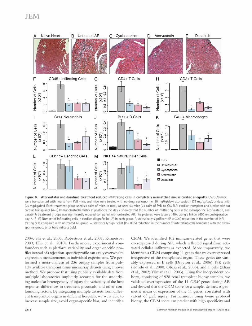
untreated AR group was 10 d, but was 17 d for atorvastatin and 24.5 d for dasatinib.
Next, we tested whether combination of cyclosporine and atorvastatin can reduce cyclosporine dose and, hence, help avoid drug toxicity. We treated a group of six mice with ator-vastatin and 50% reduced dose of cyclosporine (from 20 mg/kg/day to 10 mg/kg/day). The survival curve for this group, followed for up to 30 d, was similar to that of the mice treated with only atorvastatin (not depicted). Using Cox proportional hazard analysis, when treated with atorvastatin and cyclospo-rine compared with untreated AR, the hazard ratio for graft survival was 11.38 (P = 0.003). These results suggest that ator-vastatin and cyclosporine may work through different path-ways, each of which results in extended graft survival compared with the untreated group.
Retrospective analysis of electronic medical records shows statin treatment in renal transplant patients improves graft survivalTo validate the suggested benefits of statin use in a large clini-cal transplant population, we used electronic medical records from all 2,515 patients that received renal transplant between January 1989 and March 2012 at the University Hospitals Leuven. Out of the 2,515 patients, 1,566 received statin within the first 180 d after transplantation, with graft sur-viving at least 180 d. None of the patients started or stopped statin because of renal function evolution or intragraft phe-nomena, suggesting there is no direct bias in the association between statin use and graft outcome. In Cox proportional hazards analysis, after censoring for when a patient stopped taking statin, graft failed, or recipient death, statin use was as-sociated with improved graft survival (HR = 0.701, P = 0.01; Fig. 7 B). This effect was statistically significant after adjusting for donor and recipient age, repeat transplantation, and cal-endar year (Table S4).
DISCUSSIONThe goal of this study was to ascertain whether there are com-mon immunological and inflammatory alterations during AR in an allotransplant, regardless of tissue source. Identifying a common response could be useful for monitoring rejection and could suggest novel targets for rational immunosuppressive drug discovery and design.
Despite several transcriptional studies in organ transplan-tation, their limited sample sizes make it difficult to capture the molecular heterogeneity of AR (Chatterjee-Kishore et al., 1998; Der et al., 1998; Sarwal et al., 2003; Sharif et al.,
chose a cardiac transplant model in mouse to also illustrate that the CRM is indeed common during AR in multiple organs. We also used cyclosporine as a positive control in this model. After each drug treatment for 7 d, the grafts were evaluated by comparing against untreated AR using standard graft histology, a count of the infiltrating cell subsets in the graft, and by tran-scriptional analysis of the graft’s Q-PCR.
Gene expression profiling of nontransplanted hearts (FVB mice) and untreated, transplanted hearts showed that a major-ity of the 102 cross-organ rejection genes were significantly overexpressed in untreated AR (FDR ≤ 2%; Table S3 A), in-cluding all of the CRM genes (FDR ≤ 0.1%). Pathway analysis of down-regulated genes in each treatment group against each untreated AR group using IPA showed that only cyclospo-rine affected the T cell–related pathways. Atorvastatin affected monocyte- and macrophage-related pathways, and dasat-inib affected cell cycle–related pathways (Table S3 B). Using Q-PCR, we found that the majority of the CRM genes were down-regulated in each of the treatment groups (Table S3 C). Not all CRM genes were down-regulated by any of the drugs used, which is expected because each drug acts through differ-ent mechanisms.
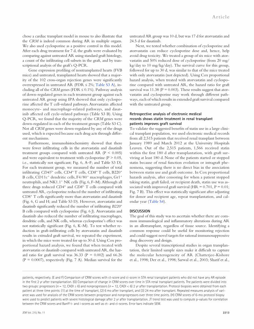
Furthermore, immunohistochemistry showed that there were fewer infiltrating cells in the atorvastatin and dasatinib treatment groups compared with untreated AR (P < 0.005) and were equivalent to treatment with cyclosporine (P > 0.05, i.e., statistically not significant; Fig. 6, A–F; and Table S3 D). For each treatment group, we measured the number of total infiltrating CD45+ cells, CD4+ T cells, CD8+ T cells, B220+ B cells, CD11c+ dendritic cells, F4/80+ macrophages, Gr1+ neutrophils, and NK1.1+ NK cells (Fig. 6, F–M). Although all three drugs reduced CD4+ and CD8+ T cells compared with untreated AR, cyclosporine reduced the number of infiltrating CD8+ T cells significantly more than atorvastatin and dasatinib (Fig. 6, G and H; and Table S3 D). However, atorvastatin and dasatinib significantly reduced the number of infiltrating B220+ B cells compared with cyclosporine (Fig. 6 J). Atorvastatin and dasatinib also reduced the number of infiltrating macrophages, dendritic cells, and NK cells, whereas cyclosporine’s effect was not statistically significant (Fig. 6, K–M). To test whether re-duction in graft-infiltrating cells by atorvastatin and dasatinib results in extruded graft survival, we repeated the experiment, in which the mice were treated for up to 30 d. Using Cox pro-portional hazard analysis, we found that when treated with atorvastatin or dasatinib compared with untreated AR, the haz-ard ratio for graft survival was 36.33 (P = 0.002) and 66.26 (P = 0.0007), respectively (Fig. 7 A). Median survival for the
patients, respectively. (E and F) Comparison of CRM scores with ct-score and ci-score in STA renal transplant patients who did not have any AR episode in the first 2 yr after transplantation. (G) Comparison of change in CRM scores over time in STA renal transplant patients. The patients were divided into two groups: progressors (n = 12, CADI ≥ 6) and nonprogressors (n = 12, CADI < 6) 2 yr after transplantation. Protocol biopsies were obtained from each patient at three time points: (1) at the time of transplant, (2) 6 mo after transplant, and (3) 24 mo after transplant. Repeated measures analysis of vari-ance was used for analysis of the CRM scores between progressor and nonprogressors over three time points. (H) CRM scores of 6-mo protocol biopsy were used to predict patients with severe histological damage after 2 yr after transplantation. JT trend test was used to compute p-values for correlation between the CRM scores and Banff t- and i-scores as well as ct- and ci-scores. Error bars indicate SEM.
2214 Common rejection module in all transplanted organs | Khatri et al.
CRM. We identified 102 immune-related genes that were overexpressed during AR, which reflected signal from acti-vated cellular infiltrates as expected. More importantly, we identified a CRM comprising 11 genes that are overexpressed irrespective of the transplanted organ. These genes are vari-ably expressed in B cells (Drayton et al., 2006), NK cells (Kondo et al., 2000; Obara et al., 2005), and T cells (Zhao et al., 2002; Yilmaz et al., 2003). Using five independent co-horts, consisting of 928 renal transplant biopsy samples, we validated overexpression of the 11 CRM genes during AR and showed that the CRM score for a sample, defined as geo-metric mean of expression of the 11 genes, correlated with extent of graft injury. Furthermore, using 6-mo protocol biopsy, the CRM score can predict with high specificity and
2004; Shi et al., 2005; Robertson et al., 2007; Kuznetsov, 2009; Ellis et al., 2010). Furthermore, experimental con-founders such as platform variability and organ-specific pro-files instead of a rejection-specific profile can easily overwhelm expression measurements in individual experiments. We per-formed a meta-analysis of 236 biopsy samples from pub-licly available transplant tissue microarray datasets using a novel method. We propose that using publicly available data from multiple laboratories implicitly accounts for the underly-ing molecular heterogeneity of injury, the variability of the host response, differences in treatment protocols, and other con-founding factors. By integrating multiple datasets from differ-ent transplanted organs in different hospitals, we were able to increase sample size, avoid organ-specific bias, and identify a
Figure 6. Atorvastatin and dasatinib treatment reduced infiltrating cells in completely mismatched mouse cardiac allografts. C57BL/6 mice were transplanted with hearts from FVB mice, and mice were treated with no drug, cyclosporine (20 mg/kg/day), atorvastatin (75 mg/kg/day), or dasatinib (25 mg/kg/day). Each treatment group used six pairs of mice. In total, we used 53 mice (24 pairs of FVB-to-C57BL/6 cardiac transplant and 5 mice without cardiac transplant). (A–E) Immunohistochemistry at postoperative day 7 showed that the number of infiltrating cells in the cyclosporine, atorvastatin, and dasatinib treatment groups was significantly reduced compared with untreated AR. The pictures were taken at 40× using a Nikon E600 on postoperative day 7. (F–M) Number of infiltrating cells in cardiac allografts (×106) in each group. *, statistically significant (P < 0.05) reduction in the number of infil-trating cells compared with untreated AR group; +, statistically significant (P < 0.05) reduction in the number of infiltrating cells compared with the cyclo-sporine group. Error bars indicate SEM.
JEM Vol. 210, No. 11
Article
2215
that use samples from the same type of organ are more likely to identify these tissue-specific triggers than the effector im-mune response that converge into a common mechanism. Our meta-analysis approach that uses samples from multiple organs in multiple independent experiments favors the common mech-anism over the tissue-specific triggers.
The CRM also provides a mechanism for understanding varying biological outcomes after transplantation and AR therapy regardless of the tissue source of the organ. Different immunosuppressive therapies are likely to have different in-fluences on the allogeneic response against distinct cell lin-eages. Most currently used immunosuppressive drugs prevent AR by inhibiting T cell activation. This is achieved through various means, such as inhibiting antigen-presenting cell de-velopment, cytokine production, or co-stimulatory signals for T cell activation (Halloran, 2004; Lechler et al., 2005; Walsh et al., 2010). This effect was also observed in our mouse model. Cyclosporine reduced infiltrating CD4+ and CD8+ T cells along with Gr1+ neutrophils but did not significantly reduce B220+ B cells and antigen-presenting cells including F480+ macrophages, CD11c+ dendritic cells, and NK1.1+ NK cells. In contrast, atorvastatin has been shown to suppress inter-feron-induced neopterin formation in monocytic cell lines (Neurauter et al., 2003). Neopterin is a metabolite of guano-sine triphosphate, which is produced by monocyte-derived macrophages. Atorvastatin reduced F480+ macrophages the most in the mouse model (Fig. 6 K).
CXCL9, CXCL10, and CXCL11 are ligands of CXCR3 that polarize CD4+ T cells toward the Th1 phenotype and promote localization of CTLs to allograft. Interaction between CTLs and B cells leads to the secretion of CXCR3 ligands by B cells, forming a positive feedback loop, which in turn ampli-fies the inflammatory signal further (Deola et al., 2008). By re-ducing CXCL9 and CXCL10 expression in serum, atorvastatin potentially inhibits this positive feedback loop, which can ex-plain lower B220+ B cells and other innate immune cells infil-trating the graft in the atorvastatin-treated group.
sensitivity patients who are likely to develop severe histologi-cal damage within 2 yr after transplantation.
It is important to note that many of the 102 genes have been previously reported in the literature, including MHC class I and II molecules, chemokines, markers of T cell activa-tion, and co-stimulatory molecules (Fig. 1). For instance, che-mokine receptor CXCR3 and its ligands, CXCL9, CXCL10, and CXCL11, are most consistently overexpressed during re-jection (Karason et al., 2006; Wang et al., 2008; Chen et al., 2010). However, the CRM genes contain only two of the CXCR3 ligands, CXCL9 and CXCL10. CXCR3 was not identified in our analysis because it was down-regulated in the liver dataset GSE13440 (not depicted), whereas CXCL11 was not identified because it was not measured in the liver dataset GSE13440 (not depicted). However, all samples in GSE13440 were HCV positive (Table S1 A). Hence, down-regulation of CXCR3 in the liver dataset does not necessarily indicate down-regulation of CXCR3 during AR in liver transplant because overexpression of CXCR3 during HCV infection could explain down-regulation or no differential expression of CXCR3 in GSE13440.
In contrast, there are several possible explanations for why our list of genes does not include many genes that have been reported in the literature. For instance, we recently proposed a five-gene set model for diagnosis of AR renal transplantation (Li et al., 2012). Although these five genes were validated in an independent randomized cohort from 12 US transplant pro-grams, there are important factors that distinguish it from our current study. First, our AJT study used peripheral blood sam-ples from transplant patients, whereas the current study used only biopsy samples from transplant patients. The set of genes that are differentially expressed during AR in blood and allo-graft can be very different. Second, the AJT study was per-formed only on pediatric samples, whereas all datasets in the current study used adult samples. Third, it has been hypothe-sized previously that the triggers, which lead to allograft rejec-tion, may be tissue specific (Wang et al., 2008). Experiments
Figure 7. Atorvastatin and dasatinib treatment significantly extended allograft survival in mice and humans. (A) C57BL/6 mice were transplanted with hearts from FVB mice, and mice were treated with no drug, cyclosporine (20 mg/kg/day), atorvastatin (75 mg/kg/day), or dasatinib (25 mg/kg/day). Each treatment group used six pairs of mice. In total, we used 48 mice (24 pairs of FVB-to-C57BL/6 cardiac transplant). Mice were treated for up to 30 d, and graft survival was monitored. (B) Electronic medical records of 2,515 renal transplant patients, transplanted between January 1989 and March 2012 at the University Hospitals Leuven, were divided into
two groups: (1) 1,566 patients who received statin within the first 180 d after transplantation, with grafts surviving at least 180 d, and (2) 949 patients who did not receive statin. None of the patients started or stopped statin because of renal function evolution or intragraft phenomena. Patients were censored when a patient stopped taking statin, graft failed, or recipient death. Cox proportional hazard analysis was used to associate statin use with graft survival while adjusting for donor and recipient age, repeat transplantation, and calendar year.
2216 Common rejection module in all transplanted organs | Khatri et al.
cytochrome P4503A4 (CYP3A4; Asberg et al., 2001). Cyclo-sporine has a significant impact on plasma concentrations of all statins, which results in a several-fold higher exposure com-pared with non-cyclosporine–treated controls (from 2-fold increase for fluvastatin to 23-fold increase for pravastatin; Arnadottir et al., 1993; Regazzi et al., 1993; Goldberg and Roth, 1996; Olbricht et al., 1997; Mück et al., 1999). Several studies have also shown that many statins tend to induce in-creased cyclosporine levels from no change for fluvastatin to 115% increase by lovastatin (Kuo et al., 1989; Cheung et al., 1993; Campana et al., 1995; Xu et al., 1998; Renders et al., 2001). However, atorvastatin has been shown to significantly reduce systematic exposure of cyclosporine by 10% in renal transplant patients (Asberg et al., 2001). Hence, bilateral phar-macokinetic interactions of different statins with cyclosporine and other widely used immunosuppressive drugs such as tacro-limus must be explored in more detail to identify an appropri-ate and effective combination of a statin and cyclosporine to treat transplant patients. It is possible that retrospective analysis of electronic medical records of multiple cohorts, similar to our analysis in Fig. 7 B, could facilitate identification of drug com-binations that should be further tested in preclinical models.
To the best of our knowledge, our retrospective analysis of statin usage in more than 2,500 patients is the largest study with patients followed for up to 22 yr. Our analysis in this large kidney transplant patient cohort showed that statin use significantly improved death-censored renal allograft survival. These data suggest that statins may be important adjunctive agents to consider for de novo treatment in all organ transplant recipients because of their propensity to reduce cardiovascular morbidity and their synergistic immuno-suppressive potential. Moreover, this analysis provides a proof of concept that identification of relevant molecules and pathways can be tested rapidly in large retrospective clinical datasets, which should then foster subsequent randomized controlled clinical trials.
Most importantly, our success in identifying the CRM in solid organ transplant rejection and the ability to reposition the existing drugs based on their interaction with the CRM targets underscores the importance of exploiting the data in public repositories for downstream clinical applications. Our results demonstrate that by broadening the sources of samples and pooling evidence from multiple studies for AR, we are able to identify a common, biologically relevant, rejection-specific signature in the tissue that can be useful for diagnostic purposes and therapeutic repositioning. NCBI GEO contains data from more than a million samples in experiments related to diseases that contribute to more than a third of human disease-related mortality in the United States (Butte, 2008). However, in re-cent years, as the amount of data in public repositories has in-creased, the number of FDA-approved drugs every year has declined, and largely stagnated to 20 drugs per year, from a peak of 53 drugs in 1996, amid an increase in the cost of drug discovery from $138 million in 1975 to $1.3 billion in 2006 and more than 15 yr needed, on average, in developing a single drug (Dudley et al., 2010). Our method can provide a rapid,
Current immune suppression therapies in transplant have been associated with increased incidence of cancer after trans-plantation (Vajdic et al., 2006). Recently, atorvastatin was shown to reduce cancer-related mortality in the cancer patients (Nielsen et al., 2012), which in turn suggests that atorvastatin could also be useful for reducing the risk of cancer in transplant patients in addition to increased graft survival. Furthermore, dasatinib is an oral BCR-ABL inhibitor for the treatment of chronic myeloid leukemia (Shah et al., 2008). Because dasat-inib is a cancer treatment drug, it may also reduce the risk of various cancers after transplantation. In addition, it has potent inhibitory effects on TNF, IFN, IL2, IL6, IL10, IL17, and IL22 and has been show to inhibit T cell activation and prolif-eration and reduce NK cell cytotoxicity (Blake et al., 2008), suggesting it would likely show efficacy as an immunosuppres-sive agent (Schade et al., 2008). Our data show that dasatinib could be an adjunctive immunosuppressive agent, given that it has potent inhibitory effects on B cells and components of in-nate immune response including macrophages, dendritic cells, and NK cells.
Our finding of atorvastatin’s immunomodulatory effect is not a complete surprise. Variants of statins have been shown to inhibit induction of MHC-II expression by IFN- and repress MHC-II–mediated T cell activation (Kwak et al., 2000). Indeed, an immunosuppressive role of statins has been sug-gested in solid organ transplant clinical trials (Navaneethan et al., 2009). Pravastatin and simvastatin have been shown to confer better survival than the statin-naive control group in cardiac transplant recipients (Kobashigawa et al., 1995; Mehra et al., 2002). It is not clear whether there are any differences in immunomodulation among different statins, as pravastatin has been shown to reduce AR incidence significantly in renal trans-plant patients (Katznelson et al., 1996), whereas simvastatin and fluvastatin have not demonstrated this effect (Holdaas et al., 2001; Kasiske et al., 2001). Atorvastatin was recently studied in renal transplant patients to show immunoregulatory effects on T cells (Guillén et al., 2010).
However, one of the significant limitations of these studies, with the exception of the ALERT study, is that they were un-derpowered (Navaneethan et al., 2009). Furthermore, almost all randomized clinical trials using statins enrolled patients im-mediately after transplantation and followed them for 3 mo, and none looked at graft survival rates. Although many of these trials have concluded that statins have no effect on AR inci-dence rates, it may be because the patients are heavily immuno-suppressed immediately after transplantation (typically using three immunosuppressive drugs).
The next logical phase of this study would be to find a combination of these drugs and appropriate dosage of each drug that is maximally beneficial. Although our results show that reducing cyclosporine dose by 50% in combination with atorvastatin does not improve graft survival as much as cyclo-sporine alone, it does not provide a complete picture. We need to consider bilateral pharmacokinetic interaction between cy-closporine and various statins. Cyclosporine and many statins (atorvastatin, lovastatin, and simvastatin) are metabolized by
JEM Vol. 210, No. 11
Article
2217
combining effect size, (c) measured in all eight datasets, and (d) when combin-ing p-values using Fisher’s test, p-value < 0.2 for a gene to be up-regulated.
Leave-one-organ-out analysisTo account for the unequal number of datasets for each organ as well as to find a set of genes that was overexpressed independently of the source organ, we performed meta-analysis by removing organ-specific datasets one organ at a time. Thus, in the first iteration, we removed the datasets from heart transplants (GDS1684, GSE2596, GSE4470, and GSE9377) and performed meta-analysis on the remaining datasets, which were from kidney, lung, and liver. In the second iteration, we removed the datasets from kidney and performed meta-analysis on the datasets from heart, lung, and liver. At each iteration, we performed meta-analysis by combining effect sizes and by combining p-values. Using FDR ≤ 20% as a threshold, at each iteration, we identified 12 genes that were overexpressed in all organs used in the given iteration.
Functional pathway analysisWe performed functional pathway analysis using Pathway-Express (Draghici et al., 2007; Khatri et al., 2007; Tarca et al., 2009). Meta-effect size was used as fold change in Pathway-Express to identify significant pathways. We used FDR ≤ 10% as a threshold for identifying significant pathways. We per-formed network analysis using IPA with an option to include only “direct relationship” to avoid spurious connections caused by “indirect relations.” Direct relationships in IPA result from publications citing experimental evi-dence for an interaction.
JT trend testWe used the JT trend test to correlate the CRM score with various Banff criteria using GSE25902. It tests for a monotone trend in terms of the class parameter. In our case, the class is defined as various values of Banff criteria (e.g., t-score, i-score, etc.). The test uses the number of times that a sample with higher Banff criterion value has higher CRM score for trend test (Flandre and O’Quigley, 2007).
RT-PCR to confirm CIRM genes and effect of drug treatment on CIRM genes in mouseRT-PCR for the CRM genes in mouse allografts was performed using a high-throughput RT-PCR instrument (BioMark; Fluidigm). Total RNA was extracted from flash-frozen apical graft portions (one third of the allograft) using TRIzol reagent (Invitrogen) according to standard protocols. cDNA was generated using Superscript II (Invitrogen) and was preamplified on an Eppendorf Thermocycler. Preamplified cDNA was mixed with TaqMan Universal PCR Master Mix (Applied Biosystems) and Sample Loading Re-agent (Fluidigm) and pipetted into the sample inlets of a Dynamic Array 96.96 chip (Fluidigm). TaqMan gene expression assays (Applied Biosystems) for the 12 genes plus 18S as endogenous control gene were diluted with Assay Load-ing Reagent (1:2; Fluidigm) and pipetted into the assay inlets of the same Dynamic Array 96.96 chip. After distributing assays and samples into the reac-tion wells of the chip in the NanoFlex controller (Fluidigm), a total of 1,876 qRT-PCR reactions were performed in the BioMark RT-PCR system in a total of 40 cycles. Data were analyzed using BioMark RT-PCR Analysis Software Version 2.0. Gene expression in each sample was calculated relative to the expression in a universal RNA sample (human universal RNA; Agilent Technologies), using the Ct method. The IDs of the assays used in PCR are as follows: 18S (Hs99999901_s1), BASP1 (Mm0234432_s1), CD6 (Mm01208285_m1), CD7 (Mm00438111_m1), CXCL9 (Mm00434946_m1), CXCL10 (Mm00445235_m1), INPP5D (Mm00494987_m1), ISG20 (Mm_00469585_m1), LCK (Mm0080297_m1), NKG7 (Mm00452524_g1), PSMB9 (Mm_00479004_m1), RUNX3 (Mm00490666_m1), and TAP1 (Mm00443188_m1).
Microarray profilingHuman renal allograft biopsies. For each kidney allograft biopsy, a sepa-rate core was stored in RNAlater (Ambion) and stored at 20°C until RNA extraction. Total RNA was extracted from each biopsy using TRIzol reagent.
economical, and targeted approach toward repositioning drugs with known human safety, for many unrelated diseases, quite different from their originally desired application.
MATERIALS AND METHODSData collection and preprocessingWe downloaded eight transplant gene expression datasets from four solid or-gans from GEO (Table S1 A). Each dataset was manually curated to select only tissue biopsy samples from AR and STA patients. For each study, we used the sample phenotypes as defined by the corresponding original published study. For all heart transplant studies, the samples were graded according to ISHLT guidelines. GDS1684, GSE2596, and GSE4470 used samples with ISHLT grade 0 as STA samples, and grade 3A or higher as AR, suggesting these studies used samples with extreme phenotype. GSE9377 also used ISHLT grade 0 as STA samples, which did not have any infections. Furthermore, GSE9377 used additional stringent criteria for sample selection (Table S1 A). GDS724 had a heterogeneous mix of STA samples that consisted of living donor controls and protocol biopsies >1 yr after transplant with good renal function and normal his-tology. In the case of GSE9493 (renal transplant) and GSE13440 (liver trans-plant), two independent pathologists classified the biopsies. Interestingly, all samples in GSE13440 were Hepatitis C virus positive. None of the studies had any antibody-mediated rejection samples or did not report this information.
Oligonucleotide arrays were checked for quality to ensure that they were free of experimental artifacts. Microarrays from a cDNA-based platform (GSE2596 and GSE4470) were not checked, as raw data were not available from GEO. Each oligonucleotide dataset was normalized using gcRMA (Irizarry et al., 2003b). Microarray probes in each dataset were mapped to Entrez Gene identifiers (IDs) to facilitate meta-analysis. If a probe matched more than one gene, the expression data for the probe were expanded to add one record for each mapped gene (Ramasamy et al., 2008).
Meta-analysis by combining effect sizeThe eight solid organ transplant datasets were analyzed using two different meta-analysis methods: (1) combining effect size and (2) combining p-values. We estimated the effect size for each gene in each dataset as Hedges’ adjusted g, which accounts for small sample bias. If multiple probes mapped to a gene, the effect size for each gene was summarized using the fixed effect inverse-variance model.
The study-specific effect sizes for each gene were then combined into a single meta-effect size using a linear combination of study-specific effect sizes, fi, where each study-specific effect size was weighted by inverse of the variance in the corresponding study (Eq. 1). After computing meta-effect size, significant genes were identified using z-statistic, and p-values were corrected for multiple hypotheses testing using Benjamini-Hochberg FDR correction.
ff w f w f w
w w ww
fmetak k
ki
i
=+ + ++ + +
=( )
1 1 2 2
1 2
1......
;var
. (1)
Meta-analysis by combining p-valueWe used Fisher’s sum of logs method (Fisher, 1934) for meta-analysis by combining p-values. For each gene, we summed the logarithm of the one-sided hypothesis testing p-values across k studies and compared the result to a 2 distribution with 2k degrees of freedom. This process allowed us to identify significant genes (Eq. 2).
χ22
1
2k ii
k
p= − ( )=∑ log . (2)
Selection of 102 significant genesWe selected 102 genes that satisfied the following criteria: (a) meta-effect size > 0 (i.e., overexpressed genes), (b) FDR ≤ 20% across all datasets when
2218 Common rejection module in all transplanted organs | Khatri et al.
6 mice for untreated AR group. Graft viability (abdominal palpation) was assessed daily for these animals until postoperative day 30.
HistologyOne third of the explanted allograft heart (postoperative day 7) was immedi-ately fixed in 20% buffered formalin for x min, embedded in paraffin, and subsequently stained with hematoxylin and eosin for histological assessment of tissue. Standard protocols were used. Pictures of the graft tissue were taken at 10 magnification using an E600 light microscope (Nikon) and SPOT version 4.6 imaging software (SPOT Imaging Solutions).
Flow cytometry analysisFITC, PE, or allophycocyanin (APC)-conjugated mAbs specific for mouse CD4 (GK1.5), CD8a (53-6.7), F4/80 (BM8), B220 (RA3-6B2), Gr1 (RB6-8C5), CD11c (N418), NK1.1 (PK136), CD45 (30-F11), and their isotype controls were purchased from BD, eBioscience, or BioLegend. Immediately after graft explantation at day 7 after transplantation, one third of each cardiac allograft was homogenized in RPMI 1640 media with 2 mg/ml Collagenase D (Worthing-ton Biochemical Corporation) and 10% FCS for 2 h at room temperature. Cells were incubated with normal hamster serum, normal mouse serum (Jackson Im-munoResearch Laboratories, Inc.), and 5 µg/ml anti-CD16/32 mAb (2.4G2; BD) and then stained with FITC-, PE-, and APC-conjugated mAbs for 30 min at 4°C. To exclude dead cells, we added 7-Amino-Actinomycin D (BD) and incubated cells for 10 min immediately before analysis. Expression of markers was determined with a FACSCalibur flow cytometer (BD) and FlowJo software (Tree Star).
Online supplemental materialTable S1, included as a separate Excel file, shows details of the gene expression datasets used for discovery of the CRM across all transplanted organs, statistically significant genes identified as differentially expressed across all datasets, and the types of blood cells these genes are expressed in. Table S2, included as a separate Excel file, shows details of the independent gene expression datasets used for validation of the CRM and effect size and p-values for the CRM genes in the validation datasets. Table S3, included as a separate Excel file, shows differential expression, pathway, and graft-infiltrating cell analysis of treated cardiac mouse transplants. Table S4, included as a separate Excel files, shows multivariate Cox proportional hazards analysis of predictors of death-censored graft survival in cohort 1 (2,664 renal allograft recipients) and cohort 2 (2,515 renal allograft recipients); statin use was significantly associated with graft survival, censored for recipient death and for statin stop, independent of recipient and donor age, repeat transplantation, and calendar year. Online supplemental material is avail-able at http://www.jem.org/cgi/content/full/jem.20122709/DC1.
P. Khatri and A.J. Butte were funded by the US National Cancer Institute (grant R01 CA138256). A.J. Butte was also funded by the Lucile Packard Foundation for Children’s Health and National Institute of General Medical Sciences (grant R01 GM079719).
The authors have no conflicting financial interests.
Submitted: 7 December 2012Accepted: 18 September 2013
REFERENCESArnadottir, M., L.O. Eriksson, H. Thysell, and J.D. Karkas. 1993. Plasma
concentration profiles of simvastatin 3-hydroxy-3-methyl-glutaryl- coenzyme A reductase inhibitory activity in kidney transplant recipi-ents with and without ciclosporin. Nephron. 65:410–413. http://dx.doi .org/10.1159/000187521
Asberg, A., A. Hartmann, E. Fjeldså, S. Bergan, and H. Holdaas. 2001. Bilateral pharmacokinetic interaction between cyclosporine A and atorvastatin in renal transplant recipients. Am. J. Transplant. 1:382–386. http://dx.doi.org/10.1034/j.1600-6143.2001.10415.x
Baron, U., S. Floess, G. Wieczorek, K. Baumann, A. Grützkau, J. Dong, A. Thiel, T.J. Boeld, P. Hoffmann, M. Edinger, et al. 2007. DNA demethylation in the human FOXP3 locus discriminates regulatory T cells from activated FOXP3(+) conventional T cells. Eur. J. Immunol. 37:2378–2389. http://dx.doi.org/10.1002/eji.200737594
RNA integrity was checked with an RNA 6000 Nano LabChip kit (Agilent Technologies) on a 2100 Bioanalyzer (Agilent Technologies). RNA was am-plified to cDNA and biotin labeled using the Ovation Biotin System (NuGEN Technologies). cDNA fragments were hybridized onto GeneChip Human Genome U133 Plus 2.0 Arrays (Affymetrix) according to the manufacturer’s protocol. The microarrays were scanned using a GeneChip Scanner 3000 (Af-fymetrix). Raw CEL files and gcRMA-normalized expression data for these 101 samples are available from the NCBI GEO series GSE50058.
Mouse heart allografts. For whole mouse genome expression analysis, we used Agilent Whole Mouse Genome 4 × 44K 60mer oligonucleotide arrays (G2519F; Agilent Technologies). A total of 100 ng of total RNA was used in the Agilent LIRAK PLUS, two-color Low RNA input Linear Amplification method, according to the manufacturer’s instructions. In brief, total RNA was reverse transcribed into cDNA using a T7-promotor primer and MMLV RT. The cDNA was transcribed into cRNA, during which it was fluorescently la-beled by incorporation of cyanine (Cy)5-CTP (exposed samples) or Cy3-CTP (negative control samples). cRNA was purified with an RNeasy mini kit, and cRNA yield and Cy incorporation efficiency (specific activity) into the cRNA was measured with a NanoDrop Spectrophotometer (Nano-Drop Technologies). cRNAs with yields > 825 ng and specific activity of 8–20 pmol/µg were selected for further processing.
Equal amounts (825 ng) of the exposed and negative control samples were competitively hybridized onto Agilent Whole Mouse GE 4 × 44K microarrays in a hybridization oven at 65°C for 17 h. Slides were washed according to the manufacturer’s instructions and dipped in Stabilization and Drying Solution (Agilent Technologies) to protect them from environmental ozone. They were scanned on an Agilent scanner and further processed using Agilent Feature Ex-traction Software. Agilent universal mouse reference RNA (#740100; Agilent Technologies) was used as a reference sample.
Animals and heterotopic heart transplantationC57BL/6J (H2b) and FVB (H2q) mice were purchased from the Jackson Laboratory. We used male mice aged 6–10 wk with an average body weight of 25 g. Animals were maintained in the animal care facility at Stanford Uni-versity. All experiments were approved by the Stanford University Institu-tional Animal Care and Use Committee, and were performed in accordance with the Guide for the Care and Use of Laboratory Animals (1996).
FVB donor hearts were implanted into the abdomen of C57BL/6 WT mice representing a complete MHC mismatch, as described previously (Corry and Russell, 1973; Fischbein et al., 2000). Animals were divided into three treatment groups (atorvastatin, dasatinib, and cyclosporine) and one nontreated control group. There were six animals in each group. Animal activity, body weight, and graft viability (abdominal palpation) were assessed daily.
Drugs and treatmentCommercially available atorvastatin (PZ0001; Sigma-Aldrich) and dasatinib (S1021; Selleck Chemicals LLC) were suspended in sterile PBS (AccuGENE; Lonza) at concentrations of 9 mg/ml and 13.5 mg/ml for low- and high-dose atorvastatin, respectively, and at 4.5 mg/ml for dasatinib. Drug suspen-sions were aliquoted and stored at 4°C (atorvastatin) or 20°C (dasatinib). We used a cyclosporine solution of 250 mg/ml in ethanol and polyoxyethyl-ated castor oil USP grade for i.v. injection (Bedford Labs). Cyclosporine was ordered through Stanford Hospital Pharmacy and diluted to 1 mg/ml in sterile saline solution.
Atorvastatin (75 mg/kg body weight/day) and dasatinib (25 mg/kg body weight/day) were administered daily by oral gavage. Cyclosporine (20 mg/kg body weight/day) was administered daily intraperitoneally. Before oral gavages of atorvastatin and dasatinib, aliquots were mixed thoroughly. Unused formulations were discarded. Treatment started the day before trans-plantation and lasted until the day before sacrifice by exsanguination at post-operative day 7. Grafts were harvested and divided into three equal parts for downstream analyses.
For the survival study, we used the same protocol described above with another group of 6 mice for each of the three treatments (total: 18 mice) and
JEM Vol. 210, No. 11
Article
2219
Flandre, P., and J. O’Quigley. 2007. Predictive strength of Jonckheere’s test for trend: an application to genotypic scores in HIV infection. Stat. Med. 26:4441–4454. http://dx.doi.org/10.1002/sim.2871
Goldberg, R., and D. Roth. 1996. Evaluation of fluvastatin in the treat-ment of hypercholesterolemia in renal transplant recipients taking cy-closporine. Transplantation. 62:1559–1564. http://dx.doi.org/10.1097/ 00007890-199612150-00005
Grip, O., and S. Janciauskiene. 2009. Atorvastatin reduces plasma levels of chemokine (CXCL10) in patients with Crohn’s disease. PLoS ONE. 4:e5263. http://dx.doi.org/10.1371/journal.pone.0005263
Guillén, D., F. Cofán, E. Ros, O. Millán, M. Cofán, and M. Brunet. 2010. Biomarker assessment of the immunomodulator effect of atorvastatin in stable renal transplant recipients and hypercholesterolemic patients. Mol. Diagn. Ther. 14:357–366. http://dx.doi.org/10.1007/BF03256393
Halloran, P.F. 2004. Immunosuppressive drugs for kidney transplanta-tion. N. Engl. J. Med. 351:2715–2729. http://dx.doi.org/10.1056/ NEJMra033540
Hartl, M., A. Nist, M.I. Khan, T. Valovka, and K. Bister. 2009. Inhibition of Myc-induced cell transformation by brain acid-soluble protein 1 (BASP1). Proc. Natl. Acad. Sci. USA. 106:5604–5609. http://dx.doi .org/10.1073/pnas.0812101106
Holdaas, H., A.G. Jardine, D.C. Wheeler, I.B. Brekke, P.J. Conlon, B. Fellstrøm, A. Hammad, I. Holme, H. Isoniemi, R. Moore, et al. 2001. Effect of fluvastatin on acute renal allograft rejection: a randomized mul-ticenter trial. Kidney Int. 60:1990–1997. http://dx.doi.org/10.1046/j.1523-1755.2001.00010.x
Irizarry, R.A., B.M. Bolstad, F. Collin, L.M. Cope, B. Hobbs, and T.P. Speed. 2003a. Summaries of Affymetrix GeneChip probe level data. Nucleic Acids Res. 31:e15. http://dx.doi.org/10.1093/nar/gng015
Irizarry, R.A., B. Hobbs, F. Collin, Y.D. Beazer-Barclay, K.J. Antonellis, U. Scherf, and T.P. Speed. 2003b. Exploration, normalization, and sum-maries of high density oligonucleotide array probe level data. Biostatistics. 4:249–264. http://dx.doi.org/10.1093/biostatistics/4.2.249
Karason, K., M. Jernås, D.A. Hägg, and P.-A. Svensson. 2006. Evaluation of CXCL9 and CXCL10 as circulating biomarkers of human cardiac allograft rejection. BMC Cardiovasc. Disord. 6:29. http://dx.doi.org/10 .1186/1471-2261-6-29
Kasiske, B.L., K.L. Heim-Duthoy, G.G. Singer, B. Watschinger, M.J. Germain, and B. Bastani. 2001. The effects of lipid-lowering agents on acute renal allograft rejection. Transplantation. 72:223–227. http://dx.doi.org/10.1097/00007890-200107270-00009
Katznelson, S., A.H. Wilkinson, J.A. Kobashigawa, X.M. Wang, D. Chia, M. Ozawa, H.P. Zhong, M. Hirata, A.H. Cohen, P.I. Teraski, et al. 1996. The effect of pravastatin on acute rejection after kidney transplantation—a pilot study. Transplantation. 61:1469–1474. http://dx.doi.org/10.1097/ 00007890-199605270-00010
Khatri, P., S. Draghici, A.L. Tarca, S.S. Hassan, and R. Romero. 2007. A system biology approach for the steady-state analysis of gene sig-naling networks. In Progress in Pattern Recognition, Image Analysis and Applications. L. Rueda, D. Mery, and J. Kittler, editors. Springer-Verlag, Berlin/Heidelberg. 32–41.
Kim, Y.C., K.K. Kim, and E.M. Shevach. 2010. Simvastatin induces Foxp3+ T regulatory cells by modulation of transforming growth factor-beta signal transduction. Immunology. 130:484–493. http://dx.doi.org/ 10.1111/j.1365-2567.2010.03269.x
Knight, S.R., N.K. Russell, L. Barcena, and P.J. Morris. 2009. Mycophenolate mofetil decreases acute rejection and may improve graft survival in renal transplant recipients when compared with azathio-prine: a systematic review. Transplantation. 87:785–794. http://dx.doi .org/10.1097/TP.0b013e3181952623
Kobashigawa, J.A., S. Katznelson, H. Laks, J.A. Johnson, L. Yeatman, X.M. Wang, D. Chia, P.I. Terasaki, A. Sabad, G.A. Cogert, et al. 1995. Effect of pravastatin on outcomes after cardiac transplantation. N. Engl. J. Med. 333:621–627. http://dx.doi.org/10.1056/NEJM199509073331003
Kondo, T., K. Morita, Y. Watarai, M.B. Auerbach, D.D. Taub, A.C. Novick, H. Toma, and R.L. Fairchild. 2000. Early increased chemo-kine expression and production in murine allogeneic skin grafts is medi-ated by natural killer cells. Transplantation. 69:969–977. http://dx.doi .org/10.1097/00007890-200003150-00051
Blake, S.J., A. Bruce Lyons, C.K. Fraser, J.D. Hayball, and T.P. Hughes. 2008. Dasatinib suppresses in vitro natural killer cell cytotoxicity. Blood. 111:4415–4416. http://dx.doi.org/10.1182/blood-2008-02-138701
Butte, A.J. 2008. Translational bioinformatics: coming of age. J. Am. Med. Inform. Assoc. 15:709–714. http://dx.doi.org/10.1197/jamia.M2824
Campana, C., I. Iacona, M.B. Regazzi, A. Gavazzi, G. Perani, V. Raddato, C. Montemartini, and M. Viganò. 1995. Efficacy and pharmacoki-netics of simvastatin in heart transplant recipients. Ann. Pharmacother. 29:235–239.
Chatterjee-Kishore, M., R. Kishore, D.J. Hicklin, F.M. Marincola, and S. Ferrone. 1998. Different requirements for signal transducer and activa-tor of transcription 1alpha and interferon regulatory factor 1 in the regu-lation of low molecular mass polypeptide 2 and transporter associated with antigen processing 1 gene expression. J. Biol. Chem. 273:16177–16183. http://dx.doi.org/10.1074/jbc.273.26.16177
Chen, R., T.K. Sigdel, L. Li, N. Kambham, J.T. Dudley, S.-C. Hsieh, R.B. Klassen, A. Chen, T. Caohuu, A.A. Morgan, et al. 2010. Differentially expressed RNA from public microarray data identifies serum protein biomarkers for cross-organ transplant rejection and other conditions. PLOS Comput. Biol. 6.
Cheung, A.K., G.A. DeVault Jr., and M.C. Gregory. 1993. A prospective study on treatment of hypercholesterolemia with lovastatin in renal trans-plant patients receiving cyclosporine. J. Am. Soc. Nephrol. 3:1884–1891.
Corry, R.J., and P.S. Russell. 1973. New possibilities for organ allografting in the mouse. In Immunological Aspects of Transplantation Surgery. R.Y. Calne, editor. Medical and Technical Publishing Co., Lancaster. 279–295.
Deola, S., M.C. Panelli, D. Maric, S. Selleri, N.I. Dmitrieva, C.Y. Voss, H. Klein, D. Stroncek, E. Wang, and F.M. Marincola. 2008. Helper B cells promote cytotoxic T cell survival and proliferation inde-pendently of antigen presentation through CD27/CD70 interactions. J. Immunol. 180:1362–1372.
Der, S.D., A. Zhou, B.R. Williams, and R.H. Silverman. 1998. Identification of genes differentially regulated by interferon alpha, beta, or gamma using oligonucleotide arrays. Proc. Natl. Acad. Sci. USA. 95:15623–15628. http://dx.doi.org/10.1073/pnas.95.26.15623
Draghici, S., P. Khatri, A.L. Tarca, K. Amin, A. Done, C. Voichita, C. Georgescu, and R. Romero. 2007. A systems biology approach for path-way level analysis. Genome Res. 17:1537–1545. http://dx.doi.org/10 .1101/gr.6202607
Drayton, D.L., S. Liao, R.H. Mounzer, and N.H. Ruddle. 2006. Lymphoid organ development: from ontogeny to neogenesis. Nat. Immunol. 7: 344–353. http://dx.doi.org/10.1038/ni1330
Dudley, J.T., E. Schadt, M. Sirota, A.J. Butte, and E. Ashley. 2010. Drug discovery in a multidimensional world: systems, patterns, and networks. J. Cardiovasc. Transl. Res. 3:438–447. http://dx.doi.org/10.1007/s12265- 010-9214-6
Einecke, G., J. Reeve, B. Sis, M. Mengel, L. Hidalgo, K.S. Famulski, A. Matas, B. Kasiske, B. Kaplan, and P.F. Halloran. 2010. A molecu-lar classifier for predicting future graft loss in late kidney transplant biopsies. J. Clin. Invest. 120:1862–1872. http://dx.doi.org/10.1172/ JCI41789
Ellis, S.L., V. Gysbers, P.M. Manders, W. Li, M.J. Hofer, M. Müller, and I.L. Campbell. 2010. The cell-specific induction of CXC chemokine li-gand 9 mediated by IFN-gamma in microglia of the central nervous sys-tem is determined by the myeloid transcription factor PU.1. J. Immunol. 185:1864–1877. http://dx.doi.org/10.4049/jimmunol.1000900
Ferreira, G.A., A.L. Teixeira, and E.I. Sato. 2010. Atorvastatin therapy re-duces interferon-regulated chemokine CXCL9 plasma levels in patients with systemic lupus erythematosus. Lupus. 19:927–934. http://dx.doi .org/10.1177/0961203310364400
Fischbein, M.P., A. Ardehali, J. Yun, S. Schoenberger, H. Laks, Y. Irie, P. Dempsey, G. Cheng, M.C. Fishbein, and B. Bonavida. 2000. CD40 signaling replaces CD4+ lymphocytes and its blocking prevents chronic rejection of heart transplants. J. Immunol. 165:7316–7322.
Fisher, R.A. 1932. Statistical Methods for Research Workers. Fourth edi-tion. Oliver and Boyd, Edinburgh. 307 pp.
Fisher, R.A. 1934. Statistical Methods for Research Workers. Fifth edition. Oliver and Boyd, Edinburgh. 319 pp.
2220 Common rejection module in all transplanted organs | Khatri et al.
Accumulation of lovastatin, but not pravastatin, in the blood of cyclo-sporine-treated kidney graft patients after multiple doses. Clin. Pharmacol. Ther. 62:311–321. http://dx.doi.org/10.1016/S0009-9236(97)90034-5
Ramasamy, A., A. Mondry, C.C. Holmes, and D.G. Altman. 2008. Key is-sues in conducting a meta-analysis of gene expression microarray datasets. PLoS Med. 5:e184. http://dx.doi.org/10.1371/journal.pmed.0050184
Reeve, J., J. Sellarés, M. Mengel, B. Sis, A. Skene, L. Hidalgo, D.G. de Freitas, K.S. Famulski, and P.F. Halloran. 2013. Molecular diagnosis of T cell-mediated rejection in human kidney transplant biopsies. Am. J. Transplant. 13:645–655. http://dx.doi.org/10.1111/ajt.12079
Regazzi, M.B., I. Iacona, C. Campana, V. Raddato, C. Lesi, G. Perani, A. Gavazzi, and M. Viganò. 1993. Altered disposition of pravastatin following concomitant drug therapy with cyclosporin A in transplant recipients. Transplant. Proc. 25:2732–2734.
Renders, L., I. Mayer-Kadner, C. Koch, S. Schärffe, K. Burkhardt, R. Veelken, R.E. Schmieder, and I.A. Hauser. 2001. Efficacy and drug interactions of the new HMG-CoA reductase inhibitors cerivastatin and atorvastatin in CsA-treated renal transplant recipients. Nephrol. Dial. Transplant. 16:141–146. http://dx.doi.org/10.1093/ndt/16.1.141
Robertson, G., M. Hirst, M. Bainbridge, M. Bilenky, Y. Zhao, T. Zeng, G. Euskirchen, B. Bernier, R. Varhol, A. Delaney, et al. 2007. Genome-wide profiles of STAT1 DNA association using chromatin immunopre-cipitation and massively parallel sequencing. Nat. Methods. 4:651–657. http://dx.doi.org/10.1038/nmeth1068
Sakaeda, Y., M. Hiroi, T. Shimojima, M. Iguchi, H. Kanegae, and Y. Ohmori. 2006. Sulindac, a nonsteroidal anti-inflammatory drug, selectively in-hibits interferon--induced expression of the chemokine CXCL9 gene in mouse macrophages. Biochem. Biophys. Res. Commun. 350:339–344. http://dx.doi.org/10.1016/j.bbrc.2006.09.058
Sarwal, M., M.-S. Chua, N. Kambham, S.-C. Hsieh, T. Satterwhite, M. Masek, and O.J. Salvatierra Jr. 2003. Molecular heterogeneity in acute renal allograft rejection identified by DNA microarray profiling. N. Engl. J. Med. 349:125–138. http://dx.doi.org/10.1056/NEJMoa035588
Schade, A.E., G.L. Schieven, R. Townsend, A.M. Jankowska, V. Susulic, R. Zhang, H. Szpurka, and J.P. Maciejewski. 2008. Dasatinib, a small-molecule protein tyrosine kinase inhibitor, inhibits T-cell activation and proliferation. Blood. 111:1366–1377. http://dx.doi.org/10.1182/blood-2007-04-084814
Shah, N.P., H.M. Kantarjian, D.W. Kim, D. Réa, P.E. Dorlhiac-Llacer, J.H. Milone, J. Vela-Ojeda, R.T. Silver, H.J. Khoury, A. Charbonnier, et al. 2008. Intermittent target inhibition with dasatinib 100 mg once daily preserves efficacy and improves tolerability in imatinib-resistant and -intolerant chronic-phase chronic myeloid leukemia. J. Clin. Oncol. 26:3204–3212. http://dx.doi.org/10.1200/JCO.2007.14.9260
Sharif, M.N., I. Tassiulas, Y. Hu, I. Mecklenbräuker, A. Tarakhovsky, and L.B. Ivashkiv. 2004. IFN-alpha priming results in a gain of proinflam-matory function by IL-10: implications for systemic lupus erythemato-sus pathogenesis. J. Immunol. 172:6476–6481.
Shi, S., A. Blumenthal, C.M. Hickey, S. Gandotra, D. Levy, and S. Ehrt. 2005. Expression of many immunologically important genes in Mycobacterium tuberculosis-infected macrophages is independent of both TLR2 and TLR4 but dependent on IFN-alphabeta receptor and STAT1. J. Immunol. 175:3318–3328.
Snyder, T.M., K.K. Khush, H.A. Valantine, and S.R. Quake. 2011. Universal noninvasive detection of solid organ transplant rejection. Proc. Natl. Acad. Sci. USA. 108:6229–6234. http://dx.doi.org/10.1073/pnas.1013924108
Storey, J.D. 2002. A direct approach to false discovery rates. J. R. Stat. Soc., B. 64:479–498. http://dx.doi.org/10.1111/1467-9868.00346
Su, A.I., T. Wiltshire, S. Batalov, H. Lapp, K.A. Ching, D. Block, J. Zhang, R. Soden, M. Hayakawa, G. Kreiman, et al. 2004. A gene atlas of the mouse and human protein-encoding transcriptomes. Proc. Natl. Acad. Sci. USA. 101:6062–6067. http://dx.doi.org/10.1073/pnas.0400782101
Tarca, A.L., S. Draghici, P. Khatri, S.S. Hassan, P. Mittal, J.-S. Kim, C.J. Kim, J.P. Kusanovic, and R. Romero. 2009. A novel signal-ing pathway impact analysis. Bioinformatics. 25:75–82. http://dx.doi .org/10.1093/bioinformatics/btn577
Vajdic, C.M., S.P. McDonald, M.R. McCredie, M.T. van Leeuwen, J.H. Stewart, M. Law, J.R. Chapman, A.C. Webster, J.M. Kaldor, and A.E. Grulich. 2006. Cancer incidence before and after kidney transplantation. JAMA. 296:2823–2831. http://dx.doi.org/10.1001/jama.296.23.2823
Kuo, P.C., J.M. Kirshenbaum, J. Gordon, G. Laffel, P. Young, V.J. DiSesa, G.H. Mudge Jr., and D.E. Vaughan. 1989. Lovastatin therapy for hy-percholesterolemia in cardiac transplant recipients. Am. J. Cardiol. 64: 631–635. http://dx.doi.org/10.1016/0002-9149(89)90492-X
Kuznetsov, V.A. 2009. Relative avidity, specificity, and sensitivity of tran-scription factor-DNA binding in genome-scale experiments. Methods Mol. Biol. 563:15–50. http://dx.doi.org/10.1007/978-1-60761-175-2_2
Kwak, B., F. Mulhaupt, S. Myit, and F. Mach. 2000. Statins as a newly recognized type of immunomodulator. Nat. Med. 6:1399–1402. http://dx.doi.org/10.1038/82219
Lechler, R.I., M. Sykes, A.W. Thomson, and L.A. Turka. 2005. Organ transplantation—how much of the promise has been realized? Nat. Med. 11:605–613. http://dx.doi.org/10.1038/nm1251
Lee, K.C., I. Ouwehand, A.L. Giannini, N.S. Thomas, N.J. Dibb, and M.J. Bijlmakers. 2010. Lck is a key target of imatinib and dasatinib in T-cell activation. Leukemia. 24:896–900. http://dx.doi.org/10.1038/ leu.2010.11
Li, L., P. Khatri, T.K. Sigdel, T. Tran, L. Ying, M.J. Vitalone, A. Chen, S. Hsieh, H. Dai, M. Zhang, et al. 2012. A peripheral blood diagnostic test for acute rejection in renal transplantation. Am. J. Transplant. 12:2710–2718. http://dx.doi.org/10.1111/j.1600-6143.2012.04253.x
Mehra, M.R., P.A. Uber, K. Vivekananthan, S. Solis, R.L. Scott, M.H. Park, R.V. Milani, and C.J. Lavie. 2002. Comparative beneficial ef-fects of simvastatin and pravastatin on cardiac allograft rejection and sur-vival. J. Am. Coll. Cardiol. 40:1609–1614. http://dx.doi.org/10.1016/ S0735-1097(02)02340-9
Morgun, A., N. Shulzhenko, A. Perez-Diez, R.V.Z. Diniz, G.F. Sanson, D.R. Almeida, P. Matzinger, and M. Gerbase-DeLima. 2006. Molecular profil-ing improves diagnoses of rejection and infection in transplanted organs. Circ. Res. 98:e74–e83. http://dx.doi.org/10.1161/01.RES.0000228714 .15691.8a
Mück, W., I. Mai, L. Fritsche, K. Ochmann, G. Rohde, S. Unger, A. Johne, S. Bauer, K. Budde, I. Roots, et al. 1999. Increase in cerivastatin sys-temic exposure after single and multiple dosing in cyclosporine-treated kidney transplant recipients. Clin. Pharmacol. Ther. 65:251–261. http://dx.doi.org/10.1016/S0009-9236(99)70104-9
Naesens, M., L. Li, L. Ying, P. Sansanwal, T.K. Sigdel, S.-C. Hsieh, N. Kambham, E. Lerut, O. Salvatierra, A.J. Butte, and M.M. Sarwal. 2009. Expression of complement components differs between kidney allografts from living and deceased donors. J. Am. Soc. Nephrol. 20:1839–1851. http://dx.doi.org/10.1681/ASN.2008111145
Naesens, M., P. Khatri, L. Li, T.K. Sigdel, M.J. Vitalone, R. Chen, A.J. Butte, O. Salvatierra, and M.M. Sarwal. 2011. Progressive histological damage in renal allografts is associated with expression of innate and adaptive immunity genes. Kidney Int. 80:1364–1376. http://dx.doi.org/10.1038/ ki.2011.245
Navaneethan, S.D., V. Perkovic, D.W. Johnson, S.U. Nigwekar, J.C. Craig, and G.F. Strippoli. 2009. HMG CoA reductase inhibitors (statins) for kidney transplant recipients. Cochrane Database Syst. Rev. 15:CD005019.
Neurauter, G., B. Wirleitner, A. Laich, H. Schennach, G. Weiss, and D. Fuchs. 2003. Atorvastatin suppresses interferon--induced neopterin formation and tryptophan degradation in human peripheral blood mononuclear cells and in monocytic cell lines. Clin. Exp. Immunol. 131:264–267. http://dx.doi.org/10.1046/j.1365-2249.2003.02021.x
Nielsen, S.F., B.G. Nordestgaard, and S.E. Bojesen. 2012. Statin use and reduced cancer-related mortality. N. Engl. J. Med. 367:1792–1802. http://dx.doi.org/10.1056/NEJMoa1201735
Obara, H., K. Nagasaki, C.L. Hsieh, Y. Ogura, C.O. Esquivel, O.M. Martinez, and S.M. Krams. 2005. IFN-gamma, produced by NK cells that infiltrate liver allografts early after transplantation, links the in-nate and adaptive immune responses. Am. J. Transplant. 5:2094–2103. http://dx.doi.org/10.1111/j.1600-6143.2005.00995.x
Ojo, A.O., H.U. Meier-Kriesche, J.A. Hanson, A.B. Leichtman, D. Cibrik, J.C. Magee, R.A. Wolfe, L.Y. Agodoa, and B. Kaplan. 2000. Mycophenolate mofetil reduces late renal allograft loss independent of acute rejection. Transplantation. 69:2405–2409. http://dx.doi.org/ 10.1097/00007890-200006150-00033
Olbricht, C., C. Wanner, T. Eisenhauer, V. Kliem, R. Doll, M. Boddaert, P. O’Grady, M. Krekler, B. Mangold, and U. Christians. 1997.
JEM Vol. 210, No. 11
Article
2221
van Leuven, S.I., D.F. van Wijk, O.L. Volger, J.-P.P.M. de Vries, C.M. van der Loos, D.V.P. de Kleijn, A.J.G. Horrevoets, P.P. Tak, A.C. van der Wal, O.J. de Boer, et al. 2010. Mycophenolate mofetil attenuates plaque in-flammation in patients with symptomatic carotid artery stenosis. Atherosclerosis. 211:231–236. http://dx.doi.org/10.1016/j.atherosclerosis.2010 .01.043
Walsh, R.C., J.J. Everly, P. Brailey, A.H. Rike, L.J. Arend, G. Mogilishetty, A. Govil, P. Roy-Chaudhury, R.R. Alloway, and E.S. Woodle. 2010. Proteasome inhibitor-based primary therapy for antibody-mediated renal allograft rejection. Transplantation. 89:277–284. http://dx.doi.org/10.1097/ TP.0b013e3181c6ff8d
Wang, E., A. Worschech, and F.M. Marincola. 2008. The immunologic constant of rejection. Trends Immunol. 29:256–262. http://dx.doi.org/ 10.1016/j.it.2008.03.002
Xu, F., Z.H. Wu, Z.Y. Zhang, and H.Q. Zou. 1998. Delay of metabolism rate of ciclosporin by simvastatin in 7 Chinese healthy men. Zhongguo Yao Li Xue Bao. 19:443–444.
Yilmaz, S., S. Tomlanovich, T. Mathew, E. Taskinen, T. Paavonen, M. Navarro, E. Ramos, L. Hooftman, and P. Häyry. 2003. Protocol core needle biopsy and histologic Chronic Allograft Damage Index (CADI) as surrogate end point for long-term graft survival in multicenter studies. J. Am. Soc. Nephrol. 14:773–779. http://dx.doi.org/10.1097/01.ASN.0000054496.68498.13
Zhao, D.X.-M., Y. Hu, G.G. Miller, A.D. Luster, R.N. Mitchell, and P. Libby. 2002. Differential expression of the IFN-gamma-inducible CXCR3-binding chemokines, IFN-inducible protein 10, monokine induced by IFN, and IFN-inducible T cell alpha chemoattractant in human cardiac allografts: association with cardiac allograft vasculopathy and acute rejection. J. Immunol. 169:1556–1560.