a cluster of antimony resistance genes on chromosome...
TRANSCRIPT

A Cluster of Antimony Resistance Genes on Chromosome 34 of Leishmania infantum and Their Properties
Dissertation with the aim of achieving a doctoral degree at the Faculty of Mathematics, Informatics and Natural Sciences
Department of Biologyof Universität Hamburg
Submitted by Paloma Tejera Nevado
2016 Hamburg

This work has been performed from May 2013 to April 2016 in the research group of PD Dr. Joachim Clos at the Bernhard-Nocht-Institute for Tropical Medicine in Hamburg.
1. Evaluator: Prof. Dr. Wilhelm SchäferBiozentrum Klein FlottbekAbteilung für Molekulare Phytopathologie und GenetikOhnhorstst. 18, 22609 Hamburg
2. Evaluator: PD Dr. Joachim ClosBernhard-Nocht-Institut für TropenmedizinAbteilung für Molekulare ParasitologieBernhard-Nocht-Straße 74, 20359 Hamburg
Day of oral defense: 15th July 2016

Hiermit erkläre ich an Eides statt, dass ich die vorliegende Dissertationsschrift selbst verfasst und keine anderen als die angegebenen Quellen und Hilfsmittel benutzt habe.
I hereby declare, on oath, that I have written the present dissertation by my own and have not used other than the acknowledged resources and aids.
Hamburg, 2016 Signature
Paloma Tejera Nevado


Acknowledgements
This thesis reflects part of the intensive work done during three years. During this time I have learnt a lot of things at the BNI. I would like to express my sincere gratitude to PD Dr Joachim Clos, who gave me the opportunity to do my doctoral studies in his lab. I would also like to thank my co supervisors at the institute PD Dr Thomas Jacobs and Dr Michael Schreiber and Prof. Dr Wihelm Schäfer at the UHH.I am especially grateful to my family, especially my parents and my sister. You have always supported my decision of choosing a research career. First, with my studies in Biology and Biochemistry and after giving me the chance to do a Master in Biotechnology.I would like to thank my colleagues in the laboratory. I spent great time with you there. I thank Dr Carola Schäfer who introduces me in the Leishmania drug resistance research during my internship at the BNI. I also thank Dr Eugenia Bifeld and Dr Antje Hombach-Barrigah for the support and the teaching during this time.In addition, I also want to thank my doctoral student colleagues Katharina Bartsch, Julia Eick and Henner Zirpel. It was really nice to have you as colleagues. I was very happy to discuss things with you.Moreover, I thank Marlis Badusche, Anne MacDonald and Dorothea Zander-Dinse for the technical support in our laboratory.I would also like to thank Janika Bartsch and Michaela Bockelmann for the indirect contributions to this work. I thank Jürgen Sievertsen and Dr Kathrin Schuldt for the support in the NGS experiments as well as Katharina Höhn at the TEM.In addition, I thank Juan Orive, James MacDonald and my colleagues for a critical reading.And last but not least, I would like to thank the European Union’s Seventh Framework Programme for research, technological development and demonstration - NMTrypI (New Medicine for Trypanosomatidic Infections) because part of the described work was supported under grant agreement n° 603240.
This thesis is dedicated to the people who have dreams, want to contribute to the society and never give up their ideas.

List of publications
Nühs, A., C. Schäfer, D. Zander, L. Trübe, P. Tejera Nevado, S. Schmidt, J. Arevalo, V. Adaui, L. Maes, J-C. Dujardin, J. Clos., 2014. A novel marker, ARM58, confers antiomny resistance to Leishmania spp. International Journal for Parasitology: Drugs and Drug Resistance 4: 37-47.
Schäfer, C., P. Tejera Nevado, D. Zander, J. Clos., 2014. Reduced Antimony Accumulation in ARM58-Overexpressing Leishmania infantum. Antimicrobial Agents and Chemotherapy. Vol 58 (3): 1565-1574.
Tejera Nevado, P., E. Bifeld, K. Höhn, J. Clos. A Telomeric Cluster of Antimony Resistance Genes on Chromosome 34 of Leishmania infantum. Antimicrobial Agents and Chemotherapy. Manuscript under revision.
Bifeld, E., P. Tejera Nevado, J. Bartsch, J. Eick, J. Clos. 2016. A Versatile qPCR Assay to Quantify Trypanosomatidic Infections in Host Cells and Tissues. Medical Microbiology and Immunology. Manuscript under revision.

“No importa el resultado sólo el esfuerzo vale”Don Quijote de la Mancha
(Miguel de Cervantes Saavedra)

Table of contents
Table of contents
Abbreviations 5Zusammenfassung 8Summary 91. Introduction 10
1.1. General facts 101.2. Life cycle of Leishmania spp 111.3. Structural organization of Leishmania 121.4. Clinical manifestations 131.5. Gene regulation and amplification in Leishmania 141.6. Treatment options 151.7. Resistance mechanisms against antimonials 151.8. Drug resistance analysis using functional cloning 181.9. Correlation between antimony and arsenic in resistance 191.10. Objectives of the thesis 19
2. Material and methods 212.1. Material 21 2.1.1. Chemicals and solutions 21
2.1.2. Parasite strains and isolates 21
2.1.3. Cell lines 21
2.1.4. Bacteria strains 21
2.1.5. Primers for PCR 21
2.1.6. Primers and Probes for qPCR (Taqman®) 22
2.1.7. Vectors 22
2.1.8. Equipment 24
2.1.9. Compounds for challenging or treatment 25
2.1.10. Kits 25
2.1.11. Enzymes and ladders 25
2.1.12. Antibodies for IFA 26
2.1.13. Dyes for IFA 26
2.1.14. Probes for FACS 26
2.1.15. Antibodies for Western blot 26
2.1.16. Medium for cell culturing 27
2.1.17. Medium composition 271
Table of contents 2.1.18. Culture flask 28
2.1.19. Antibiotics 29
2.1.20. Comercial buffer and solutions 29
2.1.21. Buffer and solutions DNA preparation 29
2.1.22. Chemicals for DNA purification 29
2.1.23. Buffer and solutions for gel electrophoresis 30
2.1.24. Buffer and solutions for protein biochemistry 30
2.1.25. Softwares and data bases 31
2.2. Methods 31 2.2.1. Leishmania culture 31
2.2.2. Trypanosoma cruzi culture 32
2.2.3. HG39 culture 32
2.2.4. L929 culture 32
2.2.5. Bone marrow-derived macrophages 33
2.2.6. Cryopreservation of cells 33
2.2.7. In vitro infection with Leishmania spp 34
2.2.8. Electrotransfection of Leishmania 34
2.2.9. Dose-inhibiton experiments 35
2.2.10. Drug selection in Leishmania 35
2.2.11. Recovery of cosmid DNA in Leishmania 36
2.2.12. Leishmania lysis to gain cell proteins 37
2.2.13. Cell fractionation 37
2.2.14. Isolation of protein detergent-resistant membranes 38
2.2.15. Secretion assay 38
2.2.16. Exosome isolation 39
2.2.17. Flow cytometry SYTOX 39
2.2.18. Flow cytometry PI/annexin V 40
2.2.19. Molecular methods 40
2.2.19.1. Point mutation PCR 40
2.2.19.2. Agarose gel electrophoresis 41
2.2.19.3. Extraction of the DNA from the agarose gel 42
2.2.19.4. Restriction of DNA 42
2.2.19.5. Ligation 42
2.2.19.6. Chemical transformation of bacteria 42
2.2.19.7. Electroporation of E. coli 43
2.2.19.8. Isolation of Plasmid DNA by alkaline lysis 43
2
Table of contents 2.2.19.9. Plasmid purification by caesium chloride 44
2.2.19.10. Purification of cosmids from E. coli 44
2.2.19.11. DNA-concentration (photometric) 45
2.2.19.12. DNA-concentration (fluorometric) 45
2.2.19.13. DNA sequencing (Sanger) 45
2.2.19.14. Next Generation Sequencing (NGS) 45
2.2.19.15. Relative parasite load quantification by semi-quantitative PCR 46
2.2.20. Protein biochemistry 47
2.2.20.1. Recombinant protein expression and purification in E. coli 47
2.2.20.2. Immunisation of laying hens for antibody production 49
2.2.20.3. Isolation of IgY 49
2.2.20.4. Non-denaturing PAGE 49
2.2.20.5. SDS-PAGE 50
2.2.20.6. Coomassie Brilliant Blue staining 50
2.2.20.7. Semi-dry Western blot (Immunoblot) 51
2.2.20.8. Trypsin digest 51
2.2.21. Microscopy 52
2.2.21.1. Transmission electron microscopy of promastigotes 52
2.2.21.2. Transmission electron microscopy of exosomes 52
2.2.21.3. Indirect immunofluorescence 52
2.2.21.4. Giemsa stain 53
2.2.21.5. Transmitted-light microscopy 53
3. Results 543.1. Functional analysis of ARM58 54 3.1.1. Verapamil does not inhibit ARM58 mechanism 54
3.1.2. Sodium orthovanadate is not an inhibitor of ARM58 mechanism 55
3.1.3. Conserved cysteines residues in ARM58 function 55
3.1.4. ARM58 does not confer resistance against arsenic 57
3.2. Detection of ARM58 58 3.2.1. Over expression of ARM58 and SbIII challenge 58
3.2.2. ARM58 is not membrane-associated 59
3.2.3. Cell fractionation 61
3.3. Drug resistance inside the macrophage 62 3.3.1. SbV treatment 62
3.3.2. Miltefosine treatment 64
3.3.3. Impact of antimony resistance genes on general virulence 64
3

Table of contents3.4. Expression of ARM56 and antibody production 65 3.4.1. Recombinant protein expression 65
3.4.2. Specific antibody production 66
3.5. Detection of ARM58 and ARM56 67 3.5.1. Denaturing conditions (SDS-PAGE) 67
3.5.2. Non-denaturing conditions (Native-PAGE) 68
3.6. Secretome and exosomes 69 3.6.1. Secretome 69
3.6.2. Detection of ARM58 in membrane-enclosed vesicles 70
3.6.3. Exosomes 71
3.7. Localization of ARM58 and ARM56 73 3.7.1. Detection by indirect immunofluorescence 73
3.7.2. Detection under over expression and antimony challenging 75
3.7.3. Localization of ARM58 by mCHERRY::ARM58 and anti-ARM58 76
3.7.4. Detection of ARM58 in intracellular amastigotes 77
3.8. ARM58 protects against SbIII-mediated cell death 78 3.8.1. Hypodiploidity quantification of DNA 78
3.8.2. Programmed cell death detection 79
3.9. Detection of heat shock protein 70 under SbIII 823.10. Cos-Seq 83 3.10.1. Selection in promastigotes 84
3.10.2. Selection in intracellular amastigotes 86
4. Discussion 884.1. Functional analysis of ARM58 894.2. Properties of ARM58 and ARM56 904.3. ARM58, ARM56 and HSP23 are involved in SbV resistance 914.4. Proteins detected in the exosomal fraction and drug resistance 924.5. Heat Shock Proteins (HSP) 934.6. Next Generation Sequencing (NGS) 934.7. ARM58 and the secretory pathway 944.8. Model for ARM58 and ARM56 mechanism 95
References 97
4

Abbreviations
Abbreviations
aa amino acidABC ATP-binding cassetteAmpR Ampicillin resistance geneAPS Ammonium persulfate AP Alkaline phosphataseAQP1 Aquaglyceroporin 1ARM56 Antimony resistance marker 56 kD (formally named ARM58rel)ARM58 Antimony resistance marker 58 kDAsIII Trivalent arsenicBNI Bernhard-Nocht-InstituteBNITM Bernhard-Nocht-Institute for Tropical MedicineBMM Bone marrow-derived macrophagesbp base pairC Cysteinec centi (10-2)ca. circa (approximately)°C degrees CelsiusCd2+ CadmiumCos-Seq Cosmid SequencingCu2+ CopperD DaltonDa DaltonDAPI 4’-6’-diamidino-2-phenylindoleDMF DimethylformamidDMSO Dimethyl sulfoxideDNA Deoxyribonucleic AcidDTT DithiothreitolDUF1935 Domain of Unknown Function 1935EDTA Ethylenediaminetetraacetic acide.g. example givenFACS Fluorescence-activated cell sortingFITC Fluorescein isothiocyanateFCaBP Flagellar calcium-binding proteing RCF - Relative Centrifugal Forceγ-GCS γ-glutamylcysteine synthethasegDNA genomic DNAGPI GlycosylphosphatidylinositolGSH GlutationeHSP(s) Heat Shock Protein(s)HIV Human immunodeficiency virus
5

AbbreviationsID Identificationi. e. From latin id est “that is”IFA Immunofluorescence AssayiFCS inactivated Fetal Calf SerumIL-10 Interleukin-10K Potassiumkb kilobaseλ Greek letter lambda, used as wavelengthLB Luria-BertaniLPG Lipophosphoglycanµ micro (10-6)m mili (10-3)m, cm, mm, µm, nm metre, centimetre, millimetre, micrometre, nanometreM, mM, µM molar [mol L-1], millimolar [mmol L-1], micromolar [µmol L-1]mA milli AmperMAPK1 Mitogen-activated protein 1M-CSF Macrophage-colony stimulating factormin minute(s)MOI Multiplicity of InfectionMRP1 Multi-drug resistance-related proteinMVB Multivesicular bodiesMW Molecular weightMWCO Molecular weight cut offNADPH Nicotinamide adenine dinucleotide phosphate (reduced)Ni NickelNGS Next Generation SequencingOD Optical densityODC Ornithine decarboxylaseΩ OhmiosP PelletPAGE Polyacrilamide gel electrophoresisPBS Phosphate-Buffered SalinePCD Programmed Cell DeathPCR Polymerase Chain ReactionPEG Polyethylene glycolP-gp P-glycoproteinPI Propidium IodidePMSF PhenylmethylsulfonylfluoridPTMs Post-translational modificationsPVDF Polyvinylidene fluorideqPCR quantitative PCR® Registered trademarkRNA Ribonucleic Acid
6

AbbreviationsRT Room TemperatureS SerineSb AntimonySbIII Trivalent antimonySbV Pentavalent antimonySDS Sodium dodecyl sulfateSMP Small myristoylated proteinSN Supernatantspp species pluralis TAE Tris/Acetate/EDTATBE Tris/Borate/EDTATBS Tris-buffered salineTEM Transmission electron microscopyTEMED TetramethylethylenediamineTM TrademarkTMD Transmembrane domainTR Trypanothione synthaseTris Tris(hydroxymethyl)aminomethaneTriton X-100 Polyethylene glycol p-(1,1,3,3-tetramethylbutyl)-phenyl etherTR Trypanothione reductaseTSH TrypanothioneTween 20 Polyoxyethylene (20) sorbitan monolaurateU enzyme unit (conversion of 1 µmol of substrate per minute)UV UltravioletV Voltvol volume(s)v/v volume/volumew/v weight/volumeWHO World Health Organization
7

Zusammenfassung
Zusammenfassung
Fünfwertige Antimonverbindungen sind die erste Wahl für die Behandlung von Leishmaniasen. In den letzten Jahrzehnten haben Antimon-resistente Infektionen stark zugenommen, besonders in den Hochendemie-Gebieten Nord-Indiens. Antimon-Resistenz ist ein multifaktorielles Phänomen und nicht vollständig aufgeklärt. Wie höhere Eukaryota besitzen Leishmania spp hochkonservierte Multi Drug-Resistenzgene, die Resistenzen gegen vielfältige Wirkstoffe verleihen. Leishmania spp besitzen jedoch auch spezifische Resistenzmarker-Gene. Durch Komplementations-genetische Untersuchungen konnten die Gene für P299 und ARM58 identifiziert werden. ARM58 wurde urspünglich in L. braziliensis identifiziert (Nuhs et al., 2014) und in L. infantum weiter charakterisiert (Schäfer et al., 2014).Das L. infantum ARM58-Gen liegt nahe dem Telomer-Ende des Chromosoms 34 und wird von den Genen ARM58rel und HSP23 flankiert. Überexpression jedes dieser Gene von episomalen Transgenen erhöht in infizierten Makrophagen die Resistenz der Erreger gegen Antimon-Behandlung. Daher wurde ARM58rel in ARM56 umbenannt.Bei Überexpression landen ARM58- und ARM56-Proteine in der Exosomen-Fraktion, einem wichtigen Teil des Parasiten-Sekretoms. Dies suggeriert, dass eine Bindung von Antimon an diese Proteine, gefolgt von Sekretion, als Entgiftungsmechanismus dient.Trotz einer vermuteten Transmembrandomäne ist ARM58 ein löslichens, zytoplasmatisches Protein und zeigt keine stabile Bindung an Lipidmembranen. ARM58 findet sich in der Geißel und nahe der Geißeltasche, während ARM56 zytoplasmatisch lokalisiert.Unter Verwendung eines Cos-Seq-Ansatzes, der Komplementationsgenetik mit Next Generation Sequencing verbindet, konnte für das ARM58/ARM56/HSP23-Gen-cluster eine spezische Selektion unter Antimon und weniger unter Kupfer, aber nicht unter Arsen-, Cadmium- oder Miltefosine-Exposition gezeigt werden. Diese Selektion war auch in intrazellulären Parasiten unter Antimon-Behandlung (SbV) sichtbar, wenn auch weniger ausgeprägt. Die Experimente zeigen die Spezifität des Drei-Gen-clusters für Antimon-Resistenz.
8

Summary
Summary
Pentavalent antimony is the first-line drug used in the treatment of Leishmaniasis. Antimony resistance has increased in the last decades, especially in the highly endemic areas of Northern India. Antimony resistance is a multifactorial phenomenon and it is not completely elucidated. Leishmania spp, like the higher eukaryotes, possess highly conserved Multi-Drug Resistance genes that confer broad resistance to many drugs. However, Leishmania also possess specific resistance marker genes. Functional cloning has been used to look for genes involved in resistance. P299 and ARM58 were described using this approach (Choudhury et al., 2008). ARM58 was first identified in L. braziliensis (Nuhs et al., 2014) and further analysis regarding the domain structure were done in L. infantum (Schäfer et al., 2014). The L. infantum ARM58 gene is flanked by ARM58rel and HSP23 genes and is located near the telomeric end of chromosome 34. L. donovani over expressing HSP23 and ARM58rel transgenes gain antimony resistance in infected macrophages; consequently ARM58rel was renamed as ARM56.Upon over expression ARM58 and ARM56 are redirected into the exosomal fraction and secreted, suggesting that sequestration of antimony followed by secretion may lead to antimony detoxification.ARM58 is a soluble protein in the cytoplasmic fraction of promastigotes after lysis, with no stable membrane interaction in spite of the putative transmembrane domain in the third domain. ARM58 localises in the flagellum and flagellar pocket in L. donovani promastigotes; while ARM56 is cytosolic.Using the Cos-seq approach, that combines functional cloning and Next Generation Sequencing, the gene cluster was selected specifically under SbIII challenge, only weakly under Cu2+ pressure, but not under AsIII, Cd2+ or miltefosine exposure. The selection was less pronounced when intracellular amastigotes were selected under sodium stibogluconate (SbV), but still detectable. The data presented show the specificity of the three-gene cluster for antimony resistance.
9

Introduction
1. Introduction
1.1. General factsThe protozoan order Trypanosomatida is part of the class Kinetoplastea (phylum Euglenozoa) and distinguished from the Bodonida by having a single flagellum instead of two. These microorganisms contain an organelle, the kinetoplast, which contains the mitochondrial genome. The genus Leishmania was established by Ross in 1903 (Ross, 1903). The name was dedicated to Sir William Boog Leishman (Leishman, 1904) who first described the causative agent of kala-azar in India. Leishmania species are separated into two subgenera, Leishmania and Viannia.Three human diseases are caused by Trypanosomatids: leishmaniasis, sleeping sickness and Chagas disease; caused by Leishmania spp., Trypanosoma cruzi and Trypanosoma brucei respectively. These three diseases are among the most important neglected tropical diseases.Leishmania spp. and T. cruzi life cycles involve an intracellular stage in mammals. These ovoid forms are called amastigotes. By contrast, T. brucei is extracellular during its complete life cycle. All three diseases are transmitted by arthropods: sand flies, tsetse flies and kissing bugs transmit leishmaniasis, sleeping sickness and Chagas disease, respectively.
It has been estimated that 2 million new Leishmania infections occur per year and leishmaniasis is present in 98 countries (Alvar et al., 2012) (Figure 1). Leishmaniasis has been designated by the WHO as a category 1 disease (emerging and uncontrolled) and prevention focusses on vector control, animal reservoir control and vaccine research.
Figure 1. Geographical distribution of leishmaniasis in the World. The graph is a representation of the data provided by Alvar et al., 2012. Visceral leishmaniasis (red), cutaneous leishmaniasis (salmon) and both (purple).
10

Introduction
Sand fly is the common name for Phlebotominae, a subfamily of the family Psychodidae. Leishmaniasis is spread by sand flies of the genera Lutzomyia and Phlebotomus, in the New and Old World, respectively. Depending on the type of reservoir host, leishmaniasis can be zoonotic (domestic or wild animal reservoirs, e.g. dogs and gerbils) or anthroponotic (human-to-human transmission).There is no vaccine available for leishmaniasis and the disease is controlled by vector control and the chemotherapeutic treatment of infected people. This is often undermined by underfinanced public health care systems and endemic poverty. Diagnosis of the disease consists of the detection of the parasite, or the DNA, in tissue specimens from skin lesions, lymph nodes, spleen or bone marrow. The analysis can be done via light-microscopic examination of stained samples, molecular methods or special culture techniques.
1.2. Life cycle of Leishmania spp
The life cycle starts when an infected female sandfly takes a blood meal from a host. The extracellular flagellated metacyclic promastigotes are injected through the proboscis and engulfed by leucocytes, particularly macrophages, neutrophils and dendritic cells. Inside the parasitophorous vacuole of macrophages, the promastigotes transform into intracellular aflagellated amastigotes and multiply by mitotic cell division. When the infected host’s cell is destroyed, the amastigotes are released into the blood and tissue and proceed to infect other mononuclear phagocytic cells, where the cycle is repeated. Sandflies become infected by ingestion of infected macrophages during a blood meal. Inside the sand flies, the amastigotes transform into promastigotes and develop in the gut (hindgut in the Viannia subgenus and midgut in the Leishmania subgenus). Promastigotes express lipophosphoglycans (LPG) and glycoconjugates to survive the hydrolytic enzymes present in the gut and migrate to the proboscis, repeating the cycle (Figure 2, (Kaye and Scott, 2011; Teixeira et al., 2013)).
11

Introduction
1.3. Structural organization of Leishmania
The Leishmania parasite has three different forms during its life cycle to adapt to the different environmental conditions (nutrients, pH, temperature and oxygen) that are found in the two hosts. Procyclic promastigotes have a size of 15 - 30 µm and are present in the sand flies’ midgut where they are able to multiply. These procyclic promastigotes then transform into a non-dividing form called metacyclic promastigotes and migrate to the thoracic midgut and proboscis of the sand fly. This is the mammalian-infective form and a sand fly can inoculate between 100 to 1000 parasites during a blood meal (Sacks and Melby, 2001). Inside the host, metacyclic promastigotes are phagocytosed by macrophages and neutrophils. Inside the parasitophorous vacuole of macrophages, Leishmania transform into amastigotes of 3 - 6 µm length, which are the non-motile but dividing form (Figure 2, (Teixeira et al., 2013)). The attachment of flagellated promastigotes to host cells seems to be random, leading to a passive uptake depending on the host cell phagocytic capacity (Forestier, 2013). In cutaneous leishmaniasis, the proliferation is initially confined to the lesion site. However, in visceral leishmaniasis, parasites circulate reaching internal organs including liver, spleen and bone marrow.
Figure 2. Life cycle of Leishmania parasites (Kaye and Scott, 2011). The life cycle starts with the bite of an infected sand fly when the metacyclic promastigotes are injected through the proboscis during a blood meal. The promastigotes are phagocytized by macrophages and other mononuclear phagocytic cells. Inside the macrophage, promastigotes transform into amastigotes and multiply by mitotic cell division. During a blood meal, sand flies become infected by ingesting infected cells. Inside the sand flies, amastigotes transform into promastigotes, divide in the gut, and migrate to the proboscis. On the right, structural organization of the main intracellular organelles from Leishmania promastigotes (upper) and amastigotes (down) (Teixeira et al., 2013). The flagellar pocket marks the anterior end of the cell.
12

IntroductionPromastigotes grow in the vector at ca. 25 °C and pH 7.4; however, inside the macrophage the temperature increases to 37 - 40°C, and the pH decreases to 5.5. Under these conditions several heat shock proteins (HSPs) are induced (Clos and Hombach, 2015). HSPs have chaperone capacity and can therefore stabilize proteins to ensure correct folding, but in Leishmania they are also involved in the cell cycle control (Hombach and Clos, 2014).
The surface membrane of kinetoplastid protozoa contains three subdomains: the flagellar membrane, the flagellar pocket and the pellicular membrane (Balber, 1990). The flagellum is located at the anterior end and it is the motility organelle that moves the parasite. The flagellum is involved in other biological processes such as the attachment of the parasites to the gut endothelium of the vector, but also in cellular organisation and sensory function.The flagellar pocket is an invagination at the base of the flagellum and it is involved in the endocytosis of larger nutrients, secretion of proteins to the extracellular space and integration of membrane proteins (Landfear and Ignatushchenko, 2001). Post-translational modifications of some proteins have been correlated with membrane association and localisation in the flagellum. The flagellar calcium-binding protein (FCaBP) of Trypanosoma cruzi is myristoylated and palmitoylated and it has been suggested that acylation and calcium dependent mechanisms could be associated with the flagellar plasma membrane (Godsel and Engman, 1999). Flagellum-associated proteins may be involved in the regulation of several cellular processes in trypanosomatids (Landfear and Ignatushchenko, 2001). The ultrastructure of Leishmania promastigotes shows a “budding zone” of vesicles between the endoplasmic reticulum and the cis-face of the Golgi. These vesicles appear to fuse and form new cis-Golgi stacks. Between the trans-Golgi and the flagellar pocket there are larger vesicles (~100 nm), probably involved in transport (Weise et al., 2000). In trypanosomatids, the major exocytosis of secretory cargo takes place in the flagellar pocket (McConville et al., 2002). The Leishmania donovani secretome contains a wide variety of proteins presumably involved in stress response and pathogenesis (Silverman et al., 2008). Leishmania exosomes are part of the secretome and these vesicles modify their cargo under elevated temperatures and lower pH; conditions that correspond to the infection of mammals and contribute to different diseases phenotypes (Silverman et al., 2010a). Several virulence factors are delivered to host cells in exosomes, modifing cell signaling (Bifeld et al., 2015; Silverman et al., 2010b).
1.4. Clinical manifestations
The manifestations of the disease depend primarily on the parasite species but also on the genetic background and the immunological status of the host. There are three types of leishmaniasis (Figure 3): cutaneous leishmaniasis (CL), mucocutaneous leishmaniasis (MCL) and visceral leishmaniasis (VL).- Cutaneous leishmaniasis occurs in the Middle East, Southwest and Central Asia, Africa, Southern Europe, Central and South America. The manifestations of CL are mostly
13

Introductionulcerating skin lesions. The sores can take weeks, months or even years to manifest after infection. The most common Old World agent is L. major.- Mucocutaneous leishmaniasis occurs mainly in Bolivia, Brazil and Peru. The disease leads to a partial or total destruction of mucous membranes of the nose, mouth and throat. The primary agent is L. braziliensis.- Visceral leishmaniasis (kala-azar) is highly endemic in India and East Africa. It is characterised by hepatosplenomegaly, irregular fever periods, pancytopenia, hypergammaglobulinemia and weight loss. It is lethal if left untreated. Visceral leishmaniasis is caused by L. donovani (anthroponotic) and L. infantum (zoonotic). After treatment and recovery from anthroponotoc visceral leishmaniasis, up to 50 % of the patients can develop post-kala-azar dermal leishmaniasis (PKDL) (Zijlstra and el-Hassan, 2001). It is a dermatitis characterized by macular, papular or nodular lesions.
The outcome of an infection depends on the parasite species and host immunological response. The balance between T-helper 1 (Th1) and T-helper 2 (Th2) responses may reflect the susceptibility and resistance observations in experimental Leishmania infections (Scott et al., 1988). Human VL has been associated with elevated levels of IL-10 (Nylen and Sacks, 2007). There are three main sources of IL-10, including Th2 cells, T regulatory cleaned dendritic cells and macrophages (Roberts, 2005).It has been reported that the geographical distribution of visceral leishmaniasis and HIV overlap, indicating that leishmaniasis may have become an opportunistic infection.
1.5. Gene regulation and amplification in Leishmania
Leishmania spp. have a diploid genome consisting of 32,8-megabases organised in 36 chromosomes per haploid set (Ivens et al., 2005). Old World Leishmania spp. have 36 chromosomes while the New World Leishmania spp. have 34 (L. mexicana) or 35 chromosomes (L. braziliensis). The order Trypanosomatida has no transcription factors or gene-specific transcription regulation, but using a polycistronic transcription instead. Gene amplification is a strategy that Leishmania use to cope with selective pressure or environmental changes. Leishmania are able to randomly and reversibly amplify genes in extrachromosomal circular or lineal amplicons (Ubeda et al., 2014). Drug pressure results in gene
Figure 3. Clinical manifestations. From left to right: child with cutaneous leishmaniasis, manifestations of mucocutaneous leishmaniasis, girl suffering from visceral leishmaniasis (markers show the enlargement of liver and spleen) and child with post-kala-azar-dermal leishmaniasis. Source: WHO.
14

Introductionamplification in Leishmania, as was described for L. tarentolae being resistant to sodium stibogluconate (Haimeur and Ouellette, 1998).
1.6. Treatment options
Pentavalent antimonials are the main drugs used to treat leishmaniasis, including meglumine antimoniate and sodium stibogluconate. There are also other drugs such as pentamidine, miltefosine, paromomycin and amphotericin B (and its lipid formulations).- Meglumine antimoniate and sodium stibogluconate (=Pentostam) belong to the group
of compounds called pentavalent antimonials. They are administered by intramuscular injection. SbV is a pro-drug that is reduced to the toxic trivalent antimony (Frezard et al., 2001). The mechanism of action of antimonials is still unknown, but it has been shown that they inhibit indispensable cell processes such as fatty acid oxidation, glycolysis and energy metabolism (Chakravarty and Sundar, 2010). The drugs also seem to have an anti-parasitic effect mediated via the macrophages and not by a direct toxic effect in the parasite (Ibrahim et al., 1994).
- Pentamidine is a synthetic derivative of amidine. It has been considered to be the second line drug to treat leishmaniasis. It is not often used due to its suboptimal efficacy and toxicity.
- Miltefosine (hexadecylphosphocholine) was originally used in cancer treatment because of its anti-proliferative effect. It is an oral drug and it has shown effectivity against Leishmania parasites and neoplastic cells. Its activity is mainly via apoptosis and disturbance of lipid-dependent cell signalling pathways (Dorlo et al., 2012).
- Paromomycin is an amino glycoside antibiotic used to treat intestinal infections caused by cryptosporidium or amoeba. Paromomycin intramuscular injection has been proposed as one of the best options in the treatment of VL and CL due to its high efficacy, low cost and shorter duration of the treatment (Jamil et al., 2015; Wiwanitkit, 2012). It works by modifiying membrane fluidity (Maarouf et al., 1997).
- Amphotericin B is a polyene antibiotic with a high affinity to ergosterol, which is the main sterol in fungi, Leishmania and Trypanosoma cruzi. It is often used intravenously; however, a lipid formulation was developed to reduce toxicity and to improve tolerability. It is used to treat CL and VL. The affinity of amphotericin B to ergosterol produces a loss of permeability of the parasite membrane (Saha et al., 1986).
1.7. Resistance mechanisms against antimonials
Pentavalent antimonial compounds (SbV) are still in wide use as first-line drugs to treat leishmaniasis. However, increased numbers of treatment failure, especially in North-Eastern India, have been reported since the early 1980s (Croft et al., 2006; Guerin et al., 2002; Mittal et al., 2007). The full mechanisms of antimony resistance still remain unknown. Several conjectures have been proposed, such as: the Indian L. donovani could have become more tolerant to SbV, inadequate treatment in Bihar in the 1980s or unknown factors in the host that determine the success of the treatment (Sundar, 2001).
15

Introduction
The most common antimony resistance mechanisms include (Figure 4): decreased uptake, increased efflux/sequestration and change of regulation in the thiol metabolism.
- AQP1: Aquaporins mediate transport of substrates down a concentration gradient, including the entry of metalloids as it has been obtained in E. coli. It was described that inactivation of the glpF gene, that codes for the glycerol facilitator GlpF, confers a SbIII resistance phenotype (Sanders et al., 1997). AQP1 is the main entry route for AsIII and SbIII into Leishmania promastigotes. It has been described that over expression of aquaglyceroporin 1 renders Leishmania hypersensitive to SbIII, and its loss of it produces resistance (Gourbal et al., 2004).Leishmania AQP1 is involved in different physiological processes: water and solute transport (including toxic compounds such as methylglyoxal, arsenite and antimonite), volume regulation and osmotaxis (Figarella et al., 2007). The Leishmania parasite has the capacity to rearrange its genome. A deletion of the region in chromosome 31 where the AQP1 gene is located, was observed in L. major antimony-resistant mutants. Transfection of the mutants with AQP1 renders the parasites sensitive to SbIII (Mukherjee et al., 2013a).
Figure 4. Antimony metabolism and laboratory resistance mechanisms in Leishmania spp. SbV is a pro-drug that needs to be reduced to SbIII to be active in Leishmania. SbV is transported into the macrophage and can enter the amastigotes as SbV (unknown mechanism) or it is reduced in the macrophages to SbIII and enters the amastigotes by transporters, such as AQP1 (1). Inside the amastigote, SbIII can be complexed spontaneously with glutathione and/or trypanothione (2). Sb-thiol complexes have two possible routes: direct efflux across the plasma membrane or sequestration in an intracellular compartment by PgpA (3). Symbol: ; indicates the three points where antimony resistance can take place in Leishmania.
MacrophageParasitophorous !vacuole (pH 5)
Amastigote
SbV
SbV
SbV
SbIII Sb
III
↓AQP1
?
TDR1/ACR2T(SH2)
T(S)2
TR
Metabolism/!oxidant stress
Orn→Spd
γGCS
ODC
T(SH)2
Cys→GSH
T(SH2)SbSb(GS)3
Trypanothione/GlutathioneSb
III complex
Sb(TS)2Sb
III complex !
in vesicles
↑MRPA/!PgpA
ATP dependentATP !dependent
1
2
3
?
↑
16

IntroductionLeishmania AQP1 was localised in the flagellar pocket, rudimentary flagellum, kinetoplast-mitochondrion and the contractile vacuole (Figarella et al., 2007). Upon phosphorylation by a MAP kinase, LmjAQP1 is relocated to the entire surface of the parasite, leading to metalloid transport and osmoregulation (Mandal et al., 2012). Genetic variation involving down-regulation of AQP1 has been correlated with clinical antimony drug-resistance in L. donovani and their increased propensity for drug unresponsiveness (Decuypere et al., 2005; Mandal et al., 2015; Mandal et al., 2010; Mishra et al., 2013). It has been shown (Mandal et al., 2015) that the AQP1 RNA is variantly expressed between species and strains. In visceral leishmaniasis expression levels are lower compared to the cutaneous species. This data matches the findings that Leishmania species causing cutaneous leishmaniasis are more sensitive to antimonials than species responsible for visceral leishmaniasis (Mandal et al., 2015).
- ABC transporters: ATP-binding cassette (ABC) transporters are responsible for multi-drug resistance in Leishmania species (El Fadili et al., 2005; Moreira et al., 2013). There are two types of ABC transporters: P-glycoprotein (PGPA/MRPA) and multi-drug resistance-related protein (MRP1). MRPA is a transporter responsible for the sequestration of metal-thiol conjugates. It is located in membranes close to the flagellar pocket, where endocytosis and exocytosis take place (Legare et al., 2001). Over expression of MRPA results in a decreased SbIII influx and antimony resistance (Callahan et al., 1994), indicating that the protein has a dominant-negative effect on antimony accumulation. It has been described that over expression of MAPK1 in promastigotes increases the sensitivity to potassium antimony tartrate by negative regulation of P-gp (P-glycoprotein) expression, resulting in an increased antimony accumulation (Garg and Goyal, 2015). It has also been described that L. donovani can induce antimony resistance in the host by the up-regulation of IL-10, resulting in an over expression of MRP1 (Mukherjee et al., 2013b).
- Thiol metabolism: Trypanothione (TSH) is exclusively found in trypanosomatids. It is involved in the protection against oxidative stress (Krauth-Siegel et al., 2003), playing an important role in the antimony mechanism of action due to the fact that SbIII produces oxidative stress (Lecureur et al., 2002).It is a conjugation of two molecules of glutathione (GSH) joined by a spermidine linker (Fairlamb and Cerami, 1992). Trypanothione synthesis is catalysed by the enzymes trypanothione synthase (TS) and trypanothione reductase (TR). Two molecules of glutathione and spermidine are needed for the synthesis of trypanothione by TS. Trypanothione is kept in its reduced form by TR in the presence of NADPH (Fairlamb et al., 1985). Trypanothione levels are regulated by the synthesis of GSH and spermidine. GSH and/or TSH form spontaneous complexes with SbIII (Mukhopadhyay et al., 1996; Sun et al., 2000; Yan et al., 2003). Thiols may have two roles in antimony resistance, i) by sensitising Leishmania through the reduction of SbV to SbIII and ii) by producing resistance with conjugate formation for efflux and sequestration (Ashutosh et al., 2007; Legare et al., 1997; Wyllie et al., 2010). Inside the amastigotes, SbIII forms a complex
17

Introductionwith GSH and/or TSH and is sequestered by MRPA and/or by efflux via unknown pumps.Natural SbV-resistant clinical isolates differ in the mechanism. SbV is taken up by the macrophage and reduced to SbIII inside the macrophage or inside the amastigotes. It has been described that a modulation of the γ-GCS expression in L. donovani natural resistant to sodium stibogluconate (SbV) and the decrease of the GSH concentration in the host produces a lower conversion of SbV to SbIII (Carter et al., 2006). Leishmania can also inhibit the activation of SbV inside the amastigote by lowering the expression of the thiol biosynthetic enzymes GCS and ODC (Decuypere et al., 2005).
1.8. Drug resistance analysis using functional cloning
Cosmid is a gene-cloning vector that contains the “cos” sites of bacteriophage lambda (λ) DNA (Collins and Hohn, 1978). Cosmids contain an origin of replication, a selectable marker gene e.g. ampicillin resistance and a site for the insertion of foreign DNA. Cosmid vectors allow the insertion of DNA fragments up to 50 kb and therefore they are suitable for the construction of genomic DNA-libraries. They can be linearised and packaged in phage capsids to be transferred to a desired E. coli host. The cosmid pcosTL was described as a shuttle vector to introduce large DNA fragments into T. cruzi and L. donovani and it can be used in functional complementation studies (Kelly et al., 1994). This includes the generation of a genomic DNA cosmid library from a donor clone that is positive for the desired trait and the transfection of a strain that is negative for the trait. This approach allows the identification of the molecular basis or the gene or genetic variation for a process, e. g. the loss of sensitivity to a drug (Clos and Choudhury, 2006).Functional cloning, or genetic complementation, was first described for the identification of factors involved in the synthesis of L. donovani lipophosphoglycans (Ryan et al., 1993) and it has allowed the identification of genes involved in different processes. This technique has been used to describe drug resistance genes in Leishmania (Choudhury et al., 2008; Nuhs et al., 2014). This technique can be combined with Next Generation Sequencing (NGS), known as Cos-Seq strategy (Gazanion et al., 2016; Leprohon et al., 2015). This allows a genome-wide analysis of selected genes in Leishmania. Cos-seq is based on the genetic complementation technique, analysing selected cosmids by Next Generation Sequencing followed by alignment of the sequence reads to the chromosome sequences. The density of the aligned reads is used as a measure of the preference with which a genomic segment is selected. Examples of functional cloning for the identification of antimony resistance markers are P299 (Choudhury et al., 2008), whose over expression confers resistance to miltefosine and SbIII, or ARM58 (Antimony resistance marker, 58 kDa) (Nuhs et al., 2014). P299 and other resistance markers show highly diverged expression patterns in L. infantum clinical isolates from the Mediterranean area and correlate with antimony resistance (Jeddi et al., 2014).There are additional metabolic adaptations in L. donovani parasites to sodium stibogluconate. The changes include a higher protection against oxidative stress, higher fluidity in the plasma membrane and better capacity to survive in the infected cells (Berg et
18

Introductional., 2013). It has also been reported that L. donovani amastigotes resistant to sodium stibogluconate have lower reduction of SbV to SbIII (Shaked-Mishan et al., 2001).Drug resistance is a multifactorial process and there are other mechanisms that can be involved, for example drug challenge produces stress response and it has been correlated with up-regulation of heat shock proteins (HSPs). HSP70 is increased constitutively in AsIII and SbIII resistant Leishmania mutants. It has been suggested that HSP70 may not be directly involved in metal resistance, but rather acts as a non-specific stress protection (Brochu et al., 2004).There is a need to predict the resistance of Leishmania in clinical cases using molecular markers (Croft et al., 2006). Antimony resistance data from L. donovani clinical isolates show that multiple genes are involved. The analysis of the expression levels of these genes can be used as a tool to distinguish resistant and sensitive forms (Kumar et al., 2012).
1.9. Correlation between antimony and arsenic in resistance
A common way to achieve artificial Leishmania antimony resistance was to expose the promastigotes to higher doses of AsIII because both are metalloids and share chemical properties.In Bihar, India, a high percentage of antimony resistant leishmaniasis cases have been observed since the 1980s. A correlation was presented between the presence of arsenic in the drinking water and the strong resistance to Pentostam (Perry et al., 2013). Chronic exposure to arsenic contributes to a shift in the balance of host-parasite interaction resulting in the increase of Leishmania fitness and modulating the immune response in favour of the parasite proliferation. Parasites with a higher fitness have substituted the sensitive parasites in the local parasite populations (Stauch et al., 2012).Other geographical areas, such as south America, are getting affected by arsenic exposure in drinking water (Bundschuh et al., 2012). In Peru, cutaneous leishmaniasis is treated as standard with pentavalent antimonials and the treatment failure is around 24% (Llanos-Cuentas et al., 2008).
1.10. Objectives of the thesis
Leishmaniasis is only controlled by chemotherapy. There are different drugs such as: pentavalent antimonials, miltefosine, paromomycin, pentamidine and amphotericin B. Pentavalent antimony is still the front-line drug used in many endemic countries. However, antimony resistance has increased sharply since the 1980s.New resistance markers have been identified in the last years. For example, P299 (Choudhury et al., 2008) and ARM58 (Nuhs et al., 2014) were identified by functional cloning. Over expression of P299 in L. infantum was found to confer resistance against antimony and miltefosine. ARM58 (Antimony resistance marker, 58 kDa) was identified in L. braziliensis and confers resistance to antimony. The selected cosmid was characterised by analytical restriction enzyme and partial sequencing, showing that ARM58
19

Introduction(LinJ34.0220) is part of the same cosmid with ARM56 (formally named ARM58rel) (LinJ34.0210) and HSP23 (LinJ34.0230).ARM58 consist of four DUF1935 domains (domain of unknown function) in its sequence (Figure 5, (Schäfer et al., 2014)). Further studies in ARM58 were done using L. infantum (Schäfer et al., 2014), where it was demonstrated that the third DUF1935 contains a putative transmembrane domain that is needed for the protein function as resistance marker. The deletion of the TMD or the mutagenesis of two critical valine residues showed that the aliphatic and hydrophobic side chains are needed for ARM58 function. Leishmania over expressing ARM56 did not show resistance to SbIII in promastigotes. However, ARM56 with the third DUF1935 from ARM58 conferred resistance to Leishmania (ARM56-DS). The opposite effect was found when the third domain of ARM56 was swapped into ARM58, causing loss of the resistance mechanism.
A recent study showed that HSP23 is also involved in the antimony tolerance (Hombach et al., 2014). L. donovani HSP23 null mutant promastigotes were more sensitive to SbIII than the wild type.
The objectives of the present thesis include i) the detection and localization of ARM58 and ARM56 in the cell; ii) the elucidation of the mechanism by which ARM58 confers resistance to antimony and the importance of the putative transmembrane domain; iii) the role of ARM58, ARM56 and HSP23 in antimony resistance in intracellular amastigotes; and iv) the possible role of these three genes under other toxic stresses.
Figure 5. Putative domain structure of LinARM58 and LinARM56 (formally named ARM58rel) (Schäfer et al., 2014). Both sequences contain four putative domains of unknown function (DUF1935) for each protein. TMD, transmembrane domain; insertion; 31 amino-acid sequence present in ARM58 but not in ARM56. The numbering below the sequence corresponds with the amino-acid sequence.
20
Material and methods
2. Material and methods
2.1. Material
2.1.1. Chemicals and solutionsAll the chemicals were purchased from Sigma-Aldrich (St. Louis, U.S.A.) and Carl Roth (Karlsruhe, Germany). The solutions were prepared in ddH2O.
2.1.2. Parasite strains and isolates-Leishmania infantum clone 35.11 was derived from isolated MHOM/FR/LEM and provided by A. Sulahian (Garin et al., 2001). -Leishmania donovani 1SR is a laboratory strain and a gift from D. Zilberstein (Rosenzweig et al., 2008).-Trypanosoma cruzi strain Y and tulahuen were gifts from T. Jacobs (BNITM, Germany).
2.1.3. Cell lines-HG39 (human glioblastoma cell line) was a gift from T. Jacobs (BNITM, Germany).-L929 is a mouse fibroblast cell line. It was derived from normal subcutaneous areolar and adipose tissue that was first isolated from a 100-day-old-male C3H/An mouse by W. R. Earle in 1940. The cells were purchased from the European Collection of Cell Cultures (ECACC).
2.1.4. Bacteria strains
2.1.5. Primers for PCR
Item Use Company
DH5α, chemically competent E. coli
DNA cloning New England Biolabs, Beverly, U.S.A.
BL21, chemically competent E. coli
Protein expression Sarstedt (Agilent Technologies), Waldbronn
XL-1 Blue cells, electroporation-competent cells
Transformation with cosmids or lambda vectors
Life Technologies, California, U.S.A.
Primer ID Sequence Use
ARM58-C27S-fwd CGGAGCTTCGACGGCCGCGACATTC Single amino acid exchange in pUC19-ARM58
ARM58-C27S-rev GTAGCACTCCTGATACGC Single amino acid exchange in pUC19-ARM58
21

Material and methods
All the primers were purchased by Sigma-Aldrich (Germany).
2.1.6. Primers and Probes for qPCR (Taqman®)
All the primers were purchased by Sigma-Aldrich (Germany).
2.1.7. Vectors
ARM58-C145S-fwd CGCAGCTTCAAGAACGGCAATGGGCTG Single amino acid exchange in pUC19-ARM58
ARM58-C145S-rev GTAGATCTTATCGTATGG Single amino acid exchange in pUC19-ARM58
ARM58-C271Sfwd AAGAGCTTCAAGGAGCACGGCAAC Single amino acid exchange in pUC19-ARM58
ARM58-C271S-rev GAACACGTGGTCTGGTGTG Single amino acid exchange in pUC19-ARM58
pUC19-Nde GCGCGTCAGCGGGTGTTG Sequencing primer for pUC19
M13-24R CGGATAACAATTTCACACAGG Sequencing primer for pUC19
PiR-P-fwd2 GGCTCTGCGTTTCACTTGC Sequencing primer for pCLN
PiR-P-rev GCGAACTGGTCGTAGAAATC Sequencing primer for pCLN
Primer ID Organism Sequence
Mouse_Acb-F Mus musculus CTGGAGAAGAGCTATGAG
Mouse_Acb-R Mus musculus CTTACCCAAGAAGGAAGGCTG
Mouse_Acb-Probe Mus musculus Cy5-CATCACTATTGGCAACGAGCGG-BHQ3
Leish_AC-F2 L. donovani BPK282A1 CAGAACCGTGAGAAGATG
Leish_AC-R L. donovani BPK282A1 ACAGCCTGAATACCAATG
Leish_AC-Probe L. donovani BPK282A1 FAM-CCTGGCTGGCCGGGACCTGAC-BHQ1
Item Use Provider
pUC19-ARM58 Template for the mutagenesis PCR C. Schäfer, BNI
pCLN Leishmania-expression vector D. Zander, BNI
pCLN-ARM58 Leishmania-expression vector for ARM58 C. Schäfer, BNI
pCLN-ARM58rel (=ARM56) Leishmania-expression vector for ARM56 C. Schäfer, BNI
pCLN2-mCHERRY::ARM58 Leishmania-expression vector for mCHERRY::ARM58
C. Schäfer, BNI
pJC45-ARM58 E. coli expression vector with 10⨉His tag ARM58
C. Schäfer, BNI
pJC45-ARM58rel (=ARM56) E. coli expression vector with 10⨉His tag ARM56
C. Schäfer, BNI
22

Material and methods
- pUC19 is a 2686 bp size plasmid, which was created by J. Messing and co-workers. It is a high copy number cloning vector. It contains the origin of replication (ori), an ampR gene (ampicillin resistance gene), a N-terminal fragment of β-galactosidase (lacZ) gene and the multiple cloning site (MCS).
- pcosTL is a cosmid shuttle vector developed by John M. Kelly and co-workers (Kelly et al., 1994). It is used to introduce large DNA fragments into Trypanosoma cruzi and Leishmania donovani. It can be selected on the basis of G418 resistance and it is suited for functional complementation studies.
- pCLN is a plasmid created by D. Zander (BNITM, Germany) to introduce DNA into Leishmania. It was derived from the vector pTLv6, which is based on the pcosTL (John M. Kelly, England). The plasmid length is 7636 bp. It contains the origin of replication (ori), a multiple cloning site (MCS), an ampR gene and a neomycin resistance gene.
- pCL2N-mCHERRY (N-terminus) is a plasmid created by D. Zander (BNITM, Germany) that derives from pCL2N. The N-terminal mCHERRY tag was introduced for the expression of a mCHERRY fusion protein. The plasmid length is 8377 bp and can be selected on the basis of G418.
- pCLS is a plasmid based on pCLN. It was created by D. Zander (BNITM, Germany) to introduce DNA into Leishmania. The plasmid has 7356 bp and can be selected on the basis of clonNAT.
- pJC45 is an expression vector (2402 bp) that derives from pJC40 (Clos and Brandau, 1994) which contains a T7/lac promoter, an ampR gene (ampicillin resistance gene) and a N-terminal histidine sequence of 10 residues that allows the purification of a recombinant gene product by metal chelate chromatography.
pCLN-ARM58-DS Leishmania-expression vector for ARM58-DS C. Schäfer, BNI
pCLN-ARM58rel-DS (=ARM56-DS) Leishmania-expression vector for ARM56-DS C. Schäfer, BNI
pCLN-ARM58 C27S Leishmania-expression vector for ARM58 C27S
In this thesis
pCLN-ARM58 C145S Leishmania-expression vector for ARM58 C145S
In this thesis
pCLN-ARM58 C271S Leishmania-expression vector for ARM58 C271S
In this thesis
pCLN-ARM58 C27S/C145S/C271S Leishmania-expression vector for ARM58 C27S/C145S/C271S
In this thesis
pcosTL-gDNAlibrary (L. infatum) cosmid library with the gDNA of L. infantum K. Choudhury, BNI
pCLS-HSP23 Leishmania-expression vector for HSP23 A. Hombach-Barrigah, BNI
23

Material and methods
2.1.8. Equipment
Item Company
BD AccuriTM C6 flow cytometer BD biosciences, California, U.S.A.
Biomate 3 Spectrophotometer Thermo Fisher Scientific, Waltham, U.S.A.
Biometra UV Band Elutor Biometra, Göttingen, Germany
CASY® Cell Counter and Analyzer Schärfe System, Reutlingen
Cooling incubator, Model number: 3324009903100 WTC Binder, Tuttlingen, Germany
Electroporation cuvette (0.4 cm) Bio-Rad, Munich, Germany
Eletroporation cuvette (0.1 cm) Bio-Rad, Munich, Germany
Electrophoresis Power Supply Biometra, Göttingen, Germany
EVOS XL Cell Imaging System Thermo Fisher Scientific, Waltham, U.S.A.
EVOS FL Auto Cell Imaging System Thermo Fisher Scientific, Waltham, U.S.A.
Eppendorf centrifuge 5810R Eppendorf, Hamburg, Germany
Eppendorf centrifuge 5417R Eppendorf, Hamburg, Germany
Eppendorf centrifuge 5415D Eppendorf, Hamburg, Germany
Eppendorf MasterCycler gradient Eppendorf, Hamburg, Germany
FluoroTrans® PVDF transfer membrane Pall, Europe, Portsmouth, U.K.
Folded filter paper (⊘ 185 mm) Roth, Karlsruhe, Germany
Gene Pulser Bio-Rad, Munich, Germany
Incubator Heraeus B 6060 Heraeus, Hannover, Germany
InnovaTM 4400 incubator shaker New Brunswick Scientific, New Yersey, U.S.A.
Invertoskop ID03 Zeiss, Oberkochen, Germany
J2-21 centrifuge Beckman Coulter, Fullerton, U.S.A.
J2-HS centrifuge Beckman Coulter, Fullerton, U.S.A.
Laminar flow cabinet HERAsafe Heraeus, Hanover, Germany
Lumox® 24 wells plate Sarstedt, Nümbrecht, Germany
Microcentrifuge® tube polyallomer Beckman Coulter, Fullerton, U.S.A.
Micro PulserTM Bio-Rad, Munich, Germany
MiSeq Illumina, San Diego, California, U.S.A.
Neubauer chamber 0.02 µm depth Assistent, Sondheim, Germany.
New Brunswick Galaxy® 170S Eppendorf, Hamburg, Germany
Ni-NTA His Bind® Resin Novagen, Madison, U.S.A.
PerfectBlue Gel System Peqlab, Erlangen, Germany
Qubit® 3.0 Fluorometer Thermo Fisher Scientific, Waltham, U.S.A.
Quickseal tubes Beckman Coulter, Fullerton, U.S.A.
24

Material and methods
2.1.9. Compounds for challenging or treatment
All the compounds were dissolved in medium (Supplemented M199 or BMMs medium).
2.1.10. Kits
2.1.11. Enzymes and ladders
Rolling shaker CAT RM5 NeoLab, Heidelberg, Germany
Rotor Gene 6000 Corbett, Sydney, Australia
Sonifier® 250 Branson Ultrasonics, Danbury, U.S.A.
Trans-Blot SD Bio-Rad, Munich, Germany
Ultracentrifuge OptimaTM XE-90 Beckman Coulter, Fullerton, U.S.A.
Ultracentrifuge OptimaTM TL Beckman Coulter, Fullerton, U.S.A.
Ultra-ClearTM centrifuge tubes Beckman Coulter, Fullerton, U.S.A.
Vortex Mixer VF2 IKA-Werke Gmbh & Co. KG, Staufen, Germany
Item Provider
Cadmium acetate dihydrate Carl Roth, Karlsruhe, Germany
Copper (II) acetate-monohydrate Carl Roth, Karlsruhe, Germany
Miltefosine Sigma-Aldrich Chemie Gmbh, Munich, Germany
Pentostam® injection (Sodium Stibogluconate) GSK (GlaxoSmithKline), Hamburg, Germany
Potassium antimonyl tartrate trihydrate Sigma-Aldrich Chemie Gmbh, Munich, Germany
Sodium (meta) arsenite Sigma-Aldrich Chemie Gmbh, Munich, Germany
Sodium orthovanadate Sigma-Aldrich Chemie Gmbh, Munich, Germany
Verapamil hydrochloride Sigma-Aldrich Chemie Gmbh, Munich, Germany
Item Provider
iProof PCR-Kit Bio-Rad, Munich, Germany
Mag Maxi Kit LGC genomics GmbH, Berlin, Germany
MiSeq Reagent kit v3 Illumina, San Diego, California, U.S.A.
Nextera XT index kit Illumina, San Diego, California, U.S.A.
Nextera XT library kit Illumina, San Diego, California, U.S.A.
Nucleo® Bond Xtra Maxi Macherey-Nagel, Düren, Germany
NucleoSpin Extract-Kit Macherey-Nagel, Düren, Germany
Item Provider
Gene Ruler 1kb DNA Ladder Fermentas, Lithuania
25

Material and methods
2.1.12. Antibodies for IFA
The antibodies were diluted in blocking solution.
2.1.13. Dyes for IFA
DAPI was diluted in blocking solution.
2.1.14. Probes for FACS
2.1.15. Antibodies for Western blot
KAPA PROBE FAST qPCR Master Mix (2⨉) VWR, Pensilvania, U.S.A.
Page Ruler Unstained Protein Ladder Fermentas, Lithuania
ProSieve QuadColor Protein Marker Lonza, Switzerland
Restriction enzymes, diverse New England Biolabs, U.S.A.
RNaseA Sigma-Aldrich Chemie Gmbh, Munich, Germany
T4-Ligase New England Biolabs, U.S.A.
Trypsin Promega, Madison, U.S.A.
Item Origin Dilution Provider
anti-ARM58 Laying hens
1:200 Schäfer C, BNI Hamburg, Germany
anti-ARM56 Laying hens
1:100 In this thesis
Monoclonal anti-tubulin Mouse 1:4000 Sigma Aldrich Chemie Gmbh, Munich, Germany
anti-mouse Alexa Fluor® 594 IgG (H+L)
Goat 1:250 Dianova, Hamburg, Germany
anti-chicken IgY (H+L) FITC Goat 1:250 Dianova, Hamburg, Germany
anti-chicken F(ab’)2 FITC Rabbit 1:250 Dianova, Hamburg, Germany
Item Dilution Provider
DAPI 1:50-1:100 Sigma Aldrich Chemie Gmbh, Munich, Germany
Item Provider
Annexin V Alexa Fluor® 488 Thermo Fisher Scientific, Waltham, U.S.A.
Propidium iodide ≥94% (HPLC) Sigma-Aldrich Chemie Gmbh, Munich, Germany
SYTOX Green Nucleic Acid Stain 5mM in DMSO Thermo Fisher Scientific, Waltham, U.S.A.
Item Origin Dilution Provider
anti-ARM58 IgY Laying hens 1:200 Schäfer C, BNI Hamburg, Germany
26
Material and methods
The antibodies were diluted in blocking solution.
2.1.16. Medium for cell culturing
2.1.17. Medium composition
anti-ARM56 IgY Laying hens 1:400 In this thesis
anti-HSL-U1 IgG Mouse 1:1000 Chrobak M, BNI Hamburg, Germany
anti-HSP90 IgY Laying hens 1:500 D. Zander, BNI Hamburg, Germany
anti-HSP70 IgY Laying hens 1:500 D. Zander, BNI Hamburg, Germany
anti-IgY (chicken)-AP Rabbit 1:2000 Dianova, Hamburg, Germany
Biotin-SP-conjugated anti-IgY Goat 1:1000 Dianova, Hamburg, Germany
Biotin-SP-conjugated anti-IgG Goat 1:1000 Dianova, Hamburg, Germany
Streptavidin-AP Purified from calf intestine
1:5000 Dianova, Hamburg, Germany
Item Company
Dulbecco’s Modified Eagle Medium (DMEM) Sigma-Aldrich Chemie Gmbh, Munich, Germany
Iscove’s Modified Dulbecco’s Medium (IMDM), without glutamin
Sigma-Aldrich Chemie Gmbh, Munich, Germany
RPMI-1640 Sigma-Aldrich Chemie Gmbh, Munich, Germany
Minimum Essential Medium (MEM) Sigma-Aldrich Chemie Gmbh, Munich, Germany
Schenider’s medium (powder) Sigma-Aldrich Chemie Gmbh, Munich, Germany
Item Kind of cells Kind of Medium Composition
L. infantum and L. donovani culture
Suspension: promastigotes
M199+ (pH 7.45) 1⨉ M19920% inactivated (30 min at 56°C) FCS2 mM L-Glutamine10,000 U Penicillin10 mg mL-1 Streptomycin40 mM HEPES (ph 7.4)15.3 µM hemin1 mM adenine5 µM 6-Biopterin
Freezing medium 30% M199+
50% inactivated FCS20% DMSO
27

Material and methods
2.1.18. Culture flaskThe culture flasks T25 cm2, T75 cm2 and T175 cm2 were purchased by Sarstedt (Germany).
L. donovani axenic amastigotes
Suspension: amastigotes
M199+ (pH 5.5) 1⨉ M19920% inactivated (30 min at 56°C) FCS2 mM L-Glutamine10,000 U Penicillin10 mg mL-1 Streptomycin40 mM HEPES (ph 7.4)15.3 µM hemin1 mM adenine5 µM 6-Biopterin
T. cruzi Intracellular amastigotes and extracellular trypomastigotes
RPMI 85% RPMI10% inactivated FCS5% L-Glutamine-Pen/Strep
Freezing medium 30% HG39 and L929 medium50% inactivated FCS20% DMSO
Epimastigotes Schneider’s medium
4.8 mM NaCO35.4 mM CaCl2 2H2O10% iFCSSchneider’s medium (for 1 L)pH 6.0
HG39 and L929 cells
Adherent, cell line
RPMI 85% RPMI10% inactivated FCS5% L-Glutamine-Pen/Strep
Freezing medium 30% HG39 and L929 medium50% inactivated FCS20% DMSO
BMMs Adherent, primary cells
IMDM+ 55% IMDM (without glutamine)10% inactivated FCS5% Horse serum30% supernatant L929 cells5% Pen/Strep
Bacteria Circlegrow medium 2% LB-Broth
LB Agar plates 2% LB-Broth1.5% LB-Agar
PBS (pH 7.4) All the cells Washing solution 0.137 M Sodium chloride10.14 mM Disodium phosphate2.64 mM Potassium chloride1.76 mM Potassium dihydrogen phosphate
28

Material and methods
2.1.19. Antibiotics
2.1.20. Comercial buffer and solutions
2.1.21. Buffer and solutions DNA preparation
2.1.22. Chemicals for DNA purification
Item Concentration stock Final concentration
Ampicillin 10 mg mL-1 in ddH2O 50 µg mL-1
G418 (Geneticin) 10 mg mL-1 50 µg mL-1
Kanamycin 10 mg mL-1 in ddH2O 10 µg mL-1
L-Glutamine Pen/Strep (Penicillin/Streptomycin)
200 mM L-Glutamine, 10,000 U Penicillin, 10 mg mL-1 Streptomycin
2 mM L-Glutamine, 100 U Penicillin, 0.1 mg mL-1
Streptomycin
Nurseothricin (clonNAT) 150 mg mL-1 in ddH2O 150 µg mL-1
Pen/Strep 10,000 U Penicillin, 10 mg mL-1 Streptomycin
100 U Penicillin, 0.1 mg mL-1
Streptomycin
Buffer Provider
10⨉ T4-DNA-Ligase buffer New England BioLabs® Inc., U.S.A.
Restriciton buffer 1-4 New England BioLabs® Inc., U.S.A.
Solution Composition
Plasmid preparation buffer 1 50 mM glucose, 25 mM Tris-HCl, 10 mM EDTA, pH 8.0
Plasmid preparation buffer 2 0.2 M sodium hydroxide, 1% SDS, pH 14.0
Plasmid preparation buffer 3 3 M potassium acetate, 2 M acetic acid
TE-RNase-buffer 10 µg mL-1 RNaseA in TE-buffer
Tris-EDTA buffer (TE-buffer) 10 mM Tris-HCl, 1 mM EDTA, pH 8.0
7.5 M ammonium acetate 57.81 g in 100 mL ddH2O
Chemical Provider
Ethanol Carl Roth, Karlsruhe, Germany
Isoamyl alcohol Carl Roth, Karlsruhe, Germany
Isopropanol Carl Roth, Karlsruhe, Germany
Phenol (Tris saturated, pH 7.0) Carl Roth, Karlsruhe, Germany
Trichloromethane (Chloroform) Sigma-Aldrich Chemie Gmbh, Munich, Germany
29

Material and methods
2.1.23. Buffer and solutions for gel electrophoresis
2.1.24. Buffer and solutions for protein biochemistry
Solution Composition
1⨉ TAE 40 mM Tris-acetate, 1 mM EDTA. Autoclaved
10⨉ Tris-Borate-EDTA (TBE) 890 mM Tris, 890 mM boric acid, 20 mM EDTA, pH 8.2. Autoclaved
6⨉ Gel tracking dye 90% formamide, 10 mM EDTA, 0.05% bromophenol blue, 0.05% xylene cyanol FF
Ethidium bromide 50 mg mL-1 in ddH2O
Solution Composition
Acrylamide/Bis-acrylamide 40% (37,5:1) Carl Roth, Karlsruhe, Germany
Acrylamide/Bis-acrylamide 40% (19:1) Carl Roth, Karlsruhe, Germany
Alkaline phosphatase (AP) buffer 100 mM Tris-HCl (pH 9.5), 100 mM NaCl, 10 mM MgCl2
Ammonium persulfate (APS) 10% in ddH20
115 mM BCIP (5-Bromo-4-chloro-3-indolyl phosphate)
100 mg BCIP, 2 mL (Dimethylformamid) DMF
Blocking solution 5% Milk powder in TBS, 0.1% Treen 20
Blot transfer buffer 48 mM Tris, 39 mM glycine, 0.04% SDS, 20% methanol
Coomassie brilliant blue staining 1 g L-1 Coomassie-brilliant blue R-250, 40% ethanol, 10% acetic acid
Coomassie destaining solution 40% ethanol, 10% acetic acid
Tripotassium phosphate buffer (KP-buffer) 71.2 mM K2HPO4, 28.3 mM KH2PO4, 100 mM NaCl
KP-buffer + 7% PEG 6000 KP-buffer + 7% w/v PEG 6000
KP-buffer + 24% PEG 6000 KP-buffer + 24% w/v PEG 6000
2⨉ Laemmli buffer 100 mM Tris-HCl (pH 6.8), 4% SDS, 0.01% bromophenol blue, 20% glycerol, 100 mM DTT (in ddH2O)
61 mM Nitroblue tetrazolium (NBT) 250 mg NBT, 3.5 mL DMF and 1.5 mL ddH20
200 mM Phenylmethane sulfonyl fluoride (PMSF)
0.35 g in 10 mL methanol
200 mM 1,10-Phenanthroline 1.8 g in 50 mL methanol
20% SDS solution 20 g SDS in 100 mL ddH2O
SDS running buffer 10⨉ 250 mM Tris, 250 glycine, 1% SDS
TBS (Tris buffered saline) 1.5 M Sodium chloride, 100 mM Tris-HCl, pH 7.2. Autoclaved
Tris buffer with Tween 20 0.02% Tween 20 in TBS
1 M Tris-HCl, pH 6.8 1 M Tris, pH 6.8
1.5 M Tris-HCl, pH 8.0 1.5 M Tris, pH 8.0
30

Material and methods
2.1.25. Softwares and data bases
2.2. Methods
2.2.1. Leishmania culture-Promastigote cultureLeishmania promastigotes are the flagellated form of the parasite. Parasites were grown in 25 cm2 culture flasks at 25°C in supplemented M199 without gas supply. Recombinant promastigotes were cultured in the presence of G418 (50 µg mL-1) or clonNAT (150 µg mL-1). The parasite growth was monitored microscopically every day. For culturing, the parasites were kept in a logarithmic growth by diluting the cultures twice per week to a cell concentration of 1 to 5 ⨉ 105 cells mL-1. The cell density was determined using a CASY® cell counter system. Measurements were performed by diluting the cultures 1:1000 in isotonic and isosmotic liquid CASY® ton. The measuring program included an uptake of 200 µL of the diluted liquid twice through a capillary. To obtain an accurate cell concentration, each culture was measured twice.
-Generation of axenic amastigotesAxenic amastigotes are the intracellular and non-flagellated form of Leishmania spp. This morphological stage can be obtained in vitro by acidification of the culture medium and elevation of the temperature, which mimics the environmental conditions of the intracellular parasites (Bates et al., 1992; Saar et al., 1998). L. donovani promastigotes were grown to the stationary phase, seeded at a cell density of 1 ⨉ 107 cells mL-1 and incubated in supplemented M199 medium at 37°C with 5% CO2 for 24 hours. The cells were sedimented (800 ⨉ g, 10 min, 4°C), and resuspended with double volume of supplemented M199 (pH 5.5). The cells were then incubated in vented culture flasks at 37°C with 5%
Data base Properties
TriTryp Functional genomic resource for the Trypanosomatidae
Programme Properties
Adobe® Photoshop® CS3 Extended, Vers. 10.0.1 Analysis of images from fluorescence microscope
CSS-Palm Prediction of Palmitoilation Site
FlowJo version 10 Data Analysis Software
Graph Pad Prism5, Vers. 5.0a 2D graphing and statistics
ImageJ 1,47q Analysis of images from fluorescence microscope
IntaglioTM, Vers. 3.9.4 vector graphics
MacVector, Inc., Vers. 13.5.1 In silico sequence analysis
31

Material and methodsCO2. Promastigotes convert into axenic amastigotes after 3 days of incubation and were diluted 1:10 with fresh acidic medium. The cultures were maintained by dilution with fresh acidic medium every 3 days and incubated in supplemented M199 at 37°C with 5% CO2.
2.2.2. Trypanosoma cruzi cultureTrypanosoma cruzi strain Y were grown as intracellular parasites in HG39 host cells in supplemented RPMI medium at 37°C with 5% CO2. Routine passages were performed every 3-4 days by infecting HG39 with sanguineous trypomastigotes released from infected cells into the supernatant. Parasite cell numbers were determined by harvesting sanguineous trypomastigotes from the infected host cell supernatant, inactivation with Proclin300 and counting with a CASY® cell counter in a 1:1000 dilution in isotonic and isosmotic liquid CASY® ton.After several passages, 1 ⨉ 107 sanguineous trypomastigotes were centrifuged at 1250 ⨉ g for 10 min at 4°C, resuspended in 10 mL supplemented Schneider’s medium, transferred to a 25 cm2 culture flask and incubated at 28°C. The sanguineous trypomastigotes transformed into epimastigotes after 10 days of incubation. Epimastigote’s growth was monitored microscopically every day. For culturing, the parasites were kept in a logarithmic growth by diluting the cultures twice per week to a cell concentration of 1 to 5 ⨉ 105 cells mL-1.
2.2.3. HG39 cultureHG39 cells were grown in supplemented RPMI medium in T75 cm2 vented cell culture flasks at 37°C with 5% CO2. At a confluence of 90% adherent cells were rinsed twice with PBS and incubated with Trypsin/EDTA (Sigma-Aldrich) at 37°C for 10 minutes. The trypsin was inactivated by adding 2 volumes of iFCS-supplemented RPMI medium. The detached cells were sedimented at 800 ⨉ g for 10 min at 4°C. The cell pellet was resuspended in supplemented RPMI medium. To determine the cell concentration, the cell suspension was diluted 1:10 in a 0.25% Trypan blue solution and counted microscopically using a Neubauer chamber. The cells were seeded into the desired cell culture vessel at an appropriate cell density.
2.2.4. L929 cultureL929 cells secrete the macrophage-colony stimulating factor (M-CSF) which triggers the differentiation of hematopoietic stem cells into resident macrophages. L929 cells were grown in T75 cm2 vented cell culture flasks at 37°C with 5% CO2. At a confluence of 90% to 100% the M-CSF containing cell supernatant was transferred into 50 mL reaction tubes, centrifuged at 3,220 ⨉ g for 10 min at 4°C, followed by sterile filtration through a 0.20 µm pore size filter and stored at -20°C. The adherent cells were harvested following the same procedure as for HG39 cells. The L929-cells from one T75 cm2 cell culture flask were divided to three T75 cm2 cell culture flasks and were incubated at 37°C
32

Material and methodswith 5% CO2 until 90% to 100% confluence was reached. The M-CSF containing L929 supernatant was collected twice a week.
2.2.5. Bone marrow-derived macrophagesBone marrow-derived macrophages (BMMs) were obtained by differentiation of hematopoietic stem cells in supplemented L929-conditioned IMDM, further referred to as BMM-medium. The bone marrow was isolated from the femurs and tibias dissected from C57BL/6 mice. The bones were disinfected by incubation in 70% isopropanol for 2 minutes in a sterile workbench. The ends of each bone were cut and the bone marrow was flushed with BMM-medium into a 50 mL reaction tube by using a 20 mL syringe coupled to a 0.4 µm needle. The cell suspension was incubated for 5 minutes on ice to let the bone pieces settle down. The upper cell suspension was transferred to a new 50 mL reaction tube and cells were sedimented at 800 ⨉ g for 10 min at 4°C. The cell pellet was resuspended in 50 mL pre-warmed BMM-medium and transferred to a T175 cm2 vented cell culture flask. The cells were grown and differentiated at 37 °C with 5% CO2 for 9 days with exchanging the BMM-medium every 2 to 3 days. The BMMs were detached following the same procedure as described for HG39 cells and harvested by centrifugation at 500 ⨉ g and 4 °C for 10 minutes. The cells were resuspended in 5 mL BMM-medium and stained with a 0.25% Trypan blue solution in a 1:10 dilution to determine the cell number with a Neubauer chamber. Depending on the experiment, the cells were seeded at a desired cell density into different cell culture vessels for up to 48 hours at 37°C with 5% CO2 prior to infection experiments.
2.2.6. Cryopreservation of cells-Leishmania promastigotesFor long term storage, cells are conserved in liquid nitrogen. To prevent crystal formation and lyses of the cells during thawing, the cell freezing medium is supplemented with dimethyl sulfoxide (DMSO).The cell density of logarithmic Leishmania promastigotes was determined using a CASY® counter system (section 2.2.1). 1 ⨉ 108 cells were sedimented (1250 ⨉ g, 10 min, 4°C), resuspended in 1 mL of freezing medium and transferred into a cryogenic tube. The tube was first transferred to a styropor box for 2 hours at -80°C and then placed into the liquid nitrogen cryogenic storage tank. When the cells were needed, the samples were rapidly thawed in a 37°C water bath and transferred to a T25 cm2 flask with 9 mL of supplemented M199 medium and incubated at 25°C. After 4 hours, the cells were diluted 1:10 and from this culture the cells were diluted 1:100 after 48 hours.
-T. cruzi trypomastigotesT. cruzi sanguineous trypomastigotes were collected from the supernatant of infected HG39 by centrifugation at 1250 ⨉ g and 4°C for 10 min, resuspended in 1 mL of freezing medium (5 ⨉ 107 to 1 ⨉ 108 cells mL-1), transferred to a cryogenic tube and first transferred
33

Material and methodsto a styropor box for 2 hours at -80°C. For long term storage the tubes were placed into the liquid nitrogen cryogenic tank.When the cells were needed, the samples were thawed in a 37°C water bath and transferred to a 15 mL reaction tube with 9 mL of supplemented RPMI to remove the DMSO. The cells were resuspended in 5 mL of supplemented RPMI and used to infect HG39 cells at a confluence of 50%. The cells were incubated at 37°C with 5% CO2.
-Mammalian cellsSemi-confluent HG39 or L929 cells were rinsed twice with pre-warmed PBS and detached with Trypsin/EDTA at 37°C for 10 minutes. Trypsin was inactivated by adding double volume of iFCS-supplemented medium. Detached cells were harvested by centrifugation (800 ⨉ g and 4°C for 10 min). The cell pellet was resuspended in ice-cold freezing medium in a cell concentration of 5 ⨉ 106 cells mL-1. The cells were transferred to cryogenic tubes. Samples were kept in a Mr. FrostyTM freezing container filled with isopropanol for 24 hours at -80°C and long term storage was done in a liquid nitrogen tank.When the cells were needed, the samples were thawed in a 37°C water bath and transferred to a 15 mL reaction tube with 9 mL of supplemented RPMI to remove the DMSO. The cells were resuspended in 10 mL of supplemented RPMI, transferred to a T25 cm2 vented flask and incubated at 37°C with 5% CO2.
2.2.7. In vitro infection with Leishmania sppBMMs were harvested, washed and seeded into 6-well plates at a density of 6 ⨉ 105 cells per well (section 2.2.5). Adherent BMMs were infected with stationary phase promastigotes at a multiplicity of infection (MOI) of 5:1. After 4 hours of incubation at 37°C with 5% CO2 in BMM-medium, extracellular parasites were washed off with PBS and incubation of the cells in BMM-medium was continued for another 24 hours at 37°C with 5% CO2. The infected cells were treated 24 hours post infection by adding 160 µg mL-1 of sodium stibogluconate or 30 µM miltefosine into BMM-medium. Untreated cells were kept as controls. After another 48 hours of incubation, the infected cells were subjected to isolation of genomic DNA (gDNA) using the mag maxiTM kit (LGC group, Berlin) following manufacturer’s instructions. The gDNA was applied to a probe-based semiquantitative real time PCR (qPCR) for the quantification of parasites in each sample.
2.2.8. Electrotransfection of LeishmaniaTransfection describes the delivery of nucleic acids into eukaryotic cells. The transient transfection is common to acquire the episomal expression of a gene in a cell. The gene of interest is localised on a circular DNA molecule (plasmid or cosmid) along with a drug resistance marker gene.Leishmania spp. late logarithmic phase promastigotes were counted using the CASY® cell counter system and harvested by centrifugation at 1250 ⨉ g and 4°C for 10 min, washed twice with 20 mL ice-cold PBS and once with 20 mL of ice-cold electroporation buffer (21 mM HEPES; pH 7.5; 137 mM NaCl; 5 mM KCl; 0.7 mM Na2HPO4, 6 mM Glucose). Cells
34

Material and methodswere then suspended at a density of 1 ⨉ 108 cells mL-1 in ice-cold electroporation buffer. Plasmid or cosmid DNA (20 µg or 50 µg, respectively) was mixed with 0.4 mL (4 ⨉ 107 cells) of the cell suspension and the “mock” did not contain DNA. The samples were immediately subjected to electroporation using a Bio-Rad Gene Pulser apparatus with 3 pulses at 3,750 V/cm, 200 Ω and 25 µF in a 4-mm electroporation cuvette. Following electroporation, the cells were kept on ice for 10 min and transferred to a T25 cm2 cell culture flask containing 10 mL of supplemented M199. The cells were cultured for 24 hours at 25°C. The selection of recombinant parasites was carried out by adding the selective antibiotic (e.g. G418 at 50 µg mL-1) to the transfected parasites medium. The selection was continued until the cells in the transfection control were killed by the selective antibiotic.
2.2.9. Dose-inhibition experimentsDose-inhibition curves show at which concentration a particular drug or other substance is efficient to inhibit a cellular biological process. In this case, the concentration of a drug or other substance that inhibits the growth by half is called IC50. Dose-inhibition curves for antimonyl tartrate (SbIII) or sodium arsenite (AsIII) were carried out as previously described (Schäfer et al., 2014). Promastigotes were seeded at 5 ⨉ 105 mL-1 in supplemented M199 with various concentrations of the toxic compounds. After 72 hours, cell densities were calculated using a CASY® cell counter and normalised against the values of the untreated cells. The experiment was repeated four times and the significance was calculated using the Mann-Whitney U nonparametric test algorithm.
2.2.10. Drug selection in LeishmaniaLeishmania drug resistant phenotype is a complex phenomenon and it has been correlated with over expression and amplification of genes (Guimond et al., 2003). The genetic complementation approach allows the identification of genes which are responsible for the resistance. Cosmids which contain the genes involved in drug resistance are selected in Leishmania harboring a cosmid-DNA library by challenging with the particular drug (Clos and Choudhury, 2006).
-Selection in promastigotesL. infantum promastigotes transfected with the L. infantum genomic DNA cosmid library (Choudhury et al., 2008) were used as a recombinant population to perform the drug selection with IC50.Dose-inhibiton experiments for cadmium acetate (Cd2+), copper acetate (Cu2+), antimonyl tartrate (SbIII), miltefosine and sodium arsenite (AsIII) were performed by seeding 5 ⨉ 105 promastigotes mL-1 in supplemented M199 containing the chemicals at varying concentrations. The cell growth was measured after 72 hours and plotted against the toxic compound concentration. The IC50 was calculated graphically by plotting the log10 of concentration against cell density and determining the point of intersection at 50% growth.
35

Material and methodsThe determination of IC50 for miltefosine and sodium stibogluconate (SbV) was performed in the same way in intracellular amastigotes by treating the infected macrophages with various concentrations of both drugs and calculating the relative parasite quantification via qPCR. L. infantum promastigotes carrying the cosmid library transgenes were seeded at a cell density of 1 ⨉ 106 cells mL-1 at the IC50 of the respective compounds. The selection was performed in duplicate. Untreated populations and selected under G418 were propagated as controls. Parasites were passaged into fresh medium with the chemicals at 2-3 day intervals for 34 days. The selected parasites were harvested for cosmid isolation.
-Selection in amastigotesLate stationary phase promastigotes carrying the cosmid library transgenes were added to 5 ⨉ 107 BMMs at a MOI 8:1 in a vented T25 cm2 flask and incubated at 37°C with 5% CO2. After 4 hours, free parasites were removed by washing three times with pre-warmed PBS. At 24 hours post infection, 160 µg mL-1 sodium stibogluconate (SbV) or 35 µM miltefosine (IC50) was added. At 72 hours post infection, the infected cells were washed twice with pre-warmed PBS followed by adding 3 mL of 0.01% SDS in PBS, pre-warmed at 37°C, to each flask. Samples were incubated at 37°C with 5% CO2 for 10 minutes. Then 2 vol of supplemented M199 was added to each flask and the cells were completely detached using a rubber cell scraper (Sarstedt). The cell lysates were sedimented at 1250 ⨉ g for 10 min at 4°C. The supernatant was discarded while the pellet was resuspended in 2 mL of supplemented M199 and passed three times through a 0.4 mm hypodermic needle. The released amastigotes were then seeded into 10 mL of supplemented M199 containing 50 µg mL-1 G418 for the maintenance of the cosmids and kept at 25°C for the conversion into promastigotes. This selection and reisolation of amastigotes was performed three times in duplicate.
2.2.11. Recovery of cosmid DNA in LeishmaniaCosmid DNA was isolated from the parasites by alkaline lysis, following the protocol for plasmid DNA mini-preparation (Sambrook, 2001). After the selection (section 2.2.10), Leishmania promastigotes were grown in 200 mL supplemented M199 with G418 until 2 - 4 ⨉ 109 cells were obtained. The cells were sedimented at 1250 ⨉ g for 8 minutes at 4°C, washed three times with PBS and resuspended in 2 mL of cold solution 1. Next, 4 mL of solution 2 were added and the samples were inverted several times followed by incubation at room temperature for 5 minutes. Then, 3 mL of cold solution 3 were added and the samples were inverted several times followed by incubation in an ice bath for 10 minutes. The samples were centrifuged at 3,220 ⨉ g for 30 minutes at 4°C and the supernatant was filtered and transferred to a new 50 mL reaction tube followed by addition of RNAseA at a concentration of 40 µg mL-1. The samples were incubated for 30 minutes up to 2 hours at 37°C. The phenol/chloroform/isoamylalcohol nucleic acids extraction was followed.
36
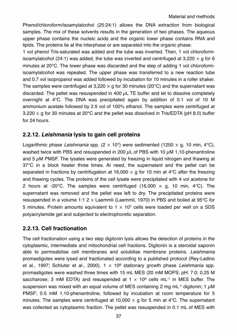
Material and methodsPhenol/chloroform/isoamylalcohol (25:24:1) allows the DNA extraction from biological samples. The mix of these solvents results in the generation of two phases. The aqueous upper phase contains the nucleic acids and the organic lower phase contains RNA and lipids. The proteins lie at the interphase or are separated into the organic phase.1 vol phenol Tris-saturated was added and the tube was inverted. Then, 1 vol chloroform-isoamylalcohol (24:1) was added, the tube was inverted and centrifuged at 3,220 ⨉ g for 6 minutes at 20°C. The lower phase was discarded and the step of adding 1 vol chloroform-isoamylalcohol was repeated. The upper phase was transferred to a new reaction tube and 0.7 vol isopropanol was added followed by incubation for 10 minutes in a roller shaker. The samples were centrifuged at 3,220 ⨉ g for 30 minutes (20°C) and the supernatant was discarded. The pellet was resuspended in 400 µL TE buffer and let to dissolve completely overnight at 4°C. The DNA was precipitated again by addition of 0.1 vol of 10 M ammonium acetate followed by 2.5 vol of 100% ethanol. The samples were centrifuged at 3,220 ⨉ g for 30 minutes at 20°C and the pellet was dissolved in Tris/EDTA (pH 8.0) buffer for 24 hours.
2.2.12. Leishmania lysis to gain cell proteinsLogarithmic phase Leishmania spp. (2 ⨉ 107) were sedimented (1250 ⨉ g, 10 min, 4°C), washed twice with PBS and resuspended in 200 µL of PBS with 10 µM 1,10-phenantroline and 5 µM PMSF. The lysates were generated by freezing in liquid nitrogen and thawing at 37°C in a block heater three times. At need, the supernatant and the pellet can be separated in fractions by centrifugation at 16,000 ⨉ g for 10 min at 4°C after the freezing and thawing cycles. The proteins of the cell lysate were precipitated with 4 vol acetone for 2 hours at -20°C. The samples were centrifuged (16,000 ⨉ g, 10 min, 4°C). The supernatant was removed and the pellet was left to dry. The precipitated proteins were resuspended in a volume 1:1 2 ⨉ Laemmli (Laemmli, 1970) in PBS and boiled at 95°C for 5 minutes. Protein amounts equivalent to 1 ⨉ 107 cells were loaded per well on a SDS polyacrylamide gel and subjected to electrophoretic separation.
2.2.13. Cell fractionationThe cell fractionation using a two step digitonin lysis allows the detection of proteins in the cytoplasmic, intermediate and mitochondrial cell fractions. Digitonin is a steroidal saponin able to permeabilise cell membranes and solubilise membrane proteins. Leishmania promastigotes were lysed and fractionated according to a published protocol (Rey-Ladino et al., 1997; Schluter et al., 2000). 1 ⨉ 108 stationary growth phase Leishmania spp. promastigotes were washed three times with 15 mL MES (20 mM MOPS; pH: 7.0; 0.25 M saccharose; 3 mM EDTA) and resuspended at 1 ⨉ 109 cells mL-1 in MES buffer. The suspension was mixed with an equal volume of MES containing 2 mg mL-1 digitonin; 1 µM PMSF; 0.5 mM 1.10-phenantroline, followed by incubation at room temperature for 5 minutes. The samples were centrifuged at 10,000 ⨉ g for 5 min at 4°C. The supernatant was collected as cytoplasmic fraction. The pellet was resuspended in 0.1 mL of MES with
37

Material and methods20 mM sodium phosphate; pH 7.0; 3 mM EDTA; 2 mg mL-1 digitonin and subjected to sonication (3 s, 50 W). Then, the samples were centrifuged at 10,000 ⨉ g for 5 minutes (4°C) and the supernatant was collected as intermediate fraction. The pellet was resuspended in 20 mM sodium phosphate (pH 7.0) and collected as mitochondrial fraction. The proteins of each fraction were precipitated with 4 vol of cold acetone for 2 hours at -20°C followed by centrifugation at 10,000 ⨉ g for 5 min at 4°C. The supernatant was discarded and the pellet was air-dried. The proteins were resuspended in 20 µL (equivalent to 1 ⨉ 108 cells) 1 ⨉ Laemmli (Laemmli, 1970) containing PBS, boiled at 95°C for 5 minutes and subjected to SDS-PAGE followed by Western blotting.
2.2.14. Isolation of protein detergent-resistant membranesProteins which contain a plasma membrane domain have been associated with insolubility in cold Triton X-100. Triton X-100 (C14H22O(C2H4O)n) is a non ionic surfactant that has a hydrophilic polyethylene chain and an aromatic hydrophobic group. Triton X-100 has been used to compare the protein extraction from insoluble micro-domains of the plasma membrane at low (0°C to 4°C) and high (25°C to 37°C) temperatures. The isolation of detergent-resistant membranes was performed by modification of a published protocol (Tull et al., 2004). 5 ⨉ 107 late logarithmic phase Leishmania spp. were sedimented at 1250 ⨉ g for 10 min (4°C) and washed twice with PBS. The cells were resuspended in 200 µL extraction buffer (1% Triton X-100; 50 mM Tris-HCl pH 7.4; 0.3 M NaCl; 5 mM EDTA, 10 µM 1,10-phenantroline; 5 µM PMSF) and kept at 0°C or 26 °C for 30 minutes. The samples were centrifuged at 14,000 ⨉ g (4°C) for 10 minutes. The supernatant was transferred to a new reaction tube and the proteins were precipitated by adding 4 vol of cold acetone for 2 hours at -20°C. The pellet was washed with PBS and frozen at -80°C. The samples containing the precipitated proteins from the supernatant fraction were centrifuged at 14,000 ⨉ g (4°C) for 10 minutes at 4°C. The supernatant was discarded and the pellet was air-dried. The proteins from each fraction were resuspended in 1 ⨉ Laemmli (Laemmli, 1970) containing PBS, boiled at 95°C for 5 minutes. The samples were subjected to SDS-PAGE followed by Western blotting.
2.2.15. Secretion assayThe identification of proteins that are secreted from the cells was performed by isolation of the supernatant fraction after incubation of the cells with serum-free medium (under 26°C or 37°C). Secreted proteins were isolated following an already described protocol (Twu et al., 2013). 2 ⨉ 108 late logarithmic phase L. donovani promastigotes were sedimented at 500 ⨉ g for 10 min (4°C) and washed thrice in 30 mL ice-cold PBS to remove any serum component from the medium. Then, the cells were seeded into 4 mL of 5% sucrose in PBS and kept at 26°C or 37°C for two hours. The cells were then sedimented by centrifugation at 500 ⨉ g for 10 min (4°C) and resuspended in 200 µL PBS with protease inhibitors (10 µM 1,10-phenantroline; 5 µM PMSF). The supernatant was transferred to a new 50 mL reaction tube with 10 µM 1,10-phenantroline, followed by filtration through a 0.20 µm pore
38

Material and methodssize membrane and concentrated using an Amicon filter system (MWCO 100,000 Da, Millipore). The concentrated secretomes and the cell pellets were stored at -80°C. For protein analysis 4 vol of ice-cold acetone were added to the samples and proteins were precipitated overnight at -20°C. The precipitated proteins were centrifuged at 13,000 ⨉ g for 5 minutes (4°C), the supernatant was discarded and the pellet was air-dried. The samples were then solubilised in PBS and 2⨉ Laemmli buffer, boiled for 10 minutes at 95°C and subjected to SDS-PAGE followed by Western blotting. Western blot analysis was performed using anti-ARM58 (1:200), anti-HSP90 (1:500) and anti-HSL-U1 (1:1000). Secondary antibodies were anti-IgY-AP (1:2000) and anti-IgG-AP (1:2000). One gel was stained with Coomassie blue as protein loading control.The verification of vesicles contained in the secretomal fraction was done with the trypsin digest (see section 2.2.20.8).
2.2.16. Exosome isolationExosomes are cell-derived vesicles with a diameter around 100 nm. They bud from the cell plasma membrane after the fusion with multivesicular bodies (MVB).Exosome isolation was performed by a modification of a published protocol (Silverman et al., 2010b). 1.2 ⨉ 109 late log-phase L. donovani promastigotes were sedimented at 500 ⨉ g for 10 min (4°C) and washed thrice in 30 mL ice-cold PBS to remove any serum component from the medium. The cells were seeded into 15 mL of 5% sucrose in PBS and kept at 37°C for 2 hours. The cells were then sedimented at 500 ⨉ g for 8 min (4°C). The pellet was resuspended in PBS and analysed microscopically to exclude cell destruction. The cell-free supernatant was transferred to a new 50 mL reaction tube with 10 µM 1,10-phenantroline, filtered through a 0.20 µm pore size membrane and concentrated using an Amicon filter system (MWCO 100,000 Da, Millipore). The concentrated exosomes were loaded on a sucrose cushion (2 mL of ice-cold PBS over 750 µL of ice-cold 1 M sucrose, 20 mM Tris-HCl pH 7.4) in an ultra-clear Beckman Coulter® 5.2 mL tube. The tubes were subjected to ultracentrifugation at 100,000 ⨉ g for 2 hours (4°C) and washed with ice-cold PBS, followed by ultracentrifugation in 1.5 mL tubes (Microfuge® Tube, Beckman Coulter®) at 125,000 ⨉ g for 1 hour (4°C). The exosomes were resuspended in 20 mM Tris-HCl (7.4) and stored at 4°C. To confirm the exosome nature of the purified vesicles, the trypsin digestion assay and visualisation under transmission electron microscopy (TEM) were performed.
2.2.17. Flow cytometry SYTOXSYTOX is a green-fluorescent nucleic acid counterstain. It allows to quantify and differentiate cells in dependence of their cell cycle phase: G1 (2n), S or G2 (4n). Leishmania spp. promastigotes were incubated at 25°C for 72 hours in supplemented M199 at a density of 1 ⨉ 106 cells mL-1 in absence or presence of miltefosine or SbIII. The inhibitory effect of the drugs on the cell growth was monitored using a CASY cell counter system. The cells were sedimented at 500 ⨉ g for 10 minutes (4°C), washed twice with
39

Material and methodsice-cold PBS and fixed with 70% ethanol for 30 minutes at 4°C. The cells were sedimented at 500 ⨉ g for 10 min at 4°C and washed with 1 mL 50 mM EDTA. The fixed cells were sedimented and resuspended in 1 mL PBS with 20 µg mL-1 RNAase A. The samples were incubated for 20 min at 37°C. After incubation, the samples were centrifuged at 13,000 ⨉ g for 2 minutes and the pellet was resuspended in 500 µL citrate buffer (45 mM MgCl2, 30 mM sodium citrate, 20 mM MOPS pH 7, 0.1% Triton X-100) with 3 µM SYTOX. The samples were incubated for 20 minutes at 26°C in the dark and washed twice with 5% PBS-iFCS (inactivated fetal calf serum). The samples were centrifuged at 13,000 ⨉ g for 4 min (4°C) and the pellet was resuspended in 1 mL 4% paraformaldehyde (PFA) in PBS. The stained samples were subjected to fluorescence-activated cell sorting analysis [λex=488 nm; λem=515 nm (FITC)] using the BD AccuriTM Flow Cytometer (BD Biosciences). Ten thousand events were analysed using the FlowJo 10 software.
2.2.18. Flow cytometry PI/annexin VPropidium iodide (PI) is a red-fluorescent DNA counterstain which stains dead cells. Annexin V is used as a marker to detect apoptosis processes since it has a strong interaction with phosphatidylserine (negatively charged). Phosphatidilserine is kept in the internal lipid layer. When cells undergo apoptosis the phosphatidilserine changes the position and is exposed to the external medium. Cells that are positive for annexin V but not for PI are in early apoptosis events. Cells that are positive for both events are dead cells. Leishmania spp. promastigotes were incubated at 25°C for 72 hours in supplemented M199 at a density of 1 ⨉ 106 cells mL-1 in absence or presence of miltefosine or SbIII. 2 ⨉ 107 cells were sedimented at 500 ⨉ g for 10 minutes at 4°C, washed twice with ice-cold PBS and resuspended in 1 mL binding buffer (10 mM HEPES pH 7.4; 140 mM NaCl, 5 mM CaCl2) containing 10 µg mL-1 propidium iodide and annexin V. The samples were incubated at room temperature for 10 min in the dark. Next, the cells were sedimented at 500 ⨉ g for 10 minutes at 4°C and resuspended in 1 mL 4% paraformaldehyde in PBS. The stained cells were subjected to FACS analysis [λex=488 nm; λem=515 nm (Annexin V) and λem=617 nm (PI)] using BD AccuriTM Flow Cytometer. Ten thousand events were analysed using the FlowJo 10 software.
2.2.19. Molecular methods2.2.19.1. Point mutation PCR The polymerase chain reaction (PCR) is a method, which uses the heat-stable polymerase for the synthesis of DNA from desoxynucleotide substrates on a single-stranded DNA template. Subsequent heating and cooling steps lead to template denaturation, annealing of primers (short DNA oligonucleotides complementary to the target sequence) and enzymatic replication of the target sequence. The usage of primers containing single nucleotide exchanges in their sequence will lead to PCR-products, which are different to the target sequence by these single nucleotide exchanges. The introduction of single
40

Material and methodsnucleotide exchanges to a DNA sequence by PCR is called mutagenesis PCR and was described before (Hombach et al., 2013).In the present thesis, mutagenesis PCR was used to exchange the codons for the amino acid (aa) cysteine at positions aa27, aa145 and aa271 of ARM58 to codons for the aa serine.The primers used in the mutagenesis PCR needed to be phosphorylated prior to the PCR. Six primers containing the required nucleotide exchanges (section 2.1.5) were applied to the phosphorylation reaction to a final concentration of 10 µM, mixed with 1 mM ATP, 1 ⨉ polynucleotide kinase A and 10 units of T4 polynucleotide kinase. The reaction volume was adjusted to 20 µL with ddH2O and incubated for 20 minutes at 37°C, followed by incubation at 75°C for 10 minutes for the enzyme inactivation. The phosphorylated primers were stored at -20°C. The PCR was performed using the iProof PCR kit (that uses a Pyrococcus-like enzyme) following the manufacturer’s instructions. The phosphorylated forward and reverse primers were added at 1.2 µM each to the provided mix, 12.3% DMSO and 2 ng of template (pUC19_ARM58) DNA were added. The reaction volume was adjusted to 25 µL with ddH2O.The PCR was performed in a thermocycler with the following program:
The PCR products were purified on an agarose gel.
2.2.19.2. Agarose gel electrophoresisAgarose gel electrophoresis allows the separation of proteins, DNA or RNA. Nucleic acids are negatively charged and migrate through the gel matrix to the anode of the gel chamber and are separated depending on their sequence length. The nucleic acids, which were amplified by PCR or were enzymatically restricted were mixed with 6 ⨉ denaturing loading buffer, loaded in a 0.8-1.5% TAE-buffered agarose gel and separated electrophoretically at 10 V/cm for 1-2 hours. A 1 kb DNA standard was loaded and separated on the same gel for the fragment length prediction in the sample. The separated DNA was stained with ethidium bromide (0.1 µg mL-1 in the gel), that intercalates in the DNA and allows the visualisation with a UV transilluminator.
Step Temperature (°C) Time
98°C 30 sec
Denaturation 98°C 1 min 29 cycles
Annealing 60°C 30 sec
Extension 72°C 1 min
72°C 5 min
41

Material and methods
2.2.19.3. Extraction of the DNA from the agarose gelThe DNA-bands of expected size were excised and purified with the Nucleospin® Gel and PCR Clean up kit according with manufacture’s instructions.
2.2.19.4. Restriction of DNAThe restriction endonucleases are able to cut DNA at specific, palindromic restriction sites thereby producing 3’-overhangs (sticky ends) or blunt ends.In the present work, the endonucleases were used either to excise a target DNA sequence for the cloning into another plasmid (preparative digest) or to confirm a plasmid sequence after cloning (analytical digest). The preparations for both reactions were performed as followed:
All restriction reactions were performed at 37°C in a water bath for 1 to 2 hours. In the preparative reaction, 30 units of the enzyme were added to the reaction after 1 hour of incubation. The reaction was stopped by adding denaturing DNA-loading buffer to the samples and subjecting them to agarose gel electrophoresis.
2.2.19.5. LigationLigation is an enzymatic reaction to covalently link two ends of DNA or RNA. There are two types of ligation: blunt-end and sticky-end, in the latter a ratio of 3:1 insert:vector is needed.The reaction was performed in a final volume of 20 µL, with T4-DNA-Ligase buffer and 80 U of the bacteriophage T4 ligase per sample, which catalyses the formation of a phosphodiester bond between juxtaposed 5’ phosphate and 3’ hydroxyl termini in duplex DNA. The reaction was incubated for 3 hours at room temperature for the blunt-end ligation or overnight at 4 °C for the sticky-end ligation. The ligation products were used to transform E. coli.
2.2.19.6. Chemical transformation of bacteriaTransformation is the process in which foreign DNA is delivered into a cell. E. coli DH5α were used for expression of plasmids and BL21 [pAPlacQ] were used for the protein expression.
Analytical digest Preparative digest
Amount of DNA 1 µg 10 µg
Restriction enzyme 2 units 60 units
Buffer 2 µL 10 µL
ddH20 ⨉ µL ⨉ µL
Final volume 20 µL 100 µL
42

Material and methodsIn the transformation of E. coli DH5α, 5 µL from the ligation sample was added to 50 µL of competent cells. A control test without plasmid DNA was also performed. The samples were incubated on ice for 30 minutes. Then a 42°C heat shock was done in a water bath for 20 seconds and the samples were incubated on ice for 2 minutes. Next 950 µL of pre-warmed circlegrow medium was added and the samples were shaked for 1 hour at 37°C. The cells were sedimented at 3,220 ⨉ g for 5 minutes at room temperature and 850 µL of the supernatant was discarded. The pellet was resuspended in the remaining volume and each sample was spread on LB plates with ampicillin. The LB plates were incubated overnight at 37°C.The ARM56 recombinant protein was expressed in the pJC45 vector that contains a deca histidine-tag. E. coli BL21 were transformed with 0.2 µg of plasmid DNA. E. coli cells were incubated on ice for 30 minutes with the plasmid or without (control). Afterwards, the cells were heat shocked for 30 seconds at 42°C followed by 2 minutes on ice. Then, 1 mL of pre-warmed circlegrow medium was added and incubated in a 37°C shaker for one hour. Then, 50 µL were plated on LB agar plates containing Kanamycin/Ampicillin. The plates were incubated at 37°C overnight.
2.2.19.7. Electroporation of E. coliApproximately 100 ng of cosmid DNA recovered from Leishmania were mixed with XL1-Blue Electroporation-Competent Cells on ice. Electroporation was performed following the manufacturer’s protocol. The transformed bacteria were transferred to 400 mL circlegrow medium under ampicillin selection and incubated in a 37°C shaker overnight.
2.2.19.8. Isolation of Plasmid DNA by alkaline lysisPlasmid DNA mini-preparation from E. coli DH5α was performed by alkaline lysis (Sambrook, 2001). 8 - 12 clones obtained from the transformation were picked, inoculated in sterile 13 mL tubes with 2 mL circlegrow medium with 0.05 µg mL-1 ampicillin and incubated at 37°C overnight. The cells were transferred to 2 mL reaction tubes and sedimented at 13,000 ⨉ g for 2 minutes (4°C). The supernatant was removed and the cells were resuspended in 100 µL of solution 1. Then 200 µL of solution 2 were added, mixed by inversion and incubated for 3 minutes at room temperature. After that 150 µl of solution 3 were added, mixed by inversion and incubated for 10 minutes at 4°C. Then the samples were centrifuged at 13,000 ⨉ g for 10 minutes (4°C). The supernatant was transferred to a new 1.5 mL reaction tube. Then 1 mL of 96% ethanol was added and incubated for 5 minutes at room temperature. The tubes were centrifuged at 13,000 ⨉ g for 10 minutes at room temperature. The supernatant was removed and the pellet was washed with 500 µL of 70% ethanol. The samples were centrifuged at 13,000 ⨉ g for 10 minutes at room temperature. The supernatant was removed, 40 µL of TE-RNaseA buffer were added and the samples were incubated for 30 minutes at 37°C.
43

Material and methods
2.2.19.9. Plasmid purification by caesium chlorideSingle clones with the correct constructs were picked and inoculated in 200 mL of circlegrow medium with 0.05 µg mL-1 ampicillin. The cultures were incubated in a 37°C shaker overnight. The cultures were transferred to centrifugation tubes and the cells were sedimented at 5,000 ⨉ g for 20 minutes at 4°C (JA 10 rotor, Beckman Coulter). The supernatant was removed and the pellets were resuspended in 5 mL buffer 1 (50 mM glucose, 10 mM EDTA, 25 mM Tris pH 8.0), followed by addition of 10 mL buffer 2 (0.2 N NaOH, 1% SDS) and the samples were incubated for 5 minutes at room temperature. Afterwards, 7.5 mL of buffer 3 (3 M potassium acetate, 2 M acetic acid) were added and the samples were incubated on ice for 10 minutes. The reaction tubes were centrifuged at 5,000 ⨉ g for 30 minutes at 4°C (JA 12 rotor, Beckman Coulter). The supernatant was filtered and transferred to a new 50 mL reaction tube followed by addition of 0.7 volumes of isopropanol. The samples were centrifuged at 3,220 ⨉ g for 20 minutes at room temperature. The supernatant was discarded and the pellet was washed with 10 mL 70% ethanol. The samples were centrifuged at 3,220 ⨉ g for 10 minutes at room temperature. The supernatant was discarded and 4 mL TE buffer (pH 8.0) were added, leaving to dissolve completely in a roller shaker at room temperature. Ethidium bromide was added to a final concentration of 0.36 mg mL-1. Next, 4.9 g of caesium chloride was added and left to dissolve completely. The samples were transferred into a 5 mL Quick-Seal tube (Beckman coulter) and centrifuged at 390,000 ⨉ g for 8 hours at 25°C (NVT90 rotor, Beckman Coulter). Due to the caesium chloride gradient, DNA appears as a horizontal band. The band corresponding to the DNA was taken using a 3 mL syringe coupled to a 0.9 µm needle and it was placed into a fresh 50 mL reaction tube. 1 volume of NH4 saturated acetate solution in isopropanol was added and mixed. Then two phases are generated and the upper band, containing ethidium bromide in the isopropanol mixture, was discarded and the step was repeated. Next, 0.1 volume of 7.5 M NH4-acetate was added. 2 volumes of ddH2O were added and mixed followed by 2.5 volumes of 96% ethanol. It was mixed properly and left for 20 minutes at room temperature. Then, the reaction tubes were centrifuged at 3,220 ⨉ g for 30 minutes (RT) and the supernatant, containing the ethanol, was removed. The pellet was dissolved in 200 - 500 µL TE buffer (pH: 8.0) and kept at 4°C for 24 hours. Then the concentration of DNA was quantified spectrophotometrically and the constructs were verified by sequencing.
2.2.19.10. Purification of cosmids from E. coliThe NucleoBond® Xtra Maxi (Macherey-Nagel) was used to isolate the cosmid population reisolated from L. infantum and electrotransformed in XL1 blue cells. The purification was performed following the manufacturer’s recommendation for low copy plasmid DNA purification.
44

Material and methods
2.2.19.11. DNA-concentration (photometric)The DNA concentration was determined by measuring the absorbance (optical density, OD) of the liquid sample at 260 nm in a spectrophotometer. An absorbance of 1.0 at 260 nm corresponds to 50 µg mL-1 of pure double strand DNA. The DNA purity can be assessed by the ratio of the OD260/OD280. A ratio of 1.8 represents pure DNA, RNA and proteins can be present as contaminants in the DNA samples. RNA absorbs light at 260 nm resulting in a higher OD260/OD280 ratio. The aromatic amino acids in proteins, however, absorb light at 280 nm and therefore lead to produce a lower OD260/OD280.
2.2.19.12. DNA-concentration (fluorometric)DNA samples that had a lower concentration of 100 ng µL-1 (e. g. for the preparation of the NGS library) were measured with a Qubit® 3.0 fluorometer. The Qubit® fluorometer uses fluorescent dyes that emit only when bound to target molecules, even at low concentrations. The preparation of the samples and measurement was performed according to manufacturer’s instructions.
2.2.19.13. DNA sequencing (Sanger)The sequence of the cloned plasmids was verified by the Sanger sequencing method by the LGC Genomics company (Berlin). For the reaction, 1 µg of plasmid-DNA and 1.4 µM forward and reverse specific primers for the target sequence in a final volume of 14 µL were prepared. The obtained sequence chromatogram was aligned to the in silico assembled plasmid using MacVector® software package.
2.2.19.14. Next Generation Sequencing (NGS)Next-Generation Sequencing is a term used to describe modern high-throughput sequencing technologies that allow a quicker and cheaper sequencing of DNA and RNA than the Sanger sequencing method. Millions of nucleic acid fragments are sequenced simultaneously in one reaction with a base pair coverage of 50 and higher, thus generating reliable results. One of these sequencing technologies is the sequencing by synthesis (SBS) from Illumina. For this technique a gDNA sample is fragmented and special adaptors are ligated to both fragment ends. Then, the library is loaded into a flow cell and the nucleic acid fragments hybridize to the surface. The template is bridge-amplified (Figure 6, (Yuan Lu, 2016)) resulting in clusters, which are composed of cloned templates. After the amplification of the template the sequencing starts with the extension of the sequencing primer. Based on the template sequence, one of the four fluorescent nucleotides is incorporated into the growing nucleic acid chain at each cycle and emits a fluorescent signal. The signals of each cluster are detected in each cycle and represent the sequence of the template. The process is called SBS. The Illumina workflows consist of library preparation and sequencing.
45

Material and methods
In this study, a cosmid-DNA library bearing Leishmania spp. were challenged with drugs and other toxic compounds for the selection of gene loci (section 2.2.10). The cosmids from selected Leishmania infantum were re-isolated (section 2.2.11) and enriched in bacteria (section 2.2.19.7). The purified cosmids were then set to a concentration of 0.5 ng µL-1 and 1 ng was used for the creation of the Nextera XT DNA library (Illumina) using the Nextera XT library prep kit and Nextera XT Index kit, following manufacturer’s protocol for the library preparation. The libraries were purified using the Agencourt AMPure XP PCR purification system (Beckman Coulter) following the manufacturer’s instructions. The quantification of each library was performed in a Qubit® 3.0 fluorometer and on a Bioanalyzer (Aligent) using the high sensitivity DNA analysis kit (Agilent), according to manufacturer’s instructions. The high sensitivity DNA kit allows the analysis of the quantity, quality and average fragment length of the DNA library. Each DNA library was set to 4 nM. The libraries were pooled, denatured with 0.2 N NaOH and diluted to 10 pmol mL-1 with chilled hybridisation buffer HT1 provided by the MiSeq reagent kit v3. A reference library (PhyX) was added to the sample at 0.2 pM. The library was heat-denatured at 95°C for 2 minutes and chilled in an ice bath. 10 pmol were loaded on the reagent cartridge (Illumina) and the sequencing of the libraries was performed in the MiSeq System (Illumina). The sequence reads were aligned to the reference genome of L. infantum (TriTrypDB-26_LinfantumJPCM5_Genome.fasta) using the assembler module of the Mac Vector® software package, Bowtie 2.0 algorithm. Settings were Fast alignment, end-to-end, 6 bases no gap, 4 threads, paired-end alignment and insert size 200-1500 bp.
2.2.19.15. Relative parasite load quantification by semi-quantitative PCRThe quantification of relative parasite load was calculated by TaqMan® probe-based qPCR. The TaqMan probes contain a fluorochrome covalently attached to the 5’-end and a quencher at the 3’-end. The probe anneals to the DNA region that will be amplified by the set of primers. During the synthesis of the nascent chain, the probe is degraded by the 5’ to 3’ exonuclease activity of the Taq polymerase. The fluorescence of the released fluorochrome is not quenched by the quencher and the emission can be detected. The
Figure 6. Drawing of Illumina genome analyser sequencing (Yuan Lu et al., 2016). NGS library is performed by fragmenting a sample and specialised adaptors are ligated to both ends of the fragment. Library is loaded into a flow cell where the fragments bind to the flow cell surface and are amplified (cluster generation). The emission wavelength and intensity are recorded to identify the base repeating “n” times (sequencing).
46

Material and methodsfluorescence emission is proportional to the amount of template. The advantage of the TaqMan qPCR method is the usage of differently labelled probes which allow the amplification of several genes in the same sample.For the relative quantification of parasites in the infected host cells (section 2.2.7), the actin genes of Leishmania spp. and mice (macrophage, host cell), respectively were amplified using primer and probe sets specific for the particular gene (section 2.1.6) (Bifeld et al., submitted). The probe, specific for the murine actin gene was labelled with the Cy5 fluorochrome and the parasite’s actin gene-specific probe was labelled with the FAM fluorochrome.Genomic DNA was isolated from BMMs (section 2.2.7) and used as a template for the TaqMan qPCR. The qPCR was performed using the KAPA probe fast qPCR kit master mix (VWR company) following the manufacturer’s instructions. Primers were applied to the master mix at a final concentration of 300 mM each. Gene-specific probes were added to the master mix at a final concentration of 200 nM. The template volume did not exceed 10% of the final reaction volume, which was adjusted with nucleic acid-free ddH2O to 20 µL and its amount did not exceed 250 ng.A one step PCR was performed on a Rotor-GeneTM Instrument with the following cycling program:
The cycle threshold (Ct) values from the exponential amplification phase were used for the relative quantification of the parasite load in the experimental samples. The Ct values from the parasites actin gene amplification were subtracted from the Ct values from the mouse actin gene amplification in the same sample (ΔCt-method). The normalised values are used to calculate the amplification difference between the sample (e. g. drug treated cells) and the control (e g. non treated cells) (ΔΔCT=ΔCtsample-ΔCtcontrol). The relative parasite load is then expressed by R (ratio)= 2-ΔΔCt. The significance was calculated by the paired student’s t-test.
2.2.20. Protein biochemistry2.2.20.1. Recombinant protein expression and purification in E. coli- Protein expressionTwo clones were picked and inoculated in 100 mL of circlegrow medium with 0.1 µg mL-1 ampicillin, 0.02 µg mL-1 kanamycin and 0.1% glucose in 1 L glass flasks and incubated at 37°C. The optical density (OD) was measured in a spectrophotometer at 600 nm wave
Step Temperature (°C) Time Cycles
Enzyme activation 95°C 7 min Hold
Denaturation 95°C 10 sec 40
Annealing/extension/data acquisition
66°C → 58°C (*) 25 sec
(*) A temperature touch down was performed to obtain specificity for the amplification, by lowering of 0.5°C for 16 cycles.
47

Material and methodslength. The induction with 0.4 mM IPTG started at OD600nm between 0.05-0.1 (approximately after 4 hours) and the flasks were incubated in a shaker at 37°C (1 mL of the medium was taken before inducing, pre-induction sample). Two hours after induction, a 1 mL aliquot of the culture was taken to be measured in the spectrophotometer (post-induction sample) and the rest of the cells were harvested at 5,000 ⨉ g for 20 minutes (4°C) (JA 10 rotor, Beckman Coulter). The supernatant was discarded and the pellet was resuspended in 20 mL of PBS and transferred to a 50 mL reaction tube. The samples were centrifuged at 3,220 ⨉ g for 20 minutes at 4°C (JA 12 rotor, Beckman Coulter). The supernatant was discarded and the pellet frozen at -70°C. The pellet was resuspended in 20 mL buffer 1 (20 mM Tris-HCl, 500 mM NaCl, 5 mM imidazole) and sonicated for 20 seconds (50 W) with a 10 seconds break, repeated 6 times. The samples were centrifuged at 10,330 ⨉ g for 30 minutes at 4°C and the supernatant was transferred to a new tube and frozen at -70°C (previously an aliquot of 50 µL was taken as post-induced soluble protein). The pellet was resuspended in 20 mL of buffer 2 (20 mM Tris-HCl, 500 mM NaCl, 5 mM imidazole, 8 M urea) and left for 1 hour in a roller shaker at 4°C. Then, the samples were centrifuged at 10,330 ⨉ g for 30 minutes at 4°C. A 1 mL aliquot was taken as induced protein-inclusion bodies. SDS PAGE was performed to detect whether (His)10-tagged ARM56 was expressed after induction and in which fraction it was. The fractions of interest were pooled for purification.
- Protein purificationThe fractions with the recombinant protein were subjected to a refolding assay. For this, 1 L of pre-cooled buffer B5 (20 mM Tris-HCl pH 8.0; 0.5 M KCl, 5 mM imidazole) was transferred to a measurement cylinder with a magnetic stir bar and kept at 4°C. The sample was added drop wise (sample in the 8 M urea solution) to the buffer B5. The (His)10-tagged ARM56 recombinant protein was purified by metal chelate affinity chromatography in a 10 mL matrix of beads charged with NiSO4 (Novagen His-Bind) in a 50 mL column body. The column was equilibrated with two volumes of 5 mL of buffer B5 with a flow rate of 0.5 mL min-1. After adding the volume to the column, it was washed with 10 mL buffer B5 followed by washing with 5 mL buffer B100 (20 mM Tris-HCl pH 8.0; 0.5 M KCl, 100 mM imidazole) and finally eluted with 10 mL buffer B1000 (20 mM Tris-HCl pH 8; 0.5 M KCl, 1 M imidazole). Then, a total of 12 fractions were collected. All the fractions were loaded onto a SDS PAGE gel to detect which fraction contains the eluted protein.The fractions that contained the protein were collected and subjected to dialysis with a pore cut of MWCO 6 - 8,000 Da against PBS at 4°C (2 changes, 4 hours). The samples were pooled and stored at -70°C. The samples were ultracentrifuged at 100,000 ⨉ g for 1 hour (4°C) to discard possible aggregates. The soluble supernatant was aliquoted and stored at -70°C.
48

Material and methods
2.2.20.2. Immunisation of laying hens for antibody productionLaying hens have been used for the immunisation and preparation of antibodies. The extraction of IgY from the egg yolk can be performed by polyethylene glycol (PEG) precipitation (Polson et al., 1985; Polson et al., 1980). The purified (His)10-tagged ARM56 was used to immunise laying hens. Immunisation of laying hens was performed in accordance to §10a of the German Animal Protection Law and registered with the Amt für Gesundheitlichen Verbraucherschutz, Behörde für Umwelt und Gesundheit, Freie und Hansestadt Hamburg. Therefore, 500 µg of recombinant protein were mixed 1:1 with the TiterMax® Gold Adjuvant. Ten days after immunisation, the IgY was isolated to test the primary titer. Then, the hens were boostered and after another 10 days, the IgY was isolated to test the antibody titer.
2.2.20.3. Isolation of IgYThe egg yolk was separated from the egg white and it was washed with distilled water. The membrane of the egg yolk was removed and the egg yolk was transferred to a 50 mL reaction tube. Then 1 volume of potassium-phosphate-buffer (10 mM K-phosphate pH 7.2, 100 mM NaCl) was added to the egg yolk and it was incubated in a roller shaker to mix, followed by the addition of 1 volume of 7% PEG 6000 in potassium-phosphate-buffer and mixed. The samples were centrifuged at 16,000 ⨉ g for 10 min (4°C) and the supernatant was filtered through a gauze swab and paper filter. Next 10% PEG 6000 was added to the supernatant and left to dissolve, followed by centrifugation at 16,000 ⨉ g for 10 minutes (4°C). The supernatant was discarded and the pellet was solved in 10 mL potassium-phosphate-buffer and left to dissolve in a roller shaker for 30 minutes. Then 1 volume of 24% PEG 6000 in potassium-phosphate-buffer was added and mixed, followed by centrifugation at 16,000 ⨉ g for 10 minutes (4°C). The supernatant was discarded and 5 mL of potassium-phosphate-buffer was added to the pellet and left to dissolve in a roller shaker. The sample was centrifuged at 16,000 ⨉ g for 60 minutes (4°C) and the supernatant was transferred to a new reaction tube with 0.02% sodium azide.
2.2.20.4. Non-denaturing PAGEIn a native gel electrophoresis, protein mobility depends on the charge-to-mass ratio, physical shape and size of the protein. Non-denaturing protein extraction, non-denaturing gradient gel electrophoresis and Western blot were performed for isolation of oligomers and monomers. Cells in logarithmic growth were counted with a CASY® counter and an equal of 2 ⨉ 107 L. donovani promastigotes were sedimented (1250 ⨉ g, 8 min, 4°C), washed twice with 5 mL cold PBS and resuspended in 40 µL extraction buffer (15% glycerol, 0.5 mM 1,10-phenanthroline, 10 mM Tris-HCl pH 8.0, 70 mM KCl). Samples were subjected to three cycles of freezing (liquid nitrogen) and thawing (block heater at 37°C) for three minutes each. Then a small amount (<10) of 2 mm ceramic beads (Ribolyser beads, Hybaid) were added to the tubes to break the DNA and it was shaken vigorously for 20 seconds. Cell lysates were subjected to centrifugation (16,000 ⨉ g, 10 min, 4°C) and
49

Material and methodsthe supernatant, which contained the soluble protein fraction, was mixed (5:1, v/v) with loading buffer (50% glycerol, 0.1% bromophenol blue). The samples were electrophoretically separated on a 4 - 18% polyacrylamide (2.5 - 6% glycerol) gradient gel in 0.5 ⨉ Tris-borate-EDTA buffer (24 h, 20 V/cm, 4°C). Then, the gel was equilibrated at 60°C in transfer buffer (48 mM Tris, 39 mM glycine, 0.5% SDS, 20% methanol, 10 mM DTT) for 30 min, followed by Western transfer and immunological detection.
2.2.20.5. SDS-PAGESDS-PAGE (Sodium Dodecyl Sulfate Polyacrylamide Gel Electrophoresis) allows the separation of proteins according to their electrophoretic mobility. SDS is an anionic surfactant that denatures and disassociates proteins by binding to the polypeptide chain which gives an approximate distribution of charge per mass. PAGE is a technique that separates macromolecules according to their electrophoretic mobility. Proteins can be separated according to their length, conformation and charge. Using SDS-PAGE, proteins migrate through the gel matrix to the anode due to the negative charge given by the SDS molecules associated to the amino acids. Electrophoresis allows the determination of an approximate size of proteins when comparing their migration with defined molecular mass standards.The lysates were loaded and separated on a discontinuous SDS-PAGE. Discontinuous gels were performed first by preparing the 10% resolving gel in 375 mM Tris-HCl (pH 8.8), 10% acrylamide/bis-acrylamide, 0.1% SDS, 0.1% APS and 0.1% TEMED. The gel solution was loaded between two glass plates of the electrophoresis chamber, followed by the addition of isopropanol. When the gel matrix was polymerised the isopropanol was discarded. Next, the 5% stacking gel that contained 125 mM Tris-HCl (pH 6.8), 5% acrylamide/bis-acrylamide, 0.1% SDS, 0.1% APS and 0.1% TEMED was loaded followed by the comb which provides the wells. After polymerisation, the comb was removed and the chamber was filled with running buffer (25 mM Tris, 250 mM glycine, 0.1% SDS). The gel was running for 15 V cm-1 for 1-2 hours. Two reference standards were used to identify the protein migration, an unstained protein marker in case of staining with Coomassie Blue and a prestained in case of being followed by Western transfer.
2.2.20.6. Coomassie Brilliant Blue stainingThe Coomassie Brilliant Blue R-250 dye allows the examination of protein bands from gels after electrophoresis. The dye has a detection sensitivity of 0.5 µg cm2 of protein present in a gel matrix. Protein gels were stained over night in staining solution (1 g L-1 Coomassie Brilliant Blue R-250, 40% ethanol; 10% acetic acid) at room temperature. To detect the protein bands, gels were destained in destaining solution (40% ethanol; 10% acetic acid) for 2 hours at room temperature. When the protein bands were visible, the gel was washed twice with ddH2O and it was scanned to digitalise the image. The gel was dried between two layers of cellophane for conservation.
50

Material and methods
2.2.20.7. Semi-dry Western blot (Immunoblot)Western blot is an analytical technique that allows the detection of specific proteins that are first separated in a gel matrix followed by transfer to a membrane, normally nitrocellulose or polyvinylidene difluoride (PVDF). The transfer of proteins was done using a PVDF membrane (Fuorotrans, 0.2 µm) equal to the size of the blot. The membrane was activated with methanol for 10 seconds and washed three times with ddH2O. The membrane was incubated shortly in blot-transfer-buffer with two pieces of 3MM Whatman paper. For the blotting, the Whatman paper was placed between the gel or membrane and the electrodes of the blotting chamber. The blotting conditions were determined by the size of the gel/membrane and the thickness of the gel. The proteins were transferred from the matrix gel to the membrane at 1 mA/cm2 for 60 min with a maximum voltage of 30 V.After Western transfer, the membranes were incubated overnight at 4°C or for 1 hour at room temperature with blocking solution (5% milk powder and 0.1% Tween 20 in Tris-buffered saline). Afterwards, the incubation with the first antibody (in blocking solution) was performed for 1 hour at room temperature. Then, the membrane was washed three times for 5 minutes with washing solution (0.1% Tween 20 in Tris-buffered saline), followed by the incubation with the second antibody-conjugated with AP (alkaline phosphatase) (in blocking solution) for 1 hour at room temperature. Then, the membrane was washed twice for 5 min with washing solution and the last incubation was done with the developing buffer. Then the blot was developed using the colorimetric detection of alkaline phosphatase activity. The substrates for the AP reaction were BCIP (5-bromo-4-chloro-3-indolyl-phosphate) and NBT (nitro blue tetrazolium). The reaction was done at room temperature until protein bands appeared, then the blot was washed twice with water and left to dry.
2.2.20.8. Trypsin digestThe trypsin digest was done in presence or absence of Triton X-100. Intact vesicles containing proteins are protected against the action of the trypsin. However, in presence of Triton X-100 the membranes are permeabilised and the trypsin can digest proteins. Trypsin (EC 3.4.21.4) is a serine protease that digests proteins into peptides. It hydrolyses the peptide bonds at the carbonyl group of the amino acids lysine or arginine. The verification of intact exosomes was performed in a modification of a published protocol (Bifeld et al., 2015; Silverman et al., 2010a). After ultra centrifugation, the intact exosomes were resuspended in a final volume of 75 µL 20 mM Tris-HCl (pH 7.4) and kept at 4°C overnight. The sample was splitted in three reaction tubes containing: i) 25 µL 20 mM Tris-HCl (pH 7.4); ii) 25 µL 20 mM Tris-HCl (pH 7.4) and 40 µg mL-1 trypsin or iii) 25 µL in Tris-HCl (pH 7.4) with 0.1% Triton X-100 and 40 µg mL-1 trypsin. The samples were then incubated at 37°C in a water bath for 2 hours. Trypsin was inactivated by adding PMSF at a final concentration of 22 mM. The sample was mixed vigorously for 10 seconds and proteins were precipitated by adding 4 vol of cold acetone for 2 hours at -20°C. The samples were centrifuged at 16.000 ⨉ g for 10 min at 4°C. The supernatant was discarded and the samples were air-dried. The samples were then subjected to SDS-PAGE and
51

Material and methodsWestern transfer. The membranes were probed with the specific antibodies anti-ARM58 (1:200), anti-ARM56 (1:400), anti-HSP90 (1:500), and anti-HSL-U1 (1:1000). It was followed by incubation with the second antibodies anti-IgY-biotin (1:2000) and anti-IgG-biotin (1:2000). The last step included the incubation of the membranes with streptavidin-AP-conjugate (1:5000) to perform the colorimetric detection.
2.2.21. Microscopy2.2.21.1. Transmission electron microscopy of promastigotesL. donovani promastigotes were incubated at 26°C or 37°C for 2 hours in serum-free medium. 2.5 ⨉ 108 parasites were sedimented at 600 ⨉ g for 8 min 4°C and washed three times with PBS. Next, the cells were fixed for 30 min with 2.5% glutaraldehyde and 4% paraformaldehyde in PBS. Then, the cells were washed three times, 5 min each, with PBS and postfixed with 2% OsO4 in PBS for 40 min on ice in the dark. Afterwards the cells were washed twice with ddH2O and treated with 0.5% uranyl acetate in ddH2O for 30 min. After rinsing thoroughly with ddH2O, the cells were dehydrated at room temperature in a graded ethanol series: 50, 60, 70, 80 and 95% for 5 min each, and then three times in 100% for 10 min each. The cells were immersed overnight at room temperature in an Epoxy resin/ethanol mix (50% v/v) and finally embedded in fresh 100% Epoxy resin. After polymerisation at 60°C for 48 hours, the embedded cell pellet was sectioned into 60-nm sections using a Leica EM UC7 ultramicrotome, and sections were collected on 300 mesh copper grids (Plano). The sections were examined with a Tecnai Spirit transmission electron microscope at 80 kV (FEI).
2.2.21.2. Transmission electron microscopy of exosomesAfter the second ultracentrifugation of the exosomes, the samples were fixed with 4% paraformaldehyde in PBS. The samples were incubated for 5 minutes on formvar-coated carbon grids (Plano) and washed once with distilled water. The samples were given contrast with 0.5% Uranil acetate for 5 minutes followed by examination with a Tecnai Spirit transmission electron microscope at 80 kV (FEI).
2.2.21.3. Indirect immunofluorescenceLog-phase promastigotes (1 ⨉ 107 cells) were sedimented at 750 ⨉ g, washed twice with PBS, and resuspended in 1 mL PBS. Aliquots containing 3 ⨉ 106 cells were applied on glass microscope slices. After drying, the cells were fixed for 8 minutes in ice-cold methanol and air-dried. The preparation was surrounded with PANPEN. The slides were washed (0.1% Triton X-100 in PBS) for 5 min followed by incubation with permeabilisation solution (0.1%Triton X-100, 50 mM NH4Cl in PBS) for 10 min. The slides were then washed three times with washing buffer for 5 min each, followed by incubation with blocking solution (2% bovine serum albumin, 0.1% Triton X-100 in PBS) for 1 hour. The slides were then incubated for 1 hour with the first antibodies diluted in blocking solution. The slides were washed three times and then incubated for 1 hour with the secondary antibodies coupled to FITC or to Alexa Fluor 594 and 4’, 6’-diamidino-2-phenylindole
52

Material and methods(DAPI). The slides were washed three times and then covered with Mowiol and cover slips. The slides were left to harden for 24 hours at 4°C. Fluorescence microscopy was performed on a Life Technologies EVOS FL Auto Cell Imaging System or on an Olympus FluViem1000 confocal microscopy (SIM-scanner and spectral detection).
2.2.21.4. Giemsa stainGiemsa’s solution is a mixture of the dyes methylene blue, eosin (acidic stain) and Azure B (basic stain). It is a standard stain used in laboratories to visualise microorganisms. In this study, Giemsa stain was used to visualise the morphology of the cells for establishing conditions and protocols. The cells were sedimented, washed and applied on glass microscope slides. Next, the slides were fixed for 3 minutes in cold methanol and stained with a 1/50 dilution of Giemsa’s azur eosin methylene blue solution for 10 minutes. The slides were washed three times with water and left to dry. Samples were visualised by transmitted-light microscopy using an EVOS XL Cell Imaging System (Thermo Fisher Scientific).
2.2.21.5. Transmitted-light microscopyCultures were visualised daily by transmitted-light microscopy to monitor the status of the cells using an EVOS XL Cell Imaging System (Thermo Fisher Scientific).
53

Results
3. Results3.1. Functional analysis of ARM58Leishmania has three well-known mechanisms involved in antimony resistance: i) down regulation of uptake transporters such as AQP1; ii) up regulation of ABC transporters and iii) increased levels of trypanothione.The SbIII resistance mechanism that involves ARM58 is unknown. This chapter describes the different approaches used to elucidate the molecular mechanism by which ARM58 confers resistance against SbIII.
3.1.1. Verapamil does not inhibit ARM58 mechanismP-glycoproteins are energy dependent multidrug efflux pumps. They consist of two identical subunits of which each has six transmembrane domain (TM) segments. The binding of the substrates takes place at the two transmembrane domains interface and this may be a common feature of multidrug efflux pumps (Pleban et al., 2005). ARM58 does not show any signature in its sequence for p-glycoproteins or for ATPases. However, ARM58 resistance mechanism may depend on P-glycoproteins or other energy-dependent transporters. This section describes the approach used to determine whether ARM58-mediated antimony resistance is depending on ATP hydrolysis. Verapamil is an inhibitor of drug efflux pumps such as p-glycoproteins. In this case, L. infantum promastigotes transfected with pCLN (empty vector) or ARM58 transgenes were challenged with various concentrations of SbIII in presence or absence of 10 µM verapamil. After 72 hours, the cell densities were recorded and normalized against the density of untreated cells to determinate the percentage of growth.
Over expression of ARM58 in the parasites protects against SbIII as it has been described previously (Schäfer et al., 2014). In the absence of SbIII, verapamil reduces the cell growth in the vector control by 17 % (Figure 7). A similar reduction can be observed at the various SbIII concentrations. Nevertheless, the protective effect of ARM58 over expression is not
Figure 7. Effect of verapamil in ARM58-mediated SbIII resistance. L. infantum promastigotes transfected with pCLN or ARM58 transgenes were grown in the presence (closed symbols) or absence (open symbols) of 10 µM verapamil and under varying concentrations of SbIII (0-400 µM). After 72 hours, cell densities were measured and plotted against the indicated SbIII concentrations. Values are the medians from four independent experiments.Data published in Schäfer et al., 2014. Sb
III (µM)
pCLN +VerapamilpCLN -VerapamilARM58 +VerapamilARM58 -Verapamil
1000 200 300 400
% G
rowt
h
0
50
100
25
75
54

Resultsaffected by verapamil addition (p = 0.0143). Therefore, ARM58-mediated antimony resistance does not depend on p-glycoprotein activity.
3.1.2. Sodium orthovanadate is not an inhibitor of ARM58 mechanismTransmembrane ATPases include transporters involved in the export of toxins across membranes. The same approach as described in the section 3.1.1. was used to see whether ARM58 function depends on energy-dependent transporters.Sodium orthovanadate is an inhibitor of protein tyrosine phosphatases, alkaline phosphatases and ATPases. L. infantum promastigotes transfected with pCLN and ARM58 transgenes were challenged with various concentrations of SbIII in the absence or presence of 50 µM sodium orthovanadate. After 72 hours, the cell densities were recorded and normalized against the density of untreated cells to determine the percentage of growth.
Sodium orthovanadate by itself reduces promastigote growth by ~30% for control (pCLN) and ARM58 over expressing parasites (Figure 8). Combined with SbIII, control cell growth is strongly reduced. Parasites over expressing ARM58 show the same 30% growth reduction under sodium orthovanadate at all SbIII concentrations (p = 0.0143), arguing against a dependence of ARM58 on ATP hydrolysis.
3.1.3. Conserved cysteines residues in ARM58 functionIt has been discussed that ARM58 could be involved in an extrusion pathway due to the fact that the protein was localized in the flagellar pocket using a mCHERRY::ARM58 fusion protein (Schäfer et al., 2014). The previous two results indicate that ARM58-mediated antimony resistance is not depending on p-glycoprotein or ATP hydrolysis. The next postulation included a possible transport of SbIII directly by ARM58.The cysteine side chains of some proteins are able to form a complex with metalloids such as AsIII and SbIII. The ArsA ATPase in Escherichia coli was reported to have three cysteine thiolates forming a complex with AsIII or SbIII (Bhattacharjee et al., 1995). Therefore it was tested whether ARM58 function depends on cysteine side chains. An alignment of the
Figure 8. Effect of sodium orthovanadate on ARM58-mediated SbIII resistance. L. infantum promastigotes transfected with pCLN or ARM58 transgenes were grown in the presence (closed symbols) or absence (open symbols) of 10 µM sodium or thovanadate and under vary ing concentrations of SbIII (0-400 µM). After 72 hours, cell densities were measured and plotted against the indicated SbIII
concentrations. Values are the medians from four independent experiments. Data published in Schäfer et al., 2014.
% G
rowt
h
0
50
100
25
75
1000 200 300 400
pCLN +O-VanadatepCLN -O-VanadateARM58 +O-VanadateARM58 -O-Vanadate
SbIII
(µM)
55

Resultsprotein sequences of ARM58 and ARM56 (previously, ARM58rel) was performed to see whether ARM58 and ARM56 have conserved amino acids (Figure 9). ARM58 and ARM56 have conserved cysteine residues in all four DUF1935. To determine whether these residues are involved in SbIII resistance, cysteines 27, 145 and 271 in DUF1935-1, -2 and -3, respectively were changed to serines (to substitute the thiol side chain and keep the electrostatic properties and tridimensional structure of the protein) by targeted mutagenesis of the expression plasmids. The three positions were chosen based on the relative importance of the DUF1935 for antimony resistance – DUF1935-4 is dispensable for resistance (Schäfer et al., 2014).
The cysteines 27, 145 and 271 were exchanged to serines using mutagenesis PCR and the mutants, bearing single or triple amino acid exchanges, were over expressed as episomes in L. infantum. The promastigotes were seeded and challenged with various concentrations of SbIII. After 72 hours, the cell densities were recorded and normalized against the density of untreated cells to determine the half maximal inhibitory concentration (IC50).
Figure 9. Protein sequence alignment between ARM58 and ARM56 (L. infantum). Cysteines in positions 27, 145 and 271 are conserved in the DUF1935.1, DUF1935.2 and DUF1935.3 (marked in red).These three amino acids were exchanged to serines in the expression vector pCLN-ARM58 by mutagenesis PCR.The ep i somes we re ove r expressed in L. in fan tum promastigotes to analyze the function of ARM58-mediated antimony resistance.The DUF1935.1, .2, .3 and .4 are marked in orange, green, blue and purple, respectively. = conserved amino acids in ARM58 and ARM56.
ARM58 1 -MTELVPPEAIRFANGEPLCAYQECYRCFDGR--DILFRLVNVEKKQWFF 47 ARM56 1 MTEEIVAPAVPQFENGEPSFEYDHIFRCFKGRGNGLLFRMVNTKQKKWAF 50 *.* * .* **** * .*** ** .***.** .*.* * ARM58 48 YNDTTDTIAHVCAVFYPGSVIRPLQRADMHVIPGPTGVADETCSIEVTLD 97 ARM56 51 YNDTADTIMQVRARFSPDCKVEKLNRARATQVPIGTEEHPDLFETVVTLE 100 ****.*** .* * * * . . *.** .* * . ***. ARM58 98 IQPGFTESFIEGEHKGFHLEFRTETAPTKEVVFEYRRPTVP-YDKIYRCF 146 ARM56 101 VHPFATEPFIKGDVNGFELEFHTEQIPVNDVKFMNRHRILPRYDKVYKCF 150 ..* ** ** *. ** ***.** * .* * *. .* ***.*.** ARM58 147 KN-GNGLLFRLVDERVRQWFFYNDTRDLMMKVSVAFANSAEVRPLGCTAL 195 ARM56 151 KSEGNGLFFRLVDEKDNKWYYYNDTHEFRMTATVSFPSADEVKPLGNTET 200 * **** ******. .*..****.. * .*.* . **.*** * ARM58 196 GVPHADTAPESVVYTLLIAPGCTEPFIEGNPVTYTLEFAAEPLDAPVTPA 245 ARM56 201 -VPVQDDESE-VAYQITVEPGNAEPFIEGVPASYHQTFNANPIDENCLDP 248 ** * * * * . . ** .****** * .* * * *.* ARM58 246 LEEPVQYLHGCPDVAVI-PHQTHVFKCFKEHGNGLLFRVVDDVNTIWAFY 294 ARM56 249 --KDVQYINGEPDSNIIDPSQCKVYKCFKENGNGLLFRLVDEKAGRWAFY 296 ***..* ** .* * *..*.*****.*******.**. **** ARM58 295 NDTPDYVMTANMRYPGTSDIRLAPGVQVIADSEREGGFVAAVEVLPLATV 344 ARM56 297 NDTHEYLMKPKVRFFGGAAVVPGPDAQVSPDPEEEDTAVITVEIPPCETR 346 *** .*.* .*. * . . * ** * * * * .**. * * ARM58 345 PFLVGAPEQYELAFAATPVVPLTPLPTTPAEPSPPPQLAAAADTATTVPP 394 ARM56 347 LFIEGRPAKYEVSVVADSIHK----------------------------- 367 *. * * .**.. * . ARM58 395 ALPVPEEAPVYCNSGPDLTVMPNIDEVYKCFKDYGNGLVFRLVDKVQRRW 444 ARM56 368 --TAPEESPEFVNGEPDRKVV-NYDAVYQCFKDKPEARLFRLVDSNRQQW 414 ***.* . * ** *. * * **.**** .***** ...* ARM58 445 AFYNDTTDVVALVRVQFSPGARIQPLGHTVLGCDLESGSVLHELRVNPLE 494 ARM56 415 GFYNDTADVFFVARFTFDAEGKVRALGDTEKEQNSDNETE-YLVSIPPME 463 *****.** . * * ... ** * . . . . *.* ARM58 495 TALFVEGDVDIFTTKFVAEQIQT----- 517 ARM56 464 TVAFVQGDVRGFKSQFTGKRRTSVPTPA 491 * **.*** * ..* . .
56

Results
L. infantum over expressing ARM58 have a higher tolerance to SbIII compared to the vector control cells as it was described previously (Schäfer et al., 2014). The single cysteine to serine exchanges at C27S, C145S and C271S produced an intermediate phenotype with partial loss of function in the protection of parasites against SbIII (Figure 10). The strongest effect was caused by the C27S mutation. However, the triple exchange showed no further reduction of activity, arguing against a cooperativity between the cysteine residues.This result shows that the cysteines 27, 145 and 271, which are conserved in the three DUF1935 might play a role in the structure of ARM58. The single amino acid exchange produced a partial reduction in the antimony resistance. However, the substitution of the three cysteines to serines did not lead to a complete loss of ARM58 function. From this data, it was concluded that binding of SbIII to the cysteine residues in positions 27, 145 and 271 is not at the core of ARM58 function in drug resistance.
3.1.4. ARM58 does not confer resistance against arsenicAntimony resistance mechanisms have been associated with arsenic resistance (Dey et al., 1996; Mukhopadhyay et al., 1996; Ouellette et al., 1998), both are metalloids and share similarities. Antimony resistance has also been associated with the presence of arsenic in the drinking water and soils in India (Perry et al., 2015; Perry et al., 2011; Perry et al., 2013).However, it was unknown whether over expression of ARM58 or ARM56 in Leishmania confers any protection against arsenic. To answer this question, L. donovani promastigotes transfected with pCLN, ARM58 and ARM56 transgenes were challenged with varying concentrations of AsIII. After 72 hours, the cell densities were recorded and normalized against the density of untreated cells to determinate the half maximal inhibitory concentration (IC50).
Figure 10. IC50 determination for SbIII in L. infantum promastigotes over expressing ARM58 transgenes with C27S, C145S and/or C271S. Promastigotes were challenged with various concentration of SbIII for 72 hours and the IC50 was determined. *, p < 0.05 (n = 4).
C27SC14
5SC27
1S
500
vecto
r
ARM58C27
SC14
5S
C271S
IC50
(µM
)400
300
200
100
0
* * *
57

Results
L. donovani promastigotes over expressing ARM58 or ARM56 were not protected against AsIII, when compared with the vector control (Figure 11). Even if SbIII and AsIII have similar properties, ARM58 is specific for SbIII.
The results obtained in this chapter using in vitro cell culture of promastigotes did not identify the mechanism of ARM58-mediated drug resistance. The data shows that i) the protection by ARM58 against antimony is not depending on p-glycoproteins or energy-dependent transporters; ii) cysteines in positions 27, 145 and 271, conserved between ARM58 and ARM56 in the DUF1935.1, -.2, and -.3 are not involved in complex formation with SbIII; and iii) over expression of ARM58 does not confer AsIII resistance.
3.2. Detection of ARM58
3.2.1. Over expression of ARM58 and SbIII challengeThe third DUF1935 (Domain of unknown function) of ARM58 contains a putative transmembrane domain which is also essential for the protein function (Schäfer et al., 2014). The production of specific anti-ARM58 IgY antibodies has been described in (Schäfer, 2013). ARM58 was expressed as recombinant protein in E. coli and used to immunize laying hens. In Western blots, the anti-ARM58 IgY recognized the (His)10-ARM58 but not the protein in Leishmania lysates. The IgY also failed in indirect immunofluorescence microscopy. It was then suggested that ARM58 may be glycosylated or subject to other post translational modifications and therefore masked against immunological detection.
In this work, the detection of ARM58 in cell lysates was performed by alternative sample preparation. The standard protocol for protein lysates includes the resuspension of the promastigotes is SDS-sample buffer before analyzing in a SDS-PAGE and Western blot. However, ARM58 could be only detected when the cells were lysated by freezing and thawing, fractionated and precipitated with acetone.ARM58 only confers antimony resistance upon over expression in Leishmania. To determine whether ARM58 is induced under SbIII challenge, L. infantum promastigotes transfected with pCLN or ARM58 transgenes were incubated with or without 200 µM antimony for 72 hours. Then, the cells were lysed and fractionated into soluble proteins
Figure 11. IC50 determination for AsIII in L. donovani promastigotes transfected with pCLN, ARM58 or ARM56 transgenes. Promastigotes were challenged with various concentration of AsIII for 72 hours and the IC50 was determined (n = 4). ns =. non-significant.
IC50
(µM
)
50
40
30
20
10
0
pCLN
ARM58
ARM56
pCLNARM58ARM56
ns ns
58

Results(supernatant) and insoluble proteins (pellet). The fractions were then analysed by SDS-PAGE and Western blot.
ARM58 migrates as a protein species of 80 kD (Figure12), mostly in the soluble fraction (Figure 12, lane 3). ARM58 levels are not induced under antimony challenge (lanes 5, 9). ARM58 is not detectable in the vector control after challenging with trivalent antimony (lane 5), probably due to cell disruption and protein loss. Upon over expression, ARM58 is still detected mainly in the soluble supernatant (lanes 7, 9). This experiment shows that i) ARM58 is a soluble protein, ii) it migrates like a 80 kD protein and iii) this is not due to post translational modifications since the bacterially expressed (His)10-ARM58 (lane 1) also migrates as a 80 kD species.ARM58 was found as a soluble protein, in spite of the putative trans-membrane domain in DUF1935.3. Therefore, its possible membrane association was investigated.
3.2.2. ARM58 is not membrane-associatedProteins can be modified covalently during or after its biosynthesis, this process is called post-translational modifications (PTMs). PTMs are involved in membrane targeting and signal transduction. Proteins can be attached to lipid molecules, for example, myristate (C14), palmitate (C16), farnesyl (C15), geranylgeranyl (C20) and glycosyl phophatidylinositol (GPI). PTMs are involved in membrane targeting and signal transduction.ARM58 does not contain a glycine residue at the N-terminus where myristoylation can take place. However, ARM58 contains a putative transmembrane domain in the DUF1935.3 which is important for the protein function (Schäfer et al., 2014). To find out a possible membrane association, a closer analysis of the protein sequence was done.Palmitoylation is a PTM where the amino acids cysteine, serine and threonine are covalently attached to palmitic acid. This process contributes to membrane association; it
Figure 12. Detection of ARM58 as recombinant protein and in lysates of L. infantum carrying pCLN and ARM58 transgenes. Promastigotes were challenged with 200 µM SbIII. After 72 hours, cells were lysed and separated into soluble supernatant (SN) and insoluble pellet (P). After SDS-PAGE and Western blot, ARM58 was detected with anti-ARM58 IgY and rabbit anti-chicken conjugated to alkaline phosphatase. Coomassie blue staining was done in parallel as loading control. Lane 2 = marker.
pCLN ARM580 200 0 200
PSN PSN PSN PSNµM SbIII
1 2 3 4 5 6 7 8
95 kDa
72 kDa
100kDa85 kDa70 kDa
9 10Coomassie Blue
anti-ARM58
59

Resultsis also involved in protein-protein interaction and sub cellular trafficking between compartments. S-palmitoylation can be reversed by a palmitoyl protein thioesterase. The ARM58 sequence was run in silico to localize possible amino acids susceptible to palmitoylation. The sequence was run in CSS-Palm, a palmitoylation site prediction (version 4.0) (Ren et al., 2008). Table 1 summarizes the data obtained from the algorithm.
Two possible cysteines in positions 192 and 477 may be palmitoylated, corresponding to the DUF1935.2 and the DUF1935.4 of ARM58. According to the algorithm, both cysteines had scores above the cutoff.The putative transmembrane domain of ARM58 is localized in the DUF1935.3, between the amino acids positions 331-351. The in silico analysis did not predict any putative palmitoylation sites in DUF1935.3.
In the previous section it was shown that ARM58 was recovered in the soluble fraction; however, upon over expression there was a partial detection in the insoluble fraction (Figure 12, lanes 8, 10). ARM58 could be palmitoylated at two cysteine residues according to the algorithm and therefore directed to the membrane. In order to find out whether ARM58 is covalent modified and membrane-bound a Triton X-100 extraction at 0 °C and 25 °C (Tull et al., 2004) was performed.L. infantum promastigotes were subjected to cell lysis in the presence of Triton X-100. The fractions were separated into supernatant (SN) and pellet (P), and they were incubated for 30 minutes at 0 or 25 °C.
The presence of Triton X-100, a nonionic detergent, at 0 °C or 25 °C did not influence ARM58 extraction (Figure 13). ARM58 was detected as a soluble protein, recovered
Position Peptide Score Cutoff
192 AEVRPLGCTALGVPH 0.252 0.196
477 LGHTVLGCDLESGSV 0.229 0.196
Table 1. Palmitoylation Site Prediction in ARM58 obtained from CSS-Palm 4.0. Cysteines at positions 192 and 477 were detected as possible S-palmitoylation sites.
Figure 13. ARM58 is not associated with detergent-resistant membranes. L. infantum promastigotes were lysed and separated into soluble supernatant (SN) and insoluble pellet (P). Proteins were extracted in the presence of Triton X-100, at 0 or 25 °C for 30 minutes. After SDS-PAGE and Western blot, ARM58 was detected with anti-ARM58 IgY and rabbit anti-chicken::AP.
31 2 4
72 kDa
95 kDa
85 kDa
100 kDa
70 kDa
0 °C 25 °CSNP PSN
Coomassie Blue
anti-ARM58
60

Resultsmainly in the supernatant (lanes 1 and 3). A difference between pellet fractions incubated at 0°C or 25°C was not observed, indicating that ARM58 is not modified covalently or membrane bound.The data indicates that ARM58 does not have an association with detergent-resistant membranes.In the next step the association of ARM58 with organelles was tested. For that, cell fractionation was performed and the fractions were analysed with specific antibodies.
3.2.3. Cell fractionationCell fractionation can be used to separate cytoplasmic and organelle compartments, using a two-step digitonin lysis of promastigotes (Rey-Ladino et al., 1997; Schluter et al., 2000). L. infantum promastigotes transfected with pCLN or ARM58 transgenes were subjected to cell fractionation. The fractions were then separated by SDS-PAGE and analysed by Western blot.
ARM58 was detected as a band that migrates like a protein species of 80 kD (Figure 14). The protein was detected in the cytoplasmic fraction both in the vector control and upon over expression of ARM58 (lanes 1, 4). An anti-CPN60.2 specific antibody (lines 3, 6) was used as a control for the confirmation of the mitochondrial fraction/membrane association (Schluter et al., 2000).ARM58 was not detected in the mitochondrial fraction. Therefore, ARM58 is not associated with membranes but detected in the cytoplasmic fraction.
The three strategies used and described in this chapter indicate that ARM58 is a soluble protein, recovered mainly in the cytoplasmic fraction and is not present in the detergent-resistant membrane fraction.
Figure 14. Detection of ARM58 in d i f fe rent f rac t ions o f L. in fa tum promastigotes transfected with pCLN or ARM58 transgenes. Fractions were named as: cytoplasm (Cyt), intermediate (Inter) or mitochondrial (Mit). After SDS-PAGE and Western blot, ARM58 was detected with anti-ARM58 IgY and anti-chicken::AP. Anti-CPN60.2 was used as a control for mitochondrial localisation. Coomassie staining was performed in parallel as loading control.
50 kDa
70 kDa
100 kDa
Coomassie Blue
anti-cpn60.255 kDa
72 kDa
anti-ARM5872 kDa
95 kDa1 2 3 4 5 6
InterCyt Mit InterCyt MitpCLN ARM58
61

Results
3.3. Drug resistance inside the macrophage
ARM58 was identified in L. braziliensis using a functional cloning strategy. Promastigotes transfected with a gDNA cosmid library were selected in vitro under SbIII and the cosmids were reisolated from the selected populations (Nuhs et al., 2014). The reisolated cosmid contained ARM58, ARM56 and HSP23. These three genes are neighboring genes in all Leishmania species (L.in34.0220, L.in34.0210 and L.in34.0230) and they are located in the telomeric region of chromosome 34 in L. infantum. ARM58 was described as the gene responsible for SbIII resistance; however, ARM56 did not have this function (Schäfer et al., 2014). HSP23 also has a function in the SbIII tolerance in promastigotes (Hombach et al., 2014). In this chapter, the effects of over expression of the three neighbouring genes on drug resistance was analysed for intracellular amastigotes. Macrophages infected with over expressing parasites were treated with SbV or miltefosine (Figure 15).
3.3.1. SbV treatment ARM58 was shown to confer resistance against SbIII to promastigotes and against SbV to intracellular amastigotes. Further studies in L. infantum showed that promastigotes over expressing ARM58 with the third domain of ARM56 (ARM58-DS) were not resistant against SbIII. However, ARM56 with the third domain of ARM58 (ARM56-DS) produces a protein with the same function as ARM58 (Schäfer et al., 2014). Promastigotes over expressing the domain swapping variants were included in the analysis of intracellular amastigotes. Bone marrow-derived macrophages (BMMs) were infected with late stationary growth L. donovani promastigotes over expressing ARM58, ARM56, ARM58-DS, ARM56-DS, ARM58+ARM56 or HSP23. 24 hours post infection, the cells were treated with 160 µg/mL sodium stibogluconate (SbV) and after 72 hours, gDNA was isolated and relative parasite load was quantified by qPCR.
Figure 15. Structures of Sodium Stibogluconate and Miltefosine, drugs used in the Leishmania treatment. Source: PubChem.
Sodium stibogluconate Miltefosine
62

Results
Over expression of ARM58 (p < 0.01) results in a higher relative parasite load compared with the vector control, confirming previous observations (Schäfer et al., 2014). Over expression of ARM56 (p < 0.001) and HSP23 (p < 0.05) also results in a higher parasite load under antimony challenge compared with the vector control (Figure 16). Parasites transfected with both ARM58 and ARM56 transgenes also cause higher parasite loads (p < 0.01), but no difference could be observed between the over expression of either proteins alone. Over expression of the variants, ARM58-DS and ARM56-DS, does not have any effect under SbV treatment in intracellular amastigotes.Parasites over expressing HSP23 transgenes are more tolerant to SbV as intracellular amastigotes. This result correlates with the observation for promastigotes challenged with SbIII (Hombach et al., 2014). Surprisingly, ARM56 causes also resistance against SbV
treatment. This effect was not observed in promastigotes challenged with SbIII (Schäfer et al., 2014). ARM56-DS did not show any effect under SbV, but the protein is functional under SbIII challenge (Schäfer et al., 2014). ARM58 and ARM56 may have similar mechanisms because the simultaneous over expression of both proteins did not have a synergistic effect. This result indicates that both proteins have similar functionality in infected macrophages.The results indicate that ARM56 is an antimony resistance marker in Leishmania and that HSP23 is also involved in antimony tolerance. The three genes at the telomeric end of the chromosome 34 in Old World leishmaniae therefore contain a cluster of antimony resistance genes.This section highlights the different findings between the use of promastigotes challenged with SbIII and intracellular amastigotes treated with SbV in the analysis of resistance patterns.
The next step was to see whether over expression of ARM58, ARM56, HSP23 and ARM58 with ARM56 causes a broad resistance against other antileishmanial drugs. To answer this question, intracellular amastigotes were treated with miltefosine.
Figure 16. In vitro infection of bone marrow-der ived macrophages and treatment with sodium stibogluconate. L. donovani transfected with vector, ARM58, ARM56, HSP23, ARM58+ARM56, ARM58-DS and ARM56-DS transgenes were used to infect BMMs at a MOI of 5:1. After 24 hours, 160 µg mL-1 sodium stibogluconate was added and incubation was continued for 48 hours. *, p < 0.05; **, p < 0.01; ***, p < 0.01 (n = 6 - 24). Relative parasite load was analysed by qPCR.
rel p
aras
ite lo
ad [%
]
vector
ARM58ARM56
HSP23
ARM58+ARM56
** *** * **
ARM58-DS
ARM56-DS
0
50
100
150
63

Results
3.3.2. Miltefosine treatmentIn this section, it was tested whether L. donovani over expressing ARM58, ARM56, HSP23 and ARM58 with ARM56 have an advantage during miltefosine treatment of infected macrophages. Experiments were carried out as described in the previous section 3.3.1. BMMs were infected with L. donovani over expressing ARM58, ARM56, HSP23 and ARM58+ARM56 transgenes. Treatment with miltefosine was started 24 hours after infection and relative parasite loads were detected by qPCR after 72 hours post infection.
No difference between the vector control and the parasites over expressing the antimony resistance genes could be observed (Figure 17). ARM58, ARM56 and HSP23 do not give protection against miltefosine. Also, the combined over expression of ARM58 and ARM56 does not protect against miltefosine. All three proteins render L. donovani tolerant against SbV but not against miltefosine.
The next question was whether over expression of ARM58 or ARM56 confers advantage to the Leishmania in infected macrophages.
3.3.3. Impact of antimony resistance genes on general virulenceDrug resistance has been correlated with general fitness and virulence in Leishmania (Vanaerschot et al., 2010). Therefore, it was tested whether ARM58 and/or ARM56 have an impact on parasite survival inside the macrophage. To answer this question, BMMs were infected with L. donovani over expressing ARM58 or ARM56. After four hours, the cells were washed and new medium was added. Relative parasite load was quantified 24 and 48 hours post infection by qPCR.
Figure 17. In vitro infection of bone marrow-derived macrophages and treatment with miltefosine. L. donovani transfected with v e c t o r , A R M 5 8 , A R M 5 6 , H S P 2 3 , ARM58+ARM56 transgenes were used to infect BMMs at a MOI of 5:1. After 24 hours, 30 µM miltefosine was added and incubation was continued for 48 hours. (n ≥ 6). Relative parasite load was analysed by qPCR.
rel p
aras
ite lo
ad [%
]
vector
ARM58ARM56
HSP23
ARM58+ARM56
0
20
40
60
80
64

Results
Over expression of ARM58 and ARM56 causes a non-significant increase of the relative parasite load 24 hours post infection, compared with the vector control (Figure 18). After 48 hours, ARM56 over expression caused significantly an increase in the parasite load, indicating that the parasites gain infectivity with increased ARM56 levels.The in vitro infection experiments show that upon over expression i) ARM58, ARM56 and HSP23 have a protective effect during SbV treatment and ii) parasites over expressing ARM56 transgene have higher proliferation activity than those that over express ARM58 transgene. Therefore, ARM56 is defined as SbV resistance marker and virulence factor.
3.4. Expression of ARM56 and antibody productionGiven that ARM56 can protect the pathogenic form of Leishmania against SbV, an ARM56-specific antibody to analyse expression, localisation and oligomerisation of the protein was needed. Therefore, (His)10-labelled ARM56 was expressed in E. coli followed by affinity purification and immunization of laying hens to obtain anti-ARM56 IgY.
3.4.1. Recombinant protein expressionARM56 was expressed in E. coli using the vector pJC45 (Schluter et al., 2000). This vector contains the T7/Lac promoter that allows controlled protein expression under IPTG induction. The protein extraction was done by sonication of the E. coli to collect the soluble protein fraction, followed by extraction with urea to recover the proteins in the inclusion bodies.
Figure 18. Parasite load quantification in in vitro infection of bone marrow-derived macrophages with L. donovani transfected w i t h p C L N , A R M 5 8 a n d A R M 5 6 transgenes. Parasite load quantification was done by qPCR. *, p > 0.05; **, p > 0.01 (n = 8).
par
asite
load
[%]
150
100
50
0
vecto
r
ARM58
ARM56ve
ctor
ARM58
ARM56
24 h 48 h
***
65

Results
Protein expression was performed with two transformed clones of E. coli BL21 (DE3) [pAPlacIQ] [pJC45-ARM56] (Figure 19; A: lanes 1-4; B: lanes 5-8). For both clones, an IPTG-inducible band of 80 kD is observed (lanes 2, 6). This apparent molecular mass of 80 kD was also observed for ARM58 and may be due to structural features of the two related proteins (Schäfer, 2013). After sonication (lanes 3,7), part of the recombinant protein is collected as soluble protein. However, the bulk of the expressed protein is found in inclusion bodies after urea extraction (lanes 4,8).
The protein was refolded using a rapid dilution method and then purified using a Nickel-affinity chromatography. 12 fractions were collected (1 mL each) and subjected to SDS-PAGE to determine which fractions contained the eluted protein (Figure 20). Again, the purified protein migrates as a band of 80 kD. The eluted protein from fractions 7 to 12 (lanes 1-6) were combined and dialysed against PBS.
The dialysed proteins were subjected to ultracentrifugation to remove protein aggregates.
3.4.2. Specific antibody productionThe purified (His)10-ARM56 protein was used to immunise laying hens. Yolk IgG (=IgY) was isolated from the eggs laid after immunisation and boostering. The specific recognition of ARM56 by these IgY was tested by Western blot (Figure 21) against recombinant (His)10-ARM56 (lane 5) and against lysates of L. donovani (lane 1) and L. major (lane 2). ARM56 also has an ortholog in T. cruzi with a predicted molecular mass of 55 kD. Therefore, lysates of T. cruzi (lanes 3,4) were also tested to see the possible recognition of
Figure 19. Expression of (His)10-ARM56 in E. coli BL21. pJC45-ARM56 was used to transform E. coli. Clons A and B were inoculated in circlegrow medium and incubated at 37 °C until OD600 = 0.1. Protein expression was induced by adding IPTG to a final concentration of 0.4 mM. Cells were sonicated to obtain soluble proteins and extraction from inclusion bodies was done with 8 M urea. Lanes 1 and 5 = Pre-induction. Lanes 2 and 6 = Post-induction. Lanes 3 and 7 = Soluble protein fraction. Lanes 4 and 8 = Proteins in inclusion bodies.
3 5 61 2 4 7 8
85 kDa120 kDa
70 kDa
Clon A Clon B
Figure 20. Purification of (His)10-ARM56. Proteins obtained in the inclusion bodies were refolded and load onto a Nickel affinity chromatography. ARM56-His10 was eluted by increasing the concentration of Imidazole up to 250 mM. 12 fractions were collected and fractions 7-12 (lanes 1-6) were pooled for dialyzation.
70 kDa
100 kDa85 kDa
3 5 61 2 4
66
Resultsthe ortholog. The pre-immune IgY does not recognize any protein (Figure 21, upper panel). Anti-ARM56 IgY recognizes the recombinant protein (lane 5). The antibodies also recognize a 75 kD protein band in the lysates of L. donovani and L. major (lanes 1, 2). The T. cruzi tulahuen and Y strain lysates show a band of lightly lower molecular mass at ~70 kD (lanes 3, 4). This band may correspond to the ARM56 ortholog present in T. cruzi.
The observations confirm that: i) ARM56 is a protein that under de-naturing conditions runs as a protein species of 75 kD; ii) anti-ARM56 specific antibodies are able to recognize the protein in Leishmania lysates and its ortholog in T. cruzi; and iii) post-translational modifications of ARM56 are not responsible for the aberrant migration in SDS-PAGE as the recombinant protein shows the same apparent molecular mass.
3.5. Detection of ARM58 and ARM56
Having anti-ARM58 and anti-ARM56 specific antibodies, further analysis in the L. donovani over-expressed variants and protein oligomerisation detection were done.
3.5.1. Denaturing conditions (SDS-PAGE)Lysates of L. donovani promastigotes over expressing pCLN, ARM58, ARM56, ARM58-DS and ARM56-DS transgenes were probed with anti-ARM58 or anti-ARM56 antibodies in a Western blot analysis.
Figure 21. Detection of ARM56 in protein lysates of L. donovani (lane 1), L. major (lane 2), T. cruzi tulahuen (lane 3),T. cruzi Y (lane 4) strains and recombinant (His)10-ARM56 (lane 5). After SDS-PAGE and Western blot, preimmune IgY (upper panel) was tested to detect a possible cross reactivity and anti-ARM56 IgY (lower panel) to detect the specificity after immunization. Both antibodies were followed by anti-chicken::AP.
72 kDa
95 kDa
72 kDa
95 kDa
3 51 2 4anti-ARM56 IgY
Preimmune IgY
L. do
novan
i
L. majo
r
T. cruz
i tulah
uen
T. cruz
i Y
ARM56-His1
0
67

Results
ARM58 and ARM56 are detected as bands that correspond to protein species of 80 kD (Figure 22). This had been observed before (section 3.2.1 for ARM58 and section 3.4.2 for ARM56). Anti-ARM58 and anti-ARM56 antibodies recognize both proteins at basal levels and upon over expression (Figure 22 lanes 1-3). ARM58-DS, where the DUF1935.3 of ARM58 has been replaced with the corresponding domain of ARM56, is not recognized by anti-ARM58 antibodies (upper panel, lane 4). However, anti-ARM56 antibodies are able to recognize ARM58-DS (middle panel, lane 4). By contrast, the ARM56-DS variant is recognized both by anti-ARM58 and anti-ARM56 antibodies, showing a slightly higher apparent molecular mass, probably due to the domain swapping (lane 5).The results show that i) anti-ARM58 and anti-ARM56 antibodies are specific for the detection of the over expressed proteins in Leishmania; ii) the third domain of ARM58 is immunogenic and it is important for the immunological recognition; and iii) anti-ARM56 antibody recognition does not depend on the third DUF1935.
3.5.2. Non-denaturing conditions (Native-PAGE)Next, the question was addressed whether ARM58 and ARM56 form oligomeric complexes or act as monomers. To answer this question, L. donovani transfected with pCLN, ARM58, ARM56, ARM58-DS or ARM56-DS transgenes were used to prepare lysates under non denaturing conditions. Proteins were then separated by non denaturing gel electrophoresis and transferred to a PVDF membrane for immunological detection of ARM58 and AMR56.
Figure 22. Detection of ARM58 and ARM56 in protein lysates of L. donovani over expressing ARM58, ARM56, ARM58-DS and ARM56-DS transgenes. The proteins were analysed by SDS-PAGE and Western blot with anti-ARM58 (top panel) or anti-ARM56 (middle panel), Coomassie blue staining was done in parallel as a loading control (bottom panel).
anti-ARM56
72 kDa
95 kDa
anti-ARM5872 kDa95 kDa
85 kDa100 kDa
60 kDa70 kDa
pCLNARM58
ARM56ARM58-
DS
ARM56-DS
1 2 3 4 5
Coomassie Blue
Figure 23. Detection of ARM58 and ARM56 in protein lysates of L. donovani over expressing ARM58, ARM56, ARM58-DS and ARM56-DS. Proteins were isolated under non-denaturing conditions, separated on a non-denaturing polyacrylamide gradient gel, denatured and transferred to a PVDF membrane. Membranes were then probed with anti-ARM58 (upper panel) or anti-ARM56 (lower panel).
anti-ARM58
anti-ARM56
66 kDa
66 kDa
pCLNARM58
ARM56ARM58-
DS
ARM56-DS
1 2 3 4 5
68

Results
ARM58 and AMR56 are recognized as bands between 55 - 60 kDa (Fig 23 lanes 2, 3). All the over expressed variants are detected at the same position. The analysis confirmed that the DUF1935.3 of ARM58 is necessary for the recognition by the ARM58 antibody (upper panel, lane 4). No higher molecular mass complexes were detected, indicating that ARM58 and ARM56 act as monomers and not as part of larger multi subunit protein complexes.
3.6. Secretome and exosomesAs it has been described in chapter 3.1, the ARM58 mechanism of action is not an energy-dependent transporter. As a possible explanation, ARM58 could be involved in SbIII sequestration followed by secretion from the cell. Two facts point at extrusion as a possible defence mechanism: i) L. infantum promastigotes over expressing ARM58 accumulate less SbIII inside the cell compared with a vector control (Schäfer et al., 2014) and ii) ARM58 was not found as part of the secretome in wild type L. donovani (Silverman et al., 2008) while ARM58 only confers resistance upon over expression. Also, the primary function of ARM58 is not antimony resistance since zoonotic Leishmania species that have never been selected under antimony treatment also harbour the ARM58 gene. This raises the possibility that ARM58 changes function and localisation upon over expression. The next step therefore was to isolate the secretome of L. donovani and analyse whether ARM58 is exported from the cell upon over expression.
3.6.1. SecretomeL. donovani promastigotes transfected with pCLN and ARM58 transgenes were incubated at 26° or 37°C in serum-free medium to isolate the secretome. Secreted proteins were concentrated from the medium and compared with cell lysates using SDS-PAGE and Western blot.
Figure 24. Detection of ARM58 in L. donovani promastigotes and in the secreted protein fraction. L. donovani transfected with pCLN or AMR58 transgenes were incubated for 2 hours at 26° or 37 °C in serum-free medium. After centrifugation, medium supernatants and sedimented cells were concentrated by acetone precipitation and dissolved in SDS sample buffer, followed by SDS-PAGE and Western blot. Immunological staining was performed with anti-ARM58, anti-HSP90 (secreted fraction marker) and anti-HSL-U1 (non-secreted control) antibodies.
anti-ARM58
anti-HSP90
anti-HSL-U1
26°C 37°C 26°C 37°CSecreted fraction Cell pellet
72 kDa
95 kDa
72 kDa95 kDa
55 kDa
pCLN
ARM58pC
LNARM58
pCLN
ARM58pC
LNARM58
3 5 6 81 2 4 7
69

Results
ARM58 is present in the secretome only upon over expression (Figure 24, lanes 2, 4) and the secretion is not heat shock-dependent. Analysis of the cellular fractions confirmed that over expressing cells contain more protein than the vector control (lanes 5-8). Detection of HSP90 was used as positive control (middle panel) for secreted proteins due to its known presence in the secretome (Silverman et al., 2008) and found in all secretome samples. Anti-HSL-U1 was used as a negative marker (lower panel) as it has not been discovered in the secretome (Silverman et al., 2008). There was no HSL-U1 signal in the secretome samples, thereby excluding contamination by cytoplasmic proteins. This result shows that ARM58 is released into the secretome upon over expression in a non-heat shock dependent manner.Protein secretion by Leishmania is largely facilitated by membrane-enclosed vesicles, the so-called exosomes (Silverman et al., 2010a). This raised the question whether ARM58, too, is secreted via exosomes. To answer this question, a trypsin assay of the secretome was performed in the presence or absence of the membrane-dissociating detergent Triton X-100.
3.6.2. Detection of ARM58 in membrane-enclosed vesiclesThe trypsin assay was carried out as described in the first description of exosome from Leishmania (Silverman et al., 2010a). The secretome protein fraction from the previous section was subjected to trypsin digest in the presence or absence of Triton X-100, a detergent that solubilizes membranes.
Over expressed ARM58 is in the secreted fraction and sensitive to a combination of trypsin and Triton X-100, but not to trypsin alone, indicating that the protein is enclosed in membrane vesicles (Figure 25). The controls using anti-HSP90 show a similar result, confirming the findings for ARM58 while detection with anti-HSL-U1 showed no specific signal for the secreted fraction.This result proves that ARM58 is secreted by the promastigotes in membrane-enclosed vesicles when over expressed. The secretome includes extracellular vesicles of variable size, including ~100 nm exosomes (Silverman et al., 2010a). The next step therefore was to see whether over expressed ARM58 is part of the exosome fraction.
Figure 25. Testing of secretome proteins for sensitivity to trypsin in the presence and absence of Triton X-100 detergent. Immunological staining was performed with anti-ARM58, anti-HSP90 (secreted fraction marker) and anti-HSL-U1 (non secreted marker) antibodies.
anti-ARM58
anti-HSP90
anti-HSL-U1
72 kDa95 kDa
72 kDa95 kDa
55 kDa
+/- +/+ -/- +/- +/+ -/-Trypsin/TritonX100
pCLN ARM58
70

Results
3.6.3. ExosomesExosomes are vesicles of approximately 100 nm and they have immune modulatory properties, thereby playing a decisive role in intracellular amastigote survival (Silverman et al., 2010b). Due to the finding that ARM56 is involved in antimony resistance under SbV and its possible virulence properties, ARM56 was included in this analysis. ARM58-DS and ARM56-DS were also included due to their similar properties in SbIII protection. Exosomes were isolated and purified from L. donovani promastigotes transfected with pCLN, ARM58, ARM56, ARM58-DS and ARM56-DS transgenes. First, the integrity of the cells and the exosomes were verified. For this, i) the cells were analyzed microscopically to exclude cell debris contaminations; and ii) exosome morphology was confirmed by transmission electron microscopy.
After incubation at 37 °C in serum-free medium to induce exosome release, L. donovani promastigotes were fixed onto microscope slides and decorated with DAPI (blue) and anti-tubulin (red) (Figure 26). The promastigotes showed morphological integrity, excluding a major contamination of the supernatant with cell debris.
To further exclude damage to the flagellum and to the flagellar pocket during the heat shock, promastigotes were fixed chemically and visualized under transmission electron microscopy (TEM). As can be seen in figure 27, the flagellum (FL) and the flagellar pocket (FLP) remained intact. Moreover, a variety of vesicles (V) are released within the flagellar pocket. At 37°C the flagellar pocket is filled with exosome-like vesicles, reflecting the increased release of exosomes at this temperature.
Figure 26. Anti-tubulin and DAPI staining of the promastigotes after harvesting of exosomes from cell supernatant. Cells were fixed and stained, followed by fluorescent microscopy. k = kinetoplast; n = nucleus.
5µm
n k
71

Results
As a third verification the isolated exosomes were visualized by TEM (Figure 28). The size of the vesicles obtained after exosome purification, 100 nm, matches what was described for the size of exosomes (Silverman et al., 2010a).
Having confirmed the isolated exosomes, they were subjected to the trypsin protection assay and analyzed by SDS-PAGE and Western blot.
Figure 27. Ultrastructure of the flagellar pocket of L. donovani promastigotes at 26 or 37 °C, showing vesicles (V) in the flagellar pocket (FLP) and the flagellum (FL).
FL FLP
V
600nm600nm
FL FLP
V
26 °C 37 °C
Figure 28. Isolated exosomes visualized by negative staining and transmission electromicroscopy. Vesicles showed higher electron dense material.
72

Results
Over expression of ARM58, ARM56, ARM58-DS and ARM56-DS is detected in the non-digested controls (-/-) (Figure 29, anti-ARM58 and anti-ARM56). Upon over expression ARM58 is present in the exosome preparation and protected against trypsin alone. However, the addition of Triton X-100 renders the protein sensitive to proteolytic digest, indicating a localization in membraneous vesicles. ARM56 was partially protected against trypsin digest in the absence of Triton X-100, also indicating at least a partial vesicular localisation. ARM58-DS was detected as ARM56, indicating a partial vesicular localisation. By contrast, ARM56-DS showed the same protection as ARM58, confirming the important role of the third domain in ARM58 function. Anti-HSP90 was used as a marker for exosomal proteins, and was present in the non-digested controls and after trypsin digest without Triton X-100 (+/-). The lack of HLS-U1 argues further against a contamination by cell debris. These controls further confirm the successful enrichment of exosomes. The results show that i) over expression of ARM58 and ARM56 in L. donovani lead to secretion of these proteins via exosome-like vesicles, ii) ARM58 is better protected against trypsin digest than ARM56, and iii) the third DUF1935 domain of ARM58 enhances exosomal localisation.
3.7. Localization of ARM58 and ARM56
3.7.1. Detection by indirect immunofluorescencePrevious localisation of ARM58 was done by the expression of an over expressed mCHERRY::ARM58 fusion protein (Schäfer et al., 2014). The availability of anti-ARM58 and anti-ARM56 specific antibodies made it possible to detect the natural localization of both proteins in L. donovani promastigotes and axenic amastigotes. While the mCherry::ARM58 fusion protein localizes at the flagellar pocket in L. infantum (Schäfer et
Figure 29. Detection of ARM58 and ARM56 in exosomes. L. donovani transfected with pCLN, ARM58, ARM56, ARM58-DS and ARM56-DS transgenes were incubated for 2 hours in free-serum medium at 37 °C. Culture supernatants were collected and subjected to exosome enrichment. Exosome fractions were then treated with trypsin alone or a combination of trypsin and Triton X-100. Samples before and after trypsin digests were precipitated with acetone, re-dissolved in SDS sample buffer, and analyzed by SDS-PAGE and Western blot. Proteins were detected with ant-ARM58, anti-ARM56, anti-HSP90 (exosomal marker) and anti-HSL-U1 (non-exosomal protein) antibodies.
72 kDaanti-ARM58
anti-ARM56
72 kDa95 kDa
anti-HSP90
72 kDa95 kDa
55 kDaanti-HSL-U1
+/- +/+ -/- +/- +/+ -/- +/- +/+ -/- +/- +/+ -/- +/- +/+ -/-Trypsin/TritonX100
pCLN
ARM58ARM56
ARM58-DS
ARM56-DS
73

Resultsal., 2014) and over expressed ARM58 and ARM56 have been detected in the exosomal fraction, the localization of both proteins under normal expression was still unknown. ARM58 is a protein exclusively present in Leishmania spp. However, ARM56 has an ortholog in T. brucei and T. cruzi. To control that the anti-ARM58 and anti-ARM56 antibodies gave a specific staining, sanguineous forms of Trypanosoma cruzi Y strain were used.
L. donovani promastigotes, L. donovani amastigotes and T. cruzi trypomastigotes were fixed with cold methanol, decorated with anti-ARM58 or antiARM56 and detected with FITC-anti-chicken antibody (green, 488 nm). Anti-alpha tubulin Alexa Fluor 594-anti-mouse (red, 594 nm) was used to view the shape of the cells while nucleus and kinetoplast were stained with DAPI (blue, 340 nm). The microscope slides were analysed under a epifluorescence microscope.
Figure 30. Subcellular localization ARM58 and ARM56. L. donovani promastigotes, L. donovani axenic amastigotes and Trypanosoma cruzi trypomastigotes were fixed and stained with anti-ARM58 or anti-ARM56 (green). DAPI (blue) and anti-alpha tubulin (red). Images were taken with an epifluorescence microscope. n = nuclei; k = kinetoplast.
n
k
5µm
n
k
5µm
n
k
5µm
n
k
5µm
n k
5µm
n k
5µm
anti-ARM58 anti-ARM56
L. d
onov
ani p
rom
astig
ote
L. d
onov
ani a
mas
tigot
eT.
cruz
i try
pom
astig
ote
74

ResultsARM58 is localized in the flagellum and in the flagellar pocket in L. donovani promastigotes (Figure 30). In axenic amastigotes, the protein is localized in the rudimentary flagellum and the flagellar pocket. By contrast, ARM56 is a cytoplasmic protein in L. donovani promastigotes and axenic amastigotes. Anti-ARM58 does not recognize any protein in T. cruzi trypomastigotes, reflecting the lack of the coding gene in that organism. However, anti-ARM56 shows a cytoplasmic distribution of its ortholog in T. cruzi, confirming its expression there.
Due to the detection of ARM58 in the flagellum and flagellar pocket together with the detection in the exosomal fraction upon over expression, there was a need to see whether the over expression changes the subcellular localization and whether antimony exposure causes changes.
3.7.2. Detection under over expression and antimony challengingL. infantum promastigotes carrying pCLN and ARM58 transgenes were subjected to fixation onto microscope slides before and after challenge with 200 µM SbIII. Samples were decorated with DAPI (blue), anti-ARM58 (green) and anti-alpha tubulin to show the cell shape.
Figure 31. Subcellular localization of ARM58. L. infantum promastigotes transfected with pCLN and ARM58 transgenes were challenged with 200 µM SbIII. After 72 hours, cells were fixed and stained with anti-ARM58 (green), DAPI (blue) and anti-alpha tubulin (red). Images were taken with an epifluorescence microscope. n = nucleus; k = kinetoplast.
0µM SbIII
L. in
fant
um pC
LNL.
infa
ntum
ARM
58
200µM SbIII
n k
n
k
n k
n k
5µm5µm
5µm 5µm
75

ResultsARM58 was detected in the flagellum and in the flagellar pocket of L. infantum promastigotes (Figure 31). Neither over expression of ARM58 nor a challenge with SbIII has a significant effect on signal intensity or localization. The localization in L. infantum corresponds with the same as in L. donovani (section 3.7.1). The increase of the ARM58 observed by Western blot (Figure 12) is not reflected in the indirect immunofluorescence microscopy. This supports the idea that over expressed ARM58 is secreted from the parasite.
3.7.3. Localization of ARM58 by mCHERRY::ARM58 and anti-ARM58The mCHERRY::ARM58 was confirmed to be a functional protein confering SbIII resistance to Leishmania promastigotes (Schäfer et al., 2014). The over expressed fusion protein was localized mostly in the anterior half of the parasite. This distribution differs with the detection by using specific antibodies. The mCHERRY fusion protein allows to distinguish between the natural protein and the over expressed protein.L. infantum promastigotes over expressing mCHERRY::ARM58 fusion protein were fixed onto microscope slices and the cells were decorated with DAPI, and anti-ARM58 (green, 488 nm). Fluorescence microscopy was carried out on an Olympus FluView 1000 confocal microscope.
Anti-ARM58 specific antibodies detect ARM58 mainly in the flagellum and in the flagellar pocket, regardless of the expression level. However, the mCHERRY::ARM58 fusion protein localizes mainly to the anterior half of the parasite's cytoplasm with a stronger presence between nucleus and kinetoplast (Figure 32). Due to the mCHERRY domain ARM58 is hindered from reaching its correct subcellular destination. Still, the mCHERRY::ARM58 is a functional resistance factor that confers SbIII resistance to promastigotes (Schäfer et al., 2014). This is proof that flagellar and flagellar pocket localization is not required for the resistance-mediating function of ARM58 and therefore not connected to its so far unknown normal function.
Figure 32. Subcellular localization or ARM58. L. infantum transfected with mCherrry::ARM58 transgene was fixed and stained with anti-ARM58 (green), and DAPI (blue). Fluorescence microscopy was carried out on an Olympus FluViem1000 confocal microscope (SIM-scanner and spectral detection).
n
k
5µm
76

Results
3.7.4. Detection of ARM58 in intracellular amastigotesOver expression of ARM58 confers resistance to promastigotes and intracellular amastigotes under SbIII and SbV treatment, respectively (Schäfer et al., 2014). ARM58 was detected in the flagellar pocket and in the flagellum of promastigotes, raising the question where ARM58 resides in the amastigote which has only a rudimentary flagellum. To detect the localization in intracellular amastigotes, late log-phase L. infantum promastigotes were used to infect BMMs. After 4 hours, the cells were washed and the infection was continued until 48 hours p.i.. The cells were fixed and the staining was done with DAPI (blue, 340 nm), anti-tubulin (red, 594 nm) and anti-ARM58 (green, 488 nm).
The DAPI staining shows that the macrophage is heavily infected. In addition to the large, blue stained macrophage nucleus, numerous small blue-stained parasite nuclei and kinetoplasts can be seen (Figure 33 A). The tubulin staining (panel B) shows the shape of the amastigotes in front of host cell cytoskeleton. Staining with anti-ARM58 antibodies shows a more general cytoplasmic distribution of ARM58 (panel C). On the basis of the images or the overlay (panel D), one cannot determine if ARM58 is also in the cytoplasm of the host cell. However, brightly stained small foci can be seen that may reflect the rudimentary flagella of the amastigotes.
Figure 33. Subcellular localization of ARM58 in intracellular amastigotes. Bone marrow-derived macrophages were infected with L infantum promastigotes. After 4 hours the extracellular parasites were removed. 48 hours post infection, cells were fixed and stained with anti-ARM58 (green), DAPI (blue) and anti-alpha tubular (red). Image was taken with an epifluorescence microscope.
15 µm
B
C
DA
77

ResultsARM58 is therefore expressed in the intracellular amastigotes. The data confirms the localization obtained by using axenic amastigotes (Figure 30).
3.8. ARM58 protects against SbIII-mediated cell deathARM58 confers a specific protection against SbIII in promastigotes. However, no protection is observed against other anti-leishmanial drugs, e.g. miltefosine (Schäfer et al., 2014). It is known that SbIII and miltefosine both cause DNA fragmentation and programmed cell death (PCD) in Leishmania ((Khademvatan et al., 2011; Lee et al., 2002; Marinho Fde et al., 2011; Paris et al., 2004; Sereno et al., 2001; Sudhandiran and Shaha, 2003)). An analysis of the effect of SbIII and miltefosine in L. donovani promastigotes was performed using fluorescent DNA staining and fluorescence-activated cell sorting (FACS) to monitor DNA fragmentation in wild type cells and in ARM58 over expressing cells.
3.8.1. Hypodiploidity quantification of DNASYTOX is a nucleic acid dye that allows the quantification of nucleic acids in each cell, typically resulting in two peaks representing the G1 (2n) and G2 (4n) cell cycle stages. The plateau between the peaks represents cells in the S phase. Cells with lower than 2n DNA content are considered hypodiploid and represent cells entering PCD.
L. donovani promastigotes transfected with vector or ARM58 transgenes were challenged with miltefosine (80 µM) or SbIII (400 and 800 µM). After 72 hours, promastigotes were fixed and the DNA was stained with the dye SYTOX. Samples were then subjected to fluorescence-activated cell sorting.
The control populations (non challenged) showed the typical two-peaks pattern corresponding to G1 (2n) and G2 (4n) cell cycle phases (Figure 34, a). Treating
Figure 34. a) Hipodiploidity detection in L. donovani transfected with pCLN or ARM58 transgenes after 72 hours challenge with 80 µM miltefosine. Promastigotes were then stained with SYTOX and subjected to FACS analysis to determine DNA content. The bars indicate the gates used, with the cell counts shown on the top.
ARM5880µM Miltefosine
0µM Miltefosine
pCLN80µM Miltefosine
0µM Miltefosine
78

Resultspromastigotes with miltefosine induces hypodiploidity in the control and in the ARM58 over expressed cells (pCLN, 59.5%; ARM58, 52.4%).
The same loss of DNA content as described before for miltefosine treatment can be seen in the control promastigotes under SbIII challenge (pCLN, 44.4%) where the decreased DNA correlates with the concentration of SbIII. Over expression of ARM58 protects against hypodiploidity under SbIII challenge (ARM58, 8.05%).This result shows the specific protection that ARM58 confers against antimony and confirms previous data showing that ARM58 has a specific effect in the protection against SbIII.
3.8.2. Programmed cell death detectionPropidium iodide (PI) and annexin V are standard markers to follow programmed cell death. PI intercalates with DNA but cannot penetrate intact cell membranes, therefore binding to the DNA of dead cells only. Annexin V has an affinity for phosphatidyl serine (PS), which is externalized only during apoptosis. Cells stained with PI or annexin V indicate damaged cells or cells at an early point of PCD, respectively. Cells positive for both markers are late in PCD.
PI and annexin V markers were used to determine the cell status of L. donovani promastigotes after 72 h challenged with miltefosine or SbIII. The same approach as described above for SYTOX analysis was used without prior fixation with ethanol, and subjected to FACS analysis.
Figure 34. b) Hipodiploidity detection in L. donovani with pCLN or ARM58 transgress after 72 hours challenge with 400 - 800 µM SbIII. Promastigotes were then stained with SYTOX and subjected to FACS analysis to determine DNA content. The bars indicate the gates used, with the cell counts shown on the top.
pCLN800µM SbIII
0µM SbIII400µM SbIII
ARM58800µM SbIII
0µM SbIII400µM SbIII
79

Results
Miltefosine induces PCD in L. donovani promastigotes in vector control cells and in ARM58 over expressing parasites (Figure 35). Miltefosine causes mainly PCD (annexin V/PI +/- 27.1 and 33.3%; annexin V/PI +/+ 17.8 and 26.7%) with few damaged cells (annexin V/PI -/+ 4.76 and 3.16 %).
Figure 35. Quantification of cell death markers. L. donovani promastigotes transfected with pCLN or ARM58 transgenes were exposed to 80 µM miltefosine for 72 hours. Cells were stained with cell death markers annexin V and PI and 10,000 events were analyzed by fluorescence-assisted cell sorting.
pCLN
ARM58
Anne
xin V
PI
0 µM Miltefosine
10 10 1010 10 100 3 6
100
103
106
80 µM Miltefosine
10 10 1010 10 100 3 6
100
103
106
5µm 5µm
Anne
xin V
PI
0 µM Miltefosine
10 10 1010 10 100 3 6
100
103
106
80 µM Miltefosine
10 10 1010 10 100 3 6
100
103
106
5µm5µm
80

Results
Promastigotes challenged with 400 µM SbIII showed increased counts of annexin V/PI +/- for the vector control (Figure 36, 4.64%); however, this effect was not observed when ARM58 was over expressed (0.55%). An increase in the amount of SbIII to 800 µM
Figure 36. Quantification of cell death markers. L. donovani promastigotes transfected with pCLN or ARM58 transgenes were exposed to 400 - 800 µM SbIII for 72 hours. Cells were stained with cell death markers annexin V and PI and 10,000 events were analyzed by fluorescence-assisted cell sorting.
ARM58
Anne
xin V
PI
10 10 1010 10 100 3 6
10 10 1010 10 100 3 6
10 10 1010 10 100 3 6
100
103
106
100
103
106
100
103
106
400 µM SBIII
800 µM SBIII
0 µM SBIII
5µm 5µm 5µm
pCLN 400 µM SBIII
800 µM SBIII
Anne
xin V
PI
100
103
106
10 10 1010 10 100 3 6
0 µM SBIII
10 10 1010 10 100 3 6
100
103
106
10 10 1010 10 100 3 6
100
103
106
5µm5µm5µm
81

Resultsproduces PCD and also damaged cells in the vector control (annexin V/PI +/+ 8.22%; annexin V/PI -/+ 10.2%). By contrast, the population over expressing ARM58 is more tolerant to PCD and cell damage (annexin V/PI +/+ 1.61%; annexin V/PI -/+ 1.45%).It can be concluded that ARM58 has an exclusive protective activity against SbIII, but not against miltefosine.
Cellular stress can be a prelude to PCD. Moreover, at least two heat shock proteins have been implicated in antimony resistance, HSP90 and HSP70 (Brochu et al., 2004; Matrangolo et al., 2013). The FACS analysis also opens the question whether heat shock proteins will be induced in Leishmania promastigotes challenged with SbIII.
3.9. Detection of heat shock protein 70 under SbIII
Chemical stress can necessitate and induce the increased expression of protective proteins, i.e. the stress proteins, namely various members of the heat shock protein families. To test whether SbIII treatment of Leishmania also causes cell stress and induction of HSP synthesis, the level of HSP70 was determined in response to a SbIII challenge. HSP70 was detected by Western blot in L. infantum promastigote lysates from cells transfected with pCLN or ARM58 transgenes and after a 72 h challenge with SbIII.
HSP70 expression is not induced in a stress response under SbIII challenge (Figure 37, lanes: 2, 4). Protein levels are constant in all the samples, indicating that HSP70 synthesis was not induced under any of the conditions. The over expression of ARM58 therefore cannot not influence a stress response.
The last question included whether ARM58 can protect against other toxic compounds. This question was addressed using the same functional cloning approach that was used to identify ARM58 (Nuhs et al., 2014). In the next chapter, different toxic compounds were used to select protective cosmids in a recombinant population of L. infantum.
Figure 37. Detection of HSP70 in lysates of L. infantum transfected with pCLN or ARM58 transgenes. Promastigotes were challenged with 200 µM of trivalent antimony. After 72 hours, cells were lysed follow by SDS-PAGE and Western blot. HSP70 was detected with anti-HSP70 IgY and rabbit anti-chicken::AP. Coomassie blue staining (lower panel) was done in parallel as loading control.
85 kDa
100 kDa
72 kDa95 kDa
55 kDa
85 kDa70 kDa
Coomassie Blue
anti-HSP70
SbIII
+ +--pC
LNARM58
pCLN
ARM58
31 2 4
82

Results
3.10. Cos-SeqThe Cos-Seq approach combines the functional cloning strategy (Clos and Choudhury, 2006) with the characterization of the selected cosmids by Next Generation Sequencing (Gazanion et al., 2016; Leprohon et al., 2015). ARM58 was described as an antimony resistance marker after selection with SbIII in L. braziliensis promastigotes bearing a gDNA cosmid library from a resistant strain (Nuhs et al., 2014).This approach was used to determine whether the telomeric region on chromosome 34 that contains ARM58, ARM56 and HSP23 was also favored under other toxic compound challenges. Two different approaches were taken: i) selection in promastigotes with Cd2+, Cu2+, SbIII, AsIII or miltefosine; and ii) selection in intracellular amastigotes with SbV or miltefosine. Both strategies included a non-selected control.
L. infantum was transfected with a gDNA cosmid librabry of L. infantum as it was described previously (Choudhury et al., 2008) (Figure 38). Genomic DNA from the donor strain was isolated and partially cut with the restriction enzyme Sau3A1 at ⋀GATC motifs. Then the fragments were ligated between the isolated arms of the cosmid vector pcosTL (Kelly et al., 1994) to produce a cosmid library which was then packaged into phage particles and used to infect E. coli. The cosmids were then isolated and used to transfect an acceptor strain of Leishmania, i.e. L. infantum strain 35.11. The recombinant parasites were then selected by challenge with compounds or drugs. The selection was done in promastigotes and in intracellular amastigotes. In the first case, the promastigotes were cultivated in liquid culture in the presence of the drugs and regular passages were performed. In the second case, promastigotes were used to infect macrophages and the selection was done by treating the infected cells with drugs. In the latter case, two aspects were involved; i) the regulatory effect of the macrophages in the system; and ii) the selection was done in the amastigotes that are the mammalian-infective form. When the selective cycles were completed, the intracellular amastigotes were converted back into promastigotes and the selected cosmids were isolated from the population. The re-isolated cosmids were used to transform E. coli to discard contaminations of gDNA, and then the characterization of the cosmids could be done by: i) restriction analysis and partial sequencing; or ii) directly by NGS.
83

Results
3.10.1. Selection in promastigotesSelection in promastigotes was used with the compounds that may have a toxic effect on macrophages too. L. infantum promastigotes transfected with the gDNA cosmid library of L. infatum was selected under Cd2+, Cu2+, SbIII, AsIII or miltefosine at the respective IC50. After 10 passages in vitro, the selected cosmids were re-isolated from the recombinant population and used to transform E. coli to remove any gDNA contaminations. The cosmids were isolated from the bacteria and subjected to Next Generation Sequencing.
Figure 38. Functional cloning in Leishmania. The donor strain was used to generate a genomic DNA cosmid library (1-4). The library was transfected in the acceptor strain (5). The recombinant population was selected under the drugs or compounds (6) in promastigotes (A) or intracellular amastigotes (B). Cosmid DNA from the surviving population was isolated (7) and transformed into E. coli (8). Cosmid from bacterial clones could be characterized by analytical restriction enzyme digest and partial sequence; or by subjecting the reisolated cosmids to NGS.
1 - Isolate gDNA2 - Cut gDNA with Sau3AI3 - Ligate into pcosTL4 - Transform E.coli
5 - Transfect Leishmania
Donor strain
A) Promastigotes B) Amastigotes6 - Selection in Leishmania
7 - Cosmids reisolation8 - Transform E. coli
Restriction analysis + Sequencing
NGS
Acceptor strain
+ Compounds + Compounds
Cosmid library!preparation
84

ResultsApproximately 6 million sequence reads were obtained which were aligned to the L. infantum genome sequence using the Bowtie 2.0 algorithm. The number of reads that aligned to chromosome 34, between base pairs 30,000 and 90,000, were recorded as a measure of the frequency with which cosmids representing this region were recovered from the selected populations.Of the 5 compounds, only SbIII and weakly Cu2+ select for this region (Figure 39, panel B and D). None of the other toxic compounds including Cd2+, SbIII, AsIII and miltefosine select for the telomeric region of chromosome 34 compared with the non-selected group.
Figure 39. Cos-Seq analysis of L. infantum carrying a L. infantum gDNA cosmid library and selected under (A) standard growth conditions, (B) antimony tartrate (SbIII) at IC50, (C) sodium arsenate (AsIII) at IC50, (D) copper acetate (Cu2+) at IC50, (E) cadmium acetate (Cd2+) at IC50, (F) miltefosine at IC50. Cosmid DNA from all selected populations was recovered and subjected to Next Generation Sequencing. Sequence reads were then aligned with L. infantum chromosome 34. Alignment frequency was plotted against chromosomal positions 30,000 to 90,000.
A
B
C
D
E
F
SbIII
31000 34100 37200 40300 43400 46500 49600 52700 55800 58900 62000 65100 68200 71300 74400 77500 80600 83700 86800 899000255075100125150175200
AsIII
31000 34100 37200 40300 43400 46500 49600 52700 55800 58900 62000 65100 68200 71300 74400 77500 80600 83700 86800 89900012345678
Cd2+
31000 34100 37200 40300 43400 46500 49600 52700 55800 86800 899000246810121416
58900 62000 65100 68200 71300 74400 77500 80600 83700
31000 34100 37200 40300 43400 46500 49600 52700 55800 58900 62000 65100 68200 71300 74400 77500 80600 83700 86800 899000246810121416
Cu2+
31000 34100 37200 40300 43400 46500 49600 52700 55800 58900 62000 65100 68200 71300 74400 77500 80600 83700 86800 899000246810121416
Ø selection
31000 34100 37200 40300 43400 46500 49600 52700 55800 58900 62000 65100 68200 71300 74400 77500 80600 83700 86800 899000510152025303540
miltefosine
85

ResultsThis result validates the previous data obtained in L. brazilensis (Nuhs et al., 2014) where promastigotes were challenged with SbIII and ARM58 was present in the re-isolated cosmids. The telomeric region of chromosome 34 containing ARM58, ARM56 and HSP23 was also weakly selected under Cu2+. Cu2+ has cytotoxic effects in eukaryotes, but there was not a strong selection as it was observed with SbIII.
3.10.2. Selection in intracellular amastigotes The same approach as described above was used to select in intracellular amastigotes under miltefosine and sodium stibogluconate (SbV). SbV was chosen due to the tolerance observed upon over expression of HSP23 and ARM56 in the intracellular amastigotes. L. infatum promastigotes transfected with the gDNA cosmid library of L. infantum were used to infect BMMs. After 4 hours, extracellular parasites were removed. Treatment with the IC50 for miltefosine and sodium stibogluconate was started 24 hours post infection. After 72 hours post infection, the amastigotes were reisolated and converted back to promastigotes. The infection and treatment procedure was repeated three times. The cosmids from the recombinant population were isolated and passaged through E. coli to remove genomic DNA contaminations. Then, the cosmids were analyzed by NGS. The sequence data was processed as above.
There was a weak selection for the region of interest under miltefosine treatment and a slightly stronger selection under SbV (Figure 40) compared with the non-select group. For
Figure 40. Cos-Seq analysis of L. infantum carrying a L. infantum gDNA cosmid library and selected in intracellular amastigotes. Bone marrow-derived macrophages were infected with the transfected population (G) and treated with miltefosine (H) at IC50 or sodium stibogluconate (SbV) at IC50. In vitro macrophages passages under drug selection were repeated three times. Cosmid DNA from all selected populations was recovered and subjected to Next Generation Sequencing. Sequence reads were then aligned with L. infantum chromosome 34. Alignment frequency was plotted against chromosomal positions 30,000 to 90,000.
G
H
J03691215182124
31000 34100 37200 40300 43400 46500 49600 52700 55800 58900 62000 65100 68200 71300 74400 77500 80600 83700 86800 89900
0246810121416
31000 34100 37200 40300 43400 46500 49600 52700 55800 58900 62000 65100 68200 71300 74400 77500 80600 83700 86800 89900
31000 34100 37200 40300 43400 46500 49600 52700 55800 58900 62000 65100 68200 71300 74400 77500 80600 83700 86800 8990003691215182124
Ø selection
miltefosine
SbV
86

Resultsstatistical evaluation, the numbers of reads aligning between base pairs 36,000-75,000 were taken and compared. As seen in the figure 41, there is a faint but weakly significant increase for the miltefosine-selected cosmids, but a two-fold increase under SbV selection.
This shows that selection in promastigotes and intracellular amastigotes leads to different results. ARM58 was not strongly selected under SbV selection in amastigotes. SbV is a pro-drug that is converted into the active form SbIII but also activates macrophages into producing microbicidal compounds to clear the Leishmania infection. Given the specificity of ARM58 against antimony, its over expression will probably not protect against the microbicidal activities of the macrophage, lessening its impact.This allows to conclude that the telomeric region of chromosome 34 in L. infantum promastigotes is selected under SbIII and slightly under Cu2+. The region was weakly selected under SbV in intracellular amastigotes; possibly due to the SbV-mediated activation of the macrophages.
Figure 41. Scatter graph analysis of alignment frequencies on chromosome 34, positions 36,000 to 75,000 for NGS reads from selected intracellular amastigotes. The bars represent the median alignment frequencies. * p < 0.05; ** p < 0.01.
K
Ø selec
tion
miltefosin
eSbV
0
5
10
15
20
25
read
s
***
87

Discussion
4. Discussion
The treatment of Leishmaniasis is based on chemotherapy and the first line drugs are pentavalent antimonials. Since 1980, antimony resistant VL cases have increased, mostly on the Indian subcontinent. There is a need to find and characterise resistance markers to avoid costly and potentially harmful treatments. Functional complementation is a strategy by a function-based selection (Descoteaux et al., 1995; Ryan et al., 1993). P299 was identified using this approach (Choudhury et al., 2008), by challenging L. infantum bearing a gDNA cosmid library of L. infantum with miltefosine. P299 is a protein that upon over expression confers resistance against miltefosine and antimony.ARM58 was identified using the same genetic complementation strategy (Nuhs et al., 2014). A L. braziliensis antimony resistant donor strain was used to isolate gDNA and generate a cosmid library, which was used to transfect a receptor strain sensitive to antimony. Further analyses of the domain structure of ARM58 were then done in L. infantum (Schäfer et al., 2014) to circumvent the RNAi-related problems in L. braziliensis (Lye et al., 2010). ARM58 was confirmed as a dominant antimony resistance marker in Leishmania spp, dependent on its over expression.
ARM58, ARM56 and HSP23 were found to be part of the same cosmid (Nuhs et al., 2014). The three genes are part of the telomeric region of chromosome 34 in L. infantum (Figure 42).
This work showed that ARM58 is part of a gene cluster at the telomeric end region of chromosome 34 in L. infantum. The cluster includes ARM56 and HSP23 and it could be demonstrated that all three genes confer resistance to intracellular amastigotes. Over expression of ARM58 and ARM56 in L. donovani results in a secretion of the proteins via exosomes, suggesting this as their mechanism of action. Using a combination of the genetic complementation strategy and Next Generation Sequencing, it was shown that in promastigotes the gene cluster was selected only under SbIII challenge and weakly under Cu2+. However, there was no selection under AsIII, Cd2+ or miltefosine. Under sodium
Figure 42. Localisation of LinJ.34.0210, LinJ.34.0220 and LinJ34.0230 in the chromosome 34 of L. infantum. The genes code for ARM56 (formally named ARM58rel), and HSP23. Three of these genes are placed in the telomeric region (Source: TriTrypDB).
88

Discussionstibogluconate (SbV) selection, the advantage was much less pronounced possibly due to the known activation of macrophages under SbV (Mookerjee Basu et al., 2006).
4.1. Functional analysis of ARM58The common antimony resistance mechanisms include: exclusion of the drug by p-glycoproteins, reduced intake, e.g. by mutation of AQP1, or increased levels of trypanothione reductase.Verapamil is a calcium channel blocker and it can reverse the chloroquine resistance in P. falciparum (Martin et al., 1987). Resistance to nifurtimox or sodium stibogluconate in T. cruzi and L. donovani is also reversed in the presence of verapamil (Neal et al., 1989). This effect has also been observed in in vitro assays using L. donovani from Indian clinical isolates that did not show any response to sodium stibogluconate (Valiathan et al., 2006).Sodium orthovanadate is an inhibitor of P-ATPases. It can be used to inhibit ATPase-dependent efflux pumps in natural resistant L. donovani isolates (Rai et al., 2013). ARM58 does not have any ATPase domain sequence motif suggesting that its mechanism may be ATP-dependent. In this study, sodium orthovanadate was used to determine whether ARM58 is ATP-dependent. The ARM58-dependent resistance was not abrogated by the presence of the inhibitors verapamil or sodium orthovanadate. From this result it can be concluded that the ARM58 mechanism is not energy-dependent.
Cysteine residues from some proteins can have affinity to trivalent metalloids such as arsenite or antimonials as shown for the ArsA ATPase in E. coli. A point mutation reveals that three of four cysteines that were conserved in the ArsA ATPase sequence are involved in the metal activation of the ArsA ATPase (Bhattacharjee et al., 1995). Also one of the two cysteines in the protein sequence of the ArsC from E. coli is required for arsenate resistance and reduction of AsIII (Liu et al., 1995). This effect has not only been described for arsenate and antimony. CadA is a P-type ATPase that transports cadmium into E. coli. The protein sequence contains two cysteines at the amino terminus and seems to be the place where the Cd2+ recognition occurs (Silver and Walderhaug, 1992). A diverse number of proteins in human cells, not directly related to the transport of arsenic, could have cysteine residues that interact with the metalloid (Yan et al., 2009). The protein sequence alignment between ARM58 and ARM56 revealed that there are four cysteines conserved in each of the four domains. The replacement of the cysteines at position 27, 145 and 271 to serines partially reduced the activity of ARM58 against trivalent antimony in promastigotes. However, the exchange of all three cysteines to serines did not abrogate the activity of ARM58. This result indicates that sequestration via cysteine side chains is not mainly the mechanism by which ARM58 confers antimony resistance.
Arsenic and antimony are both metalloids and they share similar chemical properties. The antimony resistance of Indian L. donovani strains has been correlated to the presence of arsenic agents in the drinking water in Bihar State, India (Perry et al., 2013). Earlier studies of antimony resistance were also performed by challenging promastigotes with arsenite or antimony. L. tarentolae selected under trivalent arsenicals, antimony and
89

DiscussionPentostam (SbV) showed that prominent resistance correlated with an active extrusion system (Dey et al., 1994). An AsIII-glutathione ATP-dependent pump was described in the plasma membrane which catalyses active extrusion of metal thiolates or Pentostam-glutathione conjugates (Dey et al., 1996). Further studies have also shown that As-resistant promastigotes may have increased levels of intracellular thiols instead of increased numbers of pump molecules (Mukhopadhyay et al., 1996). However, most of these experiments were done in promastigotes, i.e. the vector stage of Leishmania. ARM58 caused antimony resistance in promastigotes challenged with trivalent antimony and in infected macrophages treated with sodium stibogluconate (Schäfer et al., 2014). However, over expression of ARM58 in L. donovani promastigotes did not result in any protection against arsenic compounds indicating that ARM58 constitutes a specific resistance mechanism against antimony.
4.2. Properties of ARM58 and ARM56
ARM58 and ARM56 migrate as a 80 kD protein species under SDS-PAGE conditions both as recombinantly expressed proteins and as protein contained in Leishmania lysates. This unusual electrophoretic mobility is not due to post-translational modifications since there are no differences in the protein mobility from either source. The fact that E. coli does not modify proteins post-translationally as eukaryotes do indicates that the low mobility in SDS-PAGE is an intrinsic feature of the protein. ARM58 and ARM56 migrate with Stoke’s radii corresponding to an apparent molecular mass lower than 65 kD under non-denaturing electrophoresis. This corresponds to their predicted monomeric molecular masses, indicating that both proteins do not form multi-subunit protein complexes. The anti-ARM58 antibodies recognition depends on the third DUF1935 of ARM58, indicating a highly immunogenic structure. This domain is necessary for ARM58 function since it was shown, that ARM56 with the third DUF1935 of ARM58 was able to protect promastigotes against SbIII (Schäfer et al., 2014). However, after treatment of intracellular amastigotes with SbV this effect was not observed. This result again indicates the difference between promastigotes and intracellular amastigotes.
Some drug resistance proteins are membrane-associated, e.g. multi drug resistance proteins that consist of six or more transmembrane segments. The third domain of ARM58 contains a putative transmembrane domain which is important for the protein function (Schäfer et al., 2014). However, the predicted transmembrane domain in DUF1935-3 could not be confirmed. ARM58 is a soluble protein recovered mainly in the supernatant of lysed cells, and only upon over expression a minor fraction is detected in the cell debris pellet. Using a predictive algorithm (CSS-Palm) for the detection of palmitoylation sites, two cysteines were predicted to be susceptible to palmitoylation. This modification allows proteins to link to membranes. However, having a glycine previous to the cysteine is not the only sign for a palmitoylation site (Table 1). SMP-1 is a small myristoylated protein, associated with membrane components and it has been located in the flagellum. SMP-1 shows insolubility under cold Triton X-100 which indicates that the protein is associated
90

Discussionwith membrane components rather than the flagellar axoneme and paraflagellar rod proteins (Tull et al., 2004). This protein also shares the DUF1935 domain structure present in ARM58. ARM58 does not have a strong membrane interaction, as the protein required no Triton X-100 extraction or higher temperature. The data indicate that ARM58 is not membrane-associated.
ARM58 localises specifically in the flagellum and in the flagellar pocket as observed by indirect immune fluorescence (Figure 30). The latter localisation, flagellar pocket, was dominant when the fusion protein mCHERRY::ARM58 was expressed (Schäfer et al., 2014). The detection of the over expressed mCHERRY::ARM58 in the anterior half of the promastigotes shows that this protein does not reach the flagellum. This indicates that the flagellar localisation is not required for the antimony resistance mechanism of ARM58. Furthermore, ARM56 is also involved in antimony resistance and has a general cytoplasmic distribution. This cytoplasmic distribution was also found for the ARM56 ortholog in T. cruzi trypomastigotes. This indicates that antimony resistance is mechanistically unrelated to the normal function of ARM58.
Previous experiments for ARM58 gene replacement were not successful in L. infantum (D. Zander, unpublished) (Schäfer et al., 2014) using the classical method of two-step gene replacement (Cruz et al., 1991; Krobitsch and Clos, 1999) or the simultaneous double-allele gene replacement (Ommen et al., 2009) approach. During this study a new attempt was made in L. donovani, using another strategy where the resistance genes were flanked by the 5’- and 3’- noncoding sequences of the T. cruzi GADPH (Bifeld et al., 2015). However, this strategy also failed both in the presence or absence of an ARM58 transgene, indicating that the loss of viability was due to antibiotic selection and not by loss of ARM58 function. The ARM58 gene locus may not accommodate sufficient selection marker expression to yield viability under antibiotic pressure. Whether ARM58 is an essential gene must be found out by using inducible mutagenesis approaches (Collins et al., 2013; Zhang and Matlashewski, 2015).
4.3. ARM58, ARM56 and HSP23 are involved in SbV resistanceThe genetic complementation strategy was developed by (Ryan et al., 1993) and has been used to identify resistance marker genes in Leishmania. P299 was identified in L. infantum bearing a gDNA cosmid library of L. infantum and selected under miltefosine (Choudhury et al., 2008). Upon over expression P299 was able to confer resistance against miltefosine and trivalent antimony in promastigotes. Using the same approach, ARM58 was identified as an antimony resistance marker in L. braziliensis (Schäfer et al., 2014). Upon over expression, ARM58 confers resistance against antimony in promastigotes and intracellular amastigotes. HSP23 was also identified as a protein involved in antimony tolerance since null mutant L. donovani promastigotes were more sensitive to trivalent antimony than the wild type (Hombach et al., 2014).
91
DiscussionDrug resistant field isolates of parasites have been correlated with general fitness (Vanaerschot et al., 2011). In the in vitro infection, ARM58 and ARM56 over expressing leishmaniae showed a moderate increase of the relative parasite load 24 hours post infection which became more pronounced after 48 hours for the parasites that over express ARM56. Since ARM56 has an ortholog in T. cruzi, the role of ARM56 as a common virulence protein might be confirmed by analysing over expression of the protein in T. cruzi; however reverse genetics in T. cruzi require biosafety level 3 containment conditions. While HSP23 null mutants were not able to proliferate in macrophages (Hombach et al., 2014), presumably due to their lack of temperature and general stress tolerance, the protein's possible role as a virulence-enhancing factor still needs to be investigated. There are proteins associated with virulence and drug resistance, e.g. LABCG2 is another protein that was first described as being involved in infectivity and pathogenicity of Leishmania (Campos-Salinas et al., 2013). The protein is an ABC transporter reported to confer resistance to antimony by sequestering thiol conjugates in vesicles in an exocytosis mechanism through the flagellar pocket (Perea et al., 2016).
The most important question at this point, whether ARM58, ARM56 and HSP23 are correlated to therapeutic failure, is currently being adressed. A collaboration with the Assistance Publique Hôpitaux de Marseille (France) has been started to analyse the expression of the three genes in various L. infantum field isolates from responding and resisting clinical cases.
4.4. Proteins detected in the exosomal fraction and drug resistanceIn this study, it has been shown that ARM58 is part of the secretome upon over expression. ARM56 and ARM58 were detected inside vesicular exosomes. Neither of these two proteins were detected in the exosomal fraction in previous studies (Silverman et al., 2008; Silverman et al., 2010a; Silverman et al., 2010b). The results of this work establish that ARM58 and ARM56 are part of the exosomal fraction upon over expression, thereby linking exosomal protein export to drug resistance. In previous studies it was not possible to detect the proteins in exosomal fractions, because no ARM58 or ARM56 over expressing parasites were used.Other proteins associated with antimony resistance have been found as part of exosomes, too. Four ABC transporter family proteins are secreted via exosomes, LmjF06.0080, LmjF15.0890, LmjF27.0980, and LmjF29.0620 (Silverman et al., 2010a). A putative stibogluconate resistance protein, coded by genes LmjF31.09320, LmjF31.09330, LmjF31.09350, and LmjF31.09360, is also exported via exosomes. The exosomal proteome also contains the enzymes trypanothione synthetase and trypanothione reductase, coded by the genes LmjF27.1870 and LmjF05.0350, respectively. Both proteins are involved in the antimony metabolism. There are also two heat shock proteins, HSP70
92

Discussion(Brochu et al., 2004) and HSP90 (Vergnes et al., 2007), that have been associated with antimony resistance and established as part of the exosomal protein load.It is unknown whether there is a correlation between the proteins detected in the exosomal fraction and antimony resistance. SMP-1 has been classified in the protein data base as a protein transport and membrane protein. The protein has also been detected in the L. donovani exosomes (Silverman et al., 2010a).
In this study it was not possible to detect SbIII in the exosomes of ARM58 over expressing L. donovani after SbIII challenge. Exosomes were isolated as it has been described in this study from the secreted fraction and they were precipitated, washed, and lysed with nitric acid. The samples were analysed by inductively coupled plasma mass spectrometry (ICPMS). Another experiment was performed using the refolded recombinant protein. However, the retention is under the limit of detection of the method, not allowing a conclusion concerning the presence or absence of SbIII in exosomes.
4.5. Heat Shock Proteins (HSP)
Heat shock proteins are involved in several processes during the life cycle of Leishmania: i) temperature shift from the insect stage to the mammalian stage; ii) the intracellular survival inside the macrophage; iii) immune response of the host to the infection; and iv) parasite resistance against chemotherapy. HSP90 and HSP70 have been reported to have a correlation with antimony resistance. HSP90 (=HSP83) and HSP70 were up-regulated in SbV-resistant compared to SbV-sensitive isolates (Matrangolo et al., 2013). HSP70 and the heat shock cognate HSC70 are involved in the tolerance of Leishmania to antimonials. Both proteins are ATP-dependent molecular chaperones and provide cyto-protection under stress (Brochu et al., 2004). In this study, anti-HSP70 specific antibodies were used to detect the protein levels in promastigotes transfected with pCLN or ARM58 transgenes after challenging with trivalent antimony. There was no difference in the protein level in any sample, indicating that HSP70 is not induced by trivalent antimony in promastigotes, confirming earlier results using SbV (Clos et al., 1998).
4.6. Next Generation Sequencing (NGS)
Leishmania has the capacity to produce spontaneous gene amplification by recombination of repeated sequences in the genome (Ubeda et al., 2014). The finding that ARM58, ARM56 and HSP23 cluster on chromosome 34 and their role in antimony resistance raised the question whether this cluster is also amplified in response to other stresses. Other resistance marker genes such as ABCI4 confer resistance to Pentostam, SbIII, AsIII and Cd2+ – probably by efflux of conjugated thiol complexes (Manzano et al., 2013). Genetic complementation has been described as a useful tool to detect genes involved in processes such as drug resistance. The Cos-Seq strategy (Gazanion et al., 2016; Leprohon et al., 2015) combines the genetic complementation approach (Clos and
93

DiscussionChoudhury, 2006) and Next Generation Sequencing and was used to see whether the region that contains these three genes is selected under other chemical stresses.
The Cos-Seq analysis of the ARM58 locus argues against multiple protective roles. Only SbIII and, at a lower level, Cu2+ lead to a selection of cosmids that contain this locus. There was no selection of chromosome 34 sequences under challenge with the related metalloid AsIII and the heavy metal Cd2+. There was also no selection of this region with the anti-leishmanial drug miltefosine. This analysis shows that there is a specific protective effect against SbIII and argues against the idea of a general chemoresistance gene cluster. Moreover, the constitutive and stochastic amplification of genomic segments that includes Lin34.0210-Lin34.0230 has not been detected by bioinformatics (Ubeda et al., 2014).The exclusive protective effect of ARM58 was also observed when L. donovani promastigotes were challenged with SbIII or miltefosine and programmed cell death was measured with specific markers. ARM58 over expressing parasites were protected from SbIII-induced cell death, explained by the already described finding that these parasites retain less antimony (Schäfer et al., 2014). The Cos-Seq analysis was also done for intracellular amastigotes to monitor the effect of the two anti-leishmanial drugs sodium stibogluconate and miltefosine. The preference for cosmids coding for the three genes was lower under SbV selection, but still significantly higher compared with unchallenged control infections. This result could be explained by the fact that the selection in amastigotes was performed during three cycles of 48 hours each including a maximum of 6 generations in total. However, the in vitro selection in promastigotes was performed during 34 days with approximately 60 generations. Furthermore SbV is also able to activate the macrophage anti-microbicidal molecules (Mookerjee Basu et al., 2006). This activation may reduce the effectivity of ARM58 which is apparently an SbIII resistance gene marker. This has been already observed in previous work (Nuhs et al., 2014). The antimony specificity that has been observed upon over expression in in vitro infection was confirmed using the Cos-Seq approach – the gene locus was not selected under miltefosine treatment. The strong selection of the gene locus which includes ARM56, ARM58 and HSP23 under SbIII challenge has been reported recently using the Cos-Seq (Gazanion et al., 2016).
4.7. ARM58 and the secretory pathwayEukaryotic exosomes are secreted vesicles that are formed in the lumen of different subcellular organelles and are released from the cell. The biogenesis may occur in endosomes, the trans-Golgi or in lysosomes. Exosomes are in Multi-Vesicular Bodies (MVB) and released by the fusion of the MVB and the plasma membrane (Silverman and Reiner, 2011). Using high-pressure frozen cells (schematically in Figure 43) the distribution of GPI-anchored proteins in L. mexicana promastigotes was described (Weise et al., 2000). There is a protein concentration gradient between plasma membrane, flagellar pocket membrane, ER, and structures involved in exo- and endocytosis.
94

Discussion
A previous study of ARM58 already proposed an antimony resistance mechanism based on the observations of mCHERRY::ARM58 localisation. Over expression of ARM58 may direct the protein into the endoplasmic reticulum, Golgi apparatus and flagellar pocket (Schäfer, 2013). In this study, ARM58 was detected in the flagellum and flagellar pocket and the localisation did not change upon over expression. The increased levels of ARM58 in over expressing parasites cannot be detected by indirect immune fluorescence, possibly due to its exosomal export.However, the fully functional mCHERRY fusion protein is retained in the cells representing a possible intermediate trafficking step. This data suggests that ARM58 acts as a scavenger protein, and may be able to transport SbIII out of the cell via exosomes.Upon over expression, ARM58 and ARM56 were detected in the exosomes of L. donovani. In addition, co-expression of both proteins did not result in a further increase of antimony resistance in in vitro infections, indicating that ARM58 and ARM56 may have similar mechanisms and cannot act synergistically.
For a further look into the spatial distribution of ARM58, promastigotes were embedded in LR White Resin to perform immunocytochemistry with anti-ARM58 antibodies. Unfortunately, no specific labelling could be detected. Therefore in this study, exosome analysis was restricted to Western blot. High-pressure freeze fixation may be an option to obtain defined and immunogenic structures for specific labelling.
4.8. Model for ARM58 and ARM56 mechanismIn the course of this work a possible model for ARM58- and ARM56-mediated antimony resistance was developed (Figure 44). According to this model, SbV enters the macrophage and is reduced to SbIII either inside the macrophage or inside the amastigotes. Upon over expression, ARM58 and ARM56 are released into exosomes. Two mechanisms may take place:A) The amastigotes release their exosomal cargo containing ARM58 and ARM56 into the
macrophage’s cytoplasm and both proteins may sequester antimony there: ARM58-SbIII, ARM58-SbV or ARM56-SbV.
Figure 43. Sagittal section of the anterior part of L. mexicana promastigotes (Weise et al., 2000). az, adhesion zone; bz, budding zone; cp, coated pit; er, endoplasmic reticulum; fb, intermediate filament-like bundle; fl, flagellum; fp, flagellar pocket; g, Golgi; gl, glycoside; k, kinetoplast; lv, lipid vesicle; m, mitochondrium; mt, microtubule; mvt, multivesicular tubule; n, nucleus, t, clustered tubules; v, translucent vesicles.
95

DiscussionB) Inside amastigotes the exosomes containing ARM58 or ARM56 may sequester
antimony which is released from the parasite into the host cell. An increase of SbIII, a highly toxic substance, inside the macrophage may then induce an upregulation of host cell multidrug resistance proteins for SbIII extrusion.
Either of these two mechanisms would protect the amastigotes from antimony-induced programmed cell death and therefore produce higher parasite loads.
Further experiments need to be performed to confirm whether these models reflect the actual pathway in which ARM58 and ARM56 confer resistance to antimony. An analysis of the Sb sequestration capacity of both proteins must be performed, including an analysis of the intracellular SbIII levels in the macrophage and a quantification of multidrug resistance proteins with or without ARM58/ARM56 over expression.
Figure 44. Model for ARM58 and ARM56-mediating antimony resistance. SbV enters the macrophage and is reduced to SbIII inside the macrophage or inside the amastigote. A) ARM58 and ARM56 are released as exosomal cargo and they may be able to sequester the antimony. B) ARM58 and ARM56 may be able to sequester antimony inside the amastigotes during their secretion as exosomal proteins. The antimony is released from the amastigotes to the macrophage cytoplasm. Higher levels of SbIII inside the macrophage may produce an increase in the multi drug resistance proteins and SbIII may be taken out of the cells. SbV, pentavalent antimony; SbIII, trivalent antimony; MDR, multidrug resistance transporters; n, nucleus; k, kinetoplast.
↑SbIII
macrophage
SbV
↑MDR
ARM58-SbV/III
ARM56-SbV
A)
B)
SbV/ Sb
III
SbV/ Sb
III
SbV/ Sb
III
amastigotes
n
n
k
96

References
References
Alvar, J., Velez, I.D., Bern, C., Herrero, M., Desjeux, P., Cano, J., Jannin, J., and den Boer, M. (2012). Leishmaniasis worldwide and global estimates of its incidence. PloS one 7, e35671.
Ashutosh, Sundar, S., and Goyal, N. (2007). Molecular mechanisms of antimony resistance in Leishmania. Journal of medical microbiology 56, 143-153.
Balber, A.E. (1990). The pellicle and the membrane of the flagellum, flagellar adhesion zone, and flagellar pocket: functionally discrete surface domains of the bloodstream form of African trypanosomes. Critical reviews in immunology 10, 177-201.
Bates, P.A., Robertson, C.D., Tetley, L., and Coombs, G.H. (1992). Axenic cultivation and characterization of Leishmania mexicana amastigote-like forms. Parasitology 105 ( Pt 2), 193-202.
Berg, M., Vanaerschot, M., Jankevics, A., Cuypers, B., Maes, I., Mukherjee, S., Khanal, B., Rijal, S., Roy, S., Opperdoes, F., et al. (2013). Metabolic adaptations of Leishmania donovani in relation to differentiation, drug resistance, and drug pressure. Molecular microbiology 90, 428-442.
Bhattacharjee, H., Li, J., Ksenzenko, M.Y., and Rosen, B.P. (1995). Role of cysteinyl residues in metalloactivation of the oxyanion-translocating ArsA ATPase. The Journal of biological chemistry 270, 11245-11250.
Bifeld, E., Chrobak, M., Zander, D., Schleicher, U., Schonian, G., and Clos, J. (2015). Geographical sequence variation in the Leishmania major virulence factor P46. Infection, genetics and evolution : journal of molecular epidemiology and evolutionary genetics in infectious diseases 30, 195-205.
Brochu, C., Haimeur, A., and Ouellette, M. (2004). The heat shock protein HSP70 and heat shock cognate protein HSC70 contribute to antimony tolerance in the protozoan parasite leishmania. Cell stress & chaperones 9, 294-303.
Bundschuh, J., Litter, M.I., Parvez, F., Roman-Ross, G., Nicolli, H.B., Jean, J.S., Liu, C.W., Lopez, D., Armienta, M.A., Guilherme, L.R., et al. (2012). One century of arsenic exposure in Latin America: a review of history and occurrence from 14 countries. The Science of the total environment 429, 2-35.
Callahan, H.L., Roberts, W.L., Rainey, P.M., and Beverley, S.M. (1994). The PGPA gene of Leishmania major mediates antimony (SbIII) resistance by decreasing influx and not by increasing efflux. Molecular and biochemical parasitology 68, 145-149.
Campos-Salinas, J., León-Guerrero, D., González-Rey, E., Delgado, M., Castanys, S., Pérez-Victoria, J.M., and Gamarro, F. (2013). LABCG2, a New ABC Transporter Implicated
97

Referencesin Phosphatidylserine Exposure, Is Involved in the Infectivity and Pathogenicity of Leishmania. PLoS Neglected Tropical Diseases 7, e2179.
Carter, K.C., Hutchison, S., Henriquez, F.L., Legare, D., Ouellette, M., Roberts, C.W., and Mullen, A.B. (2006). Resistance of Leishmania donovani to sodium stibogluconate is related to the expression of host and parasite gamma-glutamylcysteine synthetase. Antimicrobial agents and chemotherapy 50, 88-95.
Chakravarty, J., and Sundar, S. (2010). Drug resistance in leishmaniasis. Journal of global infectious diseases 2, 167-176.
Choudhury, K., Zander, D., Kube, M., Reinhardt, R., and Clos, J. (2008). Identification of a Leishmania infantum gene mediating resistance to miltefosine and SbIII. International journal for parasitology 38, 1411-1423.
Clos, J., and Brandau, S. (1994). pJC20 and pJC40--two high-copy-number vectors for T7 RNA polymerase-dependent expression of recombinant genes in Escherichia coli. Protein expression and purification 5, 133-137.
Clos, J., Brandau, S., and Hoyer, C. (1998). Chemical Stress does not Induce Heat Shock Protein Synthesis in Leishmania donovani. Protist 149, 167-172.
Clos, J., and Choudhury, K. (2006). Functional cloning as a means to identify Leishmania genes involved in drug resistance. Mini reviews in medicinal chemistry 6, 123-129.
Clos, J., and Hombach, A. (2015). Heat shock proteins ofLeishmania: chaperones in the driver's seat. In Leishmania – Current Biology and Control, S. Adak, and R. Datta, eds. (Norfolk, United Kingdom: Caister Academic Press), pp. 17-36.
Collins, C.R., Das, S., Wong, E.H., Andenmatten, N., Stallmach, R., Hackett, F., Herman, J.P., Muller, S., Meissner, M., and Blackman, M.J. (2013). Robust inducible Cre recombinase activity in the human malaria parasite Plasmodium falciparum enables efficient gene deletion within a single asexual erythrocytic growth cycle. Molecular microbiology 88, 687-701.
Collins, J., and Hohn, B. (1978). Cosmids: a type of plasmid gene-cloning vector that is packageable in vitro in bacteriophage lambda heads. Proc Natl Acad Sci U S A 75, 4242-4246.
Croft, S.L., Sundar, S., and Fairlamb, A.H. (2006). Drug resistance in leishmaniasis. Clinical microbiology reviews 19, 111-126.
Cruz, A., Coburn, C.M., and Beverley, S.M. (1991). Double targeted gene replacement for creating null mutants. Proc Natl Acad Sci U S A 88, 7170-7174.
Decuypere, S., Rijal, S., Yardley, V., De Doncker, S., Laurent, T., Khanal, B., Chappuis, F., and Dujardin, J.C. (2005). Gene expression analysis of the mechanism of natural Sb(V) resistance in Leishmania donovani isolates from Nepal. Antimicrobial agents and chemotherapy 49, 4616-4621.
98

ReferencesDescoteaux, A., Luo, Y., Turco, S.J., and Beverley, S.M. (1995). A specialized pathway affecting virulence glycoconjugates of Leishmania. Science (New York, N.Y.) 269, 1869-1872.
Dey, S., Ouellette, M., Lightbody, J., Papadopoulou, B., and Rosen, B.P. (1996). An ATP-dependent As(III)-glutathione transport system in membrane vesicles of Leishmania tarentolae. Proc Natl Acad Sci U S A 93, 2192-2197.
Dey, S., Papadopoulou, B., Haimeur, A., Roy, G., Grondin, K., Dou, D., Rosen, B.P., and Ouellette, M. (1994). High level arsenite resistance in Leishmania tarentolae is mediated by an active extrusion system. Molecular and biochemical parasitology 67, 49-57.
Dorlo, T.P., Balasegaram, M., Beijnen, J.H., and de Vries, P.J. (2012). Miltefosine: a review of its pharmacology and therapeutic efficacy in the treatment of leishmaniasis. The Journal of antimicrobial chemotherapy 67, 2576-2597.
El Fadili, K., Messier, N., Leprohon, P., Roy, G., Guimond, C., Trudel, N., Saravia, N.G., Papadopoulou, B., Legare, D., and Ouellette, M. (2005). Role of the ABC transporter MRPA (PGPA) in antimony resistance in Leishmania infantum axenic and intracellular amastigotes. Antimicrobial agents and chemotherapy 49, 1988-1993.
Fairlamb, A.H., Blackburn, P., Ulrich, P., Chait, B.T., and Cerami, A. (1985). Trypanothione: a novel bis(glutathionyl)spermidine cofactor for glutathione reductase in trypanosomatids. Science (New York, N.Y.) 227, 1485-1487.
Fairlamb, A.H., and Cerami, A. (1992). Metabolism and functions of trypanothione in the Kinetoplastida. Annual review of microbiology 46, 695-729.
Figarella, K., Uzcategui, N.L., Zhou, Y., LeFurgey, A., Ouellette, M., Bhattacharjee, H., and Mukhopadhyay, R. (2007). Biochemical characterization of Leishmania major aquaglyceroporin LmAQP1: possible role in volume regulation and osmotaxis. Molecular microbiology 65, 1006-1017.
Forestier, C.L. (2013). Imaging host-Leishmania interactions: significance in visceral leishmaniasis. Parasite immunology 35, 256-266.
Frezard, F., Demicheli, C., Ferreira, C.S., and Costa, M.A. (2001). Glutathione-induced conversion of pentavalent antimony to trivalent antimony in meglumine antimoniate. Antimicrobial agents and chemotherapy 45, 913-916.
Garg, M., and Goyal, N. (2015). MAPK1 of Leishmania donovani modulates antimony susceptibility by downregulating P-glycoprotein efflux pumps. Antimicrobial agents and chemotherapy 59, 3853-3863.
Garin, Y.J., Sulahian, A., Pratlong, F., Meneceur, P., Gangneux, J.P., Prina, E., Dedet, J.P., and Derouin, F. (2001). Virulence of Leishmania infantum is expressed as a clonal and dominant phenotype in experimental infections. Infection and immunity 69, 7365-7373.
99

ReferencesGazanion, E., Fernandez-Prada, C., Papadopoulou, B., Leprohon, P., and Ouellette, M. (2016). Cos-Seq for high-throughput identification of drug target and resistance mechanisms in the protozoan parasite Leishmania. Proc Natl Acad Sci U S A.
Godsel, L.M., and Engman, D.M. (1999). Flagellar protein localization mediated by a calcium-myristoyl/palmitoyl switch mechanism. The EMBO journal 18, 2057-2065.
Gourbal, B., Sonuc, N., Bhattacharjee, H., Legare, D., Sundar, S., Ouellette, M., Rosen, B.P., and Mukhopadhyay, R. (2004). Drug uptake and modulation of drug resistance in Leishmania by an aquaglyceroporin. The Journal of biological chemistry 279, 31010-31017.
Guerin, P.J., Olliaro, P., Sundar, S., Boelaert, M., Croft, S.L., Desjeux, P., Wasunna, M.K., and Bryceson, A.D. (2002). Visceral leishmaniasis: current status of control, diagnosis, and treatment, and a proposed research and development agenda. The Lancet. Infectious diseases 2, 494-501.
Guimond, C., Trudel, N., Brochu, C., Marquis, N., El Fadili, A., Peytavi, R., Briand, G., Richard, D., Messier, N., Papadopoulou, B., et al. (2003). Modulation of gene expression in Leishmania drug resistant mutants as determined by targeted DNA microarrays. Nucleic acids research 31, 5886-5896.
Haimeur, A., and Ouellette, M. (1998). Gene amplification in Leishmania tarentolae selected for resistance to sodium stibogluconate. Antimicrobial agents and chemotherapy 42, 1689-1694.
Hombach, A., and Clos, J. (2014). No stress - Hsp90 and the signal transduction in Leishmania. Parasitology, doi:10.1017/S0031182013002151.
Hombach, A., Ommen, G., Chrobak, M., and Clos, J. (2013). The Hsp90-Sti1 interaction is critical for Leishmania donovani proliferation in both life cycle stages. Cell Microbiol 15, 585-600.
Hombach, A., Ommen, G., MacDonald, A., and Clos, J. (2014). A small heat shock protein is essential for thermotolerance and intracellular survival of Leishmania donovani. Journal of cell science 127, 4762-4773.
Ibrahim, M.E., Hag-Ali, M., el-Hassan, A.M., Theander, T.G., and Kharazmi, A. (1994). Leishmania resistant to sodium stibogluconate: drug-associated macrophage-dependent killing. Parasitology research 80, 569-574.
Ivens, A.C., Peacock, C.S., Worthey, E.A., Murphy, L., Aggarwal, G., Berriman, M., Sisk, E., Rajandream, M.A., Adlem, E., Aert, R., et al. (2005). The genome of the kinetoplastid parasite, Leishmania major. Science (New York, N.Y.) 309, 436-442.
Jamil, K.M., Haque, R., Rahman, R., Faiz, M.A., Bhuiyan, A.T., Kumar, A., Hassan, S.M., Kelly, H., Dhalaria, P., Kochhar, S., et al. (2015). Effectiveness Study of Paromomycin IM Injection (PMIM) for the Treatment of Visceral Leishmaniasis (VL) in Bangladesh. PLoS Negl Trop Dis 9, e0004118.
100

ReferencesJeddi, F., Mary, C., Aoun, K., Harrat, Z., Bouratbine, A., Faraut, F., Benikhlef, R., Pomares, C., Pratlong, F., Marty, P., et al. (2014). Heterogeneity of molecular resistance patterns in antimony-resistant field isolates of Leishmania species from the western Mediterranean area. Antimicrobial agents and chemotherapy 58, 4866-4874.
Kaye, P., and Scott, P. (2011). Leishmaniasis: complexity at the host-pathogen interface. Nature reviews. Microbiology 9, 604-615.
Kelly, J.M., Das, P., and Tomas, A.M. (1994). An approach to functional complementation by introduction of large DNA fragments into Trypanosoma cruzi and Leishmania donovani using a cosmid shuttle vector. Molecular and biochemical parasitology 65, 51-62.
Khademvatan, S., Gharavi, M.J., Rahim, F., and Saki, J. (2011). Miltefosine-induced apoptotic cell death on Leishmania major and L. tropica strains. The Korean journal of parasitology 49, 17-23.
Krauth-Siegel, R.L., Meiering, S.K., and Schmidt, H. (2003). The parasite-specific trypanothione metabolism of trypanosoma and leishmania. Biological chemistry 384, 539-549.
Krobitsch, S., and Clos, J. (1999). A novel role for 100 kD heat shock proteins in the parasite Leishmania donovani. Cell stress & chaperones 4, 191-198.
Kumar, D., Singh, R., Bhandari, V., Kulshrestha, A., Negi, N.S., and Salotra, P. (2012). Biomarkers of antimony resistance: need for expression analysis of multiple genes to distinguish resistance phenotype in clinical isolates of Leishmania donovani. Parasitology research 111, 223-230.
Laemmli, U.K. (1970). Cleavage of structural proteins during the assembly of the head of bacteriophage T4. Nature 227, 680-685.
Landfear, S.M., and Ignatushchenko, M. (2001). The flagellum and flagellar pocket of trypanosomatids. Molecular and biochemical parasitology 115, 1-17.
Lecureur, V., Le Thiec, A., Le Meur, A., Amiot, L., Drenou, B., Bernard, M., Lamy, T., Fauchet, R., and Fardel, O. (2002). Potassium antimonyl tartrate induces caspase- and reactive oxygen species-dependent apoptosis in lymphoid tumoral cells. British journal of haematology 119, 608-615.
Lee, N., Bertholet, S., Debrabant, A., Muller, J., Duncan, R., and Nakhasi, H.L. (2002). Programmed cell death in the unicellular protozoan parasite Leishmania. Cell death and differentiation 9, 53-64.
Legare, D., Papadopoulou, B., Roy, G., Mukhopadhyay, R., Haimeur, A., Dey, S., Grondin, K., Brochu, C., Rosen, B.P., and Ouellette, M. (1997). Efflux systems and increased trypanothione levels in arsenite-resistant Leishmania. Experimental parasitology 87, 275-282.
Legare, D., Richard, D., Mukhopadhyay, R., Stierhof, Y.D., Rosen, B.P., Haimeur, A., Papadopoulou, B., and Ouellette, M. (2001). The Leishmania ATP-binding cassette protein
101

ReferencesPGPA is an intracellular metal-thiol transporter ATPase. The Journal of biological chemistry 276, 26301-26307.
Leishman, W.B. (1904). Note on the Nature of the Parasitic Bodies found in Tropical Splenomegaly. Br Med J 1, 303.
Leprohon, P., Fernandez-Prada, C., Gazanion, E., Monte-Neto, R., and Ouellette, M. (2015). Drug resistance analysis by next generation sequencing in Leishmania. International journal for parasitology. Drugs and drug resistance 5, 26-35.
Liu, J., Gladysheva, T.B., Lee, L., and Rosen, B.P. (1995). Identification of an essential cysteinyl residue in the ArsC arsenate reductase of plasmid R773. Biochemistry 34, 13472-13476.
Llanos-Cuentas, A., Tulliano, G., Araujo-Castillo, R., Miranda-Verastegui, C., Santamaria-Castrellon, G., Ramirez, L., Lazo, M., De Doncker, S., Boelaert, M., Robays, J., et al. (2008). Clinical and parasite species risk factors for pentavalent antimonial treatment failure in cutaneous leishmaniasis in Peru. Clinical infectious diseases : an official publication of the Infectious Diseases Society of America 46, 223-231.
Lye, L.F., Owens, K., Shi, H., Murta, S.M., Vieira, A.C., Turco, S.J., Tschudi, C., Ullu, E., and Beverley, S.M. (2010). Retention and loss of RNA interference pathways in trypanosomatid protozoans. PLoS pathogens 6, e1001161.
Maarouf, M., Lawrence, F., Brown, S., and Robert-Gero, M. (1997). Biochemical alterations in paromomycin-treated Leishmania donovani promastigotes. Parasitology research 83, 198-202.
Mandal, G., Mandal, S., Sharma, M., Charret, K.S., Papadopoulou, B., Bhattacharjee, H., and Mukhopadhyay, R. (2015). Species-specific antimonial sensitivity in Leishmania is driven by post-transcriptional regulation of AQP1. PLoS Negl Trop Dis 9, e0003500.
Mandal, G., Sharma, M., Kruse, M., Sander-Juelch, C., Munro, L.A., Wang, Y., Vilg, J.V., Tamas, M.J., Bhattacharjee, H., Wiese, M., et al. (2012). Modulation of Leishmania major aquaglyceroporin activity by a mitogen-activated protein kinase. Molecular microbiology 85, 1204-1218.
Mandal, S., Maharjan, M., Singh, S., Chatterjee, M., and Madhubala, R. (2010). Assessing aquaglyceroporin gene status and expression profile in antimony-susceptible and -resistant clinical isolates of Leishmania donovani from India. The Journal of antimicrobial chemotherapy 65, 496-507.
Manzano, J.I., Garcia-Hernandez, R., Castanys, S., and Gamarro, F. (2013). A new ABC half-transporter in Leishmania major is involved in resistance to antimony. Antimicrobial agents and chemotherapy 57, 3719-3730.
Marinho Fde, A., Goncalves, K.C., Oliveira, S.S., Oliveira, A.C., Bellio, M., d'Avila-Levy, C.M., Santos, A.L., and Branquinha, M.H. (2011). Miltefosine induces programmed cell
102

Referencesdeath in Leishmania amazonensis promastigotes. Memorias do Instituto Oswaldo Cruz 106, 507-509.
Martin, S.K., Oduola, A.M., and Milhous, W.K. (1987). Reversal of chloroquine resistance in Plasmodium falciparum by verapamil. Science (New York, N.Y.) 235, 899-901.
Matrangolo, F.S., Liarte, D.B., Andrade, L.C., de Melo, M.F., Andrade, J.M., Ferreira, R.F., Santiago, A.S., Pirovani, C.P., Silva-Pereira, R.A., and Murta, S.M. (2013). Comparative proteomic analysis of antimony-resistant and -susceptible Leishmania braziliensis and Leishmania infantum chagasi lines. Molecular and biochemical parasitology 190, 63-75.
Mishra, J., Madhubala, R., and Singh, S. (2013). Visceral and post-Kala-Azar dermal leishmaniasis isolates show significant difference in their in vitro drug susceptibility pattern. Parasitology research 112, 1001-1009.
Mittal, M.K., Rai, S., Ashutosh, Ravinder, Gupta, S., Sundar, S., and Goyal, N. (2007). Characterization of natural antimony resistance in Leishmania donovani isolates. The American journal of tropical medicine and hygiene 76, 681-688.
Mookerjee Basu, J., Mookerjee, A., Sen, P., Bhaumik, S., Sen, P., Banerjee, S., Naskar, K., Choudhuri, S.K., Saha, B., Raha, S., et al. (2006). Sodium antimony gluconate induces generation of reactive oxygen species and nitric oxide via phosphoinositide 3-kinase and mitogen-activated protein kinase activation in Leishmania donovani-infected macrophages. Antimicrobial agents and chemotherapy 50, 1788-1797.
Moreira, D.S., Monte Neto, R.L., Andrade, J.M., Santi, A.M., Reis, P.G., Frezard, F., and Murta, S.M. (2013). Molecular characterization of the MRPA transporter and antimony uptake in four New World Leishmania spp. susceptible and resistant to antimony. International journal for parasitology. Drugs and drug resistance 3, 143-153.
Mukherjee, A., Boisvert, S., Monte-Neto, R.L., Coelho, A.C., Raymond, F., Mukhopadhyay, R., Corbeil, J., and Ouellette, M. (2013a). Telomeric gene deletion and intrachromosomal amplification in antimony-resistant Leishmania. Molecular microbiology 88, 189-202.
Mukherjee, B., Mukhopadhyay, R., Bannerjee, B., Chowdhury, S., Mukherjee, S., Naskar, K., Allam, U.S., Chakravortty, D., Sundar, S., Dujardin, J.C., et al. (2013b). Antimony-resistant but not antimony-sensitive Leishmania donovani up-regulates host IL-10 to overexpress multidrug-resistant protein 1. Proc Natl Acad Sci U S A 110, E575-582.
Mukhopadhyay, R., Dey, S., Xu, N., Gage, D., Lightbody, J., Ouellette, M., and Rosen, B.P. (1996). Trypanothione overproduction and resistance to antimonials and arsenicals in Leishmania. Proc Natl Acad Sci U S A 93, 10383-10387.
Neal, R.A., van Bueren, J., McCoy, N.G., and Iwobi, M. (1989). Reversal of drug resistance in Trypanosoma cruzi and Leishmania donovani by verapamil. Transactions of the Royal Society of Tropical Medicine and Hygiene 83, 197-198.
Nuhs, A., Schafer, C., Zander, D., Trube, L., Tejera Nevado, P., Schmidt, S., Arevalo, J., Adaui, V., Maes, L., Dujardin, J.C., et al. (2014). A novel marker, ARM58, confers antimony
103

Referencesresistance to Leishmania spp. International journal for parasitology. Drugs and drug resistance 4, 37-47.
Nylen, S., and Sacks, D. (2007). Interleukin-10 and the pathogenesis of human visceral leishmaniasis. Trends in immunology 28, 378-384.
Ommen, G., Lorenz, S., and Clos, J. (2009). One-step generation of double-allele gene replacement mutants in Leishmania donovani. International journal for parasitology 39, 541-546.
Ouellette, M., Legare, D., Haimeur, A., Grondin, K., Roy, G., Brochu, C., and Papadopoulou, B. (1998). ABC transporters in Leishmania and their role in drug resistance. Drug resistance updates : reviews and commentaries in antimicrobial and anticancer chemotherapy 1, 43-48.
Paris, C., Loiseau, P.M., Bories, C., and Breard, J. (2004). Miltefosine induces apoptosis-like death in Leishmania donovani promastigotes. Antimicrobial agents and chemotherapy 48, 852-859.
Perea, A., Manzano, J.I., Castanys, S., and Gamarro, F. (2016). LABCG2 transporter from the protozoan parasite Leishmania is involved in antimony resistance. Antimicrobial agents and chemotherapy.
Perry, M., Wyllie, S., Prajapati, V., Menten, J., Raab, A., Feldmann, J., Chakraborti, D., Sundar, S., Boelaert, M., Picado, A., et al. (2015). Arsenic, antimony, and Leishmania: has arsenic contamination of drinking water in India led to treatment- resistant kala-azar? Lancet (London, England) 385 Suppl 1, S80.
Perry, M.R., Wyllie, S., Prajapati, V.K., Feldmann, J., Sundar, S., Boelaert, M., and Fairlamb, A.H. (2011). Visceral leishmaniasis and arsenic: an ancient poison contributing to antimonial treatment failure in the Indian subcontinent? PLoS Negl Trop Dis 5, e1227.
Perry, M.R., Wyllie, S., Raab, A., Feldmann, J., and Fairlamb, A.H. (2013). Chronic exposure to arsenic in drinking water can lead to resistance to antimonial drugs in a mouse model of visceral leishmaniasis. Proc Natl Acad Sci U S A 110, 19932-19937.
Pleban, K., Kopp, S., Csaszar, E., Peer, M., Hrebicek, T., Rizzi, A., Ecker, G.F., and Chiba, P. (2005). P-glycoprotein substrate binding domains are located at the transmembrane domain/transmembrane domain interfaces: a combined photoaffinity labeling-protein homology modeling approach. Molecular pharmacology 67, 365-374.
Polson, A., Coetzer, T., Kruger, J., von Maltzahn, E., and van der Merwe, K.J. (1985). Improvements in the isolation of IgY from the yolks of eggs laid by immunized hens. Immunological investigations 14, 323-327.
Polson, A., von Wechmar, M.B., and van Regenmortel, M.H. (1980). Isolation of viral IgY antibodies from yolks of immunized hens. Immunological communications 9, 475-493.
104

ReferencesRai, S., Bhaskar, Goel, S.K., Nath Dwivedi, U., Sundar, S., and Goyal, N. (2013). Role of efflux pumps and intracellular thiols in natural antimony resistant isolates of Leishmania donovani. PloS one 8, e74862.
Ren, J., Wen, L., Gao, X., Jin, C., Xue, Y., and Yao, X. (2008). CSS-Palm 2.0: an updated software for palmitoylation sites prediction. Protein engineering, design & selection : PEDS 21, 639-644.
Rey-Ladino, J.A., Joshi, P.B., Singh, B., Gupta, R., and Reiner, N.E. (1997). Leishmania major: molecular cloning, sequencing, and expression of the heat shock protein 60 gene reveals unique carboxy terminal peptide sequences. Experimental parasitology 85, 249-263.
Roberts, M.T. (2005). Current understandings on the immunology of leishmaniasis and recent developments in prevention and treatment. British medical bulletin 75-76, 115-130.
Rosenzweig, D., Smith, D., Opperdoes, F., Stern, S., Olafson, R.W., and Zilberstein, D. (2008). Retooling Leishmania metabolism: from sand fly gut to human macrophage. FASEB journal : official publication of the Federation of American Societies for Experimental Biology 22, 590-602.
Ross, R. (1903). Note on the Bodies Recently Described by Leishman and Donovan. Br Med J 2, 1261-1262.
Ryan, K.A., Garraway, L.A., Descoteaux, A., Turco, S.J., and Beverley, S.M. (1993). Isolation of virulence genes directing surface glycosyl-phosphatidylinositol synthesis by functional complementation of Leishmania. Proc Natl Acad Sci U S A 90, 8609-8613.
Saar, Y., Ransford, A., Waldman, E., Mazareb, S., Amin-Spector, S., Plumblee, J., Turco, S.J., and Zilberstein, D. (1998). Characterization of developmentally-regulated activities in axenic amastigotes of Leishmania donovani. Molecular and biochemical parasitology 95, 9-20.
Sacks, D.L., and Melby, P.C. (2001). Animal models for the analysis of immune responses to leishmaniasis. Current protocols in immunology / edited by John E. Coligan ... [et al.] Chapter 19, Unit 19.12.
Saha, A.K., Mukherjee, T., and Bhaduri, A. (1986). Mechanism of action of amphotericin B on Leishmania donovani promastigotes. Molecular and biochemical parasitology 19, 195-200.
Sambrook, J.R., D. W. (2001). Molecular cloning: A Laboratory Manual. Cold Spring Harbor Laboratory Press. Cold Spring Harbor. 3rd ed, 2100.
Sanders, O.I., Rensing, C., Kuroda, M., Mitra, B., and Rosen, B.P. (1997). Antimonite is accumulated by the glycerol facilitator GlpF in Escherichia coli. Journal of bacteriology 179, 3365-3367.
Schäfer, C. (2013). Analyse des Antimon-Resistenzmarkers ARM58 aus Leishmania infantum. 109.
105

References
Schäfer, C., Tejera Nevado, P., Zander, D., and Clos, J. (2014). Reduced Antimony Accumulation in ARM58-Overexpressing Leishmania infantum. Antimicrobial agents and chemotherapy 58, 1565-1574.
Schluter, A., Wiesgigl, M., Hoyer, C., Fleischer, S., Klaholz, L., Schmetz, C., and Clos, J. (2000). Expression and subcellular localization of cpn60 protein family members in Leishmania donovani. Biochimica et biophysica acta 1491, 65-74.
Scott, P., Natovitz, P., Coffman, R.L., Pearce, E., and Sher, A. (1988). Immunoregulation of cutaneous leishmaniasis. T cell lines that transfer protective immunity or exacerbation belong to different T helper subsets and respond to distinct parasite antigens. The Journal of experimental medicine 168, 1675-1684.
Sereno, D., Holzmuller, P., Mangot, I., Cuny, G., Ouaissi, A., and Lemesre, J.L. (2001). Antimonial-mediated DNA fragmentation in Leishmania infantum amastigotes. Antimicrobial agents and chemotherapy 45, 2064-2069.
Shaked-Mishan, P., Ulrich, N., Ephros, M., and Zilberstein, D. (2001). Novel Intracellular SbV reducing activity correlates with antimony susceptibility in Leishmania donovani. The Journal of biological chemistry 276, 3971-3976.
Silver, S., and Walderhaug, M. (1992). Gene regulation of plasmid- and chromosome-determined inorganic ion transport in bacteria. Microbiological reviews 56, 195-228.
Silverman, J.M., Chan, S.K., Robinson, D.P., Dwyer, D.M., Nandan, D., Foster, L.J., and Reiner, N.E. (2008). Proteomic analysis of the secretome of Leishmania donovani. Genome biology 9, R35.
Silverman, J.M., Clos, J., de'Oliveira, C.C., Shirvani, O., Fang, Y., Wang, C., Foster, L.J., and Reiner, N.E. (2010a). An exosome-based secretion pathway is responsible for protein export from Leishmania and communication with macrophages. Journal of cell science 123, 842-852.
Silverman, J.M., Clos, J., Horakova, E., Wang, A.Y., Wiesgigl, M., Kelly, I., Lynn, M.A., McMaster, W.R., Foster, L.J., Levings, M.K., et al. (2010b). Leishmania exosomes modulate innate and adaptive immune responses through effects on monocytes and dendritic cells. J Immunol 185, 5011-5022.
Silverman, J.M., and Reiner, N.E. (2011). Leishmania exosomes deliver preemptive strikes to create an environment permissive for early infection. Front Cell Infect Microbiol 1, 26.
Stauch, A., Duerr, H.P., Dujardin, J.C., Vanaerschot, M., Sundar, S., and Eichner, M. (2012). Treatment of visceral leishmaniasis: model-based analyses on the spread of antimony-resistant L. donovani in Bihar, India. PLoS Negl Trop Dis 6, e1973.
Sudhandiran, G., and Shaha, C. (2003). Antimonial-induced increase in intracellular Ca2+ through non-selective cation channels in the host and the parasite is responsible for apoptosis of intracellular Leishmania donovani amastigotes. The Journal of biological chemistry 278, 25120-25132.
106

References
Sun, H., Yan, S.C., and Cheng, W.S. (2000). Interaction of antimony tartrate with the tripeptide glutathione implication for its mode of action. European journal of biochemistry / FEBS 267, 5450-5457.
Sundar, S. (2001). Drug resistance in Indian visceral leishmaniasis. Trop Med Int Health 6, 849-854.
Teixeira, D.E., Benchimol, M., Rodrigues, J.C., Crepaldi, P.H., Pimenta, P.F., and de Souza, W. (2013). The cell biology of Leishmania: how to teach using animations. PLoS pathogens 9, e1003594.
Tull, D., Vince, J.E., Callaghan, J.M., Naderer, T., Spurck, T., McFadden, G.I., Currie, G., Ferguson, K., Bacic, A., and McConville, M.J. (2004). SMP-1, a member of a new family of small myristoylated proteins in kinetoplastid parasites, is targeted to the flagellum membrane in Leishmania. Molecular biology of the cell 15, 4775-4786.
Twu, O., de Miguel, N., Lustig, G., Stevens, G.C., Vashisht, A.A., Wohlschlegel, J.A., and Johnson, P.J. (2013). Trichomonas vaginalis exosomes deliver cargo to host cells and mediate hostratioparasite interactions. PLoS pathogens 9, e1003482.
Ubeda, J.M., Raymond, F., Mukherjee, A., Plourde, M., Gingras, H., Roy, G., Lapointe, A., Leprohon, P., Papadopoulou, B., Corbeil, J., et al. (2014). Genome-wide stochastic adaptive DNA amplification at direct and inverted DNA repeats in the parasite Leishmania. PLoS biology 12, e1001868.
Valiathan, R., Dubey, M.L., Mahajan, R.C., and Malla, N. (2006). Leishmania donovani: effect of verapamil on in vitro susceptibility of promastigote and amastigote stages of Indian clinical isolates to sodium stibogluconate. Experimental parasitology 114, 103-108.
Vanaerschot, M., De Doncker, S., Rijal, S., Maes, L., Dujardin, J.C., and Decuypere, S. (2011). Antimonial resistance in Leishmania donovani is associated with increased in vivo parasite burden. PloS one 6, e23120.
Vanaerschot, M., Maes, I., Ouakad, M., Adaui, V., Maes, L., De Doncker, S., Rijal, S., Chappuis, F., Dujardin, J.C., and Decuypere, S. (2010). Linking in vitro and in vivo survival of clinical Leishmania donovani strains. PloS one 5, e12211.
Vergnes, B., Gourbal, B., Girard, I., Sundar, S., Drummelsmith, J., and Ouellette, M. (2007). A proteomics screen implicates HSP83 and a small kinetoplastid calpain-related protein in drug resistance in Leishmania donovani clinical field isolates by modulating drug-induced programmed cell death. Molecular & cellular proteomics : MCP 6, 88-101.
Weise, F., Stierhof, Y.D., Kuhn, C., Wiese, M., and Overath, P. (2000). Distribution of GPI-anchored proteins in the protozoan parasite Leishmania, based on an improved ultrastructural description using high-pressure frozen cells. Journal of cell science 113 Pt 24, 4587-4603.
107

ReferencesWiwanitkit, V. (2012). Interest in paromomycin for the treatment of visceral leishmaniasis (kala-azar). Therapeutics and clinical risk management 8, 323-328.
Wyllie, S., Mandal, G., Singh, N., Sundar, S., Fairlamb, A.H., and Chatterjee, M. (2010). Elevated levels of tryparedoxin peroxidase in antimony unresponsive Leishmania donovani field isolates. Molecular and biochemical parasitology 173, 162-164.
Yan, H., Wang, N., Weinfeld, M., Cullen, W.R., and Le, X.C. (2009). Identification of arsenic-binding proteins in human cells by affinity chromatography and mass spectrometry. Analytical chemistry 81, 4144-4152.
Yan, S., Li, F., Ding, K., and Sun, H. (2003). Reduction of pentavalent antimony by trypanothione and formation of a binary and ternary complex of antimony(III) and trypanothione. Journal of biological inorganic chemistry : JBIC : a publication of the Society of Biological Inorganic Chemistry 8, 689-697.
Yuan Lu, Y.S., Wesley Warren and Ronald Walter (2016). Biochemistry, Genetics and Molecular Biology "Next Generation Sequencing - Advances, Applications and Challenges". edited by Jerzy K Kulski.
Zhang, W.W., and Matlashewski, G. (2015). CRISPR-Cas9-Mediated Genome Editing in Leishmania donovani. mBio 6, e00861.
Zijlstra, E.E., and el-Hassan, A.M. (2001). Leishmaniasis in Sudan. Post kala-azar dermal leishmaniasis. Transactions of the Royal Society of Tropical Medicine and Hygiene 95 Suppl 1, S59-76.
108