a central role for heme iron in colon carcinogenesis...
TRANSCRIPT
1
A central role for heme iron in colon carcinogenesis associated
with red meat intake
Nadia Bastide*1,4
, Fatima Chenni*1
, Marc Audebert1, Raphaelle Santarelli
1, Sylviane
Taché1, Nathalie Naud
1, Maryse Baradat
1, Isabelle Jouanin
1, Reggie Surya
1, Ditte A
Hobbs2, Gunter G Kuhnle
2, Isabelle Raymond-Letron
3, Françoise Gueraud
1, Denis
Corpet1 and Fabrice Pierre
1.
*NB and CF
contributed equally to this work
Authors’ Affiliations:
1INRA UMR1331; TOXALIM (Research Center in Food Toxicology); Université de
Toulouse; ENVT; INP; UPS; TOXALIM; 180 chemin de Tournefeuille, F-31027 Toulouse,
France
2Department of Food and Nutritional Sciences, University of Reading, Whiteknights, PO Box
226, Reading, RG6 6AP, UK
3National Veterinary School of Toulouse; Histology – Pathology; 23 chemin des Capelles, F-
31076 Toulouse, France
4INSERM UMR-S1018; Gustave Roussy; Université Paris-Sud; 114, rue Edouard Vaillant, F-
94805 Villejuif, France
5Faculty of Sciences, department of Biology, University of Djilali Liabes, Sidi Bel Abbes,
Algeria
Running Title: Heme, unsaturated aldehydes and colon cancer.
Keywords: colorectal cancer, red and processed meat, heme iron, lipoperoxidation,
Adenomatous Polyposis Coli gene
Research. on May 28, 2018. © 2015 American Association for Cancercancerres.aacrjournals.org Downloaded from
Author manuscripts have been peer reviewed and accepted for publication but have not yet been edited. Author Manuscript Published OnlineFirst on January 15, 2015; DOI: 10.1158/0008-5472.CAN-14-2554
2
Financial support: This research was supported by grants from the French National Research
Agency (ANR-06-PNRA-5E14 HemeCancer and ANR-10-ALIA-014 SecuriViande). The
funders had no role in study design, data collection and analysis, the decision to publish, or
preparation of the manuscript.
Correspondence: Dr Fabrice PIERRE; INRA UMR1331, 180 chemin de Tournefeuille, F-
31027 Toulouse, France; [email protected]; + 33 (0)5 61 28 55 18
Conflict of Interest: The authors disclose no potential conflicts of interest.
Research. on May 28, 2018. © 2015 American Association for Cancercancerres.aacrjournals.org Downloaded from
Author manuscripts have been peer reviewed and accepted for publication but have not yet been edited. Author Manuscript Published OnlineFirst on January 15, 2015; DOI: 10.1158/0008-5472.CAN-14-2554
3
ABSTRACT
Epidemiology shows that red and processed meat intake is associated with an increased risk
of colorectal cancer. Heme iron, heterocyclic amines and endogenous N-nitroso compounds
(NOC) are proposed to explain this effect, but their relative contribution is unknown. Our
study aimed at determining, at nutritional doses, which is the main factor involved and
proposing a mechanism of cancer promotion by red meat. The relative part of heme iron (1%
in diet), heterocyclic amines (PhIP+MeIQx, 50+25 µg/kg in diet) and NOC (induced by
NaNO2+NaNO3 0.17+0.23 g/l of drinking water) was determined by a factorial design and
preneoplastic endpoints in chemically-induced rats and validated on tumors in Min mice. The
molecular mechanisms (genotoxicity, cytotoxicity) were analyzed in vitro in normal and Apc-
deficient cell lines and confirmed on colon mucosa. Heme iron increased the number of
preneoplastic lesions but dietary heterocyclic amines and NOC had no effect on
carcinogenesis in rats. Dietary hemoglobin increased tumor load in Min mice (control diet:
67±39 mm2; 2,5% hemoglobin diet: 114±47 mm
2, p=0.004). In vitro, fecal water from rats
given hemoglobin was rich in aldehydes and was cytotoxic to normal cells, but not to
premalignant cells. The aldehydes 4-hydroxynonenal and 4-hydroxyhexenal were more toxic
to normal versus mutated cells and were only genotoxic to normal cells. Genotoxicity was
also observed in colon mucosa of mice given hemoglobin. These results highlight the role of
heme iron in the promotion of colon cancer by red meat and suggest that heme iron could
initiate carcinogenesis through lipid peroxidation.
Precis: Elevated risk of colon cancer associated with red meat consumption is linked to heme
iron, which may initiate carcinogenesis by enabling lipid peroxidation, providing a possible
etiological basis to understand this connection.
Research. on May 28, 2018. © 2015 American Association for Cancercancerres.aacrjournals.org Downloaded from
Author manuscripts have been peer reviewed and accepted for publication but have not yet been edited. Author Manuscript Published OnlineFirst on January 15, 2015; DOI: 10.1158/0008-5472.CAN-14-2554
4
INTRODUCTION
Colorectal cancer (CRC) is the third most common type of cancer worldwide after lung and
prostate cancer in men and after lung and breast cancer in women (1). Environmental factors,
particularly diet, play roles in the development of CRC (2,3). Based on epidemiological
studies, the World Cancer Research Fund panel considers the colorectal cancer risk associated
with red and processed meat intake to be convincing and recommends limiting the
consumption of red meat and avoiding the consumption of processed meat (2,3). Our previous
works showed that red and processed meats promote precancerous lesions (aberrant crypt foci
and mucin-depleted foci, MDF) in the colons of rats fed a low-calcium diet (4-6). These data
strongly support the results from epidemiological studies.
Three major mechanisms may explain the association between meat and CRC (7).
First, potentially carcinogenic N-nitroso-compounds can form in the gastrointestinal tract by
N-nitrosation of peptide-derived amine or by nitrosylation yielding S-nitrosothiols and
nitrosyl iron (FeNO). Collectively, these are measured as the apparent total N-nitroso-
compounds (ATNCs) (8). Second, meat cooked at high temperatures contains mutagenic
heterocyclic amines (HCA) like 2-amino-1-methyl-6-phenylimidazo(4,5-b)pyridine (PhIP)
and 2-amino-3,8-dimethylimidazo [4,5-f]quinoxaline (MeIQx) (9). Third, epidemiological
and experimental data support the hypothesis that heme iron present in red and processed
meats promotes CRC (5-7,10). This effect can be explained by the direct cytotoxic, genotoxic
effects of heme on epithelial cells, and by the catalytic effect of heme iron on the formation of
ATNC, and lipid peroxidation end-products like 4-hydroxynonenal (4-HNE) (4,5,7,11-14).
Cross et al. investigated these three hypotheses in a cohort study and found a significant
association between CRC and the intake of heme iron, nitrate from processed meat, and HCA
(15). Nevertheless, numerous biases are possible in the determination of risk factors using
Research. on May 28, 2018. © 2015 American Association for Cancercancerres.aacrjournals.org Downloaded from
Author manuscripts have been peer reviewed and accepted for publication but have not yet been edited. Author Manuscript Published OnlineFirst on January 15, 2015; DOI: 10.1158/0008-5472.CAN-14-2554
5
epidemiological approach, and contribution of each of these factors has never been evaluated
experimentally in the same study.
The present study aimed at investigating the roles of these three potential mechanisms,
namely heme iron, NOC and HCA, in CRC in vivo at a precancerous lesion stage (MDF) in
carcinogen-induced rats (see a flow chart in Supplementary Fig 1). Doses were chosen to
mimic red meat consumption. Subsequently, the results were confirmed at the tumor stage
using C57BL/6J ApcMin/+
mice, a genetic model of CRC. The tumor incidence was associated
with genotoxicity endpoints in mucosa as H2AX and anaphase bridges. Like tumors in the
majority of human CRC cases and in ApcMin/+
mice, the preneoplastic lesion MDF in rats
show activation of the Wnt signaling pathway driven by mutations in Apc and/or in the beta-
catenin gene. We also used an intestinal cellular model with normal and premalignant cells
(Apc+/+ and Apc-/+
cells) to complement the in vivo studies. In combination with animal
models, such cellular models allowed us (i) to understand the effect of dietary compounds on
cancer promotion at early stages of carcinogenesis and (ii) to explain and further investigate
the effects observed in vivo.
Research. on May 28, 2018. © 2015 American Association for Cancercancerres.aacrjournals.org Downloaded from
Author manuscripts have been peer reviewed and accepted for publication but have not yet been edited. Author Manuscript Published OnlineFirst on January 15, 2015; DOI: 10.1158/0008-5472.CAN-14-2554
6
Materials and Methods
Animals and diets. Male 4-week-old F344 rats (n=80; Charles Rivers), male and female 4-
week-old C57BL/6J ApcMin/+
mice (Jackson, Laboratory, n=35) and Apc+/+
mice (Charles
River, n=33), and male and female C57BL/6J mice (n=10) were housed (two rats per cage
and two to three mice per cage) under standard laboratory conditions with free access to food
and water. After acclimatization with AIN76 diet, rats were fed experimental diets for 100
days, ApcMin/+
and Apc+/+
mice were fed experimental diets for 49 days, and C57BL/6J mice
were fed experimental diets for 14 days. Rats were killed by CO2 asphyxiation, and mice were
killed by cervical dislocation. Animal care was in accordance with the European Council and
ARRIVE guidelines.
To assess the relative contributions of the three potential mechanisms (heme iron, NOC, and
HCA), we conducted a 222 protocol on azoxymethane-induced F344 rats fed a diet
containing 1% hemoglobin, HCA (PhIP, 50 µg/kg; MeIQx, 25 µg/kg), or both. To induce a
strong endogenous NOC formation, drinking water was supplemented with sodium nitrate and
nitrite (0.17 g/l of NaNO2 and 0.23 g/l of NaNO3) and compared with a nitrate-free water,
according to the experimental groups described in Table 1A. (16). Mice were fed a control
diet or a 2.5 % hemoglobin diet (Table 1B).
Azoxymethane-induced Colon Carcinogenesis in Rat. After one week on the experimental
diet, rats received intraperitoneal injection of azoxymethane (Sigma; 20 mg/kg body weight).
Neoplastic Lesions. The large intestines of rats and large and small intestines of mice were
removed and fixed in 0.05% buffered formalin (Sigma).
Research. on May 28, 2018. © 2015 American Association for Cancercancerres.aacrjournals.org Downloaded from
Author manuscripts have been peer reviewed and accepted for publication but have not yet been edited. Author Manuscript Published OnlineFirst on January 15, 2015; DOI: 10.1158/0008-5472.CAN-14-2554
7
MDF Scoring in Colon of Rats
MDF were scored in duplicate by two readers who were blinded to the origin of the colon
following the high-iron diamine Alcian blue procedure (17) described by Santarelli et al (4).
Tumor Scoring in Small Intestine and Colon of Mice
At sacrifice, the intestinal tract from duodenum to colon was removed. Sections of duodenum,
jejunum, and ileum were harvested, opened along the longitudinal axis, and washed in
phosphate-buffered saline. After fixation in 10% formalin, mouse colons were stained for 6
minutes in a 0.05% filtered methylene blue solution, and small intestines were stained for 48
hours in a 300 ppm solution of methylene blue in formalin. One reader who was blinded to
the origin of the sample scored tumors and determined their diameters using a binocular
microscope at 25x magnification. All tumors in each section of the intestines were counted,
the smallest tumors that could be counted were approximately 0.5 mm in diameter.
Fecal Assays in Rat and Mice. Feces were collected during the last 10 days and frozen at -
20°C. Urine was collected on days 67–70 for rats and on days 44–45 for mice and frozen at -
20°C before DHN-MA assay (Supplementary Materials and Methods).
Fecal Water Preparation
Feces of 24h were collected. To prepare fecal water, distilled water (1 ml for rats or 0.85 ml
for mice) was added to 0.3 g of dried feces. Fecal water was prepared as described by Pierre
et al (5).
Heme, TBARs in Fecal Water of Rats and Mice
The heme concentration in the fecal water was measured by fluorescence according to Van
den Berg et al (18) and as described by Pierre et al (5). To determine the lipid peroxides in the
lumen, TBARS were quantified in fecal water according to the technique of Ohkawa et al
(19) as described previously (20). The results are expressed as the MDA equivalent.
Research. on May 28, 2018. © 2015 American Association for Cancercancerres.aacrjournals.org Downloaded from
Author manuscripts have been peer reviewed and accepted for publication but have not yet been edited. Author Manuscript Published OnlineFirst on January 15, 2015; DOI: 10.1158/0008-5472.CAN-14-2554
8
Apparent Total N-nitroso Compound in Fecal Water of Rats
ATNCs include N-nitroso compounds, S-nitrosothiols, and FeNO nitrosyl heme. They were
analyzed as described previously (11) with an Ecomedics CLD Exhalyzer (Ecomedics,
Duernten, Switzerland). The values measured in 100 µL of the sample are expressed as
concentration (in µM).
Cell Lines. Apc+/+ (derived from C57BL/6J mice) and Apc-/+ (derived from C57BL/6J
ApcMin/+
mice) colon epithelial cells (21) express the heat-labile SV40 large T antigen (AgT
tsa58) under the control of an interferon γ-inducible promoter. Both cell lines expressed
cytokeratin 18, a marker of their epithelial phenotype (Forest 2003). Consequences of the Apc
mutation were also detected in the Apc-/+ cell line. As expected, actin network was
disorganized in Apc-/+ cells (21,22); Supplementary Fig 2A), accumulation of
multinucleated cells was observed in in Apc-/+ cells (Supplementary Fig 2A). As expected,
the culture conditions affected cell proliferation due to the thermolabile tsA58 T antigen,
which confers conditional immortalization: at 33°C with interferon γ, the large T antigen is
active and drives cellular proliferation, and at 37°C, the temperature-sensitive mutation yields
an inactive protein, and cells act like non-proliferating epithelial cells (Supplementary Fig
2B).
Aldehydes for Cytotoxicity and Genotoxicity Assays. 4-HNE derived from the oxidation of
n-6 poly-unsaturated fatty acids and 4-hydroxy-2-hexenal (4-HHE) derived n-3 poly-
unsaturated fatty acids were synthesized as described by Chandra and Srivastava (23).
Malondialdehyde (MDA) derived from poly-unsaturated fatty acids with three or more double
bonds was prepared as described by Fenaille et al (24).
Research. on May 28, 2018. © 2015 American Association for Cancercancerres.aacrjournals.org Downloaded from
Author manuscripts have been peer reviewed and accepted for publication but have not yet been edited. Author Manuscript Published OnlineFirst on January 15, 2015; DOI: 10.1158/0008-5472.CAN-14-2554
9
Aldehyde Trapping of Fecal Water from Hemoglobin fed Rats for Cytotoxicity Assay.
Polymer resin (4-Fmoc-hydrazinobenzoyl AM NovaGel™, Nobabiochem ® Merck
Chemicals, Nottingham, UK) contains hydrazine functional groups protected by Fmoc groups.
In order to unprotect them, the resin was washed with 0.8 ml DMSO + 0.2 ml piperidine,
vortexed for 1 minute, and allowed to settle for 15 minutes. The settled resin was washed
twice with DMSO, 4 times with ethanol, and with distilled water. The amount of polymer
resin used for each sample was based on using 100x the amount of MDA equivalents present
in the fecal water sample. Polymer resin was added to fecal water, and the samples were
agitated for 2 hours at 4°C. After letting the resin settle for 15 minutes, supernatant was
transferred to a new tube with polymer resin and agitated for 2 hours at 4°C. After settling,
the supernatant was diluted into culture medium without fetal calf serum and used for the
MTT assay.
Cytotoxicity and Genotoxicity Assays on Cell Lines Treated with Fecal Water, Heme or
Aldehydes. To determine cytotoxic activity of fecal water, of HNE (20M) and of hemin
(100 M), the MTT assay on Apc+/+ and Apc-/+ cells was used, as described previously (5).
H2AX phosphorylation (H2AX) is a rapid and sensitive cellular response to genotoxicity
(25,26). Genotoxicity and cytotoxicity of aldehydes were measured after 24 hours of
treatment of Apc+/+ and Apc-/+ cells using a H2AX in-cell Western assay according to
Audebert et al (27,28). Graillot et al (28) demonstrated that this assay can be used to measure
cell viability via DNA quantification. Cells were seeded into 96-well plates at 5x10³ cells per
well in DMEM supplemented with 10% (v/v) fetal calf serum, 1% (v/v)
penicillin/streptomycin, and 10 U/mL interferon γ at the permissive temperature of 33°C.
After 72 h, cells were transferred to 37°C without interferon γ for 24 h then treated with
Research. on May 28, 2018. © 2015 American Association for Cancercancerres.aacrjournals.org Downloaded from
Author manuscripts have been peer reviewed and accepted for publication but have not yet been edited. Author Manuscript Published OnlineFirst on January 15, 2015; DOI: 10.1158/0008-5472.CAN-14-2554
10
aldehyde (5, 10 and 20 µM) in duplicate. Culture medium without interferon γ and fetal calf
serum was used for untreated control wells. This assay was repeated 4 times.
Apoptosis Assay on Cell Lines Treated Aldehydes. Apoptosis was measured in Apc+/+ and
Apc-/+ cells using a luminescent assay (Caspase-Glo3/7; Promega). Cells were treated with
aldehydes for 6 h. After cell lysis, plates were incubated at RT for 2 hours, and the
luminescence intensity of each well was determined using an INFINITEM200 plate reader
(TECAN). This measure was performed in triplicate with aldehydes at 2.5, 5, 10, 20, 40, and
80 µM.
Histological analyses of the Small Intestine of Mice
Immunohistochemistry H2AX
Four-micrometer paraffin-embedded sections from formalin-fixed mouse small intestine
(Swiss rolls) specimens were de-waxed in toluene and rehydrated. Sections were incubated in
Dako peroxidase blocking solution (Dako S2023) and in goat serum (1:10, Dako X0907) for
20 min at room temperature (RT). Sections were incubated with the rabbit polyclonal anti-
H2AX antibody (1:400, Cell Signaling Technology #9718) 50 min at RT. The secondary
antibody (biotinylated goat anti-rabbit, Thermo Scientific TR-060-BN) was applied for 30
min at RT followed by HRP-streptavidin solution (DAB, Dako K0675) for 25 min.
Peroxidase activity was revealed by DAB substrate (DAKO, K3468). Sections were
counterstained with Harris hematoxylin, dehydrated, and coverslipped.
Enterocytes with nuclear γH2AX-positive foci or complete nuclear labeling were considered
positive cells. Cells were assessed by counting the positive nuclei in segments of the small
intestine specimen that were at least 200 glands long. The positive counts were expressed as
counts per one villi-gland unit.
Research. on May 28, 2018. © 2015 American Association for Cancercancerres.aacrjournals.org Downloaded from
Author manuscripts have been peer reviewed and accepted for publication but have not yet been edited. Author Manuscript Published OnlineFirst on January 15, 2015; DOI: 10.1158/0008-5472.CAN-14-2554
11
Anaphase bridges (AB)
Chromosomal or mitotic alterations can arise from numerous events, including errors during
cell division or repair of damaged DNA. As a consequence, the separating sister chromatids
are often connected by DNA bridges in anaphase. AB were evaluated on four-micrometer
paraffin-embedded sections from formalin-fixed mouse small intestine (Swiss rolls). Sections
were stained with hematoxylin and eosin and AB were evaluated under light microscope
using 400x magnification. Four segments from the duodenum, jejunum, and ileum that were
at least 100 consecutive glands long were selected for counting. Criteria for anaphase bridges
included having a well-separated parallel anaphase plate displaying a perpendicularly aligned
amphophilic (stretched) connecting filament (29). The scores were expressed as number of
AB per villi-gland unit.
Statistical analysis. Results were analyzed using Systat 10 software for Windows, and all
data are reported as mean ± SEM. For the in vivo experiments on chemically-induced rats, the
importance of each factor was tested independently of the experimental groups (ANOVA per
factor). If a significant difference was found between groups (p<0.05), each experimental
group was compared with the control using Dunnett’s test, the difference between control and
hemoglobin diets effect on Min mice tumors was analyzed using the Student’s t-test. For the
in vitro study, the dose-effect of aldehydes was analyzed using one-way analysis of variance
(ANOVA). If a significant difference was found between groups (p<0.05), each experimental
group was compared with the control treatment using Dunnett’s test. Secondly, the effect of
the mutation effect at each concentration of aldehyde was analyzed with the comparison
between the Apc+/+ and Apc-/+ cell lines using the Student’s t-test.
Research. on May 28, 2018. © 2015 American Association for Cancercancerres.aacrjournals.org Downloaded from
Author manuscripts have been peer reviewed and accepted for publication but have not yet been edited. Author Manuscript Published OnlineFirst on January 15, 2015; DOI: 10.1158/0008-5472.CAN-14-2554
12
RESULTS
Heme Iron Plays a Major Role in Mucin-depleted Foci Formation
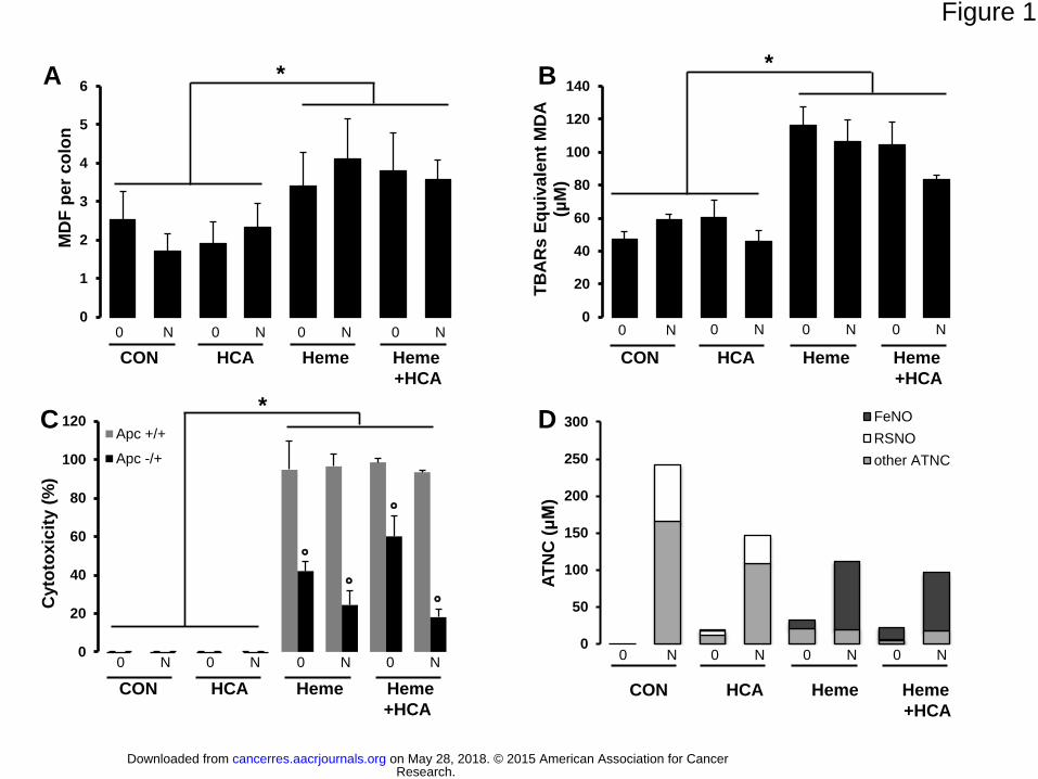
Only diets containing hemoglobin significantly increased the number of MDF per colon
(p<0.001) independent of the two other factors (Fig 1A). Indeed, although nitrates/nitrites in
drinking water induced a considerable increase in fecal ATNCs in all groups, they failed to
increase the number of MDF per colon (Fig 1A, D). Nevertheless, we noticed that the ATNC
composition was different between groups, containing 30–80% FeNO and no S-nitrosothiols
in the hemoglobin-fed groups compared to no FeNO and about 30% of S-nitrosothiols in
other groups.
Diets containing hemoglobin significantly increase the amount of thiobarbituric acid
reactive substances (TBARS) in fecal water (Fig 1B) and the amount of urinary 1,4-
dihydroxynonemercapturic acid (DHN-MA), a metabolite of the lipid oxidation product 4-
HNE (Supplementary Fig 3B). These oxidation biomarkers depended only on dietary and
fecal heme (Supplementary Fig 3A) and remained unchanged when the diet contained
nitrates/nitrites or HCA without hemoglobin (Fig 1B, Supplementary Fig 3B).
Premalignant Epithelial Cells Resist Cytotoxicity Induced by Fecal Water from Heme-
fed Rats: The Central Role of Aldehydes
Fecal water from rats fed hemoglobin-containing diets was more cytotoxic to the non-mutated
Apc+/+ cells than to premalignant Apc-/+ cells (Fig 1C). This data is consistent with previous
results (20). Fecal water from rats fed HCA or nitrites/nitrates without hemoglobin was not
cytotoxic to these cells (Fig 1C).
With the trapping of aldehydes from fecal water of rats fed heme with a polymer resin with
hydrazine functional groups, we found that in Apc+/+ and Apc-/+ cells, a 95% reduction in
Research. on May 28, 2018. © 2015 American Association for Cancercancerres.aacrjournals.org Downloaded from
Author manuscripts have been peer reviewed and accepted for publication but have not yet been edited. Author Manuscript Published OnlineFirst on January 15, 2015; DOI: 10.1158/0008-5472.CAN-14-2554
13
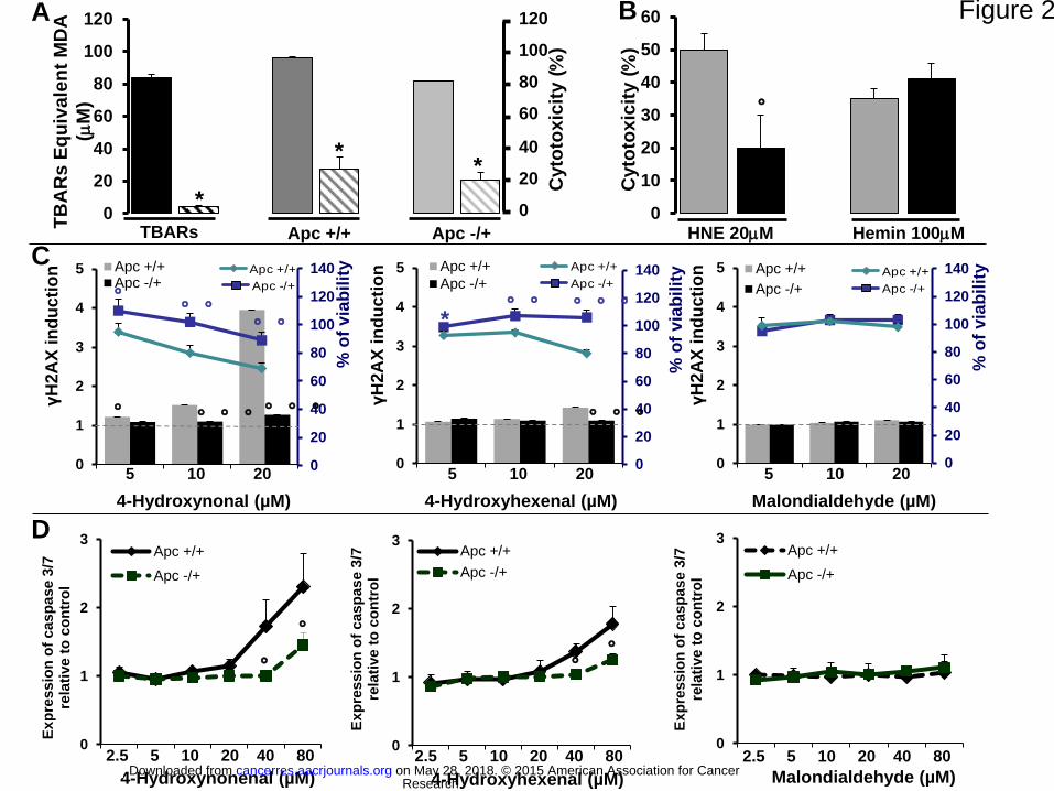
fecal water peroxidation was associated with a 75% reduction in cytotoxicity (Fig 2A).
Furthermore, we observed that only 4-HNE, but not heme, had differential cytotoxic effects in
Apc+/+ and Apc-/+ cells that was similar to that observed with fecal water of rats fed heme
(Fig 2B).
We then measured the cytotoxic and genotoxic effects of three main lipid peroxidation end
products (4-HNE, 4-HHE and MDA) on Apc+/+ and Apc-/+ cells using a H2AX in-cell
Western assay. HNE and HHE were more cytotoxic and more genotoxic to normal Apc+/+
cells than to premalignant Apc-/+ ones (Fig 2C, p<0.05). HNE at 10 and 20 µM and HHE at
20 µM were significantly more genotoxic to Apc+/+ cells than to Apc-/+ ones (Fig 2C,
p<0.05). MDA was neither cytotoxic nor genotoxic in the tested cell lines (Fig 2C). We
confirmed these viability results with the MTT assay and with the CellTiter-Glo® assay
(Supplementary Fig 4), and we used an expanded range of treatment concentrations (from 0
to 80 µM) in these assays. We confirmed that HNE and HHE were more cytotoxic (from 10 to
80 µM and from 40 to 80 µM, respectively) to normal cells than to premalignant ones, while
MDA had no effect (Supplementary Fig 4).
We determined the caspase 3/7 activity and again found a significant difference between
Apc+/+ and Apc-/+ cells after HNE and HHE treatment at 80 and 40 µM, respectively (Fig
3D, p<0.05). Specifically, caspase 3/7 activity was higher in Apc+/+ cells than in Apc-/+ cells,
and MDA treatment had no effect (Fig 2D).
Heme Iron and Tumoral promotion: Hemoglobin Increases Intestinal Tumorigenesis in
ApcMin/+
Mice
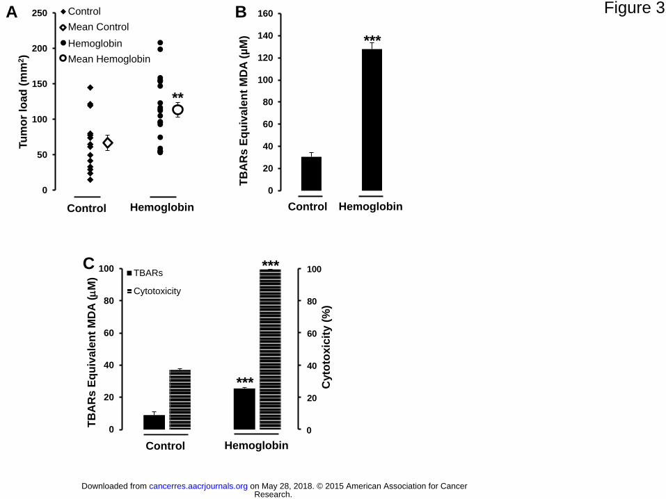
A diet containing 2.5% hemoglobin given to ApcMin/+
mice significantly increased the
intestinal tumor load (control diet: 67±39 mm2; hemoglobin diet: 114±47 mm
2, p=0.004; Fig
3A). These mice develop polyps mainly in the small intestine, and in our study we did not
Research. on May 28, 2018. © 2015 American Association for Cancercancerres.aacrjournals.org Downloaded from
Author manuscripts have been peer reviewed and accepted for publication but have not yet been edited. Author Manuscript Published OnlineFirst on January 15, 2015; DOI: 10.1158/0008-5472.CAN-14-2554
14
observe any effects of hemoglobin diet in the colon. The hemoglobin diet also significantly
increased the number of all tumors in the jejunum (Supplementary Fig 5A). In the entire
small intestine, the hemoglobin diet significantly increased the number of tumors with a
diameter greater than 1 mm (1 mm<tumor size≤2 mm, p=0.04; 2 mm<tumor size, p=0.006)
(Supplementary Fig 5B). Giving the same hemoglobin diet to normal C57BL/6J Apc+/+
mice
induced no neoplasia. As observed in rats, the effect of dietary heme on mice tumors was
associated with a significant increase in fecal heme (from 20±22 to 198±29 M) and with
increases in lipoperoxidation biomarkers: fecal TBARs (Fig 3B) and urinary DHN-MA
(Supplementary Fig 5C). We also assessed TBARs and the cytotoxicity of fecal water in the
small intestine to measure biomarkers at the same location as tumors. As Min mice have a
mutation in the Apc gene, we decided to use the mouse Apc-/+ model to investigate the
cytotoxic activity of fecal water in vivo. Heme diet was associated with a significant increase
in fecal TBARS and cytotoxicity in the small intestine (Fig 3C).
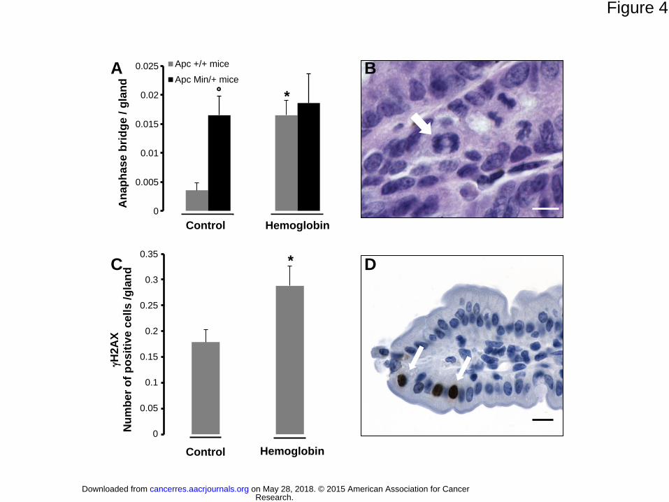
A Heme Diet is Genotoxic in vivo in the Epithelium of C57BL/6J Apc+/+
Mice
The induction of luminal lipid peroxidation by the hemoglobin diet (Fig 3B, C) was
associated with increased genotoxicity only in non-mutated C57BL/6J Apc+/+
mice with a
higher anaphase bridge index in the epithelium (Fig 4A and 4B). As expected the anaphase
bridge index was higher in C57BL/6J ApcMin/+
mice than in C57BL/6J Apc+/+
mice fed a
control diet (Fig 4A) because of the Apc mutation. In ApcMin/+
mice, the hemoglobin diet had
no additional genotoxic effect (Fig 4A). The genotoxic effect of the hemoglobin diet in
C57BL/6J Apc+/+
was confirmed by H2AX induction (Fig 4C, D).
Research. on May 28, 2018. © 2015 American Association for Cancercancerres.aacrjournals.org Downloaded from
Author manuscripts have been peer reviewed and accepted for publication but have not yet been edited. Author Manuscript Published OnlineFirst on January 15, 2015; DOI: 10.1158/0008-5472.CAN-14-2554
15
DISCUSSION
This study examines in vivo the relative contributions of the three main factors that may
explain how consumption of red and processed meat promotes CRC. Cross et al. showed that
these factors, i.e. heme, HCA, and NOC, were associated with CRC in a prospective cohort
study in humans (15). However, the identification of risk factors using an epidemiological
approach has to be correlated with the experimental approach in order to establish the
causative effect of such factors. Here, heme iron was the only experimental factor associated
with a significant increase in precancerous lesions (MDF) in rats. Heme iron showed no
additive or synergic effects with nitrates/nitrites or with HCA. Using a complementary
approach that included two animal models and a cellular model, we found that heme is the
determining factor in the promotion of colorectal carcinogenesis, and that the selective
toxicity of heme-induced alkenals to non-mutated cells seemed to play an important role in
this mechanism.
HCA are complete carcinogens that induce colon, mammary, and prostate tumors in
rodents and monkeys (30). The absence of effects of HCA in this study could be explained by
the dose we chose, which was based on the estimated dietary exposure to HCA in a diet that is
high in red meat and was relevant of the human food exposure. Indeed, carcinogenic doses of
HCAs in rodents are 1000 to 100,000 times higher than levels found in human foods (31).
Nitrite undergoes an enterosalivary cycle in humans but not in rats. We hypothesized that the
addition of sodium nitrates/nitrites to the rodents’ drinking water, which mimics human
saliva, would increase the effects of heme iron in rats by boosting nitrosation in the gut. In
humans, red meat consumption increases fecal ATNC concentrations (11), as in our study
with rats. Nevertheless, we could not detect any association between the ATNC level (Fig
1D) and carcinogenesis (Fig 1A). The highest level of ATNC was seen in the control group
Research. on May 28, 2018. © 2015 American Association for Cancercancerres.aacrjournals.org Downloaded from
Author manuscripts have been peer reviewed and accepted for publication but have not yet been edited. Author Manuscript Published OnlineFirst on January 15, 2015; DOI: 10.1158/0008-5472.CAN-14-2554
16
given nitrates/nitrites-supplemented water; this group had the fewest MDF. The lack of a
relationship between ATNCs and the number of MDF does not support a strong role for
ATNCs in the promotion of colon carcinogenesis by red meat.
The present results strongly suggest that at concentrations that are in line with human
red meat consumption, heme iron is associated with the promotion of colon carcinogenesis at
a preneoplastic stage. Most human colon cancers have an Apc mutation, as do MDF in
humans and rats (32). In order to unravel the mechanisms, we used a cellular model that
represented the CRC stages that we investigated in vivo. We chose a cellular model that
mimicked the early steps of carcinogenesis. This conditionally immortalized intestinal cellular
model uses premalignant Apc-/+ cells derived from C57BL/6J ApcMin/+
mice and “normal”
Apc+/+ cells from C57BL/6J mice (33). Characterization of both cell lines showed the
expected consequences of Apc mutation, such as actin network disassembly, aneuploidy, and
multi-nucleated cells (Supplementary Fig 2). These cell lines can therefore be used to study
the mechanisms involved in the early steps of CRC and thus, comprise a cellular model that is
a relevant complement to our in vivo model.
In rats, promotion of colon carcinogenesis by dietary hemoglobin was associated with
changes in non-invasive biomarkers: fecal water heme iron, TBARs, and cytotoxic activity.
Only the hemoglobin diet increased TBARs levels in fecal water (Fig 1B). We speculated that
the cytotoxic effects of fecal water on normal and premalignant colonic cells in vitro mimics
the in vivo situation with normal epithelium (Apc+/+) and with Apc-mutated MDF. In this
study, only fecal water from hemoglobin-fed rats was more cytotoxic to Apc+/+ cells than to
mutated cells (Fig 1C). We propose that premalignant cell selection explains the heme-
induced promotion of MDF. Aldehydes or heme iron itself, both present at high concentration
in feces from hemoglobin-fed rats, might be responsible for this differential cytotoxicity.
Using a resin to specifically trap fecal aldehydes, we showed that aldehydes alone are
Research. on May 28, 2018. © 2015 American Association for Cancercancerres.aacrjournals.org Downloaded from
Author manuscripts have been peer reviewed and accepted for publication but have not yet been edited. Author Manuscript Published OnlineFirst on January 15, 2015; DOI: 10.1158/0008-5472.CAN-14-2554
17
responsible for fecal water cytotoxicity. In addition, we observed that 4-HNE, but not heme
iron, induced differential cytotoxicity in Apc+/+ and Apc-/+ cells similar to that observed with
fecal water (Fig 2B). Therefore, we propose that heme-induced lipid peroxidation in the gut
explains the observed differential cytotoxicity and the CRC-promoting effects of heme that
are observed in vivo.
To explore the link between aldehydes and the promotion of colon carcinogenesis, we
tested the effects of three relevant aldehydes, 4-HNE, HHE, and MDA, in Apc+/+ and Apc-/+
cells. These α,β-unsaturated hydroxyalkenals are highly reactive compounds with proteins and
nucleic acids (34), and they are potentially cytotoxic and genotoxic. In our cellular model, 4-
HNE and HHE were more cytotoxic to normal cells than to premalignant cells, and induced
higher levels of apoptosis in normal cells than in premalignant cells. HNE was more cytotoxic
than HHE, as reported previously (35). Furthermore, HNE, like HHE, was more genotoxic to
normal cells than to premalignant ones (Fig 2C), with a higher index of DNA double-strand
breaks as revealed by the phosphorylation of histone H2AX. DNA double-strand breaks pose
a critical hazard to the genome, and erroneous rejoining of DNA double-strand breaks can
lead to mutation. These results thus suggest that at concentrations higher than 20 µM, HNE
and HHE will kill normal cells, while at lower concentration; they could create mutations in
Apc+/+ cells and might thus initiate carcinogenesis. Therefore, Apc-mutated cells are resistant
to apoptosis and can survive to contact with cytotoxic and genotoxic aldehydes, which allows
them to undergo further mutation and to become more malignant. Surprisingly, MDA was not
toxic to the cells tested in this study, but others found also that MDA had little or no toxicity
in cells (35,36). The results obtained by aldehyde trapping and in vitro with HNE and HHE,
confirmed our hypothesis that aldehydes are responsible for the differential cytotoxic effects
of fecal water from heme-fed rats. Heme iron catalyzes the formation of aldehydes in the
Research. on May 28, 2018. © 2015 American Association for Cancercancerres.aacrjournals.org Downloaded from
Author manuscripts have been peer reviewed and accepted for publication but have not yet been edited. Author Manuscript Published OnlineFirst on January 15, 2015; DOI: 10.1158/0008-5472.CAN-14-2554
18
gastrointestinal tract, which would “select” premalignant cells and also increase the mutation
frequency in normal cells (22).
This study shows that a hemoglobin-rich diet significantly increased the tumor load in
the small intestine of ApcMin/+
mice. In contrast, tumor load was not changed by heme diet in
the colon of mice, despite the expected modulation of biochemical markers. The number of
tumors in the colon of Min mice is low (less than 0.5 tumors per mouse), which reduces
statistical power (37). These mice have a truncated Apc gene as in human familial
adenomatous polyposis (FAP) (38). Moreover, sporadic CRCs tumors have the same early
Apc mutation in 50–80 % of cases (37). This mutation is also present in MDF (39). Our
nutritional experiments in rats and mice were thus conducted in the defined genetic context of
the Apc mutation. Promotion of carcinogenesis in rats and in ApcMin/+
mice (Fig 3A) was
associated with two non-invasive biomarkers, fecal water TBARs and cytotoxic activity, in
colon and in small intestine (Fig 3B, C).
Furthermore, as we found that HNE is more genotoxic in vitro to wild type cells than
to Apc-/+ cells (Fig 2C), we decided to investigate the genotoxicity of dietary heme in vivo by
measuring (i) the anaphase bridge index in the epithelium of C57BL/6J Apc+/+
mice and
C57BL/6J ApcMin/+
mice and (ii) by assessing H2AX induction in the epithelium of
C57BL/6J Apc+/+
mice (Fig 4). In these two studies, the induction of lipid peroxidation in the
gut by heme (Fig 3B, C) was associated with increased epithelial genotoxicity in Apc+/+
mice
but not in ApcMin/+
mice. Together with in vitro data, these data show that dietary hemoglobin
can induce DNA damage. We also observed anaphase bridges, which are biomarkers of
chromosomal instability and a major consequence of Apc mutation. As expected, in mice fed
the control diet, more anaphase bridges were seen in ApcMin/+
mice than in Apc+/+
mice (Fig
4A). Moreover, the hemoglobin diet increased the anaphase bridge index in Apc+/+
mice (Fig
4A). The hemoglobin diet induced the same number of anaphase bridges as the Apc mutation,
Research. on May 28, 2018. © 2015 American Association for Cancercancerres.aacrjournals.org Downloaded from
Author manuscripts have been peer reviewed and accepted for publication but have not yet been edited. Author Manuscript Published OnlineFirst on January 15, 2015; DOI: 10.1158/0008-5472.CAN-14-2554
19
suggesting that dietary hemoglobin generates strong initiators. Taken together, these data
suggest that heme-induced aldehydes can induce mutations in vitro and in vivo and may
initiate carcinogenesis.
In conclusion, we identified heme iron as the main factor responsible for the promotion of
CRC by red meat and showed that aldehydes such as 4-HNE or HHE play roles in the
underlying mechanism of action. Furthermore, we suggest that dietary heme could result in
initiating agents in the gut. Improved dietary recommendations should focus (i) on the amount
of heme iron in meat-based diets rather than on the modes of cooking or preparation and (ii)
on dietary changes that could reduce the heme effect in the gut (i.e. on changes that limit the
bioavailability of heme and of heme-induced peroxidation) (40).
Acknowledgments:
The authors thank F. Blas-Y-Estrada for animal care, M.L. Jourdain for cell cycle data and J-
Ph. Nougayrède for immunofluorescence microscopy.
Research. on May 28, 2018. © 2015 American Association for Cancercancerres.aacrjournals.org Downloaded from
Author manuscripts have been peer reviewed and accepted for publication but have not yet been edited. Author Manuscript Published OnlineFirst on January 15, 2015; DOI: 10.1158/0008-5472.CAN-14-2554
20
References
1. DeSantis CE, Lin CC, Mariotto AB, Siegel RL, Stein KD, Kramer JL, et al. Cancer
treatment and survivorship statistics, 2014. CA Cancer J Clin 2014;64:252-71.
2. WCRF. Food, nutrition, physical activity, and the prevention of cancer: a global
perspective. WCRF and American Institute for Cancer Research, Washington DC
2007:1-537.
3. WCRF. WCRF/AICR Systematic Literature Review Continuous Update Project
Report : The Associations between Food, Nutrition and Physical Activity and the Risk
of Colorectal Cancer. WCRF and American Institute for Cancer Research,
Washington DC 2010:1-855.
4. Pierre FH, Santarelli RL, Allam O, Tache S, Naud N, Gueraud F, et al. Freeze-dried
ham promotes azoxymethane-induced mucin-depleted foci and aberrant crypt foci in
rat colon. Nutr Cancer 2010;62:567-73.
5. Pierre F, Freeman A, Tache S, Van der Meer R, Corpet DE. Beef meat and blood
sausage promote the formation of azoxymethane-induced mucin-depleted foci and
aberrant crypt foci in rat colons. J Nutr 2004;134:2711-16.
6. Corpet DE, Pierre F. Point: from animal models to prevention of colon cancer.
systematic review of chemoprevention in min mice and choice of the model system.
Cancer Epidemiology Biomarkers & Prevention 2003;12:391-400.
7. Bastide NM, Pierre FH, Corpet DE. Heme iron from meat and risk of colorectal
cancer: a meta-analysis and a review of the mechanisms involved. Cancer Prev Res
(Phila) 2012;4:177-84.
Research. on May 28, 2018. © 2015 American Association for Cancercancerres.aacrjournals.org Downloaded from
Author manuscripts have been peer reviewed and accepted for publication but have not yet been edited. Author Manuscript Published OnlineFirst on January 15, 2015; DOI: 10.1158/0008-5472.CAN-14-2554
21
8. Bingham SA, Hughes R, Cross AJ. Effect of white versus red meat on endogenous N-
nitrosation in the human colon and further evidence of a dose response. J Nutr
2002;132:3522S-25S.
9. Sinha R, Knize MG, Salmon CP, Brown ED, Rhodes D, Felton JS, et al. Heterocyclic
amine content of pork products cooked by different methods and to varying degrees of
doneness. Food Chem Toxicol 1998;36:289-97.
10. Sesink ALA, Termont DSML, Kleibeuker JH, Vandermeer R. Red meat and colon
cancer: the cytotoxic and hyperproliferative effects of dietary heme. Cancer Res
1999;59:5704-09.
11. Kuhnle GG, Story GW, Reda T, Mani AR, Moore KP, Lunn JC, et al. Diet-induced
endogenous formation of nitroso compounds in the GI tract. Free Radic Biol Med
2007;43:1040-7.
12. Glei M, Klenow S, Sauer J, Wegewitz U, Richter K, Pool-Zobel BL. Hemoglobin and
hemin induce DNA damage in human colon tumor cells HT29 clone 19A and in
primary human colonocytes. Mutat Res 2006;594:162-71.
13. Pierre FHF, Martin OCB, Santarelli R, Tache S, Naud N, Gueraud F, et al. Calcium
and α-Tocopherol Suppress Cured Meat Promotion of Chemically-Induced Colon
Carcinogenesis in Rats and Reduce Associated Biomarkers in Human Volunteers. Am
Journal of Clin Nut 2013;98:1255-62.
14. Santarelli RL, Naud N, Taché S, Guéraud F, Nassy G, Vendeuvre JL, et al. Calcium
Inhibits Promotion by Hot Dog of 1-2-Dimethylhydrazine-Induced Mucin-Depleted
Foci in Rat Colon. Int J of Cancer 2013;133:2533-41.
15. Cross AJ, Ferrucci LM, Risch A, Graubard BI, Ward MH, Park Y, et al. A large
prospective study of meat consumption and colorectal cancer risk: an investigation of
potential mechanisms underlying this association. Cancer Res 2010;70:2406-14.
Research. on May 28, 2018. © 2015 American Association for Cancercancerres.aacrjournals.org Downloaded from
Author manuscripts have been peer reviewed and accepted for publication but have not yet been edited. Author Manuscript Published OnlineFirst on January 15, 2015; DOI: 10.1158/0008-5472.CAN-14-2554
22
16. Mirvish SS, Davis ME, Lisowyj MP, Gaikwad NW. Effect of feeding nitrite,
ascorbate, hemin, and omeprazole on excretion of fecal total apparent N-nitroso
compounds in mice. Chem Res Toxicol 2008;21:2344-51.
17. Caderni G, Femia AP, Giannini A, Favuzza A, Luceri C, Salvadori M, et al.
Identification of mucin-depleted foci in the unsectioned colon of azoxymethane-
treated rats: correlation with carcinogenesis. Cancer Res 2003;63:2388-92.
18. Van den Berg JW, Koole-Lesuis R, Edixhoven-Bosdijk A, Brouwers N. Automating
the quantification of heme in feces. Clin Chem 1988;34:2125-6.
19. Ohkawa H, Ohishi N, Yagi K. Assay for lipid peroxides in animal tissues by
thiobarbituric acid reaction. Anal Bioch 1979;95:351-8.
20. Pierre F, Santarelli R, Tache S, Gueraud F, Corpet DE. Beef meat promotion of
dimethylhydrazine-induced colorectal carcinogenesis biomarkers is suppressed by
dietary calcium. Br J Nutr 2008;99:1000-6.
21. Forest V, Pierre F, Bassonga E, Meflah K, Olivier C, Menanteau J. Apc+/min colonic
epithelial cells express TNF receptors and ICAM-1 when they are co-cultured with
large intestine intra-epithelial lymphocytes. Cell Immunol 2003;223:70-76.
22. Pierre F, Tache S, Guéraud F, Rerole AL, Jourdan ML, Petit C. Apc mutation induces
resistance of colonic cells to lipoperoxide-triggered apoptosis induced by faecal water
from haem-fed rats. Carcinogenesis 2007;28:321-27.
23. Chandra A, Srivastava SK. A synthesis of 4-hydroxy-2-trans-nonenal and 4-(3H) 4-
hydroxy-2-trans-nonenal. Lipids 1997;32:779-82.
24. Fenaille F, Mottier P, Turesky RJ, Ali S, Guy PA. Comparison of analytical
techniques to quantify malondialdehyde in milk powders. J Chromatogr A
2001;921:237-45.
Research. on May 28, 2018. © 2015 American Association for Cancercancerres.aacrjournals.org Downloaded from
Author manuscripts have been peer reviewed and accepted for publication but have not yet been edited. Author Manuscript Published OnlineFirst on January 15, 2015; DOI: 10.1158/0008-5472.CAN-14-2554
23
25. Rogakou EP, Pilch DR, Orr AH, Ivanova VS, Bonner WM. DNA double-stranded
breaks induce histone H2AX phosphorylation on serine 139. J Biol Chem
1998;273:5858-68.
26. Khoury L, Zalko D, Audebert M. Validation of high-throughput genotoxicity assay
screening using γH2AX in-cell western assay on HepG2 cells. Environ Mol Mutagen
2013;54:737-46.
27. Audebert M, Dolo L, Perdu E, Cravedi JP, Zalko D. Use of the gammaH2AX assay
for assessing the genotoxicity of bisphenol A and bisphenol F in human cell lines.
Arch Toxicol 2011;85:1463-73.
28. Graillot V, Takakura N, Hegarat LL, Fessard V, Audebert M, Cravedi JP.
Genotoxicity of pesticide mixtures present in the diet of the French population.
Environ Mol Mutagen 2012;53:173-84.
29. Montgomery E, Wilentz RE, Argani P, Fisher C, Hruban RH, Kern SE, et al. Analysis
of anaphase figures in routine histologic sections distinguishes chromosomally
unstable from chromosomally stable malignancies. Cancer Biology and Therapy
2003;2:248-52.
30. Sugimura T, Wakabayashi K, Nakagama H, Nagao M. Heterocyclic amines:
Mutagens/carcinogens produced during cooking of meat and fish. Cancer Sci
2004;95:290-9.
31. Schwabn CE, Huber WW, Parzefall W, Hietsch G, Kassie F, SchulteHermann R, et al.
Search for compoiunds that inhibit the genotoxic anc carcinogenic effects of
heterocyclic aromatic amines. Crit Rev Toxicol 2000;30:1-69.
32. Femia AP, Giannini A, Fazi M, Tarquini E, Salvadori M, Roncucci L, et al.
Identification of mucin depleted foci in the human colon. Cancer Prev Res (Phila)
2008;1:562-67.
Research. on May 28, 2018. © 2015 American Association for Cancercancerres.aacrjournals.org Downloaded from
Author manuscripts have been peer reviewed and accepted for publication but have not yet been edited. Author Manuscript Published OnlineFirst on January 15, 2015; DOI: 10.1158/0008-5472.CAN-14-2554
24
33. Jamin EL, Riu A, Douki T, Debrauwer L, Cravedi JP, Zalko D, et al. Combined
genotoxic effects of a polycyclic aromatic hydrocarbon (B(a)P) and an heterocyclic
amine (PhIP) in relation to colorectal carcinogenesis. PLoS One 2013;8:e58591.
34. Schaur RJ. Basic aspects of the biochemical reactivity of 4-hydroxynonenal. Mol
Aspects Med 2003;24:149-59.
35. Eckl PM, Ortner A, Esterbauer H. Genotoxic properties of 4-hydroxyalkenals and
analogous aldehydes. Mutat Res 1993;290:183-92.
36. Fischer SM, Ogle S, Marnett LJ, Nesnow S, Slaga TJ. The lack of initiating and/or
promoting activity of sodium malondialdehyde on SENCAR mouse skin. Cancer Lett
1983;19:61-6.
37. Corpet DE, Pierre F. How good are rodent models of carcinogenesis in predicting
efficacy in humans? A systematic review and meta-analysis of colon chemoprevention
in rats, mice and men. Eur J Cancer 2005;41:1911-22.
38. Su LK, Kinzler KW, Vogelstein B, Preisinger AC, Moser AR, Luongo C, et al.
Multiple intestinal neoplasia caused by a mutation in the murine homolog of the APC
gene. Science 1992;256:668-70.
39. Femia AP, Dolara P, Giannini A, Salvadori M, Biggeri A, Caderni G. Frequent
mutation of Apc gene in rat colon tumors and mucin-depleted foci, preneoplastic
lesions in experimental colon carcinogenesis. Cancer Res 2007;67:445-9.
40. Corpet DE. Red meat and colon cancer: should we become vegetarians, or can we
make meat safer? Meat Sci 2011;89:310-6.
Research. on May 28, 2018. © 2015 American Association for Cancercancerres.aacrjournals.org Downloaded from
Author manuscripts have been peer reviewed and accepted for publication but have not yet been edited. Author Manuscript Published OnlineFirst on January 15, 2015; DOI: 10.1158/0008-5472.CAN-14-2554
25
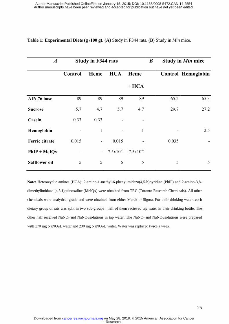
Table 1: Experimental Diets (g /100 g). (A) Study in F344 rats. (B) Study in Min mice.
A Study in F344 rats B Study in Min mice
Control Heme HCA Heme
+ HCA
Control Hemoglobin
AIN 76 base 89 89 89 89 65.2 65.3
Sucrose 5.7 4.7 5.7 4.7 29.7 27.2
Casein 0.33 0.33 - -
Hemoglobin - 1 - 1 - 2.5
Ferric citrate 0.015 - 0.015 - 0.035 -
PhIP + MeIQx - - 7.5x10⁻⁶
7.5x10⁻⁶
Safflower oil 5 5 5 5 5 5
Note: Heterocyclic amines (HCA): 2-amino-1-methyl-6-phenylimidazo(4,5-b)pyridine (PhIP) and 2-amino-3,8-
dimethylimidazo [4,5-f]quinoxaline (MeIQx) were obtained from TRC (Toronto Research Chemicals). All other
chemicals were analytical grade and were obtained from either Merck or Sigma. For their drinking water, each
dietary group of rats was split in two sub-groups : half of them recieved tap water in their drinking bottle. The
other half received NaNO2 and NaNO3 solutions in tap water. The NaNO2 and NaNO3 solutions were prepared
with 170 mg NaNO2/L water and 230 mg NaNO3/L water. Water was replaced twice a week.
Research. on May 28, 2018. © 2015 American Association for Cancercancerres.aacrjournals.org Downloaded from
Author manuscripts have been peer reviewed and accepted for publication but have not yet been edited. Author Manuscript Published OnlineFirst on January 15, 2015; DOI: 10.1158/0008-5472.CAN-14-2554
26
Figure 1. Heme iron plays a major role in mucin-depleted foci (MDF) formation in rats
and is associated with fecal water lipid peroxidation biomarkers independent of the fecal
concentration of apparent total N-nitroso-compounds (ATNC). (A) The number of MDF
per rat colon. A three-factor Anova was used and showed only an effect of heme. (B) Lipid
peroxidation in fecal water [measured as TBARs (MDA equivalents)]. (C) Fecal water
cytotoxicity. The cytotoxic effects of fecal water from the eight groups of rats on Apc+/+ cells
(gray bars) and on Apc-/+ cells (black bars). Viability was measured by MTT assay after 24 h
of incubation with a 1/50 dilution of fecal water. (D) ATNCs in fecal water shown as nitrosyl
iron (FeNO), S-nitrosothiols (RSNO), and other ATNCs. TEM: control diet; HCA: diet with
PhIP (50 µg/kg) and MeIQx (25 µg/kg); Heme: diet containing 1% hemoglobin; Heme +
HCA: diet containing 1% hemoglobin, PhIP, and MeIQx: 50+25 µg/kg; 0: drinking water
control; N: drinking water with nitrites and nitrates at 0.4 g/l. Values are means ± SEM, n=10;
*Significantly different from TEM and HCA, p<0.001. °Significantly different from Apc-/+
cells, p<0.01.
Figure 2. The central role of aldehydes in the cytotoxic effect of fecal water of
hemoglobin fed rats and the cytotoxic, genotoxic, and pro-apoptotic dose-dependent
effects of HNE, HHE, and MDA in Apc+/+ and Apc-/+ cells. (A) TBARs and cytotoxicity
on Apc+/+ and Apc-/+ cells of fecal water of hemoglobin fed rats treated or not with trapping
aldehydes resin. Solid bars: fecal water of hemoglobin group, hatched bars: fecal water of
hemoglobin group treated with resin. Values are means ± SEM, n=3. (B) Cytotoxicity on
Apc+/+ (gray bars) and Apc-/+ (black bars) cells of HNE (20 M) and hemin (100 M).
Values are means ± SEM, n=3. (C) Genotoxic and cytotoxic effects of aldehydes on Apc+/+
and Apc-/+ cells. Values are means ± SEM, n=4; (D) Pro-apoptotic dose-dependent effects of
aldehydes on Apc+/+ and Apc-/+ cells. Values are means ± SEM, n=3;
Research. on May 28, 2018. © 2015 American Association for Cancercancerres.aacrjournals.org Downloaded from
Author manuscripts have been peer reviewed and accepted for publication but have not yet been edited. Author Manuscript Published OnlineFirst on January 15, 2015; DOI: 10.1158/0008-5472.CAN-14-2554
27
*Significant difference between fecal water treated or not with resin, using Student’s test
(*p<0.05). °Significant difference between Apc+/+ and Apc-/+ using Student’s test (°p<0.05;
°°p<0.01; °°°p<0.001).
Figure 3. The effect of hemoglobin on intestinal tumorigenesis in Min mice and changes
in fecal and luminal biomarkers associated with this effect. (A) Intestinal tumor load (mm2
per intestine). Values are means ± SEM, n= 14 for the control diet and n=21 for the
haemoglobin diet. (B) Lipid peroxidation in fecal water from feces [measured as TBARs
(MDA equivalents)]. n= 5 for the control diet and n=7 for the haemoglobin diet. (C)
Cytotoxic activity and peroxidation in small intestine contents from mouse. Cytotoxicity was
measured in the Apc-/+ cell line (fecal water diluted 1:50, MTT assay). n= 5 for the control
diet and n=7 for the haemoglobin diet. ** and ***Significantly different from control diet
(**p<0.01; ***p<0.001).
Figure 4. The effect of a hemoglobin-containing diet on anaphase bridges and on the
induction of H2AX in the small intestinal epithelium in mice. (A) The number of
anaphase bridges per gland in ApcMin/+
and Apc+/+
mice after 50 days of the indicated
experimental diet. *Significantly different from the control diet in the same genetic
context, °Significantly different from Apc+/+
mice on the same diet. (B) An anaphase bridge
(arrow) in an Apc+/+
mouse fed a hemoglobin diet, scale bar: 2 m. (C) The number of
H2AX positive cells per gland in C57BL/6J mice after 14 days on the experimental diet. (D)
H2AX-positive cells (arrows) in Apc+/+
mice fed the heme diet, scale bar: 4 m.
Research. on May 28, 2018. © 2015 American Association for Cancercancerres.aacrjournals.org Downloaded from
Author manuscripts have been peer reviewed and accepted for publication but have not yet been edited. Author Manuscript Published OnlineFirst on January 15, 2015; DOI: 10.1158/0008-5472.CAN-14-2554
0
1
2
3
4
5
6
MD
F p
er
co
lon
0
20
40
60
80
100
120
140
TB
AR
s E
qu
ivale
nt
MD
A
(µM
)
0
50
100
150
200
250
300
AT
NC
(μM
)
FeNO
RSNO
other ATNC
A B
D
* *
CON HCA Heme Heme
+HCA
0 N 0 N 0 N 0 N
CON HCA Heme Heme
+HCA
C
0
20
40
60
80
100
120
% o
f cyto
toxic
ity
(M
TT
assay)
Apc +/+
Apc -/+
*
°
°
0 N 0 N 0 N 0 N
CON HCA Heme Heme
+HCA
0 N 0 N 0 N 0 N
CON HCA Heme Heme
+HCA
0 N 0 N 0 N 0 N
°
° Cyto
toxic
ity (
%)
Figure 1
Research. on May 28, 2018. © 2015 American Association for Cancercancerres.aacrjournals.org Downloaded from
Author manuscripts have been peer reviewed and accepted for publication but have not yet been edited. Author Manuscript Published OnlineFirst on January 15, 2015; DOI: 10.1158/0008-5472.CAN-14-2554
0
1
2
3
4
5
γH
2A
X i
nd
ucti
on
Apc +/+Apc -/+
0
20
40
60
80
100
120
140
% o
f via
bil
ity
5 10 20 0
1
2
3
4
5
γH
2A
X i
nd
ucti
on
Apc +/+
Apc -/+
0
20
40
60
80
100
120
140
% o
f via
bil
ity
0
1
2
3
4
5
γH
2A
X i
nd
ucti
on
Apc +/+
Apc -/+
0
20
40
60
80
100
120
140
% o
f via
bil
ity
4-Hydroxynonal (µM) 4-Hydroxyhexenal (µM) Malondialdehyde (µM)
0
1
2
3
Ex
pre
ss
ion
of
ca
sp
as
e 3
/7
rela
tive
to
co
ntr
ol
4-Hydroxyhexenal (µM)
Apc +/+
Apc -/+
° °
0
1
2
3
Ex
pre
ss
ion
of
ca
sp
as
e 3
/7
rela
tive
to
co
ntr
ol
4-Hydroxynonenal (µM)
Apc +/+
Apc -/+
°
°
0
1
2
3
Ex
pre
ss
ion
of
ca
sp
as
e 3
/7
rela
tive
to
co
ntr
ol
Malondialdehyde (µM)
Apc +/+
Apc -/+
°°° °°° °°° °
°° °°
° °°° °° *
2.5 5 10 20 40 80
0
20
40
60
80
100
120
140
% o
f v
iab
ility
Apc -/+
Apc +/+
0
20
40
60
80
100
120
140
% o
f v
iab
ility
Apc -/+
Apc +/+
0
20
40
60
80
100
120
140
% o
f v
iab
ility
Apc -/+
Apc +/+
0
20
40
60
80
100
120
140
% o
f v
iab
ility
Apc -/+
Apc +/+
0
20
40
60
80
100
120
140
% o
f v
iab
ility
Apc -/+
Apc +/+
0
20
40
60
80
100
120
140
% o
f v
iab
ility
Apc -/+
Apc +/+
0
20
40
60
80
100
120
TB
AR
s E
qu
iva
len
t M
DA
(mM
)
Cyto
tox
icit
y (%
)
Cyto
tox
icit
y (%
)
TBARs Apc +/+ Apc -/+ HNE 20mM Hemin 100mM
*
* *
°
A B
C
D
0
10
20
30
40
50
60120
100
80
60
40
20
0
2.5 5 10 20 40 80 2.5 5 10 20 40 80
5 10 20 5 10 20
Figure 2
Research. on May 28, 2018. © 2015 American Association for Cancercancerres.aacrjournals.org Downloaded from
Author manuscripts have been peer reviewed and accepted for publication but have not yet been edited. Author Manuscript Published OnlineFirst on January 15, 2015; DOI: 10.1158/0008-5472.CAN-14-2554
***
B
Hemoglobin Control
0
20
40
60
80
100
120
140
160
TB
AR
s E
qu
ivale
nt
MD
A (
µM
)
A
Hemoglobin
0
50
100
150
200
250Tu
mo
r lo
ad
(m
m2)
CON
Mean CON
Heme
Mean Heme
**
Control
0
20
40
60
80
100
TB
AR
s E
qu
ivale
nt
MD
A (m
M) TBARs
Cytotoxicity
Hemoglobin Control
C
0
20
40
60
80
100
0
20
40
60
80
100
% o
f c
yto
xic
ity
on
Ap
c
Min
/+ c
ells
(M
TT
as
sa
y)
TB
AR
s (µ
M e
qu
iva
len
t M
DA
)
***
***
C
yto
toxic
ity (
%)
Control
Mean Control
Hemoglobin
Mean Hemoglobin
Figure 3
Research. on May 28, 2018. © 2015 American Association for Cancercancerres.aacrjournals.org Downloaded from
Author manuscripts have been peer reviewed and accepted for publication but have not yet been edited. Author Manuscript Published OnlineFirst on January 15, 2015; DOI: 10.1158/0008-5472.CAN-14-2554
An
ap
hase b
rid
ge / g
lan
d
Apc +/+ mice
Apc Min/+ miceA B
D
° *
*
gH2A
X
Nu
mb
er
of
po
sit
ive c
ells /
gla
nd
C
Hemoglobin
Hemoglobin Control
Control
0.025
0.02
0.015
0.01
0.005
0
0.35
0.3
0.25
0.2
0.15
0.1
0.05
0
Figure 4
Research. on May 28, 2018. © 2015 American Association for Cancercancerres.aacrjournals.org Downloaded from
Author manuscripts have been peer reviewed and accepted for publication but have not yet been edited. Author Manuscript Published OnlineFirst on January 15, 2015; DOI: 10.1158/0008-5472.CAN-14-2554
Published OnlineFirst January 15, 2015.Cancer Res Nadia Bastide, Fatima Chenni, Marc Audebert, et al. associated with red meat intakeA central role for heme iron in colon carcinogenesis
Updated version
10.1158/0008-5472.CAN-14-2554doi:
Access the most recent version of this article at:
Material
Supplementary
http://cancerres.aacrjournals.org/content/suppl/2015/01/16/0008-5472.CAN-14-2554.DC1
Access the most recent supplemental material at:
Manuscript
Authorbeen edited. Author manuscripts have been peer reviewed and accepted for publication but have not yet
E-mail alerts related to this article or journal.Sign up to receive free email-alerts
Subscriptions
Reprints and
To order reprints of this article or to subscribe to the journal, contact the AACR Publications
Permissions
Rightslink site. Click on "Request Permissions" which will take you to the Copyright Clearance Center's (CCC)
.http://cancerres.aacrjournals.org/content/early/2015/01/15/0008-5472.CAN-14-2554To request permission to re-use all or part of this article, use this link
Research. on May 28, 2018. © 2015 American Association for Cancercancerres.aacrjournals.org Downloaded from
Author manuscripts have been peer reviewed and accepted for publication but have not yet been edited. Author Manuscript Published OnlineFirst on January 15, 2015; DOI: 10.1158/0008-5472.CAN-14-2554