a case report of manipulation under anesthesia of posttraumatic type ii occipital-atlantoaxial...
TRANSCRIPT

A CASE REPORT OF MANIPULATION UNDER ANESTHESIA
OF POSTTRAUMATIC TYPE II OCCIPITAL-ATLANTOAXIAL
ROTATORY SUBLUXATION IN A 4-YEAR-OLD GIRL
Sen-Wei Tsai, MD,a,b and Chorng-Sonq Chou, MDa
ABSTRACT
352
a Attending PhyRehabilitation, TaTaiwan.
b Lecturer, Depaversity, Taichung,Sources of suppSubmit requests
ment of Physical MGeneral Hospital,Taichung, Taiwan
Paper submitted2004.0161-4754/$30.Copyright D 20doi:10.1016/j.jm
Objective: To discuss a case of occipital-atlantoaxial rotatory subluxation successfully treated with manipulation under
general anesthesia.
Clinical Features: A 4-year-old girl presented to the Taichung Veterans General Hospital with acute torticollis and
neck stiffness for 1 week after she had fallen. Although some nonsteroidal anti-inflammatory drugs had been prescribed for
her, her neck still tilted to the right side and her chin inclined to the left side. There were no neurological signs, no
significant pain if she did not move her neck and head, and no muscular hypertonocity. There was painful guarding in the
right sternocleidomatoid muscle when manipulation was attempted. Three-dimensional computer tomography revealed
uneven joint space between the C1 anterior arch and odontoid process and confirmed a type II atlantoaxial rotatory
subluxation.
Intervention and Outcome: Manipulation under anesthesia was performed by a medical doctor trained in manual
therapy. The low-velocity, right rotational manipulation applied to the occiput included axial traction. The neck symptoms
were relieved immediately after treatment.
Conclusion: Under general anesthesia, manipulation may be a good method for treating noncomplicated type II
atlantoaxial rotatory subluxation. Additional investigations may be necessary to evaluate the treatment effect.
(J Manipulative Physiol Ther 2005;28:352-355)
Key Indexing Terms: Atlantoaxial Joint; Rotatory Subluxation; Manipulation; Spinal
Sudden onset of acute torticollis is a rare condition in
children.1 One possible cause is atlantoaxial rotatory
subluxation. The common clinical characteristic is
the patient presenting with the head tilted to one side and
rotated to the other side, the so-called cock robin position.
Ligament laxity secondary to minor trauma, or a preceding
history of upper respiratory infection, has been reported to
be the etiology.2,3 Treatment options include medication,
traction, manipulation, bracing, or operation,4,5 but it
sician, Department of Physical Medicine andichung Veterans General Hospital, Taichung,
rtment of Physical Therapy, HungKuang Uni-Taiwan.ort: none.for reprints to: Dr Sen-Wei Tsai, MD, Depart-edicine and Rehabilitation, Taichung VeteransNo 160, Section 3, Taichung Harbor Road,(e-mail: [email protected]).February 3, 2004; in revised form August 10,
0005 by National University Health Sciences.pt.2005.04.006
usually takes several weeks for symptoms to resolve. We
present this case treated successfully with manipulation
under general anesthesia and with immediate relief after this
maneuver, and then review our other 3 cases.
CASE REPORT
A 4-year-old girl presented to the Taichung Veterans
General Hospital with neck stiffness, and her head was tilted
to the right for 1 week after she fell down on a playground.
She had taken some nonsteroidal anti-inflammatory drugs,
but the symptoms remained. Observation showed that her
head tilted 308 to the right with a slight rotation to the left.
Her active range of motion was less than 108 in rotation and
lateral bending. Palpation showed the atlas in a right rotated
position with left border posterior. The axis spinous process
was in a relatively neutral position. Muscle guarding was
found in the paraspinal C1-2 muscles when mobilization of
the C1-2 joints was attempted. There were no neurological
signs, no fever, no significant pain, and no muscular
hypertonocity, but there was some painful guarding in
the right sternocleidomatoid muscle when manipulation
was attempted. This pain guarding of the neck muscles

Fig 1. Uneven joint space between the C1 anterior arch andodontoid process, with the atlas in a left-rotated position.
Fig 2. Posterior view of 3D CT scan; the atlas-axis joint is tilted tothe right about 128.
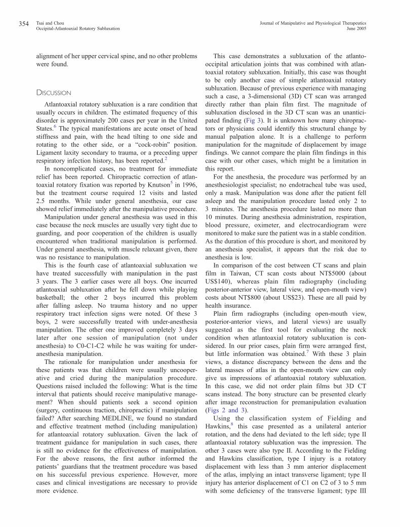
Fig 3. Top view of 3D CT scan shows subluxation of the right andleft atlanto-occipital articulation joints and a 758 left-posteriorrotated positional change.
Tsai and ChouJournal of Manipulative and Physiological Therapeutics
Occipital-Atlantoaxial Rotatory SubluxationVolume 28, Number 5353
made the child irritable and uncooperative and impeded
manipulation. According to previous experience with
such cases, atlantoaxial rotatory subluxation was the first
diagnostic impression.
Plain film radiographs for her neck were not taken
because of our first diagnostic impression. Instead, a
traditional computed tomography (CT) scan was taken and
showed uneven joint space between the C1 anterior arch and
odontoid process and a left rotated atlas (Fig 1). Three-
dimensional CT scan was done; the bony structure of the
occipital-atlantoaxial rotatory subluxation was recon-
structed. The posterior view showed the atlas tilted about
128 to the right side (Fig 2). The top view showed the atlas
in a unilateral left-rotated position, with uneven space
between the odontoid process and anterior arch of C1; the
occiput was in a left-rotated position of about 758 (Fig 3).
According to the classification of Fielding and Hawkins,8
the diagnosis of this patient was atlantoaxial rotatory
subluxation type II.
Consent to proceed with treatment was obtained from the
patient’s parents. An anesthesiologist was consulted for
general anesthesia, and a muscle relaxant was given. The
anesthesia was delivered through a mask; thus, no endo-
tracheal tube was inserted. The manipulation procedure was
performed in the operating room by the primary author who
has a specialty of physical medicine and rehabilitation and
was trained by the chief of department who learned
manipulative skills from Cyriax.
The patient was placed in a supine position, with some
occipital traction vector. With the left index finger fixing on
the left C2 articular pillar, a gentle rotary manipulation was
given to the right side. Passive stretching of the right neck
muscles and mobilization manipulation of the cervical
spine were done after this manipulation. The symptoms
were relieved immediately after this maneuver. Three
months later, a follow-up examination showed normal
354 Journal of Manipulative and Physiological TherapeuticsTsai and Chou
June 2005Occipital-Atlantoaxial Rotatory Subluxation
alignment of her upper cervical spine, and no other problems
were found.
DISCUSSION
Atlantoaxial rotatory subluxation is a rare condition that
usually occurs in children. The estimated frequency of this
disorder is approximately 200 cases per year in the United
States.6 The typical manifestations are acute onset of head
stiffness and pain, with the head tilting to one side and
rotating to the other side, or a bcock-robinQ position.
Ligament laxity secondary to trauma, or a preceding upper
respiratory infection history, has been reported.2
In noncomplicated cases, no treatment for immediate
relief has been reported. Chiropractic correction of atlan-
toaxial rotatory fixation was reported by Knutson5 in 1996,
but the treatment course required 12 visits and lasted
2.5 months. While under general anesthesia, our case
showed relief immediately after the manipulative procedure.
Manipulation under general anesthesia was used in this
case because the neck muscles are usually very tight due to
guarding, and poor cooperation of the children is usually
encountered when traditional manipulation is performed.
Under general anesthesia, with muscle relaxant given, there
was no resistance to manipulation.
This is the fourth case of atlantoaxial subluxation we
have treated successfully with manipulation in the past
3 years. The 3 earlier cases were all boys. One incurred
atlantoaxial subluxation after he fell down while playing
basketball; the other 2 boys incurred this problem
after falling asleep. No trauma history and no upper
respiratory tract infection signs were noted. Of these 3
boys, 2 were successfully treated with under-anesthesia
manipulation. The other one improved completely 3 days
later after one session of manipulation (not under
anesthesia) to C0-C1-C2 while he was waiting for under-
anesthesia manipulation.
The rationale for manipulation under anesthesia for
these patients was that children were usually uncooper-
ative and cried during the manipulation procedure.
Questions raised included the following: What is the time
interval that patients should receive manipulative manage-
ment? When should patients seek a second opinion
(surgery, continuous traction, chiropractic) if manipulation
failed? After searching MEDLINE, we found no standard
and effective treatment method (including manipulation)
for atlantoaxial rotatory subluxation. Given the lack of
treatment guidance for manipulation in such cases, there
is still no evidence for the effectiveness of manipulation.
For the above reasons, the first author informed the
patients’ guardians that the treatment procedure was based
on his successful previous experience. However, more
cases and clinical investigations are necessary to provide
more evidence.
This case demonstrates a subluxation of the atlanto-
occipital articulation joints that was combined with atlan-
toaxial rotatory subluxation. Initially, this case was thought
to be only another case of simple atlantoaxial rotatory
subluxation. Because of previous experience with managing
such a case, a 3-dimensional (3D) CT scan was arranged
directly rather than plain film first. The magnitude of
subluxation disclosed in the 3D CT scan was an unantici-
pated finding (Fig 3). It is unknown how many chiroprac-
tors or physicians could identify this structural change by
manual palpation alone. It is a challenge to perform
manipulation for the magnitude of displacement by image
findings. We cannot compare the plain film findings in this
case with our other cases, which might be a limitation in
this report.
For the anesthesia, the procedure was performed by an
anesthesiologist specialist; no endotracheal tube was used,
only a mask. Manipulation was done after the patient fell
asleep and the manipulation procedure lasted only 2 to
3 minutes. The anesthesia procedure lasted no more than
10 minutes. During anesthesia administration, respiration,
blood pressure, oximeter, and electrocardiogram were
monitored to make sure the patient was in a stable condition.
As the duration of this procedure is short, and monitored by
an anesthesia specialist, it appears that the risk due to
anesthesia is low.
In comparison of the cost between CT scans and plain
film in Taiwan, CT scan costs about NT$5000 (about
US$140), whereas plain film radiography (including
posterior-anterior view, lateral view, and open-mouth view)
costs about NT$800 (about US$23). These are all paid by
health insurance.
Plain firm radiographs (including open-mouth view,
posterior-anterior views, and lateral views) are usually
suggested as the first tool for evaluating the neck
condition when atlantoaxial rotatory subluxation is con-
sidered. In our prior cases, plain firm were arranged first,
but little information was obtained.7 With these 3 plain
views, a distance discrepancy between the dens and the
lateral masses of atlas in the open-mouth view can only
give us impressions of atlantoaxial rotatory subluxation.
In this case, we did not order plain films but 3D CT
scans instead. The bony structure can be presented clearly
after image reconstruction for premanipulation evaluation
(Figs 2 and 3).
Using the classification system of Fielding and
Hawkins,8 this case presented as a unilateral anterior
rotation, and the dens had deviated to the left side; type II
atlantoaxial rotatory subluxation was the impression. The
other 3 cases were also type II. According to the Fielding
and Hawkins classification, type I injury is a rotatory
displacement with less than 3 mm anterior displacement
of the atlas, implying an intact transverse ligament; type II
injury has anterior displacement of C1 on C2 of 3 to 5 mm
with some deficiency of the transverse ligament; type III

Tsai and ChouJournal of Manipulative and Physiological Therapeutics
Occipital-Atlantoaxial Rotatory SubluxationVolume 28, Number 5355
injury has greater than 5 mm of anterior displacement;
type IV injury has posterior displacement of C1 on C2.
The positional change of the occiput was not mentioned
in previously published literature on atlantoaxial rotatory
subluxation. The atlanto-occipital articulation joins the
elliptic and convex occipital condyles with the superior
articular cavities of the atlas. In Fig 3, subluxation of the
atlanto-occipital articulation can be found. Occipital atlan-
toaxial rotatory subluxation may be a more proper diagnosis
for this rare disorder.
CONCLUSION
Occipital-atlantoaxial rotatory subluxation is a rare, more
complex condition than atlantoaxial rotatory subluxation.
With a 3D CT scan, a precise structural diagnosis of the
upper cervical spine can be obtained before performing
manipulation. Using traditional manipulation, the resistance
of ligaments and guarding muscles may be difficult to
overcome in children. Under general anesthesia, the
resistance of ligaments and muscles in the upper neck is
reduced, and manipulation is easy to perform. Precise
structural diagnosis of the spine and careful manipulation
RECEIVE TABLES OF CO
To receive the tables of contents by e-m
http://www.mosb
Choose E-mail Notification.Simply type your e-mail address in the b
Alternatively, you may send an e-mail methe subject line blank, and type the follow
subscribe
You will receive an e-mail message conthe mailing list.
Note that TOC e-mails will be sent ouWeb site.
may be a good method for treating a type II occipital-
atlantoaxial rotatory subluxation.
REFERENCES
1. Wortaman G, DeWar FP. Rotatory fixation of the atlantoaxialjoint: rotational atlantoaxial subluxation. Radiology 1968;90:479-87.
2. Kawabe N, Hirotani H, Tanaka O. Pathomechanism ofatlanto-axial rotatory fixation in children. J Pediatr Orthop1989;9:569-74.
3. Sullivan CR, Bruwer AJ, Harris LE. Hypermobility of thecervical spine in children: a pitfall in the diagnosis of cervicaldislocation. Am J Surg 1958;95:636-40.
4. Philips WA, Hensinger RN. The management of rotatoryatlanto-axial subluxation in children. J Bone Joint Surg 1989;71A:664-8.
5. Knutson GA. Chiropractic correction of atlantoaxial rotatoryfixation. J Manipulative Physiol Ther 1996;19:268-72.
6. Goddard NJ, Stabler J, Albert JS. Atlanto-axial rotatory fixationand fracture of the clavicle. J Bone Joint Surg Br 1990;72:72-5.
7. Nicholson P, Higgins T, Forgarty E. Three-dimensional spiralCT scanning in children with acute torticollis. Int Orthop1999;23:47-50.
8. Fielding JW, Hawkins RJ. Atlanto-axial rotatory fixation. J BoneJoint Surg 1997;59A:37-44.
NTENTS BY E-MAIL
ail, sign up through our Web site at
y.com/jmpt
ox and click the Subscribe button.
ssage to [email protected]. Leaveing as the body of your message:
jmpt_toc
firming that you have been added to
t when a new issue is posted to the