446: maternal obesity and long-term cognitive function of offspring

1
71.8% and preterm labor with intact membranes and cervical length < 15 mm n ¼ 11, 28.2%) . Gestational age at study was 26.94 2.61 weeks. Amniocentesis, universal and specic PCR and microbio- logical cultures were performed. Amniotic uid IL18, IL 2, IL4, IL6, IL10, IL12, TNF-alpha, IFN-g and MMP-8 were measured by Multiplex method. After delivery a detailed histological study of placenta and umbilical cord was done. Comparisons were performed among women without and with chorioamnionitis with and without funisitis. Students t test, ANOVA, DMS and binary logistic regres- sion were used. RESULTS: In 24 cases the results of the histological studies were normal (61.5%). In the remaining 15 there were 9 cases of cho- rioamnionitis alone (9/39; 23.1%) and 6 cases of chorioamnionitis plus funisitis (6/39; 15.4%). All amniotic uid cytokine levels were signicantly higher in those cases with chorioamnionitis with the exception of IFNg with a borderline difference (p ¼ 0.05). ANOVA test comparing three groups (normal, chorioamnionitis without funisitis and choriomanionitis with funisitis) again show signicat differences in all the studied cytokines. Post hoc comparisons showed signicant differences between chorioamnionitis alone and chorioamnionitis plus funisitis in IL 18, IL6, IL10, IL12, IL8, y TNF- alpha which were increased when funisitis was present. Logistic regression found a model with a high predictive value for funisitis (R2 ¼ 1, p ¼ 0.0000009) which included the following cytokines: IL4, IL10, IL12 e IL8. CONCLUSION: Measurements of amniotic uid interleukins 4, 10, 12 and 18 allow distinguishing fetuses with funisitis in women at high risk of chorioamnionitis. 446 Maternal obesity and long-term cognitive function of offspring James Paulson 1 , Shobha Mehta 2 , Robert Sokol 3 , Suneet Chauhan 4 1 Old Dominion College, Psychology, Norfolk, VA, 2 Henry Ford Health System, Obstetrics and Gynecology, Detroit, MI, 3 Wayne State University, Obstetrics and Gynecology, Detroit, MI, 4 Eastern Virginia Medical School, Obstetrics and Gynecology, Norfolk, VA OBJECTIVE: A recent study (Krakowiak, Pediatrics 2012) found an association between maternal obesity and offspring neuro- developmental delay and autism spectrum disorder. We sought to determine if cognitive delay is noted among offspring of obese mothers [BMI (Body mass index)30 kg/m2] vs. non-obese women in a nationally representative birth cohort. STUDY DESIGN: The Early Childhood Longitudinal Study-Birth Cohort (US Center for Education Statistics) data set was utilized to identify non-anomalous, term singletons, with pre-pregnancy or rst prenatal visit weight and height. An analysis compared cognitive function among newborns of women with BMI vs. <30. Cognitive and academic functioning was measured at 4 time points: 9 months, 2 years, 3.5 years, and 5.5 years. The association between maternal obesity and offspring cognitive function at each time point was examined using a series of confounder-adjusted General Linear Models based methods to best characterize the potential develop- mental impact of maternal obesity. RESULTS: Of 2,867 cases that met criteria, 393 (14%) had pre- pregnancy BMI30. Using repeated measures ANOVA that controlled for maternal age, education, father education, socio- economic status, offspring race and gender, we examined cognitive performance in children in the 2 groups. Children of obese mothers showed cognitive performance across the observed time- frame that was slightly poorer than their counterparts (p<0.001). At 9 months, a moderately-sized difference was noted (difference ¼ 6.7%ile points; p¼0.034) with variable ndings at other time points: 2 years (difference ¼ 2.6%ile; p¼NS), Preschool (difference ¼ 6.5%ile; p¼0.012), and Kindergarten (difference ¼ 5.5%ile; p¼0.066). CONCLUSION: There is a signicant decrease in cognitive function of children of obese women compared to those of non-obese women. Maternal obesity has been consistently associated with metabolic ndings in offspring; now a clear association between maternal obesity and neurocognitive delay has been demonstrated. The mechanism is yet unclear. 447 Cervical length screening in asymptomatic, low-risk nulliparous women David Cole 1 , Nicolle Underwood 1 , Mario Vega 1 , Lois Brustman 1 1 St. Lukes Roosevelt Hospital Center, OB/GYN, New York, NY OBJECTIVE: Cervical length screening of asymptomatic, low risk, nulliparous women with singleton gestations is proposed to decrease the rate of preterm birth. STUDY DESIGN: Asymptomatic, low-risk, nulliparous women with singleton gestations undergoing Level II ultrasound in the Roosevelt Fetal Unit between Jan 2010 and Dec 2012 with a cervical length (CL) of <3.0 cm were identied. Controls were identied with a CL of 3.0 cm or greater. Treatment for short CL, preterm labor and gestational age (GA) at delivery among those treated and untreated was noted. RESULTS: 121 asymptomatic, low-risk, nulliparous women with singleton gestations were identied with cervical length (CL) at the time of Level II ultrasound, 41 with a CL < 3.0cm, 40 with a CL of 3.0cm, and 40 with CL > 3.0 cm. The median GA at delivery differed; women with a CL < 3.0cm and those with a CL 3.0cm, 38.2 wks vs. 39.5 wks, p¼0.0003. Risk of delivery prior to 37 wks was different; a CL <3.0cm was 2.31 times more likely to deliver prior to term than a CL 3.0cm, (95% CI 1.13- 4.69, p¼0.02). 22 women received treatment for a shortened CL. Excluding these women, the risk of preterm birth further increased. Untreated women with a CL < 3.0cm were 3.16 times more likely to deliver before 37 wks compared to those with a CL 3.0cm (95% CI 1.43- 6.96, p¼0.004) with the median GA of delivery earlier in the CL < 3.0cm group compared to the 3.0cm CL group (37.7 wks vs 39.5 wks, p¼0.007). Women with a CL < 3.0cm treated for a short CL did not have a higher relative risk of delivery before 37 wks compared to those with www.AJOG.org Epidemiology, Ob Quality, Operative Obstetrics, Public Health, Infectious Disease, Academic Issues Poster Session III Supplement to JANUARY 2014 American Journal of Obstetrics & Gynecology S225

Upload: suneet

Post on 30-Dec-2016

215 views

Category:

Documents


0 download

TRANSCRIPT

www.AJOG.org Epidemiology, Ob Quality, Operative Obstetrics, Public Health, Infectious Disease, Academic Issues Poster Session III

71.8% and preterm labor with intact membranes and cervical length< 15 mm n ¼ 11, 28.2%) . Gestational age at study was 26.94 � 2.61weeks. Amniocentesis, universal and specific PCR and microbio-logical cultures were performed. Amniotic fluid IL18, IL 2, IL4, IL6,IL10, IL12, TNF-alpha, IFN-g and MMP-8 were measured byMultiplex method. After delivery a detailed histological study ofplacenta and umbilical cord was done. Comparisons were performedamong women without and with chorioamnionitis with and withoutfunisitis. Student’s t test, ANOVA, DMS and binary logistic regres-sion were used.RESULTS: In 24 cases the results of the histological studies werenormal (61.5%). In the remaining 15 there were 9 cases of cho-rioamnionitis alone (9/39; 23.1%) and 6 cases of chorioamnionitisplus funisitis (6/39; 15.4%). All amniotic fluid cytokine levels weresignificantly higher in those cases with chorioamnionitis with theexception of IFNg with a borderline difference (p ¼ 0.05). ANOVAtest comparing three groups (normal, chorioamnionitis withoutfunisitis and choriomanionitis with funisitis) again show significatdifferences in all the studied cytokines. Post hoc comparisonsshowed significant differences between chorioamnionitis alone andchorioamnionitis plus funisitis in IL 18, IL6, IL10, IL12, IL8, y TNF-alpha which were increased when funisitis was present. Logisticregression found a model with a high predictive value for funisitis(R2 ¼ 1, p ¼ 0.0000009) which included the following cytokines:IL4, IL10, IL12 e IL8.CONCLUSION: Measurements of amniotic fluid interleukins 4, 10, 12and 18 allow distinguishing fetuses with funisitis in women at highrisk of chorioamnionitis.

446

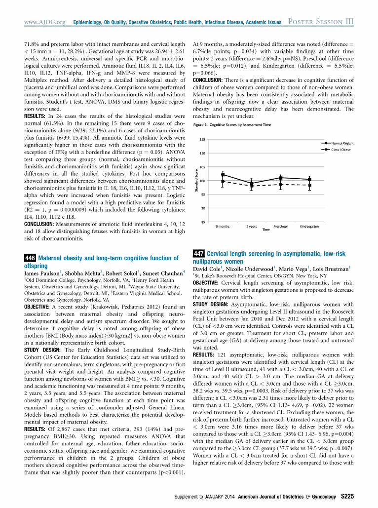
Maternal obesity and long-term cognitive function ofoffspringJames Paulson1, Shobha Mehta2, Robert Sokol3, Suneet Chauhan41Old Dominion College, Psychology, Norfolk, VA, 2Henry Ford HealthSystem, Obstetrics and Gynecology, Detroit, MI, 3Wayne State University,Obstetrics and Gynecology, Detroit, MI, 4Eastern Virginia Medical School,Obstetrics and Gynecology, Norfolk, VA

OBJECTIVE: A recent study (Krakowiak, Pediatrics 2012) found anassociation between maternal obesity and offspring neuro-developmental delay and autism spectrum disorder. We sought todetermine if cognitive delay is noted among offspring of obesemothers [BMI (Body mass index)�30 kg/m2] vs. non-obese womenin a nationally representative birth cohort.STUDY DESIGN: The Early Childhood Longitudinal Study-BirthCohort (US Center for Education Statistics) data set was utilized toidentify non-anomalous, term singletons, with pre-pregnancy or firstprenatal visit weight and height. An analysis compared cognitivefunction among newborns of women with BMI� vs. <30. Cognitiveand academic functioning was measured at 4 time points: 9 months,2 years, 3.5 years, and 5.5 years. The association between maternalobesity and offspring cognitive function at each time point wasexamined using a series of confounder-adjusted General LinearModels based methods to best characterize the potential develop-mental impact of maternal obesity.RESULTS: Of 2,867 cases that met criteria, 393 (14%) had pre-pregnancy BMI�30. Using repeated measures ANOVA thatcontrolled for maternal age, education, father education, socio-economic status, offspring race and gender, we examined cognitiveperformance in children in the 2 groups. Children of obesemothers showed cognitive performance across the observed time-frame that was slightly poorer than their counterparts (p<0.001).

Supplem

At 9 months, a moderately-sized difference was noted (difference ¼6.7%ile points; p¼0.034) with variable findings at other timepoints: 2 years (difference ¼ 2.6%ile; p¼NS), Preschool (difference¼ 6.5%ile; p¼0.012), and Kindergarten (difference ¼ 5.5%ile;p¼0.066).CONCLUSION: There is a significant decrease in cognitive function ofchildren of obese women compared to those of non-obese women.Maternal obesity has been consistently associated with metabolicfindings in offspring; now a clear association between maternalobesity and neurocognitive delay has been demonstrated. Themechanism is yet unclear.

447

Cervical length screening in asymptomatic, low-risknulliparous womenDavid Cole1, Nicolle Underwood1, Mario Vega1, Lois Brustman11St. Luke’s Roosevelt Hospital Center, OB/GYN, New York, NY

OBJECTIVE: Cervical length screening of asymptomatic, low risk,nulliparous women with singleton gestations is proposed to decreasethe rate of preterm birth.STUDY DESIGN: Asymptomatic, low-risk, nulliparous women withsingleton gestations undergoing Level II ultrasound in the RooseveltFetal Unit between Jan 2010 and Dec 2012 with a cervical length(CL) of <3.0 cm were identified. Controls were identified with a CLof 3.0 cm or greater. Treatment for short CL, preterm labor andgestational age (GA) at delivery among those treated and untreatedwas noted.RESULTS: 121 asymptomatic, low-risk, nulliparous women withsingleton gestations were identified with cervical length (CL) at thetime of Level II ultrasound, 41 with a CL < 3.0cm, 40 with a CL of3.0cm, and 40 with CL > 3.0 cm. The median GA at deliverydiffered; women with a CL < 3.0cm and those with a CL �3.0cm,38.2 wks vs. 39.5 wks, p¼0.0003. Risk of delivery prior to 37 wks wasdifferent; a CL <3.0cm was 2.31 times more likely to deliver prior toterm than a CL �3.0cm, (95% CI 1.13- 4.69, p¼0.02). 22 womenreceived treatment for a shortened CL. Excluding these women, therisk of preterm birth further increased. Untreated women with a CL< 3.0cm were 3.16 times more likely to deliver before 37 wkscompared to those with a CL �3.0cm (95% CI 1.43- 6.96, p¼0.004)with the median GA of delivery earlier in the CL < 3.0cm groupcompared to the �3.0cm CL group (37.7 wks vs 39.5 wks, p¼0.007).Women with a CL < 3.0cm treated for a short CL did not have ahigher relative risk of delivery before 37 wks compared to those with

ent to JANUARY 2014 American Journal of Obstetrics & Gynecology S225