3d carboxymethyl cellulose/hydroxyapatite (cmc/ha ... · / journal of applied pharmaceutical...
TRANSCRIPT

Journal of Applied Pharmaceutical Science Vol. 8(03), pp. 023-030, March, 2018Available online at http://www.japsonline.comDOI: 10.7324/JAPS.2018.8304
ISSN 2231-3354
© 2018 M. Sayed et al. This is an open access article distributed under the terms of the Creative Commons Attribution License -NonCommercial- ShareAlikeUnported License (http://creativecommons.org/licenses/by-nc-sa/3.0/).
*Corresponding AuthorS. M. Naga, National Research Center, Ceramics Department, 12622 El-Bohouth Str., Dokki, Cairo, Egypt. E-mail: salmanaga @ yahoo.com
3D Carboxymethyl Cellulose/Hydroxyapatite (CMC/HA) Scaffold Composites Based on Recycled Eggshell
M. Sayed1, H.F. El-Maghraby1, F. Bondioli2, S.M. Naga1,*
1National Research Center, Ceramics Dept., El-Bohous Str., 12622 - Cairo, Egypt.2Department of Industrial Engineering, Parco delle Scienze 181/A, 43124 - Parma, Italy.
ARTICLE INFO ABSTRACTArticle history:Received on: 31/10/2017Accepted on: 08/01/2018Available online: 30/03/2018
In this study, recycling of eggshell, which possesses valuable biominerals was utilized for production of nanocrystalline hydroxyapatite powder. Hydroxyapatite was then converted into 3D porous scaffolds by an eco-friendly method. For enhancing the mechanical resistance and improving the biodegradability of HA porous scaffold, CMC was used to coat the scaffolds with different concentration (0.5, 1.0, 1.5, 2.0% w/v) via infiltration method. The produced carboxymethyl cellulose/hydroxyapatite (CMC/HA) scaffold composites were evaluated for biomedical applications. The results revealed that the compressive strength of CMC/HA composite scaffolds increases with the increase in the CMC content up to 1.5% w/v. Furthermore, the results obtained from thin film XRD, SEM and EDS analysis after immersion in SBF solution for 28 days indicate the high bioactivity of the scaffolds. The biodegradability test points out that the CMC/HA are degradable naturally over the time; which is essential for tissue growth.
Key words: Recycling, Carboxymethylcellulose, Hydroxyapatite, Composite, Bioactivity.
INTRODUCTIONThe increasing demand for bone implants worldwide and
the lack of available organs and tissues suitable for transplantation forced the researchers to look for new biosources. Synthetic biomaterial especially those of biological origin are biocompatible, bioactive, inexpensive and eco-friendly to the environment. One of the promising biological origin that can be utilize for extraction of some valuable biominerals is the biowaste natural resources that generated from our daily operations with the huge amount such as bovine bone, fish scale, fish bone and eggshell (Sri Asliza et al., 2009; Alparslan et al., 2017; Naga et al., 2015a, 2015b). Recently, some authors have obtained biomaterials from eggshell and urine as an innovated waste management approach through, either simple chemical process (Kannan and Ronan, 2017) or a novel sustainable route via electrosynthesis (Ronan and Kannan, 2017).
The biogenic natural resources can be used for production hydroxyapatite; the main inorganic component of natural bone.
Hydroxyapatite is widely used for bone repair and replacement due to its favourable bioactivity and biocompatibility, besides its chemical composition similar to human bone (Mauludin et al., 2012). However, the brittleness and poor mechanical properties limited its clinical applications (Constanda et al., 2016). Therefore, a combination of HA with some materials like polymers will lead to increase the mechanical resistance as well as improve the biodegradability and osteoconductivity. On the other hand, this combination will mimic the phase composition of natural bone that consists of; mineralized hydroxyapatite inorganic phase and non-mineralized natural polymer organic phase (Raucci et al., 2012). Many studies reported the preparation of composites containing HA in conjugation with some bioactive polymer or proteins, such as collagen, gelatin, chitosan, silk fibroin and chondroitin sulphate (Zhongguo et al., 2012; Chiu et al., 2012; Hunter and Ma 2013; Kweon et al., 2011). Selection of the polymeric system is based on its water solubility, polarizability (anionic/cationic) and hydrogen bond, as well as Ca2+ ions chelation capacity of the functional group associated with the polymer matrix. Carboxymethyl cellulose (CMC) is one of the most commonly employed polymers used in this process (Sinha et al., 2007). It possess excellent properties such as biocompatibility, low degradability and

Sayed et al. / Journal of Applied Pharmaceutical Science 8 (03); 2018: 023-030024
low cost compared to the other naturally derivative polymers. All these properties promoted the selection of CMC to combine with the HA to suit better the mechanical and physiological demands of the host tissue in biomedical field (Ramli and Wong, 2011). Many methods have been reported for fabrication the carboxymethyl cellulose (CMC)/hydroxyapatite (HA) composite scaffolds.
Garai and Sinha (2014) synthesized three-dimensional micro/macroporous CMC/HA nanocomposites by in suit formation method. They reported that their method not only controls the size of HA particles in the range of 15-50 nm, but it also assists the formation of a mechanically strong three-dimensional structure exhibiting a systematic dependence of structure-property correlations and cytocompatibility with composition. They added that the ionic/polar or hydrogen bonding/electrostatic interaction is the main driving force for the formation of three-dimensional porous nanocomposites. Zakharov et al. (2005) synthesized CMC/HA nanocomposites via coprecipitation technique. The prepared hydroxyapatite particles are agglomerated and the agglomerate size was about 200 nm. The interaction between the nano-HA particles and the CMC macromolecules led to the formation of a pore structure suitable for biomedical applications. The microwave-assisted method was used to prepare cellulose/HA nanocomposites at 1250°C (Ma et al., 2010; Jia et al., 2010a, 2010b; Ma et al., 2011). Mixing of CMC and HA for the preparation of CMC/HA composites were carried out by Sadiasa et al. (2013) and Aly et al. (2011). On the other hand, Pasqui et al. (2014) inserted HA nanocrystals within the CMC matrix, rather than mixing them to a polymer solution. CMC/HA and HA/chitosan/CMC composites were also prepared by freeze-drying method (Jiang et al., 2008; Basu et al., 2014). Polymeric sponge method was used by Aly et al. (2011) to prepare porous CMC/HA-β-tricalcium phosphate scaffolds. They reported that the use of HA and TCP powders together allows controlling of the degradation rate and the mechanical properties. The prepared scaffolds have a highly porous interconnected microstructure. The increase in the powder content increases the compressive strength, but it decreases the pore size of the scaffolds.
To the best of the authors knowledge, the fabrication of CMC/HA composite scaffolds were mainly based on chemically hydroxyapatite sources. In our study we propose fabrication of porous CMC/HA composite scaffolds based on eggshell as a natural source for HA. The eggshell is attractive biosource not only due to its high content of calcium carbonate but also due to the presence of trace elements such as Mg2+, Na+, Sr2+ and Si4+,which have great value in biomineralization during bone formation. The study focused on the developing of CMC/HA composite scaffolds via coating the nano HA scaffold based on eggshell natural sources with different concentrations (0.5, 1.0, 1.5, 2.0% w/v) of carboxymethyl cellulose (CMC) solutions for 10 minutes. Investigation of the microstructure and the mechanical properties of the different concentrations CMC/HA composite scaffolds were carried out. Physical, mechanical properties, as well as the bioactivity and biodegradability of the selected CMC/HA composite scaffolds, were accomplished.
MATERIALS AND METHODSStarting materials
Hen eggshell collected from bakery shops, orthophosphoric acid H3PO4 (Elgoumhouria Co. for trading Medicines Ameria Cairo, Egypt), ammonia solution NH4OH
(Wessex House, Shaftesbury, Dorset, UK-Molekula), polyvinyl alcohol LR (Laboratory Rasyan), highly dense polyethylene sponge, Carboxymethyl cellulose CMC (RnOCH2COONa, 250,000 M.Wt., United Company for Chem. and Med) and reagent-grade NaCl, NaHCO3, KCl, Na2HPO4∙2H2O, MgCl2∙6H2O, CaCl2, and Na2SO4 were used as starting materials in the present study.
Methods
Preparation of hydroxyapatite powder from eggshellThe hen eggshells were collected from bakery shops,
washed, dried then crushed to fine powder. The eggshell powder was calcined at 900°C for 2h with a heating rate of 5°C/min. The decomposition of eggshell (CaCO3) to CaO takes place according to the equation:
900°C, 2hCaCO3 CaO + CO2
The freshly prepared CaO was mixed with the stoichiometric amount of distilled water to prepare Ca(OH)2, according to the equation:
CaO + H2O Ca(OH)2
0.5 M Ca(OH)2 suspension and 0.3 M orthophosphoric acid were prepared by the appropriate dilution. The 0.5 M Ca(OH)2 suspension was vigorously stirred at 70°C for an hour. Then the 0.3 M H3PO4 solution was added drop wisely with a slow stirring rate. The pH of the solution was kept at the range of 10.5−11 by the addition of ammonia solution. The formed slurry was left at room temperature overnight for aging. The precipitate was dried at 105°C for 24 h. The HA formation reaction was taking place according to the following equation:
10Ca(OH)2 + 6H3PO4 Ca10 (PO4)6(OH)2 + 18H2O
The formed powder was calcined at 900ºC for 2h with a firing rate of 10°C/min. The calcined powder was finely ground completely to pass 90 microns sieve as described in our previous work (Naga et al., 2015b).
Preparation of 3D porous hydroxyapatite scaffoldsA set of cubic and rectangular specimens, with
dimensions of 10 × 10 × 10 mm and 50 × 15 × 10 mm respectively, were cut from high-density polyethylene sponge. Specimens were immersed in the HA slurry under vacuum for 3 h to form porous HA scaffolds after firing. The soaked samples were dried slowly at 50ºC for 6 h, then at 80ºC for 6 h and finally at 110ºC overnight. To accelerate the oxidation of the formed carbon produced by the sponge fiber and to sweep the gases formed, the samples were fired up to 600ºC under static air. A slow heating rate was adopted at low temperature to prevent bodies cracking during the burnout process, i.e., a rate of 2ºC/min from room temperature to 600ºC followed by a higher rate of 5ºC/min to the sintering temperature. The samples were fired at 1250ºC with a soaking time of 2h.
Preparation of hydroxyapatite scaffolds coated with car-boxymethyl cellulose (CMC)
Carboxymethyl cellulose (CMC) solutions with different concentrations (0.5, 1.0, 1.5, 2.0% w/v) were prepared

Sayed et al. / Journal of Applied Pharmaceutical Science 8 (03); 2018: 023-030 025
by dissolving the appropriate amounts of CMC in double distilled water. Sintered HA scaffolds (sintered at 1250ºC for 2h) were infiltrated with carboxymethyl cellulose (CMC) solutions having different concentrations (0.5, 1.0, 1.5, 2.0% w/v). The scaffolds were immersed for 10 minutes. A vacuum was applied to force the CMC solution to penetrate the pores of the scaffolds. The coated scaffolds were then removed from the solutions and kept in air for 24 hours to dry.
In vitro testHA scaffold coated with 1.5% w/v CMC specimens
were immersed in simulated body fluid (SBF) for 1, 3, 6, 12 hrs and 1, 3, 7, 14, 21, 28 days at 37ºC. The used SBF solution has ions concentration nearly equal to that of human blood plasma as described by Kokubo et al. (1990). 0.5 gm of the sample was immersed in 100 ml SBF at pH 7.4. The temperature was kept at 37˚C during the test. After immersing process, the concentration of both calcium and phosphorus ions in the solution was analyzed. The microstructure of the immersed samples was investigated by SEM attached to EDS, while the formed apatite layer on the surface of scaffolds was characterized using thin film XRD analysis.
Biodegradability testWeight loss (WL%) of pure HA and HA scaffold coated
with 1.5% w/v CMC specimens can be calculated according to the following equation:
WL = [(M0 − Mt)/M0] × 100,
where, M0: the initial weight of each sample, Mt: the dry weight of the same sample measured at time t.
Weight loss measurements were carried out for the samples by immersion in SBF solution for 1, 3, 7, 14, 21, 28 days at 37ºC in order to study the degradation of those samples at mentioned time periods.
CharacterizationBulk density and apparent porosity were evaluated using
the Archimedes method (ASTM C-20). The microstructure of the of the HA scaffolds coated with different CMC concentrations was investigated via scanning electron microscopy (Jeol JSM- T20, Japan) attached with EDX unit. The particle size and the morphology of the HA powder calcined at 900°C for 2h along with its agglomeration tendency were studied by transmission electron microscopy, TEM (JEOL, JEM-2100-HR, Japan, Electron probe micro-analyzer) working at 200 kV. The phase composition of the prepared powder was analyzed using X-ray diffraction analysis (XRD) using monochromated CuKα radiation (D 500, Siemens, Mannheim, Germany). Compressive strength was measured using universal testing machine (LRLR10KPlus 10 kN (2248 lbf) type, Japan), with speed range 0.01 mm/min. FTIR analysis was carried out using FTIR-6100 Fourier transform Infrared Spectrometer from JASCO. Bending strength was measured using a three-point bending test on a universal testing machine (Model 4204, Instron Corp., Danvers, MA) at a crosshead speed of 0.5 mm/min and support distance of 40 mm. 10 specimens were measured for each data point. The Change of Ca2+ and PO4
3− ions concentration determined with Inductively Coupled Plasma Spectrometer (ICP); Model Ultima-2 JY 2000-2, France; while the formed apatite layer
on the surface of the immersed samples in SBF was characterized using thin film XRD (XʼPert Pro. PANlxtical, target Cu-Kα with second monochromator Kυ = 45 mA; 4, Holland).
RESULTS AND DISCUSSION
Characterization of hydroxyapatite powder obtained from eggshell bio waste
Phase composition (XRD)The XRD analysis of the as-prepared powder, calcined
at 900°C for 2h is shown in Figure (1). The figure indicates that the powder is composed completely of HA phase with sharp and well-defined peaks. It reveals that HA is present as a pure phase without any secondary phases. The indexed d-spacing of the samples were found to be very close to the values obtained from the JCPDS (009-0432) standard data.
Fig. 1: XRD analysis of HA powder calcined at 900°C for 2h.
Particle size analysis (TEM)The TEM observation of the as prepared HA powder
calcined at 900°C for 2h., Figure (2); revealed that the particles have a crystalline size in the nano-scale ranging between 61 to 130 nm.
Fig. 2: TEM image of the HA powder calcined at 900°C for 2h.

Sayed et al. / Journal of Applied Pharmaceutical Science 8 (03); 2018: 023-030026
Characterization of porous 3D CMC/HA scaffolds
Compressive strength The compressive strength and the apparent porosity
of the coated porous CMC/HA scaffolds are given in Figure (3). It is clear from the figure that there is a close relationship between the bodies porosity and their compressive strength. The figure shows that the compressive strength increases with the increase in the CMC content up to 1.5% w/v. The increase in the CMC concentration to 2.0% w/v decreases the compressive strength of the scaffolds significantly. The presence of the polymer in the coated scaffolds not only prevents the catastrophic failure of the material but also retains and increases the mechanical integrity. CMC coat can penetrate some micropores and small cracks in the HA scaffolds, which result in an increase in compressive strength. CMC coat played a critical role in improving mechanical properties of the scaffolds by binding the scaffold into a whole body. The increase in the 2.0% w/v CMC solution viscosity hinders its diffusion throughout HA scaffolds, which in turn leads to an increase in the scaffolds porosity and a decrease in their compressive strength, Figure 3. Based on the above-mentioned results it is worth to conclude that the CMC/HA composite scaffolds can simultaneously retain high porosity and moderate mechanical properties by using an appropriate polymer concentration (Zhao et al., 2009; Kang et al., 2011), which is 1.5% w/v in this study. Accordingly, 1.5% w/v CMC is selected for further testing to throw light on the physical, bioactivity and biodegradability properties of the scaffolds.
Characterization of the 1.5% w/v CMC/HA porous scaffolds
Infrared analysis (IR)The FTIR spectrum for the HA scaffolds coated with
1.5% w/v CMC is illustrated in Figure (4). A broad band centred at 3865 cm−1 was ascribed to the hydrogen-bonded hydroxyl groups distribution. The broad band at 3424 cm−1 confirms the presence of strong hydrogen bonding between HA and CMC. This result indicates that a cross linking between HA and CMC has occurred. On the other hand, the absorption band of C-O (1061 cm−1 in CMC) disappeared because of the overlapping of the absorption band of P-OH (Garai and Sinha, 2014). The bands corresponding to PO4
3− group at 1028 cm−1 (υ3), 957 cm−1 (υ1), 590 cm−1 (υ4) and 3679 cm−1 (υ) (liberation mode) are assigned to HA. Bands at 1454, 1426 and 799 cm−1 are corresponding to vibration mode υ3, υ3 and υ2, respectively, of carbonate group. The positions of the carbonate bands indicate that CO3
2− groups are substituting the PO43− position (Elliott,
1994). The liberation and stretching mode of the OH− were detected at around 3757 and 1633 cm−1 respectively. The stretching vibration, ascribed to CO3
2− present around 1426 cm−1 is corresponding to the symmetrical stretching vibration of the carboxylate group (Rosca et al., 2005). It is suggested that CO3
2− ions are incorporated during the synthesis of HA from eggshell (Koutsopoulos, 2002). While, the bands at 2924 and 2857 cm−1 are assigned to the CH2 stretching vibration (Li et al., 2005). The additional peak at 2423 cm−1 may be due to the existence of some impurities or combination band with
water. The bands around 1272 and 1386 cm−1 are assigned to methyl (-CH2) scissoring and hydroxyl group (-OH) bending vibration, respectively (Mohkami and Talaeipour, 1988).
Fig. 3: The effect of CMC concentration on the compressive strength and the apparent porosity of the prepared CMC/HA porous scaffolds.
Fig. 4: The FTIR spectrum for the HA scaffolds coated with 1.5% w/v CMC.
Table 1: Physical and mechanical properties of porous HA scaffold uncoated and coated with 1.5% w/v CMC.
PorousHA
Scaffold
Bulk density,g/cm3
Apparent porosity,
%
Bending strength,
MPa
Compressive strength,
MPa
Uncoated with CMC 0.98 ± 0.11 73 ± 1.9 1.72 ± 0.11 0.82 ± 0.01
Coated with 1.5% CMC 1.04 ± 0.08 50 ± 1.03 3.43 ± 0.17 1.49 ± 0.06
Morphology Figure (5-a) represents the typical morphology of
untreated sponge, while Figures (5-b) and (5-c) represent the uncoated and 1.5% w/v CMC coated HA scaffolds prepared by polymeric sponge method. Both uncoated and coated HA scaffolds possess round interconnected macropores and maintain the initial sponge structure.

Sayed et al. / Journal of Applied Pharmaceutical Science 8 (03); 2018: 023-030 027
Fig. 5: SEM images of (a: untreated sponge, b: uncoated HA scaffold and c: HA scaffold coated with 1.5 % w/v of CMC).
Fig. 6: The mean value of Ca2+ and PO43− ions concentrations (mg/L) after
immersion of 1.5% w/v CMC/HA porous scaffolds in SBF for 28 days (a: first region, b: second and third region and c: collected graph for both (a) and (b)).
Fig. 7: Thin film XRD pattern of the surface 1.5% CMC coated porous HA scaffold composite surface; immersed in SBF for 28 days.
Physical and mechanical properties The effect of coating HA scaffolds with 1.5% w/v CMC
on the scaffolds physical properties is illustrated in Table (1). The bulk density of the coated scaffolds increased from 0.98 ± 0.11 g/cm3 for uncoated scaffolds to1.04 ± 0.08 g/cm3 for samples coated with 1.5% w/v CMC. On the other hand, the apparent porosity and the median pore diameter of the coated scaffolds decreased from 73.0 ± 1.9 and 41.72 µm for the uncoated scaffold to 50.0 ± 1.03% and 0.05 µm for coated scaffolds, respectively. The obtained values of both compressive and bending strength are reported in Table (1). It is noticed that the compressive strength of the CMC coated scaffolds increased from 0.82 ± 0.01 MPa for uncoated scaffolds to 1.49 ± 0.06 MPa for coated scaffolds. The bending strength also, increased from 1.72 ± 0.11 for HA scaffolds to reach 3.43 ± 0.17 for CMC coated scaffolds. The obtained results are in agreement with the results of Follet et al. (2004) and Garai and Sinha (2014). They reported that the compressive strength is a function of the mineral content. The presence of unmineralized polymer chains around the HA crystals led to the sluggish of the crack propagation process and the enhancement of the mechanical properties. The decrease in the porosity figures of the coated scaffolds is an additional factor that enhanced the mechanical properties of the coated scaffolds.
Bioactivity measurements, in vitro test of HA scaffold coated with 1.5% w/v CMC
The bioactivity of HA porous scaffolds coated with 1.5% w/v CMC were evaluated after soaking in SBF solution for

Sayed et al. / Journal of Applied Pharmaceutical Science 8 (03); 2018: 023-030028
28 days. The mean value of Ca2+ and PO43− ions concentrations
(mg/l) as a function of the immersion time are given in Figure (6). The figure shows three different regions; in the first one, both Ca2+ and PO4
3− ions concentrations decrease sharply till 12 h due to the quick formation of calcium phosphate layer on the scaffolds surfaces (Figures 6a&c). In the second region, they slightly increase from the second half of the first day till the 3rd day, which is attributed to an intermediate in equilibrium between the formation and dissolution of HA till the stage of the three-dimensional structure formation. A slight decrease is observed till the end of the first week followed by a stability of the ions concentration up to the end of the fourth week of the experiment; third region (Figures 6 b&c). The decrease in the Ca2+ and PO4
3− ions concentration in the first region is attributed to the early formation of apatite layer on the coated scaffold surfaces. Where, the availability of functional groups, such as carboxyl (-COO) and hydroxyl (-OH) groups of the CMC, promotes the attraction
of Ca2+ and PO43− ions to form HA nuclei. Once HA nuclei
formed, a spontaneous and quick consuming of both Ca2+ and PO4
3− occurs causing a sharp decrease in the ions concentration in the first region. In this region, the calcium ions are attracted to carboxyl groups (COO-ions) to form a gel like solution due to electrostatic interactions as well as physical cross linking. Then, phosphate ions react with calcium ions to form calcium phosphates including hydroxyapatite, depending on the existing pH conditions. Under unsaturated conditions for precipitation, the nuclei of HA crystals are believed to be formed and stabilized by heterogeneous nucleation process as well as hydrogen bonding between hydroxyl groups of CMC and HA. Once HA has been nucleated and grown to a size, controlled by the geometrical space provided by the CMC gel, it gets entrapped in the gel structure by steric entrapment and cross linking forming a three-dimensional structure (Garai and Sinha, 2014).
Fig. 8: SEM micrograph of the 1.5% w/v CMC/ HA porous scaffold immersed in SBF for 28 days (a: at magnification X 2500 and b: at high magnification X 10,000 with Energy-dispersive XRD spectra (EDX)).
Thin film XRD analysis: Thin film XRD analysis of HA deposited on the surface of the 1.5% w/v CMC coated scaffolds immersed in SBF for 28 days is shown in Figure (7). XRD pattern confirms the deposition of HA as a main phase together with a modicum quantity of beta-tricalcium phosphate.
Microstructure of 1.5% CMC/HA scaffold surface after immer-sion in SBF: SEM micrograph of the coated samples immersed in SBF for 28 days; Figure (8-a); shows apatite particles aggre-gates forming a bead like structure deposit on the surface of the scaffolds. It confirms that the CMC/HA composites have high bioactivity and have the ability to form an apatite layer on their surfaces, which can make direct bond to the living bone when implanted in the body (Rezwan et al., 2006). Figure (8-b) with higher magnification indicates the uniform distribution of irreg-ular pores having a size range of 0.8-2.4 µm. According to Kim et al. (2006) the presence of micro-pores in the HA/polymer composite scaffolds enhances bone generation. EDX analysis was performed in order to determine the molar ratio between Ca2+ and P3− ions after immersing 1.5% w/v CMC sample in SBF solution for 28 days. EDX analysis revealed that Ca, P and O accumulated on the surface of HA scaffold coated with 1.5% w/v CMC, which can be identified as a calcium phosphate
layer; Figure (8-b). The Ca/P ratio confirmed the formation of non-stoichiometric apatite (1.51), which might be indicative of the presence of a Ca deficient apatite. This Ca–P deposition is of greater biological interest than stoichiometric HA since the Ca/P ratio in natural bone is lower than 1.67 (Kobayashi et al., 2001).
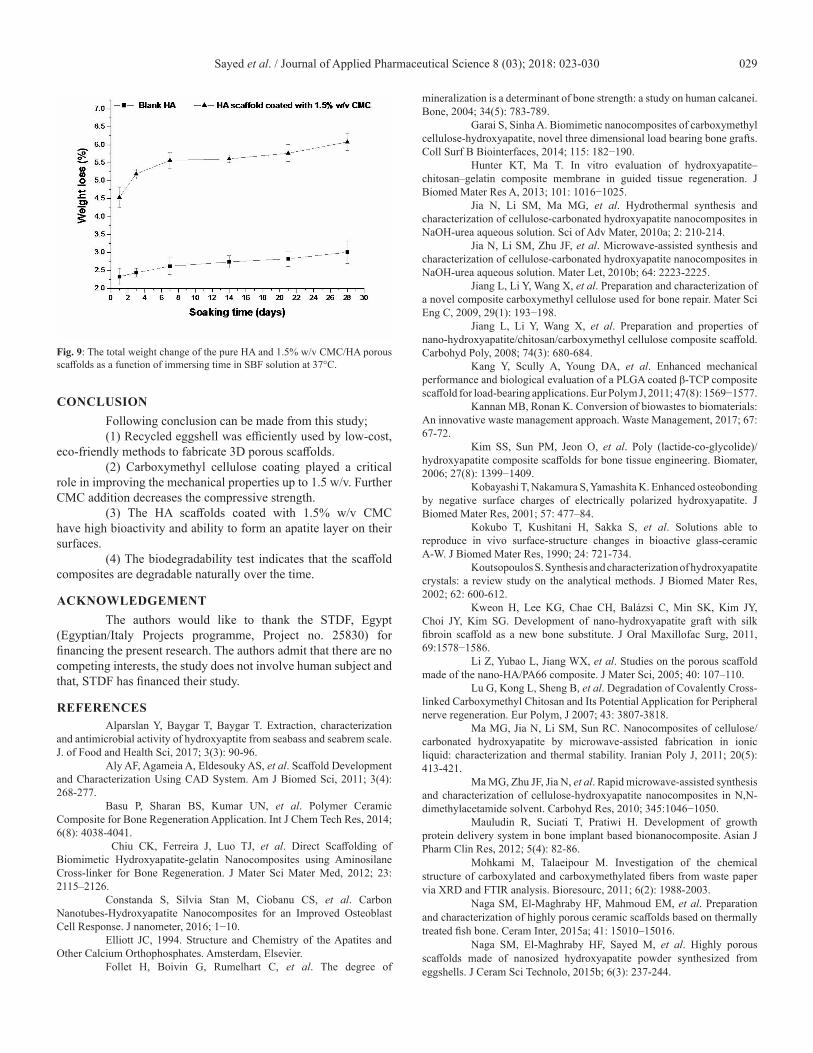
Biodegradability Biodegradability of the 3D organic/inorganic scaffold
has an important role in biomedical applications. Scaffolds should be degradable naturally over the time to permit new tissues to grow. Since CMC is a water-soluble polymer, we carried out in vitro degradation test to determine the weight loss of the developed composite scaffolds. Figure (9) represents the weight change of pure HA and HA scaffold coated with 1.5% w/v CMC as a function of immersing time in SBF solution at 37°C. The figure shows that the rate of weight loss increases from 4.54% in the first day to 6.08 in the 28th day. It is noticeable that the 1.5% w/v CMC composites scaffolds have higher weight loss 6.08% in comparison to HA scaffold samples without coating, 3.01%. It is due to the presence of the cross-linking of the carboxymethyl, which is soluble in water (Jiang et al., 2009; Lu et al., 2007).
Sayed et al. / Journal of Applied Pharmaceutical Science 8 (03); 2018: 023-030 029
Fig. 9: The total weight change of the pure HA and 1.5% w/v CMC/HA porous scaffolds as a function of immersing time in SBF solution at 37°C.
CONCLUSION Following conclusion can be made from this study;(1) Recycled eggshell was efficiently used by low-cost,
eco-friendly methods to fabricate 3D porous scaffolds.(2) Carboxymethyl cellulose coating played a critical
role in improving the mechanical properties up to 1.5 w/v. Further CMC addition decreases the compressive strength.
(3) The HA scaffolds coated with 1.5% w/v CMC have high bioactivity and ability to form an apatite layer on their surfaces.
(4) The biodegradability test indicates that the scaffold composites are degradable naturally over the time.
ACKNOWLEDGEMENTThe authors would like to thank the STDF, Egypt
(Egyptian/Italy Projects programme, Project no. 25830) for financing the present research. The authors admit that there are no competing interests, the study does not involve human subject and that, STDF has financed their study.
REFERENCESAlparslan Y, Baygar T, Baygar T. Extraction, characterization
and antimicrobial activity of hydroxyaptite from seabass and seabrem scale. J. of Food and Health Sci, 2017; 3(3): 90-96.
Aly AF, Agameia A, Eldesouky AS, et al. Scaffold Development and Characterization Using CAD System. Am J Biomed Sci, 2011; 3(4): 268-277.
Basu P, Sharan BS, Kumar UN, et al. Polymer Ceramic Composite for Bone Regeneration Application. Int J Chem Tech Res, 2014; 6(8): 4038-4041.
Chiu CK, Ferreira J, Luo TJ, et al. Direct Scaffolding of Biomimetic Hydroxyapatite-gelatin Nanocomposites using Aminosilane Cross-linker for Bone Regeneration. J Mater Sci Mater Med, 2012; 23: 2115–2126.
Constanda S, Silvia Stan M, Ciobanu CS, et al. Carbon Nanotubes-Hydroxyapatite Nanocomposites for an Improved Osteoblast Cell Response. J nanometer, 2016; 1−10.
Elliott JC, 1994. Structure and Chemistry of the Apatites and Other Calcium Orthophosphates. Amsterdam, Elsevier.
Follet H, Boivin G, Rumelhart C, et al. The degree of
mineralization is a determinant of bone strength: a study on human calcanei. Bone, 2004; 34(5): 783-789.
Garai S, Sinha A. Biomimetic nanocomposites of carboxymethyl cellulose-hydroxyapatite, novel three dimensional load bearing bone grafts. Coll Surf B Biointerfaces, 2014; 115: 182−190.
Hunter KT, Ma T. In vitro evaluation of hydroxyapatite–chitosan–gelatin composite membrane in guided tissue regeneration. J Biomed Mater Res A, 2013; 101: 1016−1025.
Jia N, Li SM, Ma MG, et al. Hydrothermal synthesis and characterization of cellulose-carbonated hydroxyapatite nanocomposites in NaOH-urea aqueous solution. Sci of Adv Mater, 2010a; 2: 210-214.
Jia N, Li SM, Zhu JF, et al. Microwave-assisted synthesis and characterization of cellulose-carbonated hydroxyapatite nanocomposites in NaOH-urea aqueous solution. Mater Let, 2010b; 64: 2223-2225.
Jiang L, Li Y, Wang X, et al. Preparation and characterization of a novel composite carboxymethyl cellulose used for bone repair. Mater Sci Eng C, 2009, 29(1): 193−198.
Jiang L, Li Y, Wang X, et al. Preparation and properties of nano-hydroxyapatite/chitosan/carboxymethyl cellulose composite scaffold. Carbohyd Poly, 2008; 74(3): 680-684.
Kang Y, Scully A, Young DA, et al. Enhanced mechanical performance and biological evaluation of a PLGA coated β-TCP composite scaffold for load-bearing applications. Eur Polym J, 2011; 47(8): 1569−1577.
Kannan MB, Ronan K. Conversion of biowastes to biomaterials: An innovative waste management approach. Waste Management, 2017; 67: 67-72.
Kim SS, Sun PM, Jeon O, et al. Poly (lactide-co-glycolide)/hydroxyapatite composite scaffolds for bone tissue engineering. Biomater, 2006; 27(8): 1399−1409.
Kobayashi T, Nakamura S, Yamashita K. Enhanced osteobonding by negative surface charges of electrically polarized hydroxyapatite. J Biomed Mater Res, 2001; 57: 477–84.
Kokubo T, Kushitani H, Sakka S, et al. Solutions able to reproduce in vivo surface-structure changes in bioactive glass-ceramic A-W. J Biomed Mater Res, 1990; 24: 721-734.
Koutsopoulos S. Synthesis and characterization of hydroxyapatite crystals: a review study on the analytical methods. J Biomed Mater Res, 2002; 62: 600-612.
Kweon H, Lee KG, Chae CH, Balázsi C, Min SK, Kim JY, Choi JY, Kim SG. Development of nano-hydroxyapatite graft with silk fibroin scaffold as a new bone substitute. J Oral Maxillofac Surg, 2011, 69:1578−1586.
Li Z, Yubao L, Jiang WX, et al. Studies on the porous scaffold made of the nano-HA/PA66 composite. J Mater Sci, 2005; 40: 107–110.
Lu G, Kong L, Sheng B, et al. Degradation of Covalently Cross-linked Carboxymethyl Chitosan and Its Potential Application for Peripheral nerve regeneration. Eur Polym, J 2007; 43: 3807-3818.
Ma MG, Jia N, Li SM, Sun RC. Nanocomposites of cellulose/carbonated hydroxyapatite by microwave-assisted fabrication in ionic liquid: characterization and thermal stability. Iranian Poly J, 2011; 20(5): 413-421.
Ma MG, Zhu JF, Jia N, et al. Rapid microwave-assisted synthesis and characterization of cellulose-hydroxyapatite nanocomposites in N,N-dimethylacetamide solvent. Carbohyd Res, 2010; 345:1046−1050.
Mauludin R, Suciati T, Pratiwi H. Development of growth protein delivery system in bone implant based bionanocomposite. Asian J Pharm Clin Res, 2012; 5(4): 82-86.
Mohkami M, Talaeipour M. Investigation of the chemical structure of carboxylated and carboxymethylated fibers from waste paper via XRD and FTIR analysis. Bioresourc, 2011; 6(2): 1988-2003.
Naga SM, El-Maghraby HF, Mahmoud EM, et al. Preparation and characterization of highly porous ceramic scaffolds based on thermally treated fish bone. Ceram Inter, 2015a; 41: 15010–15016.
Naga SM, El-Maghraby HF, Sayed M, et al. Highly porous scaffolds made of nanosized hydroxyapatite powder synthesized from eggshells. J Ceram Sci Technolo, 2015b; 6(3): 237-244.
Sayed et al. / Journal of Applied Pharmaceutical Science 8 (03); 2018: 023-030030
Pasqui D, Torricelli P, De Cagna M, et al. Carboxymethyl cellulose—hydroxyapatite hybrid hydrogel as a composite material for bone tissue engineering applications. J Biomed Mater Res, 2014; 102(5): 1568−1579.
Ramli NA, Wong TW. Sodium carboxymethyl cellulose scaffolds and their physicochemical effects on partial thickness wound healing. Int J Pharm, 2011; 403: 73-82.
Raucci MG, Guarino V, Ambrosio L. Biomimetic strategies for bone repair and regeneration. J Funct Biomater, 2012; 3: 688-705.
Rezwan K, Chen QZ, Blaker JJ, et al. Biodegradable and bioactive porous polymer/inorganic composite scaffolds for bone tissue engineering. Biomater, 2006; 27(18): 3413-3431.
Ronan K, Kannan BM. A novel sustainable route for synthesis of hydroxyapatite biomaterial from biowastes. ACS Sustainable Chem & Eng, 2017; 5: 2237-2245.
Rosca C, Popa MI, Lisa G, et al. Interaction of chitosan with natural or synthetic anionic polyelectrolytes. 1. The chitosan-carboxymethylcellulose complex. Carbohydrate Poly, 2005; 62(1)*: 35–41.
Sadiasa A, Franco RA, Seo HS, et al. Hydroxyapatite delivery to dentine tubules using carboxymethyl cellulose dental hydrogel for treatment of dentine hypersensitivity. J Biomed Sci and Eng, 2013; 6: 987-995.
Sinha A, Das G, Shanna BK, et al. Poly (vinyl alcohol) hydroxyapatite biomimetic scaffold for tissue regeneration. Mater Sci Eng
C, 2007; 27: 70–74.Sri Asliza MA, Zaheruddin K, Shahrizal H. Study the properties
of dense hydroxyapatite extract from cow bone. J Nuclear and Related Technolo, 2009; 6: 175−182.
Zakharov NA, Ezhova Zh A, Koval EM, et al. Hydroxyapatite-Carboxymethyl Cellulose nanocomposite biomaterial. Inorg Mater, 2005; 41: 509-515.
Zhao J, Lu X, Duan K, et al. Improving mechanical and biological properties of macroporous HA scaffolds through composite coatings. Colloid Surface B, 2009; 74: 159−166.
Zhongguo XF, Chong J, Wai K, et al. Preparation and biocompatibility of a novel biomimetic osteochondral scaffold: collagen-chitosan/nano-hydroxyapatite-collagen-polylactic acid. Chin J Repear Recon Surg, 2012; 26: 1001−1006.
How to cite this article: Sayed M, El-Maghraby HF, Bondioli F, Naga SM. 3D Car-boxymethyl Cellulose/Hydroxyapatite (CMC/HA) Scaffold Composites Based on Recycled Eggshell. J App Pharm Sci, 2018; 8(03): 023-030.