3- nutrition and digestion
DESCRIPTION
nutrition and digestionTRANSCRIPT

Animal Nutrition and digestion

An animal’s diet must provide
– Chemical energy for cellular processes - ATP– Organic building blocks for macromolecules * Carbon– Essential nutrients

Essential Nutrients
• Criteria to be an Essential nutrient:1. They must be essential to the health2. They can not be synthesized by the body
• These must be obtained from an animal’s diet• There are four classes
– Essential amino acids– Essential fatty acids– Vitamins– Minerals

Essential Amino Acids
• Animals require 20 amino acids and can synthesize about half from molecules in their diet
• The remaining amino acids, the essential amino acids, must be obtained from food in preassembled form
• Meat, eggs, and cheese provide all the essential amino acids and are thus “complete” proteins

Figure 5.16a
Valine(Val or V)
Leucine(Leu or L)
Isoleucine(Ile or I)
Figure 5.16a
Methionine(Met or M)
Phenylalanine(Phe or F)
Tryptophan(Trp or W)
Lysine(Lys or K)
Threonine(Thr or T)
Essential Amino Acids

Essential Fatty Acids
• Animals can synthesize most of the fatty acids they need
• The essential fatty acids must be obtained from the diet and include certain unsaturated fatty acids (i.e., fatty acids with one or more double bonds)
• Deficiencies in fatty acids are rare

What is a fatty acid?
- Animals do not produce unsaturated fats, and therefore must obtain them from the diet.
- Unsaturated fatty acids are acquired by ingesting oils produced in plants. (olive oil, corn oil)
Because of the “kinks” when double bonds are present, unsaturated fats do not pack close together and are therefore liquid at room temperature

Saturated Fatty Acid are a Solid at Room Temperature:
Unsaturated Fatty Acids are liquid at Room Temperature:

Vitamins
• Vitamins are organic molecules required in the diet in very small amounts
• Vitamins are grouped into two categories: fat-soluble and water-soluble
• Importance: needed for coenzymes, and for production of cellular proteins, nucleic acids, and other cell processes.

Excess Water-soluble vitamins are not stored in the body, therefore not toxic in excess

Table 41.1
Excess Fat-soluble vitamins are stored in adipose fat, and therefore subject to
toxicity Dietary Source major function in body symptoms of deficiency

Minerals
• Minerals are simple inorganic nutrients, usually required in small amounts
• Ingesting large amounts of some minerals, such as NaCl, can upset homeostatic balance
• Sodium, potassium and chloride are needed for nerve function, where they are important for the establishment of membrane potential
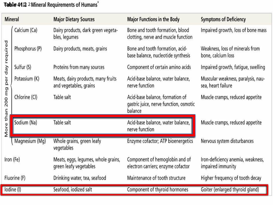
Table 41.2

Vitamin B3Iron
Linoleic acid
NADH
Fatty acid desaturase
ESSENTIALAMINO ACIDS
Phospholipids
-Linoleic acid
Prostaglandins
GlyIle
LeuPhe
PheTyr
Glu
Essential Fatty Acid
Essential MineralEssential vitamin
Example of roles of Essential Nutrients

Anemia• Reduced red blood count due to diet deficient
in iron, folate (B9) and/or B12
• Degeneration of skin and teeth• Due to lack of Vitamin C
Scurvy

Ingestion and digestion
• The taking in and breaking down of nutrients• Differs with the species

Intracellular digestion,
Breaking down of food inside of cellfood particles are engulfed by phagocytosis
- Food vacuoles, containing food, fuse with lysosomes containing hydrolytic enzymes

Mouth
Tentacles
Food
Epidermis Gastrodermis
Digestive enzymesare released from agland cell.
Enzymes breakfood down into smallparticles.
Food particles areengulfed and digestedin food vacuoles.
1
2
3
Simple organisms have a gastrovascular cavity
gastrovascular cavity functions in both digestion and distribution of nutrients

Figure 41.8
Esophagus
Esophagus
Esophagus
Crop
Crop
Crop
Gizzard
Gizzard
Intestine
Intestine
Anus
Anus
Anus
Mouth
Mouth
Mouth
Stomach
Foregut Midgut Hindgut
Rectum
Gastric cecae
(a) Earthworm
(b) Grasshopper
(c) Bird
Pharynx
More complex animals have a digestive tube with two openings.
Alimentary canal

The Main Stages of Food Processing
Mechanicaldigestion
Chemicaldigestion(enzymatichydrolysis)
Nutrientmoleculesenterbody cells
Undigestedmaterial
INGESTION
DIGESTION
ABSORPTION
ELIMINATION
1
2
3
4

• Digestion is the process of breaking food down into molecules small enough to absorb
• Chemical digestion splits food into small molecules that can pass through membranes; these are used to build larger molecules
• In chemical digestion, the process of enzymatic hydrolysis splits bonds in molecules with the addition of water
Mammalian Digestion
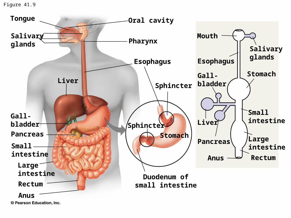
Figure 41.9
Tongue
Salivaryglands
Oral cavity
Pharynx
Esophagus
SphincterLiver
Stomach
Gall-bladder
Smallintestine
PancreasPancreas
Smallintestine
Largeintestine
Largeintestine
Gall-bladder
Stomach
LiverSphincter
Esophagus
Rectum
Anus
Mouth
Anus
Rectum
Salivaryglands
Duodenum ofsmall intestine

Tongue
Pharynx
GlottisLarynx
Trachea
Bolus offood
Epiglottisup
Esophagealsphinctercontracted
Esophagus
Tolungs
Tostomach
(a) Trachea open
EpiglottisdownGlottis upand closed
Esophagealsphincterrelaxed
(b) Esophagus open
Digestion Begins in the Mouth- Mechanical digestion, chewing, increases the surface area of food- Salivary Amylase begins carbohydrate digestion- Tongue shapes food into a bolus and provides help with
swallowing.- Swallowing causes the epiglottis to block entry to the trachea

Food is pushed along by peristalsis, rhythmic contractions of smooth muscles through the
esophagus,…

…where it encounters a sphincter before it enters the stomach
Gastroesophageal Sphincter
Pyloric Sphincter

Chemical Digestion in the Stomach
• Gastric juice has a low pH of about 2, which kills bacteria and denatures proteins
• Gastric juice is made up of hydrochloric acid (HCl) and pepsin
• Pepsin is a protease, or protein-digesting enzyme, that cleaves proteins into smaller peptides

Why doesn’t gastric juice destroy the stomach cells that make it?

Figure 41.11
Stomach
Gastric piton the interiorsurface ofstomach
Gastric gland
Mucous cell
Chief cell
Parietal cell
Epithelium
Hydrogen and Chloride molecules are secreted separately from Parietal Cells, and they form HCl in lumen of gastric gland.
Chief Cells secrete the inactive enzyme precursor, pepsinogen
Three cell types responsible for secretions 1. Chief cells 2. Parietal cells 3. mucus cells

Pepsinogen
Chiefcell
Parietalcell
HCl
Cl−H+
1
1
Pepsinogen andHCl secreted intolumen
HCl denatures pepsinogen, exposing sites that the molecules self-cleaves, resulting in Pepsin.
2
2Pepsin(activeenzyme)
Pepsin activatesmore pepsinogen,starting a chainreaction.
3
3
Activation of Pepsin

Roles of HCl
1. Activation of pepsinogen into pepsin
2. Denatures proteins therefore exposing peptide bonds
3. Kills microorganisms

Role of PepsinInitiates protein digestion through cleavage between
specific amino acids.
Cleaves peptide bonds between hydrophobic amino acids
Pepsin

Stomach Dynamics
• Coordinated contraction and relaxation of stomach muscle churn the stomach’s contents
• Sphincters prevent chyme from entering the esophagus and regulate its entry into the small intestine

Small IntestineSite at which most digestion and absorption
of nutrients occurs
1. Duodenum – Site of entry for pancreatic, liver and gall bladder digestive juices.
Some absorption
2. Jejunum – Comprises nearly half of the small intestine. Site for most absorption
3. ileum – end portion of small intestine.

• The first portion of the small intestine is the duodenum, where chyme from the stomach mixes with digestive juices from the pancreas, liver, gallbladder, and the small intestine itself

Bile Production by the Liver• Bile is made in the liver and stored in the gallbladder• In the small intestine, bile aids in digestion and
absorption of fats: Bile exhibits detergent-like action, and breaks
apart large fat molecules, exposing them to pancreatic lipase

Pancreatic Secretions• alkaline solution that neutralizes acidic chyme
• trypsin and chymotrypsin- partial digestion of proteins. Activated in the lumen of the duodenum. These enzymes differ in their specificity to amino acids.
• pancreatic amylase – digests starch, producing disaccharides.
• Pancreactic Lipase- breaks down fats

• Enzymes that cleave small peptides into amino acids are found at the brush border of the small intestine
• The intestinal enzymes maltase, sucrase, and lactase cleave disaccharides into monosaccharides.
Intestinal Enzymes
maltase

Figure 41.12a
ORAL CAVITY, PHARYNX, ESOPHAGUS
CARBOHYDRATEDIGESTION
Polysaccha-rides(starch, glycogen)
Disaccha-rides(sucrose, lactose)
MaltoseSmallerpolysaccharides
Salivary amylase
Carbohydrate digestion only: disaccharides produced

Figure 41.12b
STOMACH
CARBOHYDRATEDIGESTION
ProteinsDisaccha-rides(sucrose, lactose)
MaltoseSmallerpolysaccha-rides Pepsin
Smallpolypeptides
PROTEINDIGESTION
Protein digestion only

Figure 41.12c
SMALL INTESTINE (enzymes from pancreas)
CARBOHYDRATEDIGESTION
Smallerpolypeptides
Disaccha-rides(sucrose, lactose, maltose)
Disaccharides
Smallerpolysaccha-rides
Pancreaticamylases
Smallpolypeptides
PROTEINDIGESTION
NUCLEIC ACIDDIGESTION
FATDIGESTION
Pancreatictrypsin andchymotrypsin
Pancreaticcarboxy-peptidase
Smallpeptides
Aminoacids
DNA, RNA
Nucleotides
Fat(triglycerides)
Pancreaticlipase
Glycerol,fatty acids,monoglycerides
Pancreaticnucleases
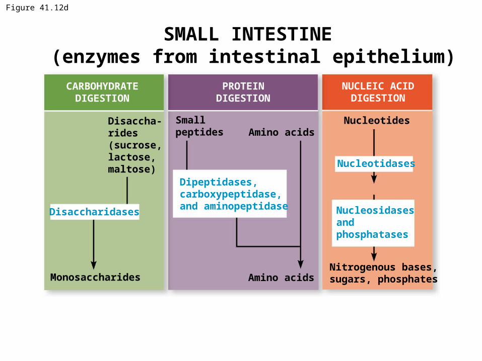
Figure 41.12d
SMALL INTESTINE (enzymes from intestinal epithelium)
CARBOHYDRATEDIGESTION
Disaccha-rides(sucrose, lactose, maltose)
Monosaccharides
Disaccharidases
PROTEINDIGESTION
NUCLEIC ACIDDIGESTION
Nucleosidasesandphosphatases
Dipeptidases,carboxypeptidase,and aminopeptidase
Smallpeptides
Amino acids
Nucleotides
Nucleotidases
Amino acids
Nitrogenous bases,sugars, phosphates

Villi of Small Intestine

Absorption in the Small Intestine
• The small intestine has a huge surface area, due to villi and microvilli that are exposed to the intestinal lumen
• The enormous microvillar surface creates a brush border that greatly increases the rate of nutrient absorption
• Transport across the epithelial cells can be passive or active depending on the nutrient

Vein carryingblood to liver
Bloodcapillaries
Epithelialcells
Largecircularfolds
Muscle layersVilli
Intestinalwall
Nutrientabsorption
LactealLymphvessel
Villi
(towardcapillary)
LumenEpithelialcells
Microvilli(brush border)at apical (lumenal)surface
Basalsurface
Nutrient Absorption in the Small Intestine

Epithelial cells at the brush border are polar. Nutrients move in one direction, out
of the lumen and toward the capillaries
(towardcapillary)
LumenEpithelialcells
Microvilli(brush border)at apical (lumenal)surface
Basalsurface
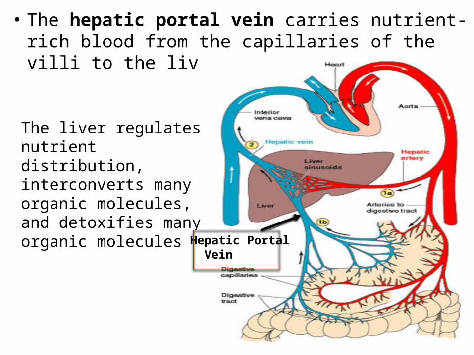
• The hepatic portal vein carries nutrient-rich blood from the capillaries of the villi to the liver, then to the heart
The liver regulates nutrient distribution, interconverts many organic molecules, and detoxifies many organic molecules
Hepatic Portal Vein

Figure 41.14LUMENOF SMALLINTESTINEEpithelialcell
Triglycerides
Fatty acids Monoglycerides
Triglycerides
Phospholipids,cholesterol,andproteins
Chylomicron
Lacteal
Triglyceridesare brokendown to fattyacids andmonoglyceridesby lipase.
Monoglyceridesand fatty acidsdiffuse intoepithelial cellsand are reformedinto triglycerides.
Triglycerides areincorporated intochylomicrons.
Chylomicronsenter lactealsand are carriedaway by lymph.
1
2
3
4
Fatty acids form chylomicrons, which pass through the epithelial cells and enter the lacteals, a lymphatic vessel that bypasses the liver, emptying the chylomicrons into the general circulation. They are then delivered to cells throughout the body

The Large Intestine
- Removes water and salt- Storage of fecal material.
Colon, cecum and rectum
Cellulose, a component of the plant cell wall, is not digested by mammals and contributes to bulk.
Some anaerobic bacteria that live in the hind gut of many mammals secrete cellulases, enzymes that break down
cellulose.

Ascendingportionof colon
Smallintestine
Appendix
Cecum
The cecum aids in the fermentation of plant material and connects where the small and large intestines meet
The human cecum has an extension called the appendix, which plays a minor role in immunity

• The colon completes the reabsorption of water that began in the small intestine
• Feces, including undigested material and bacteria, become more solid as they move through the colon
• Two sphincters between the rectum and anus control bowel movements

Regulation of Digestion• The enteric division of the nervous system
– Autonomic nerve control– Nerve cell bodies found in the wall of the gut– Reflex pathway

Endocrine Regulation
• Gastrin – secreted by specialized cells in the stomach wall in response to presence of amino acid and stomach distention.
• Secretin – secreted by specialized cells in the small intestines in response to amino acids and fats.
• Cholecystokinin (CCK) – secreted by specialized cells in the intestines in response to amino acids and fat.

Gallbladder Liver Food
Stomach
Gastricjuices
Gastrin
Pancreas
Duodenum ofsmall intestine
StimulationInhibition
1
Gastrin is released into the blood before finding its target cell on the stomach wall
Gastrin stimulates the release of gastric juices (HCl) into the lumen of the stomach

StimulationInhibition
Bile
CCK
Chyme
HCO3, enzymes
Secretin CCK
The acidic nature of chyme in the intestine stimulates release of secretin and CCK
There is an additive effect of secretin and CCK on cells of the pancreas which release HCO3
- and other digestive enzymes

Figure 41.20c
StimulationInhibition
Secretinand CCK
Gastricjuices
Feedback inhibition of CCK

Evolutionary adaptations of vertebrate digestive systems correlate with diet
Carnivore Herbivore
Omnivore
Key Incisors Canines Premolars Molars
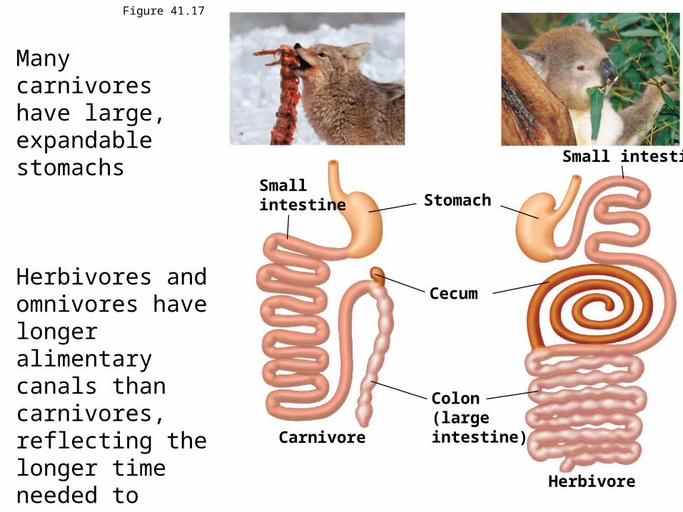
Figure 41.17
Smallintestine
Carnivore
Stomach
Cecum
Colon(largeintestine)
Small intestine
Herbivore
Many carnivores have large, expandable stomachs
Herbivores and omnivores have longer alimentary canals than carnivores, reflecting the longer time needed to digest vegetation

Mutualistic Adaptations in Herbivores
Reticulum
Esophagus
Rumen
OmasumAbomasum
Intestine
4
3
2
1
• Many herbivores have fermentation chambers, where mutualistic microorganisms digest cellulose