2009 abstracts summer undergraduate … programs in the biomedical sciences 2009 abstracts summer...
TRANSCRIPT
Graduate Programs in the Biomedical Sciences
2009
ABSTRACTS
Summer Undergraduate
Research Program
www.einstein.yu.edu/phd
ALBERT EINSTEIN COLLEGE OF MEDICINE OF YESHIVA UNIVERSITY
GRADUATE DIVISION OF BIOMEDICAL SCIENCES
SUMMER UNDERGRADUATE RESEARCH PROGRAM
2009
Victoria H. Freedman, Ph.D. Assistant Dean for Graduate Studies Director, Summer Undergraduate Research Program

2009 SUMMER UNDERGRADUATE RESEARCH PROGRAM
Student Name Undergraduate School Albert Einstein Faculty Advisor
Avital Bauman Yeshiva University Dr. Sunhee Lee Daniel Biro The Cooper Union Dr. Myles Akabas Alexandra M. Bobe College of Mt. Saint Vincent Dr. Richard N. Kitsis Julian Botta Cornell University Dr. Deyou Zheng Ashleigh Bouchelion University of Maryland - Baltimore Dr. David Hall Jessica Brown University Of Arizona Dr. Nicholas Sibinga Darren Bryk New York University Dr. Amy Fox Fay Burekhovich Yeshiva University Dr. Louis Hodgson Gaurab Chakrabarti Brown University Dr. Chandan Guha Rachel Chess University Of Rochester Dr. Florence Marlow Saralin Davis Colby College Dr. Kamran Khodakhah Naomi-Liza Denning Lehigh University Dr. S. Horwitz, Dr. H. McDaid Dalal Eldick University Of Miami Dr. Simon Spivak Kyle Ellefsen Brigham Young University Dr. Adam Kohn Farzana Faisal Yale University Dr. Brian Currie Erin Finn Rice University Dr. Hannes Buelow Amanda Franklin Harvard University Dr. Roy Sillitoe Brandi D. Freeman Bowling Green State University Dr. Chi-Wing Chow Matthew Friedman Yeshiva University Dr. Ekaterina Dadachova Jared Gans Emory University Dr. Frederick Kaskel Chaim Golfeiz Yeshiva University Dr. Bhaskar Das Alexander Goodell University of Oregon Dr. Bettina Fries David T. Guerrero College of Mount Saint Vincent Dr. Markus Bitzer Yizheng He Duke University Dr. Peng Wu Kelsey Hoidal Fordham University Dr. Sridhar Mani Amy Jobe Cornell University Dr. Jonathan Warner Terrika C. Jones Xavier University of Louisiana Dr. Ekaterine Dadachova Michael Klein Pomona College Dr. Jack Lenz Ross Kristal University of Florida Dr. Meredith Hawkins David Kuppermann Yeshiva University Dr. Bernice Morrow Nai aka Robert Li Fordham University Dr. Xingxing Zang Emily Liebling Yeshiva University Dr. Hernando Sosa Alison Liss Haverford College Dr. Saleem Nicola Deepti Mathur Cornell University Dr. Ganjam Kalpana David Melnick University of Michigan Dr. Robert Burk Carlisdania Mendoza Fordham University Dr. Elyse S. Sussman Esther Montanes Universitat Rovira I Virgili – Spain Dr. Hernando Sosa Maxine Owusu Syracuse University Dr. Roy V. Sillitoe Shanka Paramananda CUNY-Lehman College Dr. John Chan Franklin E. Paulino Montclair State University Dr. Kartik Chandran Brandilyn Peters SUNY At Stony Brook Dr. Richard Stanley Scott Philips Brown University Dr. S. Horwitz, Dr. H. McDaid Michael Rothschild Bowdoin College Dr. Nir Barzilai Chava Ruderman Yeshiva University Dr. Lloyd Fricker David A. Sanchez Polytechnic Institute of NYU Dr. Joshua D. Nosanchuk Yona Saperstein Yeshiva University Dr. Ben Ovryn Zackary Scholl University Of Washington-Seattle Dr. Matthew Levy Michael Shusterman Tufts University Dr. Michael Brenowitz Kevin Smith University Of Wisconsin-Milwaukee Dr. Kostantin Dobrenis Daniel Southren Washington University in St. Louis Dr. Bridget Shafit-Zagardo Tirtza Spiegel Yeshiva University Dr. Jeffrey Segall Jasmine C. Stephens Clarkson University Dr. Sherry Downie Terrence N. Turner Marist College Dr. John M. Greally Thomas Vogler Bucknell University Dr. Duncan Wilson Ezra Weinblatt University Of Pennsylvania Dr. Richard Gorlick Rebecca Weiss Yeshiva University Dr. Ana Maria Cuervo Shira Wieder New York University Dr. Liang Zhu Paul Winograd University Of Wisconsin-Madison Dr. V. Prasad, V. Rao Xiaomeng Xu CUNY- City College of New York Dr. Michael Keogh Elaine Yang Johns Hopkins University Dr. Jil Tardiff Shoshana Zitter Yeshiva University Dr. Amy Fox
Endocannabinoid Protein Expression in Human Immunodeficiency Virus Encephalitis
1Avital Bauman, 1Meng-Liang Zhao, 3Susan Morgello, 1Sunhee C. Lee, 1, 2Melissa A. Cosenza-Nashat
1Department of Pathology, Albert Einstein College of Medicine, Bronx, NY
2Department of Science, Borough of Manhattan Community College, New York, NY 3Departments of Pathology and Neuroscience, Mount Sinai Medical Center, New York, NY
Cannabinoid receptors 1 and 2 (CB1 and CB2, respectively) are part of the endocannabinoid system along with intracellular enzymes (such as fatty acid amide hydrolase, FAAH) that degrade endocannabinoid ligands. Exogenous cannabinoids are potential therapeutics for treating neurological sequalae in HIV-infected patients because cannabinoids can suppress the immune system. Human immunodeficiency virus encephalitis (HIVE) is a pathological correlate to HIV-associated dementia, a condition that occurs in some HIV-infected individuals. Recently, Benito et. al. reported that CB2 receptors are upregulated in simian immunodeficiency virus encephalitis, a model for HIVE. We therefore sought to explore endocannabinoid protein expression in HIVE. We obtained paraffin-embedded human autopsy brain tissue sections from the National NeuroAIDS Tissue Consortium and divided them into four groups, HIV-seronegative (HIV-, n = 6, HIV-seropositive without brain pathology (HIV+, n = 12), HIV-seropositive with encephalitis (HIVE, n = 4) and HIV+ with co-infections/co-morbidities (HIV+/Coinfection, n = 5). Tissue sections were subjected to immunohistochemistry with several antibodies: anti-CB1, anti-CB2 and anti-FAAH. Immunolabeled sections were analyzed with microscopy and analysis of digital images was performed. Results indicate that CB1 and FAAH are present in neurons in all cases, while white matter CB1 staining in HIVE and HIV+/Coinfection cases was significantly above control levels. CB1 is upregulated in glia and perivascular macrophages based on morphology. Staining for CB2 illustrated immunoreactive perivascular macrophages, astrocytes and some microglia. Our results indicate that cannabinoid receptors are strongly expressed in HIVE brains and this may inform clinicians who are considering cannabinoids as adjunctive therapies for HIV-associated neurologic disorders. Acknowledgements: This research was supported by R01 MH55477 (Sunhee C. Lee, P.I.), 5 R25 MH080663-02; Sub award no: 0253-6141-4609 (Melissa Cosenza-Nashat, P.I.), the AECOM Center for AIDS Research, the Roth Scholars Program and the Summer Undergraduate Research Program.

Interactions of Thymidine and Immucillin-H with PfENT1 Daniel Biro, I.J. Frame, Myles Akabas, MD, PhD. Plasmodium falciparum is the most dangerous of the malaria causing parasites in humans. It is responsible for over 300 million cases of malaria annually, including approximately one million deaths, primarily children in sub-Saharan Africa. It is known that P. falciparum is incapable of de novo purine synthesis, and relies on transport of host derived purines to remain viable. The main purine transporter in P. falciparum has been identified as the plasma membrane protein PfENT1. It has also been previously demonstrated that cells expressing PfENT1 show increased accumulation of thymidine and Immucillin-H (ImmH), although intracellular concentrations have not been shown to reach equilibrium with extracellular concentrations. The goal of this study was to determine whether thymidine and ImmH were binding to PfENT1 as opposed to being transported by it. If these compounds bind to PfENT1, then homologues of these substances could potentially be effective drugs in combating malaria by eliminating the parasite’s ability to transport purines through PfENT1. Xenopus oocytes were injected with PfENT1 mRNA and the uptake of radio-labeled thymidine was measured and compared against uptake by uninjected oocytes. Using this method we constructed a concentration-response curve for thymidine uptake showing that thymidine uptake follows Michaelis-Mentin kinetics. The magnitude of the uptake implies thymidine is not binding but is in fact a transported substrate of PfENT1. ImmH did not inhibit thymidine uptake. Further experiments are in progress to determine why thymidine uptake reaches a plateau level that is significantly below the level expected for equilibration with the external thymidine concentration. Acknowledgments: Moez Bali, PhD, Julius Miltante, PhD, Nicole McKinnon, Rishi Parikh, Asif Rahman, Dana Harrison Additional funding provided by: AECOM SURP
Pancreatic β-cell ER Stress-Induced Apoptosis Alexandria Bobe1,2, Wendy Mckimpson3, Richard Kitsis3,4
1College of Mount Saint Vincent, 2MSSROP, 3Department of Cell Biology, 4Department of Medicine [Division of Cardiology]
Albert Einstein College of Medicine, Bronx, NY, USA
Approximately eight percent of the U.S. population suffers from Type 2 Diabetes (T2D). This
disease is mediated by pancreatic β-cell dysfunction and/or insulin resistance. In vitro
experiments with mouse βTC-tet cells were performed to imitate similar effects of T2D
stressors, such as obesity, in order to analyze the effects of prolonged endoplasmic reticulum
(ER) stress as a pro-apoptotic stimulus in β-cells. Treatment of βTC-tet cells over expressing
apoptosis repressor with CARD (CAspase Recruitment Domain) (ARC), an inhibitor of both the
intrinsic and extrinsic cell death pathways, with the free fatty acid palmitate, induced lipotoxicity
and triggered β-cell apoptosis. Higher doses of palmitate resulted in decreased cell viability,
reduced levels of both ARC and Bak, a protein involved in ER stress, and increased presence of
insulin within the cell. Future experiments will further explore the mechanisms behind ER stress
induced apoptosis in β-cells, ARC’s location within this pathway, as well as clinical implications
in T2D.
Acknowledgments: MSSROP, SURP, Wendy Mckimpson, Kitsis Lab
1 Barski, Artem and Zhao, Keji: “Genomic Location Analysis by ChIP-Seq” (Journal of Cellular Biochemistry Vol. 107 [2009], pgs. 11-18) 2 Ji, Hongkai et al.: “An integrated software system for analyzing ChIP-chip and ChIP-seq data” (Nature Biotechnology Vol. 26 [2008], pgs. 1293-1300) 3 Rozowski, Joel et al.: “PeakSeq enables systematic scoring of ChIP-seq experiments relative to controls” (Nature Biotechnology Vol. 27 [2009], pgs. 66-75)
Identification of Gata-1 Binding Sites by ChIP-Seq
Julian S. Botta, Xingyi Guo, Sandeep N. Wontakal, Arthur I. Skoultchi, Deyou Zheng
Departments of Genetics and Cell Biology, Albert Einstein College of Medicine, Bronx, NY
Gata-1 is a master regulatory transcription factor of the erythroid lineage. Though Gata-1 is known to play an essential role in the development of red blood cells, the transcriptional program that it regulates is not fully understood. In this study, we used chromatin immunoprecipitation coupled with high-throughput sequencing (ChIP-Seq1) to identify genomic sites that are bound by Gata-1 in both normal and malignant murine erythroid progenitor cells, as well as malignant cells chemically induced to re-enter the differentiation program. Sequence reads obtained from ChIP-Seq represent short genomic sequences and computational methods must be applied in order to determine true binding sites versus background noise. We first applied the software CisGenome2, which scans the mouse genome for regions enriched with Gata-1 ChIP-Seq reads, corresponding to the Gata-1 in vivo binding sites (i.e. peaks). After this analysis was done for the three different cellular conditions, we developed software to determine the overlap between Gata-1 peak lists and to find genes that are potentially differentially bound by Gata-1 in these cells. Surprisingly, we found that Gata-1 bound to many more sites in normal cells than in malignant cells, suggesting that this differential binding may play a role in the formation of erythroleukemias. Moreover, malignant cells that were induced to differentiate showed a binding pattern significantly different from that of the parental cells, further suggesting that abnormal binding of Gata-1 may play a role in tumorigenesis. We also used PeakSeq3, another program for peak identification, and obtained similar results, although more analysis is necessary for comparison. Our results provide novel insight into the transcriptional network regulated by Gata-1 in erythroid cells, and suggest that abnormal Gata-1 binding maybe an important factor in either the establishment and/or maintenance of erythroleukemia.
Acknowledgements: This research was funded in part by the AECOM Summer Undergraduate Research Program.

Electron Tomography of C.elegans Ashleigh Bouchelion 1, David Hall 2
1University of Maryland, Baltimore County 2 Albert Einstein School of Medicine, Department of Neuroscience
Caenorhabdidis elegans are used as a model system to study the genetic control of cellular development. David Hall’s Lab specializes in ultrastructural studies of the nervous system. Currently, we are using electron tomography to further characterize the anatomy of the organelles of the C.elegans. Electron tomography is a technique for obtaining 3D structures of subcellular macromolecular objects. It is an extension of traditional transmission electron microscopy and uses a transmission electron microscope to collect the data, which is used to assemble a 3D image of the organelles and cellular structures. My project, in particular focuses on modeling (the secretory canal), (intestine), and (muscle structures) of the animals. Data collection for electron tomography involves collecting images while titling the specimen around a single axis. Complete data collecting would require a tilt at an angle as high as 90o. Most of our data consist of thin specimens, which allow for greater resolution. In order to model the organisms we use IMOD, which is an image processing and modeling display program used to construct a 3D structure from EM serial sections. From this technique, we hope to further characterize the anatomical features of C.elegans.
THE ROLE OF AIF-1 IN ARTERIAL NEOINTIMA ACCUMULATION Brown JN, Casimiro I, Riascos-Bernal DF, Sibinga NE Albert Einstein College of Medicine at Yeshiva University Developmental and Molecular Biology Department, Cardiology Department Abstract Cardiovascular disease (CVD) dominates as the leading cause of death in western-style societies. Inflammatory CVDs such as atherosclerosis and restenosis may be affected by allograft inflammatory factor-1 (AIF-1), which is reported to function as a pro-migratory, pro-proliferative, and pro-survival protein in vascular smooth muscle cells (VSMCs) and macrophages. Several preceding studies suggest that, through these effects on VSMCs and macrophages, AIF-1 acts to induce greater accumulation of neointima. We used a surgical carotid arterial ligation model to mimic aspects of the pathogenesis of atherosclerosis using AIF-1 knockout (KO) and WT mice on a C57BL/6J genetic background (n = 6 KO, n = 4 WT). We hypothesized that AIF-1 would induce greater neointima accumulation, thus the KO model would have smaller neointimas. The mice were anesthetized, and the ventral aspect of the neck was shaved, and cleaned with ethanol. A longitudinal incision was made in the skin overlying the left carotid artery. The artery was completely occluded by ligation near its bifurcation. The skin incision was closed using super glue, and the mice recovered amongst their cagemates. After 28 days, the mice were killed and the ligated arteries were harvested. The arteries were perfused and fixed with 4 % paraformaldehyde for 16 hours and embedded in parrafin. The paraffin-embedded arteries were sectioned 5 μm thick using a microtome. The sections were mounted and stained with hemotoxylin-eosin and Verhoeff’s Elastic Stain. Morphometric analysis was performed with Photoshop® by calculating the area of the lumen and the areas inside the internal elastic and external elastic laminae. Contrary to our hypothesis, the KO mice showed a trend toward more neointima formation than WT mice, although this difference was not statistically significant (neointima/media area 0.5155 ± 0.1892 vs. 0.1446 ± 0.04673, p = 0.1599). These data suggest that AIF-1 may act to limit neointimal formation. AIF-1 has been implicated, based on its overexpression in VSMCs, as a promoter of neointima accumulation in previous publications. Our findings using mice completely lacking AIF-1 suggests that AIF-1 has a protective function that limits neointimal formation since total loss of AIF-1 was hypothesized, based on previous literature, to increase neointima. Future investigations would benefit by examining overexpressed AIF-1, AIF-1 KO, and WT mice. These studies could compare a broad range of AIF-1 expression levels, because it may be that AIF-1 functions best under optimal expression conditions. I would like to thank the Sibinga Laboratory, Nicholas Sibinga, MD, Yueting Shang, MD, PhD, Isabel Casimiro, MS, Prameladevi Chinnasamy, MS, and Dario Riascos-Bernal, MD. Jessica Brown was supported by NIH Grant No. T34 GM008718 and Isabel Casimiro was supported by the Cell Molecular Biology & Genetics Training Grant T32 GM007491.
Swine Influenza or Seasonal Influenza? An Evaluation of Pediatric Patients at the Beginning of the 2009 Pandemic Bryk, D., Zitter, S., Narlieva, M. MS, Akid, I., Pan, Q. PhD, Fox, A. MD, MS
Montefiore Medical Center Bronx, NY 10467
There have been four major influenza pandemics in the twentieth century. The
most recent one was the swine influenza pandemic in 1977 until novel swine influenza
was identified in the United States in April 2009. Over the next two months, the situation
escalated and the World Health Organization declared a swine influenza pandemic on
June 11. The first 40 Pediatric Emergency Room (PER) patients who were positive on the
rapid influenza A test (Quidel Corp., San Diego, CA) at the beginning of the pandemic
were located in the hospital’s database. Thirty-one specimens (viral transport media)
were available for testing on an automated platform. The dates of these samples ranged
from April 22 to May 18.
Viral RNA from the 31 specimens was extracted using the Abbott m2000sp
(Abbott Molecular, Chicago, IL)∗ and then was analyzed by RT-PCR using the Abbott
m2000rt with CDC approved primers and probes1. The primers and probes were for
influenza A, swine influenza A, swine H1N1, and an internal positive control, RnaseP.
Cycle number threshold (Ct) < 37 indicated a positive result.
Of the 31 patients, 13 had swine influenza while 18 had seasonal influenza. From
April 22 to May 13, 0/13 cases were swine flu. The first swine influenza case was
detected on May 14. Following this, there were 12 more swine influenza cases and only
5 seasonal influenza cases. The prevalence of swine influenza increased as the month
progressed ultimately becoming the dominant strain.
Acknowledgements: SURP 2009 for support. Dr Gavin Cloherty and Dr. Danijela Lucic of Abbott Molecular Laboratories for advice and technical assistance. Dr. Tylis Chang for advice and technical support.
∗ For research use only 1 http://www.who.int/csr/resources/publications/swineflu/realtimeptpcr/en/index.html
Novel biosensor for Cdc42 – N-WASP interaction, based on solvatochromic dyes
Fay Burekhovich1, Louis Hodgson2
1Stern College for Women, Yeshiva University, New York, NY 10016 2Gruss-Lipper Biophontics Center, Department of Anatomy and Structural Biology, Albert
Einstein College of Medicine, Bronx, NY 10461
Cdc43, a member of Rho-family GTPase, regulates critical cellular functions including cell polarity maintenance and actin cytoskeleton rearrangement. Interaction of Cdc42 with one of its key downstream effectors, neuronal isoform of Wiskott Aldrich Syndrome Protein (N-WASP), has important implications with respect to cancer, where Cdc42-N-WASP binding plays a principal role in the establishment and function of invadopodia, invasive membrane extensions with matrix degrading activity vital for metastatic invasion of carcinoma cells. Studies utilizing fluorescent biosensors in live cancer cells have shown that N-WASP is active only at invadopodia, and does not impact normal cellular operations. Thus, inhibiting Cdc42-N-WASP interaction could be useful as a possible anti-metastatic therapy.
Based on previous research that detected endogenous Cdc42 activation in living cells by its binging to the Cdc42 binding domain (CBD) of hematopoietic WASP. The derivatized GBD is attached to a solvent-sensitive dye that undergoes a great change in fluorescence emission intensity upon binding to activated, endogenous Cdc42. The new probe will be valuable in development of potential approaches for high-throughput screening of inhibitor libraries targeting Cdc42-N-WASP interaction, while minimizing spurious inhibition of hematopoietic WASP. Using recombinant DNA techniques, a novel biosensor for Cdc42-N-WASP interaction was produced and characterized in vitro.

FOCUSED ULTRASOUND-INDUCED UNFOLDED PROTEIN RESPONSE
Gaurab Chakrabarti, Subhrajit Saha and Chandan Guha
Department of Radiation Oncology, Albert Einstein College of Medicine and Montefiore Medical Center, Bronx, NY
Unfolded protein response (UPR) is a stress response induced in cells because of defects in protein folding during protein synthesis, in the endoplasmic reticulum. UPR induces the synthesis of chaperone molecules, such as , heat-shock proteins that attempt to correct misfolded proteins. On prolonged stress, unfolded proteins are targeted for proteosomal degradation. We hypothesized that therapeutic ultrasound would generate mechanical vibrational and thermal stress in cells, thereby, inducing UPR. The murine prostate cancer cell line RM1 and rat oval cells (ROC, hepatic stem cells) were treated with low intensity (LOFU: focal intensity ~500 W/cm2, frequency, 1 Mhz) and high intensity focused ultrasound (HIFU: focal intensity ~1300-2000 W/cm2 at 4 Mhz). Using qRT-PCR, we found that LOFU increased the expression of UPR target genes (GRP78 and EDEM) by 10-20 folds in RM1 cells, which was futher augmented by combination therapy of LOFU-HIFU to 200-800 folds. ELISA demonstrated an increase in HSP70 protein levels with subsequent release of HSPs from LOFU-HIFU treated cells (390±62.9ng/mil and 8-fold increase). LFU alone increased membrane bound HSP70 levels in ROC by 7.5 folds, as noted in FACS. The results indicate that focused ultrasound induces a UPR stress response in tumor and stem cells. Our long-term goals is to exploit the ultrasound-induced UPR stress response for generating tumor-derived peptide antigens for an autologous in situ tumor vaccine. Further investigation of UPR on differentiation of hepatic ROC progenitor cells is underway.
Acknowledgements
We would like to thank the Summer Undergraduate Research Program for financial support. Thanks are due to Hongchou Zhang for his support throughout.
Comparative analysis of frl gene expression and function in the developing zebrafish embryo
Rachel Chess1,2, Gretchen Dollar3, Andreas Jenny3, Florence Marlow3 1SURP Graduate Division of Einstein, 2University of Rochester, 3Department of
Developmental and Molecular Biology at Einstein College of Medicine of Yeshiva University, Bronx, NY
Gastrulation is a conserved developmental process in which major cell movements establish the basic animal body plan. An important regulator of gastrulation is Planar Cell Polarity (PCP) signaling, a highly conserved non-canonical Wnt pathway. PCP regulates cytoskeletal rearrangements, in part through Rho Kinase (Rok). Formin related in Leukocytes (Frl), was identified as a Rok target that modulates PCP pathway phenotypes in Drosophila (Dollar and Jenny unpublished). We are investigating whether Frl is a conserved PCP component, which contributes to convergent extension (CE) in zebrafish. First, we determined that zebrafish frl gene expression is temporally and spatially consistent with a potential function during gastrulation. During early and middle stages of gastrulation the four zebrafish frl genes have similar expression patterns, however, after gastrulation, the frl genes show distinct and overlapping expression domains. Therefore, frl genes may be functionally redundant or cooperate during early development, but have distinct functions in later processes. To test whether frl function is necessary during gastrulation we microinjected Morpholino Oligomers to deplete Frl2. Depletion of Frl2 did not cause specific developmental defects, and in general did not adversely affect embryonic development. Therefore, frl2 is not essential for gastrulation, other frl genes have redundant functions, or maternal Frl2 is sufficient for normal embryonic development. We are currently investigating whether gain of Frl function disrupts CE. In future studies, the developmental function of the remaining frl genes will be investigated. Further experiments will be needed to determine whether Frl functions as a conserved target of Rho kinase. Acknowledgements: Funding provided by SURP. Thank you to Sophie Von Eisner, Amanda Heim and Spartak Kalinin for all your help in the lab.
LACK OF REBOUND FIRING IN NEURONS OF THE DEEP CEREBELLUM IN VIVO Saralin Davis, Esra Tara, and Kamran Khodakhah Dominick P. Purpura Department of Neuroscience, Albert Einstein College of Medicine, Bronx, NY
After strong inhibitory input neurons of the deep cerebellar nuclei (DCN) are capable of a transient increase in firing rate, termed rebound firing. Although rebound firing has been incorporated into various models of cerebellar function, it remains unclear whether it occurs under strictly physiological conditions. The current study examined the degree of rebound firing in DCN neurons in vivo in awake WT mice and considered the parameters necessary for rebound firing to encode information at this site. We found minimal changes in firing rate after pauses in baseline firing, indicating a lack of significant rebound firing. Furthermore, the parameters necessary for a reasonable signal-to-noise ratio require either a large change in firing rate or a low interspike interval CV, both outside the range typically seen in DCN neurons in vivo. These observations indicate that rebound firing in DCN neurons is limited if present in vivo and that the role of rebound firing in cerebellar function should be reconsidered. This project was supported by SURP of Albert Einstein College of Medicine and grants from NIH.
The Effect of p90RSK Inhibition on the Proliferation of Human Cancer Cells Naomi-Liza Denning1, Jack Taunton2, Susan Horwitz3, Hayley McDaid3,4 1Albert Einstein College of Medicine Summer Undergraduate Research Program 2Dept. of Cellular & Molecular Pharmacology, UCSF, CA; Dept of 3Molecular Pharmacology; 4Medicine, Albert Einstein College of Medicine, Bronx NY RSKs (p90 Ribosomal S6 kinases) are kinases that are activated by MAPK (Mitogen-Activated Protein Kinases) and PDK1 (phosphoinositide-dependent kinase). RSKs have cytosolic and nuclear targets and are implicated in a diverse range of cellular processes. RSKs contain two kinase domains in a single polypeptide rendering them structurally unique. Activated MAPKs dock and phosphorylate the C-terminal kinase domain (CTKD), which recruits PDK1 into the linker region. Docked PDK1 then phosphorylates the N-terminal kinase domain (NTKD), which phosphorylates all known RSK substrates; although speculation exists that there is a CTKD-independent mechanism of RSK activation. FMK is a CTKD inhibitor of RSK 1/2, which covalently binds two residues in the ATP binding site, thereby preventing NTKD activation. Since RSKs act downstream of MAPK, which are hyperactivated in RAS and RAF mutant tumors, there has been interest in the therapeutic potential of RSK inhibitors. We used the SRB assay, used to measure protein concentration, to test the anti-proliferative effect of FMK in approximately 20 human cancer cell lines with various genotypes. There were minimal effects on proliferation with no discernable correlation with RAS or RAF status. In PMA-stimulated B-Raf mutant cells, FMK partially inhibited RSK380 phosphorylation at 3µM suggesting incomplete inhibition, or a CTKD-independent mechanism of RSK activation. FMK did not reverse the phosphorylation status of PMA-induced RSK effectors, again suggesting incomplete suppression of RSK. Preliminary evidence of additivity between FMK and a rapalog (mTOR inhibitor) was observed in cell lines with constitutive activation of PIK3. Additional studies will further explore these pilot data.
MODULATION OF GSTP1, NQO1 AND CYP1B1 BY PLANT-DERIVED CHEMOPREVENTATIVE AGENTS IN HUMAN LUNG NORMAL AND IMMORTALIZED CELLS
Dalal A. Eldick Xiang-Lin Tan Miao Shi Shengli Xiong Nandita Mullapudi Weiguo Han Simon D. Spivack
Division of Pulmonary Medicine, Albert Einstein College of Medicine, Bronx, NY
The carcinogens present in cigarette smoke, the leading risk factor for lung cancer, are metabolized by phase I and phase II enzymes. Within human lung tissue, CYP1B1 (phase I enzyme) bioactivates various carcinogens present in tobacco smoke, while GSTP1 (phase II enzyme) deactivates many carcinogens. The role of NQO1 phase II enzyme is more ambiguous. Dietary intake of antioxidant agents endogenous to fruits and vegetables, such as SFN and reservatrol, protect against lung cancer through phase II enzyme induction and possible phase I enzyme inhibition. Our in-vitro study targeted the effect of mRNA and protein expression of the aforementioned phase I and II enzyme genes with cigarette smoke extract (CSE), SFN, reservatrol and their appropriate combinations. Analysis was achieved through real-time PCR and western blot from 24hr and 48hr exposures of all conditions to normal human bronchial epithelial cells (NHBE) and human bronchial epithelial cells (HBEC). All conditions containing CSE have shown induction effects of CYP1B1 and GSTP1 mRNA expression. However, reservatrol and SFN to a lesser extent reduced mRNA induction of CSE in CYP1B1 in HBEC cells but not in NHBE cells. GSTP1 protein expression remained unaffected. NQO1 mRNA expression in NHBE cells was induced by CSE+SFN and RES+CSE+SFN. NQO1 mRNA expression in HBEC cells was also induced by CSE+SFN. This effect however, was partially confirmed by western blot. Our results have shown that mild phase I reduction occurs in HBEC cells by reservatrol. Because sulforaphane did not affect mRNA and protein expression, its mechanism is more ambiguous.
Acknowledgments:
I would like to thank Dr. Simon Spivack and Dr. Xiang-Lin Tan for all of their help and guidance throughout this process. This work was also supported by the Summer Undergraduate Research Program (SURP) and the Depart of Pulmonary Division at Albert Einstein College of Medicine and NIH-R21 CA 94714 (to SD Spivack); NIH-R01 CA 10618 (to SD Spivack).
PERCEPTUAL EFFECTS OF ADAPTATION WITH SIMPLE AND COMPLEX STIMULI
Kyle Ellefsen, Stephanie Wissig, Adam Kohn
Dept Neuroscience, Albert Einstein College of Medicine, Bronx NY
Adaptation, which is classically described as a reduction in firing rate with
prolonged stimulation, is a fundamental property of sensory neurons. It can be measured
psychophysically in humans as a decrease in perceived contrast or an increase in
threshold detection, the contrast necessary to detect a stimulus. The properties of
adaptation are generally studied with simple laboratory stimuli such as sinewave gratings.
Early psychophysical experiments suggested that spreading the stimulus energy across
spatial frequencies by adapting with square wave or compound gratings reduces the effect
of adaptation as compared to the effects of adaptation with sinewave gratings. Likewise,
recent extracellular recordings in V1 of macaque monkeys from our lab show reduced
effects of adaptation with compound gratings as compared to adaptation with sinwave
gratings. To investigate the consequences of this neural effect on perception, we
measured the effects of adaptation with sinwave and compound gratings using threshold
detection and contrast matching tests. Using the threshold detection measure, our results
agree with the above findings. However, the results of our contrast matching tests failed
to show a difference in the strength of adaptation with these stimuli.
This work was supported by the SURP program.
K. pneumoniae, Imipenem, and other Hodge-Podge: Can Patient Clinical Risk Factors Predict KPC Positivity?
F. Faisal1, A. Kaltsas MD2, B.P. Currie MD MPH2
1Albert Einstein SURP. 2Montefiore Medical Center, Bronx, NY.
K. pneumoniae carbapenemase (KPC) is a plasmid-borne resistant factor that is rapidly spreading in NYC hospitals. Automated methods do not accurately detect KPC; the phenotypic Modified Hodge test (MHT) is now recommended. According to current lab standards for automated systems, an imipenem MIC>4 is considered intermediate or resistant, often leading patients with MIC≤4 to be inappropriately considered KPC negative. The aim of this study was to use the MHT to understand the prevalence, epidemiology, and risk factors for KPC positive K. pneumoniae. Ertapenem resistant and imipenem sensitive (by automated methods) K. pneumoniae isolates were collected from patients at our three affiliated hospitals from 2007 to 2009. The MHT was performed on a random sample of 28 of these isolates. Corresponding clinical data was extracted from patient charts associated with these isolates. Bivariate statistical analysis used the chi2/Fisher’s exact, Mann-Whitney, and Student’s t tests. Logistic regression estimated the effect of variables on MHT positivity. Of the 28 isolates, 18 were from urine cultures, 6 sputum, 2 blood, and 2 wound/other. Seven cultures tested MHT negative, and 21 tested MHT positive. Of the negative MHT, 6 had an imipenem MIC≤1 and 1 had an imipenem MIC>1. Of the positive MHT, 17 had an imipenem MIC>1 and 4 had an imipenem MIC≤1. Vitek 2 imipenem MIC>1 was the only variable evaluated in this study that significantly predicted a positive MHT for K. pneumoniae. Patients with positive MHT were also more likely to be a nursing home resident, have been previously admitted to the hospital, have a decubitus ulcer or chronic foley catheter at admission, have prior multidrug resistant Klebsiella, or fail first antibiotic treatment; although these trends were not statistically significant.
Investigation of the in vivo role of 3-O-Sulfated Heparan Sulfate Erin Finn, Eillen Tecle, Hannes E. Büelow Department of Molecular Genetics, Albert Einstein College of Medicine, Bronx, NY 10461 Heperan sulfate (HS) is an unbranched polysaccharide chain linked to cell membrane bound and extracellular matrix proteins. HS is extensively modified via epimerization, de-acetylation, and sulfation enzymes resident to the Golgi. Many of the HS modifications have been shown to play specific and instructive roles for neuronal development in Caenorhabditis elegans (Bülow and Hobert, 2004; Bülow et al., 2009) . However, the role of the 3-O sulfation modification of HS has not been established. In C. elegans only two genes (termed hst-3.1 and hst-3.2) encode the enzymatic activity that introduces the 3-O modification in HS. In vertebrates, this enzymatic activity is encoded by seven different genes thus making C. elegans an excellent model system to establish the in vivo function of 3-O sulfated HS. Preliminary analysis of mutant worms lacking hst-3.1 and/or hst-3.2 reveled that the 3-O sulfation modification is required for the proper patterning of a subset of C. elegans neurons. My project focused on two hst-3.2 mutant alleles: tm3006 and tm3206. I generated worms carrying the tm3206 allele with neuronal gfp reporters that were previously analyzed in the tm3006 background. The worms I generated will be tested to see if both hst-3.2 alleles have similar phenotypes. In addition, I generated worms carrying two previously untested GFP reporters in the tm3006 background. Using these reporters, I investigated coelemocyte migration and VC neuron connectivity in the hst-3.2(tm3006) mutant background. My data indicates that hst-3.2 is critical for the ventral posterior coelemoctyes migration but dispensable for the VC patterning Acknoelwedgements: Albert Einstein College of Medicine Student Undergraduate Research Program
Topographical Organization of Climbing Fibers, Bergmann Glia and Purkinje Cells in the Cerebellum
Amanda Franklin, Stacey Reeber, Roy Sillitoe
Dominick P. Purpura Department of Neuroscience, Albert Einstein College of Medicine, Bronx, NY 10461
The adult cerebellum (Cb) is organized into ten anatomically distinct lobules in the anterior-posterior axis, and a complex molecular map consisting of striped Purkinje cell (PC) gene expression domains in the medial-lateral (ML) axis. Recent immunohistochemical studies demonstrated that ZebrinII and PhospholipaseCβ4 (Plcβ4) are expressed in complementary PC stripes in lobules I-V and VIII while the small Heat shock protein 25 (Hsp25) is expressed in stripes in lobules VI/VII and IX/X. It is well known that climbing fiber afferents terminate and chemically interact with the dendrites of PCs and the processes of Bergmann glia astrocytes. In addition, previous studies demonstrated intimate anatomical associations and intercellular signaling between Bergmann glia and PCs. However, very little is known about the topographical relationship between genetically defined subsets of climbing fibers, PC stripe domains and Bergmann glia. Here we used a transgenic mouse line expressing green fluorescent protein driven by the Neuropeptide Y gene promoter (Npy-Gfp) to label climbing fibers and Bergmann glia in the mouse Cb. We identified that Npy-Gfp was expressed in ML climbing fiber bands that terminated upon the Plcβ4 immunopositive subset of PCs in lobules I-V and VIII. In addition, Npy-Gfp was expressed in Bergmann glia stripes that were positionally related to Hsp25 expressing PCs in lobules VI/VII and IX/X. Importantly, we found that within any given stripe domain both Bergmann glia and PCs co-existed as targets for particular climbing fibers. We propose the theory that functional Cb “modules” consist of interacting cell types that simultaneously process incoming information. This work was funded by SURP (to AF) and AECOM New Investigator Start-Up funds (to RVS).
Subcellular Localization of C. Elegans Simple – Implications for Conserved Role in Charcot-Marie-Tooth Disease C1 Neuropathy
Brandi D. Freeman, Hong Zhu, Poh Choo How, and Chi-Wing Chow Department of Molecular Pharmacology
Albert Einstein College of Medicine, Bronx, New York, USA
Charcot-Marie-Tooth (CMT) disease 1C is an inherited neuropathy that causes demyelination in the peripheral nervous system. It is caused by a
mutation in the protein SIMPLE (Small Integral Membrane Protein of the Lysosome/ late Endosome). SIMPLE is mainly localized to multi-vesicular bodies with markers denoting late endosome/ lysosome. We found that
SIMPLE is evolutionally conserved in C. elegans (ceSIMPLE). Although SIMPLE is highly conserved, its function remains elusive. Here, we examined the localization of ceSIMPLE in cells. We performed immunofluorescence and
found that endogenous SIMPLE co-localizes with LAMP2, a marker for late endosome/ lysosome. Double-staining indicated that ceSIMPLE co-localizes with the mouse ortholog. We also found that ceSIMPLE is co-localized with
different markers denoting ER, Golgi, and endosome. We also performed biochemical fractionation and found ceSIMPLE is secreted and present in exosomes. These data indicate that ceSIMIPLE exhibited similar properties
as its vertebrate orthologs. The conserved role of SIMPLE will allow the use of the power of C. elegans genetics to elucidate the function of SIMPLE, which accounts for the CMT1C neuropathy.
Acknowledgments: This work was supported by the Minority Student Summer Research Opportunity Program (MSSROP) and Summer Undergraduate Research Program (SURP) of Albert Einstein College of
Medicine to BF and grant 114376 from the Muscular Dystrophy Association (MDA) to CWC.
Assessing the radiotrophic characteristic of Cryptococcus
neoformans Matthew Friedman¹, Ruth Bryan¹, Richard Magliozzo², Abdelahad Khajo², Arturo Casadevall³ Ekaterina Dadachova¹ ¹Departments of Nuclear Medicine and Microbiology and Immunology, Albert Einstein College of Medicine, 1695A Eastchester
Bronx, NY 10461 ²Department of Chemistry, Brooklyn College CUNY, 2900 Bedford Avenue Brooklyn, NY 11210
³Departments of Microbiology and Immunology and Medicine, Albert Einstein College of Medicine 1300 Morris Park Avenue
Bronx, NY 10461
Electromagnetic radiation is ubiquitous in our environment. When melanized, several genera within the fungal kingdom, including Cryptococcus neoformans, appear to be able to harness large doses of radiation and thrive in its presence. This radiotropism has been correlated with the presence of the pigment melanin within the fungal cell. Using an acapsular strain of C. neoformans, CAP67, we set out to further demonstrate that only melanized fungal cells are capable of responding positively to radiation. When grown in a minimal media containing L-Dopa, CAP67 is able to synthesize and incorporate melanin within its cell wall. Using melanized and non-melanized cultures of CAP67, we performed Electron Paramagnetic Spin (EPR) spectroscopy of the samples illuminated with a white light source. We subsequently observed a significant increase in the strength of the EPR signal that was proportional to the time of illumination. These changes, which were six times greater in the melanized samples spectra, demonstrate an alteration of electronic composition of the cell as a result of exposure to light. To further establish the radiotrophic tendencies of melanized CAP67, we also performed colorimetric assays of internal ATP levels. These assays were inhibited and we believe this was caused by membrane-bound ecto-ATPase as well as inefficient lysis methods. Future assays will hopefully circumvent these issues and show a relationship between ATP levels and increasing radiation in CAP67. We also hope in the near future to do EPR spectroscopy on cell samples irradiated with ionizing radiation. Positive results would further support the hypothesis of melanin acting as a chlorophyll-like radiosynthetic pigment in fungal cells. Acknowledgments: I would like to express my sincere gratitude to the Dadachova lab and in particular to Drs.
Dadachova and Bryan for their mentorship throughout this summer experience. I would also like to thank the Magliozzo
Lab of CUNY Brooklyn College for their collaboration in this project and for the use of their EPR spectrometer. Lastly, I
would like to thank the Summer Undergraduate Research Program, of the Sue Golding Graduate Division of Albert
Einstein College of Medicine for this unique summer experience.
ASSOCIATIONS BETWEEN KIDNEY LENGTH ADJUSTED FOR BODY SURFACE AREA AND AMBULATORY BLOOD PRESSURE IN CHILDREN Gans, JH1,2, Yang, S3, Kaskel, FJ4, Woroniecki, RP4 1) Summer Undergraduate Research Program (SURP), Albert Einstein College of Medicine (AECOM), Bronx, NY 2) Emory University, Atlanta, GA 3) SUNY Albany, Albany, NY 4) Pediatric Nephrology, Children’s Hospital at Montefiore (CHAM), AECOM, Bronx, NY Blood pressure (BP) has been correlated with reductions in nephron number. Kidney Length (KL) measured by ultrasonography (US) has been correlated with age, height, and obesity. Studies associating KL with BP have shown conflicting results. However, these studies have not adjusted KL to body surface area (K_S) to correct for already established anthropometric correlations and have note used 24 hours Ambulatory Blood Pressure Monitoring (ABPM). We hypothesized that K_S would be correlated with ABPM parameters. We reviewed records of 48 subjects evaluated in the Pediatric Hypertension Program at CHAM between January 2004 and July 2009 who underwent ABPM and KL determination by US. Subjects with kidney transplant and significant hydronephrosis or cysts (N=7) were excluded. We confirmed anthropometric associations with KL. We found that ABPM average dipping was correlated with K_S in hypertensive subjects (r=-0.45, P=0.03) by bivariate analysis and in our multiple regression analysis model with Systolic BP and Systolic BP load (R2=0.34, P=0.04). This association was absent in normotensive subjects. Association between Nocturnal Dipping and K_S in subjects who are at increased risk for end organ damage (kidney, heart) needs further investigation. We thank SURP at AECOM for providing funds.
Design and Synthesis of Novel Boron Containing Alkene
Derivatives to Study TGF-β Signaling Pathways Chaim Golfeiz, Sakkarapalayam M. Mahalingam, Jaime Anguiano, Bhaskar C. Das* Department of Developmental and Molecular Biology Albert Einstein College of Medicine of Yeshiva University, Bronx, New York USA It is well documented that biological pathways that govern embryonic development continue to be used in controlling adult physiology, and that deregulation of these pathways can lead to disease. Therefore, identification of small molecule modulators of gene networks active in early development can lead to a better understanding of component specificity for signaling pathways and ultimately the design of novel therapeutic and diagnostic agents for adult diseases. The TGF-β signaling pathway deregulated in cancer and disease, and represents a prime candidate pathway for development of pharmacological modulators. We are interested in developing new compounds that interact with developmentally important receptor-mediated pathways such as the TGF-β pathway, acting as antagonist or agonist.
From our previous chemical genetic screening on developing zebrafish embryos, we identified a lead molecule BT7 that modulates specifically a Smad-independent TGF-β-regulated MAPK pathway, namely p-SAPK/JNK. In this project we focused to increase the potency and biological activity of our lead molecule BT7. So we synthesized functionally oriented boron containing alkene-derivatives of BT7 analogues. To this end, we designed a protocol to synthesize these highly useful molecules. The desired products were synthesized with moderate to good yield by mixing the Wittig salts of various substituted benzyl phosphonium ylides, aldehydes, bases and DMF as solvent at room temperature. The products were purified using column chromatography techniques and then verified with NMR and HRMS analysis. This novel procedure not only streamlined the synthesis of the boron containing alkene derivatives efficiently, but also increased its yield and selectivity. We used this procedure to synthesize combretastatin analogues (antimitotic and TGF-beat signaling modulator). We are currently testing their relevance in the TGF- beta signaling pathways. In the future, an understanding of the mechanism behind this specificity could expedite development of new drug discovery, for example relevant to cardiovascular and cancer diseases.
Acknowledgments: This work was supported by the SURP program to CG and AECOM start up funding to BCD.
Characterization of the capsule phenotype in all1∆ Cryptococcus neoformans Alex Goodell1,2,3, Neena Jain1, Mythri Subramaniam1, Bettina Fries1 1 Department of Medicine & Microbiology and Immunology, Albert Einstein College of Medicine, Bronx, NY, USA; 2 SURP , Sue Golding Graduate Division of Biomedical Sciences, Albert Einstein College of Medicine, Bronx, NY, USA; 3 Department of Biology, University of Oregon, Eugene, OR, USA Cryptococcus neoformans is an encapsulated fungal pathogen that infects mostly immunocompromised patients presenting chronic meningoencephalitis, causing upwards of 600,000 deaths annually (1,2). The organism has a unique polysaccharide capsule which varies in size according to environmental conditions such as carbon dioxide levels, iron starvation and pH (3,4) . Increased capsule size has been linked to virulence in mammalian hosts. By shedding the capsule, excess PS causes agitation of the meninges and is associated with high inter-cranial pressure (ICP) (5,6). Increased capsule size has also been shown to cause decreased susceptibility to both anti-fungal medication and phagocytosis by immune cells (7). The capsule character of C. neoformans var. neoformans has been shown to microevolve, undergoing a "phenotypic switch," from a smooth (SM) to a mucoid (MC) appearance invivo (Figure 1) (8). MC switch variants are more virulent and were shown to down-regulate one gene in particular, allergen 1 (ALL1), of which homologs have been found in other strains, such as C. neoformans var. grubii (9). This project analyzes the character of the PS capsule in wildtype, ALL1 knockout mutant, and reconstituted strains in RC-2 and H99 strains in a variety of medium. Characterization is done through photography and cryptocrit analysis. Results show increased capsule size when incubation conditions have low iron, high thiamine, or minimal media. All1∆ consistently show smaller capsule size. This suggests that the ALL1 protein plays a role in capsule formation during inducing conditions. Future experiments on the ALL1 protein interactions may yield interesting results. Word count: 247
MicroRNA-21 does not inhibit WWP1 protein expression through 3’ UTR activity
David Guerrero, Ujunwa Cynthia Okoye, Minkyung Kim, Wenjun Ju, and Markus Bitzer Departments of Medicine and Developmental & Molecular Biology
Albert Einstein College of Medicine, Bronx, New York
MicroRNAs (miRNAs) target genes by binding to a region in the 3’UTR of
messenger RNAs leading to mRNA degradation or transcriptional repression. miRNA
(miR)-21 has been shown be expressed at high levels in most tumors and modulates
apoptosis, proliferation and extracellular matrix homeostasis by targeting PDCD4, PTEN,
Sprouty1, among others. WW domain containing E3 ubiquitin protein ligase 1 or WWP1
is a predicted target gene of miR-21 and plays a significant role in protein degradation,
RNA splicing and transcription.
We hypothesize that an increase in miR-21 inhibits the expression of WWP1 by
targeting a putative miR-21 binding site in the 3’ untranslated region (3’UTR). First we
confirmed that WWP1 protein expression decreased in kidneys of TG using
immunohistochemistry. WWP1 was strongly expressed in tubular epithelial cells (TEC)
of WT but less so in TEC of TG. To explore whether miR-21 regulates WWP1
expression through targeting its 3’UTR, we transfected 293 cells with a plasmid
containing the 3’UTR of WWP1 coupled to a luciferase reporter along with a miR-21
over expression plasmid or a miR-21 inhibitor to determine if its expression would be
affected by the varying amount of miR-21. Manipulation of miR-21 levels in human
embryonic kidney (HEK) 293 cells showed no significant change of WWP1 after
inhibition of miR-21 using Western blot analysis. Overall the data shows that the miR21
does not directly target the 3’ UTR of WWP1.
Acknowledgments: MSSROP, SURP, and Dr. Bitzer Lab This project is supported by the AMGEN Nephrology Institute, the Nephcure Foundation and the American Society of Nephrology. AMOs were provided by Regulus Therapeutics.
Chemoenzymatic Synthesis of Sialyl Lewis X and its Derivatives
Yizheng He*, Christen Besanceney**, Wei Wang**, Peng Wu**
*Duke University and **Department of Biochemistry, Albert Einstein College of Medicine
Sialyl Lewis X (sLex), a tetrasaccharide glycan, plays a vital role in many cell-cell
recognition processes. The expression of sLex-bearing glycoconjugates is also a common feature
shared by numerous cancers. However, the absence of robust, facile and cost-effective methods
for the synthesis of sLex and its structurally related analogues has severely hampered the
elucidation of the specific functions of these glycan epitopes. Here we demonstrate that
chemically defined sLex and its derivatives can be synthesized on preparative scales using a
chemoenzymatic approach.
Our strategy involves constructing sialyl N-acetyllactosamine, the acceptor glycan, using
a one-pot three-enzyme reaction that combines aldolase, CMP-sialic acid synthetase and α2,3
sialyltransferase. Next, we introduced L-fucose and its derivatives to the acceptor substrate
using a bifunctional fucokinase/GDP-fucose pyrophosphorylase (FKP) and a Helicobacter pylori
α1,3 fucosyltransferase to yield the desired sLex tetrasaccharide and its derivatives. The purity
and identity of the sLex derivatives have been confirmed by Thin Layer Chromatography, NMR
and High Resolution MS analyses.
The association of aberrant sialylation with malignancy has prompted the development of
numerous carbohydrate-based tumor vaccines. Unnatural sLex derivatives synthesized using this
approach may have altered immunological properties than their natural counterparts. Toward
this end, we will conjugate these unnatural glycans to carrier proteins and evaluate their efficacy
for tumor vaccine therapy.
Acknowledgements:
This work was supported by the National Institutes of Health (4R00 GM080585-03) and the Summer Undergraduate Research Program at Albert Einstein College of Medicine.
Role of Pregnane X Receptor (PXR) in Cancer and Pathophysiologic States Kelsey Hoidal, Sridhar Mani (PI), Subhajit Mukherjee, and Madhukumar Venkatesh Department of Medicine, Genetics, and Cancer Center Albert Einstein College of Medicine – Yeshiva University, Bronx, NY In eukaryotes, orphan nuclear receptors (ONRs) have large and relatively non-specific ligand-binding domains, and they control most major physiological and biochemical processes including: cell metabolism, xenobiotic detoxification, cell differentiation, cancer cell growth, and apoptosis. Pregnane X Receptor (PXR) is an ONR with enigmatic function, having implications in xenobiotic metabolism, cancer drug resistance, carcinogenesis, and pathophysiologic states, such as inflammatory bowel disease (IBD). In order to probe PXR function as a xenosensor, we have established a method of discovering novel antagonists of activated PXR; it is hypothesized that PXR antagonists, such as ketoconazole, allosterically inhibit PXR at the AF-2 domain by preventing coactivator SRC-1 from binding. Project 1 entails screening over 10,000 compounds for weak agonists with the potential to fit into the antagonist pharmacophore, then testing these compounds for antagonist activity at the AF-2 docking station. This assay utilizes DPX2 cells, human hepatocytes which contain PXR and a luciferase-CYP3A4 promoter. Cells are plated in 96-well plates, the test compound is applied in titrations, and luminescence is recorded using an electronic reader. If correlation is seen between the concentrations at which 50% of PXR is inhibited (IC50) and the docking scores, the “best” antagonists will be retested for binding and toxicity. Future experiments will also employ radioligands to obtain Kd, and Ki may be calculated using the Cheng-Prusoff equation. In addition to PXR’s role in xenobiotic resistance, it has also been implicated in innate immunity in the mammalian gut; PXR protects the gut from aberrant signals, such as pathogenic bacteria, that induce inflammation. Therefore, Project 2 is designed to study whether this function is conserved in non-mammalian systems. Caenorhabditis elegans have 284 well-documented nuclear receptors, two of which—NHR 8 and 48—have PXR-like function in nematodes (Antebi A., 2006). In this assay, ten Day 1 C. elegans are plated on lawns of bacteria expressing GFP, and behavior and survival are measured at 24-hour intervals. To date, no conclusive results have been obtained, although the researchers have noted NHR-8 mutants are more sluggish than wild-type worms on plates of Salmonella enterica. This survival study is a continuing experiment in the Mani lab and several pathogenic bacteria remain to be tested. Thank you to Dr. Mani, Subhajit, and Madhu for their support and patience this summer, as well as Hao, Hong Wei, Arunima, and Myra for welcoming me to the lab; additional thanks to SURP for funding.
Interactions in the Pathway to Diamond-Blackfan Anemia Amy Jobe*, Kerri B. McIntosh, Jonathan R. Warner Department of Cell Biology, Albert Einstein College of Medicine, Bronx, NY *Cornell University, Ithaca, NY Diamond-Blackfan Anemia (DBA) is an inherited anemia characterized by low levels of erythropoeisis, or formation of blood cell components. Of cases with a known genetic cause, 25 percent are attributed to mutation in the ribosomal protein (RP) gene RPS19; mutation in RPS24 and RPL11A as well as several other RP genes account for smaller proportions of incidence. These patients are haploinsufficient, meaning that their having only a single functional copy of any of those genes yields a DBA phenotype. Although it is known that such haploinsufficiency leads to apoptosis, it remains unclear why the erythrocyte is apparently the most immediately and severely compromised cell type in patients. To identify putative genetic interactions arising in the poorly understood ribosomal assembly pathway affected in DBA, we executed a synthetic genetic array (SGA) screen, a method unique to yeast. Our screen involved crossing a single-knockout Saccharomyces cerevisiae query strain to a whole-genome array of approximately 4,700 viable single-knockout strains followed by selection for doubly mutant haploids and screening for changes in colony fitness. In S. cerevisiae, and particularly in these strains, colony size is an initial indicator of fitness. After the appropriate rounds of selection, colonies that appeared significantly small or large relative to a control were deemed “synthetic lethal” or “sick” or “synthetic alive”, respectively. These labels suggest certain types of interactions between the genes deleted in a given colony’s’ parent strains, which may elucidate the pathway affected by the query strains’ deletions. Evidence of absent, slow or robust growth was confirmed by streaking labeled strains and performing a Bioscreen assay, in which growth rate of labeled strains and their parent strains was measured over time. We found that genes related to cell cycle regulation, proliferation, cell membrane biogenesis, transmembrane movement and ribosomal proteins often corresponded to synthetic lethal phenotypes, and DNA repair, ribosomal proteins and iron- or heme- related pathways were generally associated with synthetic alive phenotypes. The apoptosis characteristic of DBA might be explained by the many putative connections to cell cycle regulation and telomere maintenance seen in both SS/L and SA hits. Lastly, associations with proliferation and, again, with cell cycle regulation may hint at a reason for the correlation between DBA and certain types of cancer. Yeast two-hybrid experiments may confirm the most interesting of our results, thereby illuminating both the mechanism of ribosome assembly and that of DBA. We thank Ian Willis and Robyn Moir for providing and assisting with the use of the Singer robot and the Colony Imager and Scorer software, and for further advice throughout the project. Thanks are due to Arpita Bhattacharya and Saqui Huq for assistance as well. This project was funded by NIH grant GM25532 to JRW, by a NSERC Postdoctoral Fellowship to KBM and by the AE SURP to AJ.
Investigation of Immunoreactivity towards melanin of monoclonal antibody fragments used in radioimmunotherapy (RIT) of metastatic melanoma
Terrika C. Jones, Ekaterina Revskaya, Ekaterina Dadachova Department of Nuclear Medicine
Albert Einstein College of Medicine of Yeshiva University; Bronx, NY 10461
Metastatic melanoma is a skin cancer that overtakes the patient’s body through rapid tumor growth. To date, there is no known cure for metastatic melanoma. In past studies, our lab has addressed the treatment of melanoma with radioimmunotherapy (RIT). In this technique lethal radiation is delivered by the antibodies, labeled with radioisotopes, to designated antigens on the tumors. Antibody, 6D2 mAb, labeled with 188-Rhenium has been confirmed in the laboratory to possess binding capabilities to melanoma melanin and effective in suppressing the growth of melanoma tumors in mice. However, some 188Re-6D2 molecules have been found to undergo small fragmentation. Now that this form of treatment has entered into clinical trials, our goal is to find the molecular weight, concentration, and binding to melanin capability of these fragments.
A non-radioactive sample of Re-6D2 was prepared and tested for two simulated patient cohorts, 100mCi Re, 50mg 6D2 and 50mCi Re, 50mg 6D2. The antibody underwent column purification and was injected into high performance liquid chromatograph (HPLC). Four peaks were rendered: 6D2 peak itself followed by peaks of high, medium, and low molecular weight. The HPLC fragments were used in protein assay against standards (6D2, Ferritin, IgG, and BSA) of similar molecular weight to calculate the concentration of the fragments. The protein assay of both cohorts proved that the samples alongside their standards had a 10% concentration recovery of ~1.00mg/ml. Enzyme-linked immunosorbent assay (ELISA) was performed using 96-well plates coated with melanin to test the binding capabilities of the fragments. ELISA confirmed binding of all samples to melanin which assures the suitability of the labeled antibody for clinical trials. Acknowledgements: This project would not have been possible without the opportunity presented by MSSROP and SURP of Albert Einstein College of Medicine of Yeshiva University, the dedication and support of the Nuclear Medicine lab headed by Kate Dadachova, and funding from Pain Therapeutics, Inc.
ELUCIDATING MECHANISMS OF RETROVIRAL TUMORIGENESIS THROUGH MASSIVELY PARALLEL DNA SEQUENCING Michael Klein, Koyel Mitra, and Jack Lenz Department of Genetics, Albert Einstein College of Medicine, Bronx, NY 10461
Retroviruses replicate through DNA intermediates known as proviruses that are
inserted in the genome of infected cells. Proviruses inserted adjacent to cancer-causing proto-oncogenes can have cancerous effects on host organisms due to enhancer elements within the proviruses increasing proto-oncogene transcription. Retroviruses used as delivery vectors in human gene therapy have caused tumors in patients by this identical mechanism of oncogene activation. It is unknown whether oncogenes are preferred for retroviral DNA insertion, whether retroviruses with different tumorigenic capacities target different sites in the host genome, in which organs and tissues tumors actually originate, and when in the process of tumorigenesis tumor cell clones first emerge. Massively parallel DNA sequencing offers a means to address these questions. Ligation mediated PCR was performed on genomic DNA samples at different time points from the thymus, bone marrow and spleen of mice infected with a strongly or a weakly lymphomagenic retrovirus to obtain a large number of retroviral insertion sites for analysis. Prior to performing massively parallel sequencing, we performed a pilot study using conventional sequencing on a subset of samples taken six weeks after viral infection at a time point about one month before the mice would succumb to lymphomas following infection with the strongly oncogenic virus. Of the 27 sequences that underwent analysis, three provirus inserts were positioned in proximity to three previously reported common insertion sites, none of which, however, has been previously reported in lymphomas resulting from the strongly lymphomagenic retrovirus used here. Only one viral insertion site was identified twice in a thymus sample from a mouse infected with the weakly lymphoma virus. Thus there was at most only a small indication of possible emergence of a tumor clone at the six-week time point. Completion of this pilot study demonstrated that the samples I prepared are ready for massively parallel DNA sequencing that will provide a far greater level of insight into the genomic target sites for viral insertion and the evolution of tumorigenesis over time. Acknowledgements Authors thank Summer Undergraduate Research Program (SURP) at the Albert Einstein College of Medicine. This work was supported by NIH grant CA44822.
Central leptin activation of adipose tissue macrophage is coincident with reactive microglia in the medial basal hypothalamus (MBH): Preliminary findings.
Ross Kristal, Emilce Carrasco, Meredith Hawkins Department of Medicine/Endocrinology
Albert Einstein College of Medicine, Bronx, NY The lineage of macrophages appears to contribute to systemic inflammation and insulin resistance. Given the evidence for central regulation of macrophage activation and possible central effects of leptin on immunity, we hypothesized that this fat-derived protein could contribute to activation of microglia (central nervous system (CNS) resident macrophage) and adipose macrophages in obesity. In this short pilot study we investigated the effect of low dose intracerebroventricular (ICV) administration of leptin (n=7) vs. vehicle (n=5) for 18 hours on visceral adipose tissue and CNS inflammation in ~9 week old (X= 300 g), male Sprague-Dawley rats. As anticipated, this acute low dose administration of leptin did not significantly alter body weight (P=0.9), or food intake (P=0.1) in either treatment groups. Adipose macrophage content was not affected by acute leptin administration (macrophage content: leptin=28 +/- 2.6% vs. vehicle=24 +/- 3.6%, P= 0.2). However, leptin did show a trend towards activating adipose macrophage, as evidenced by a 1.7 fold increase in these cells of the pro-inflammatory marker inducible nitric oxide (iNOS) (leptin=38 +/- 11% vs. vehicle 22 +/- 10%, P=0.3). Next we asked if this peripheral inflammatory effect of central leptin was coincident with reactive microglia. We assessed the quantity of reactive microglia based on their morphology (ramified, characteristic of quiescent, versus amoeboid, characteristic of activated) in the medial basal hypothalamus. We found a ~2 fold increase in amoeboid shaped microglia in leptin treated compared to vehicle treated rats (35+/- 11% vs. 16 +/- 7.9 %, respectively, P= 0.07). In summary, central leptin administration rapidly induced adipose macrophage and CNS microglia activation in normal rats. Increased production of this fat derived protein in obesity could contribute to adipose tissue and CNS inflammation, specifically in the MBH, causing dysregulation of the orexigenic/anorexigenic neurons in this area, thereby exacerbating the metabolic and inflammatory consequences. Acknowledgements: This research was supported by the Albert Einstein Summer Research Program, grants from the NIH to Meredith Hawkins (1P01 AG021654, DK069861), Einstein’s Clinical Research Center (MO1-RR12248), and Einstein’s Diabetes Research and Training Center (P60 DK020541). Special thanks to Kahao Zhang, Preeti Kishore, Sudha Koppakay, Sylvia Kehlenbrink, and Weijie Li for their support.
Investigating the Role of Tbx1 in Chondrogenesis of the Periotic Mesenchyme David Kuppermann¹, Dennis C. Monks², Bernice E. Morrow³ ¹Yeshiva College, Yeshiva University, New York, NY 10033 ² ³ Department of Genetics, Albert Einstein College of Medicine of Yeshiva University, Bronx, NY 10461 Tbx1 is a transcription factor of the T-box family whose haploinsufficiency
has been implicated in velo-cardio-facial syndrome/DiGeorge syndrome in
human patients. Tbx1 -/- mice exhibit severe defects in structures of the
outer, middle, and inner ear. Since Tbx1 is expressed in both the otic
vesicle (OV) epithelium as well as the periotic mesenchyme (POM)
surrounding the inner ear, we utilize a conditional mutant approach to
dissect the roles of Tbx1 in both of these domains. When Tbx1 is ablated
specifically in the POM using TCre to inactivate a floxed Tbx1 allele (TCre
KO), defects in structures derived from both the OV and POM are
apparent; including a shortened cochlear duct and smaller cartilaginous
otic capsule surrounding the cochlea. This suggests both signaling to the
OV as well as defects in the POM cells themselves contribute to the
phenotype. We hypothesize that loss of Tbx1 in POM leads to premature
chondrogenesis and thus prevents the cochlea from properly coiling. To
test this hypothesis, we have successfully synthesized antisense
riboprobes to assay for expression of chondrogenic markers (Sox5, Sox6,
Sox9, Col9a2, Col11a2, Mia1, and Pbx1) via whole mount in situ
hybridization. These probes will now be used to test for premature initiation
of chondrogenesis in the POM of TCre KO embryos. This data will help us
to better understand the molecular basis of hearing loss in both model
organisms and human patients.
Funded by SURP of Albert Einstein College of Medicine (DK) and grant DC05186-06 (BEM)
T CELL COINHIBITORY MOLECULE PD-L1 PLAYS A PROTECTIVE ROLE IN COLITIS Robert Nai Li, Lisa Scandiuzzi, Xingxing Zang Department of Microbiology and Immunology, Cancer Center, Diabetes Center Albert Einstein College of Medicine, Bronx, New York PD-L1 (B7-H1) is a B7 family-member playing a negative regulation on T cell responses. PD-L1 is highly expressed on hematopoietic cells and on epithelial cells of several organs such as the pancreas, intestine, heart and brain. We evaluated the role of PD-L1 in a murine model of colitis induced by oral administration of Dextran Sodium Sulfate (DSS) in the drinking water for 5 days. DSS is a polysaccharide that mimics the human Inflammatory Bowel Disease (IBD) by inducing destruction of colon epithelium, inflammation and alteration of colon absorptive functions characterized by bloody stool formation, weight lost and, ultimately, death. During DSS-treatment, PD-L1 -/- mice showed lower survival as compared to PD-L1 expressing mice. Moreover, eosin-hematoxylin staining on the colon tissue sections demonstrated that PD-L1-/- mice exhibited higher destruction of colon crypts and colon epithelium, which were milder in wild type mice. Surprisingly, further analysis revealed that PD-L1-/- naïve mice have reduced numbers of white blood cells such as neutrophils and basophiles. By contrast, flow cytometry analysis showed an increased number of activated B cells (CD62Llow) in mesenteric lymphonodes. These data could suggest that PD-L1 could play an important role in the differentiation and regulation of immune cells that participate in gut homeostasis and therefore affect the gut response upon DSS-treatment. However, additional experiments need to be performed to confirm this hypothesis. Funded by the SURP of Albert Einstein College of Medicine and grant NIH DP2DK083076. Special thanks to the members of the Zang Lab, Jun Sik Lee, Nousheen Zaidi, Kimberly Anne Hofmeyer, Yael Saden Barach, Anjana Ray, and Sang C. Lee for helping and answering my numerous questions.
Interactions between microtubules and kinesin-13 Emily Liebling, Ana B. Asenjo, Vania De Paoli, Uttama Rath, David Sharp, Hernando Sosa
Department of Physiology and Biophysics, Albert Einstein College of Medicine, Bronx, NY 10461
Kinesin, a superfamily of motor proteins, uses ATP to propel itself along microtubules. Kinesin-13’s behave differently than other families, such as kinesin-1, and do not undergo unidirectional movement. Instead, they diffuse to the ends of the microtubules where they induce depolymerization, an essential component of chromosomal segregation during cellular mitosis. Kinesins are comprised of a motor domain, neck, and coiled coil. The motor domain, which serves as the location for microtubule and ATP binding, is the minimal domain necessary for the depolymerization activity of kinesin-13’s.
The mechanism by which kinesin-13’s achieve depolymerization is believed to involve the curving of tubulin protofilaments at the microtubule ends. We examined the interactions between kinesin-13 and microtubules in the ATP hydrolytic cycle using various nucleotide conditions. Previous research has shown that conditions of high affinity of kinesins for microtubules produce a regular protein-microtubule decoration pattern. Electron microscopy, from ongoing studies in this lab, revealed that in the presence of AMP-PNP, a non-hydrolyzable ATP analogue, some kinesin-13’s form oligomeric rings and spirals around microtubules. We are currently exploring KLP59D, which is known not to form rings during depolymerization. Surprisingly, however, it exhibits severing of the microtubules, a phenomenon not previously described for kinesin-13. In addition to initiating depolymerization at the ends, KLP59D cuts microtubules in the middle. These experiments are the first to demonstrate such findings, shedding light on the mechanism of kinesin-13 activity.
Appreciation is expressed to the Summer Undergraduate Research Program at
AECOM, the Sosa and Sharp labs, and the NIH for funding this research.

Activity of the ventral pallidum during cue-motivated reward seeking behavior
Sylvie Lardeux, Alison L Liss, Saleem M Nicola
Departments of Psychiatry and Neuroscience
Albert Einstein College of Medicine, Bronx, NY, USA
The ventral pallidum (VP) and the nucleus accumbens (NAc) are two key structures that
influence reward-seeking behavior. The NAc encodes reward-predicting stimuli. The
VP is the primary target for the neurons of the nucleus accumbens. Therefore, we
hypothesize that the VP is also involved in encoding cue-motivated reward seeking
behaviors. Using both pharmacological and electrophysiological methods, we aim to
understand the role of the VP in reward-seeking behavior.
Rats were first trained to perform a lever-press in response to an auditory cue in order to
obtain a sucrose reward, and were then surgically implanted with microinjection cannulae
or recording electrodes in the VP. We injected GABA receptor agonists or dopamine
antagonists into the VP to determine their effects on reward-seeking behavior.
Additionally, we recorded electrical activity from individual neurons in the VP during a
reward-seeking task to determine the information being encoded in the VP.
No results have been achieved to date; however, previous research indicates that VP
neurons fire in anticipation of reward and show excitation in response to reward-
predictive cues. We hypothesize that both dopamine receptor antagonists and GABA
agonists will decrease reward-seeking behavior. These experiments may delineate a role
for VP neurons in promoting reward-seeking.
This work was supported by the SURP program and NIH grant R01DA19473 to SMN.
The authors thank Vince McGinty, Johann Duhoffmann and James Kim for helpful
discussions and instruction.
Inhibition of rhabdoid tumor cell growth by Flavopiridol and 4-OH Tamoxifen Deepti Mathur1, Melissa E. Smith2, Dr. Ganjam V. Kalpana2,3 1Cornell University, Ithaca, NY; Department of Genetics, 2Albert Einstein College of Medicine, NY; 3Albert Einstein Cancer Center Rhabdoid tumors (RTs) are rare but highly aggressive and incurable pediatric
malignancies. Our laboratory is interested in developing novel targeted therapies for RTs based on the understanding of the genesis of these tumors. The majority (>95%) of RTs are caused by the homozygous loss of INI1/hSNF5 tumor suppressor gene, a regulator of transcription. INI1 directly represses cyclin D1, a mediator of G1-S progression and activator of cyclin-dependent kinases (CDKs). Cyclin D1 is essential for genesis and survival of RTs in vitro and in vivo, suggesting that targeting cyclin D1 and the cyclin/cdk-axis could be effective in inhibiting RT growth. Consistent with this hypothesis, Flavopiridol, a pan-CDK inhibitor, inhibits RT growth in vitro and in vivo mouse models. Unfortunately, Flavopiridol can be toxic at high concentrations. Therefore, we are testing the effect of combining Flavopiridol with other drugs. We combined Flavopiridol with varying concentrations of 4-hydroxy-tamoxifen (4OH-Tam), to determine the effect of combination of the two drugs on four different RT cell lines. Combining 10μM 4OH-Tam with low concentrations of Flavopiridol resulted in dramatic reduction of cell growth four RT cell lines. Flavopiridol alone exhibited an IC50 of 45-100nM and addition of 10uM 4OH-Tam lowered this to 0.15-35nM in various RT cell lines. Flavopiridol and 4OH-Tam induced both G1 arrest and apoptosis in a concentration and cell type dependent manner. Our results demonstrate that 4OH-Tam potentiates the effects of Flavopiridol in different human RT cell lines. We propose that combining Flavopiridol with 4OH-Tam could be a novel therapeutic strategy for RT in humans.
Investigation into the Effects of HPV 16 E6 and E7 on Primary Cilia Formation David Melnick, Koenraad Van Doorslaer, and Robert Burk Department of Microbiology and Immunology Albert Einstein College of Medicine The primary cilia is a singular non-motile organelle that projects from vertebrate cells and plays an important role in signal pathways of the cell, including regulation of cell development and cell cycle. Cells that lose primary cilia have been found to lose regulation of the cell cycle and are associated with malignant transformation. Human papillomavirus (HPV) type 16 is a high-risk type that is known to cause cervical cancer. Transformation by HPV 16 is associated with expression of the E6 and E7 oncoproteins. We hypothesized that HPV 16 oncoproteins E6 and E7 disrupt primary cilia formation causing loss of cell cycle regulation facilitating malignant transformation. Cells were grown on type I collagen coated surfaces in DMEM containing 10% Fetal Bovine Serum (FBS) except when inducing cilia formation. Cilia formation occurs when the cell differentiates and exits the cell cycle. To force cells in culture to exit the cell cycle, the cells were first grown to confluence and thereafter were grown without FBS or maintained in low levels (0.1%, 0.5%) of FBS for 1-5 days. Immunoflourescence microscopy was used to visualize primary cilia and cell nuclei using mouse anti-acetylated tubulin primary antibody (1:10,000) with goat anti-mouse Alexa Flour 488-conjugated secondary antibody (1:1,000), and 4'-6-Diamidino-2-phenylindole (DAPI) staining, respectively. Cells were visualized by wide-field, fluorescence microscopy using a Zeiss Axiovert 200M with Apotome. Imaging of the HPV negative C33A cervical carcinoma cell line revealed formation of primary cilia whereas, imaging of the HPV 16 positive SiHa cervical carcinoma cell line did not detect primary cilia formation. The results support the notion that HPV 16 reduces cilia formation and might contribute to the oncogenic potential of this human carcinogen. We have infected NIH 3T3 mouse embryonic fibroblast cells with HPV 16 E6 and E7 and experiments are in process to evaluate cilia formation. Acknowledgements: SURP 2009, Mallory S. Lutz, Erik Snapp

Mathematical Analysis of the Neurophysiology of Multisensory Auditory Cortical Processing Carlisdania Mendoza, Latha Swamy, Elyse Sussman Dominick P. Purpura Department of Neuroscience, Albert Einstein College of Medicine, Bronx, NY Intricate mechanisms for integration of sensory information are necessary for our brains to be able to generate appropriate responses to the massive amount of information that it processes. Little is known about the areas of the brains that create a unified perception of reality using the different types of information that it gathers from different senses or the mechanisms by which this is done. Event Related Potential (ERP) analysis allows scientists to study high order processing in humans that are impossible to study in animals. Information from these measurements can be analyzed by using their second spatial derivative to obtain information about current source generators and aid in the understanding of multisensory integration. The surface Laplacian provides an estimate of local, two-dimensional polarity of the current density on the surface of the scalp. This estimate results from applying a spatial filter that isolates aspects of EEG data that can be associated with superficial cortical tissue in the electrode’s immediate surroundings. Calculating the surface Laplacian involves three steps: projecting from the real scalp surface onto the sphere of computation, interpolating the potential data using spherical spline surfaces, and projecting from the sphere onto the plane to obtain waveforms. Using the current source density maps, which are visual representations of the surface Laplacian, allows for a more precise analysis of ERP data by eliminating noise from volume conducted signals that come from distant regions of the brain and are not associated with the potential current source generators. Acknowledgements: This study was funded by the National Institutes of Health (NIH). Thanks to Dr. Sussman and all of the members of the lab, as well as the Summer Undergraduate Research Program (SURP) and Minority Student Summer Research Opportunity Program (MSSROP).
Searching for the in vivo rol of the new site of interaction between kinesin-13 and tubulin Esther Montanes, Ana Asenjo, Uttama Rath, David Sharp and Hernando Sosa. Department Physiology & Biophysics, Albert Einstein College of Medicine of Yeshiva University, Bronx NY 10461 Kinesin is a group of motor proteins that uses energy from ATP hydrolysis to move along the microtubule. Unlike most kinesins, the kinesin-13 family uses that energy to destabilize the microtubule structure. They are regulators of the microtubules dynamic and are responsible of the proper spindle assembly and chromosome segregation during mitosis. Kinesin-13 form oligomeric rings and spirals around the microtubules in vitro in presence of AMP-PNP or ATPγS.1 It has been shown by cryo-electromicroscopy that kinesin-13 from Drosophila melanogaster (KLP10A) has a new site of interaction with tubulin when it makes this oligomeric ring. The amino acids responsible for this interaction are Lysines at positions 306, 350 and 399. When these residues are substituted by Alanines, the mutant depolymerizes microtubules but does not form rings.2 To test the role of the second site of interaction of KLP10A with tubulin in vivo, we transfected Drosophila S2 cells with mutant KLP10A green fluorescence protein (GFP) as well as KLP10A wild type GFP and monitored at the effect on mitosis. We found that over expression of the mutant protein reduces the number of cells that are in mitosis (mitotic index) whereas over expression of the wild type KLP10A GFP has no effect on mitotic index. In addition, we observed that the over expression of the mutant protein results in spindle abnormalities. Therefore we conclude that this new site of interaction has an important function in the mitotic process. Further experiments have to be done in this theme such as depleting endogenous KLP10A by an RNAi treatment. Acknowledgements: All members in the Sosa and Sharp labs for their scientific support and assistance. The SURP 2009 and especially to Rosa Garcia. The Analytical Imaging Facility at AECOM. 1. Tan, D., Asenjo, A. B., Mennella, V., Sharp, D.J., and Sosa, H. (2006). Kinesin-13s form rings around microtubules. The journal of cell Biology. 2. Tan, D., Rice, W.J., and Sosa, H. (2008). Structure of the Kinesin13-Microtubule Ring Complex. Cell Press
Topographic Organization of Unipolar Brush Cell Connectivity in the Mouse Cerebellum Maxine Owusu, Samrawit Gebre, Roy V. Sillitoe
Summer Undergraduate Research Program Dominick P. Purpura Dept. of Neuroscience, Albert Einstein College of Medicine, Bronx,
NY 10461
Unipolar brush cells (UBCs) are small excitatory interneurons of the cerebellum. UBCs are innervated by extrinsic mossy fiber afferents and in turn project axons that innervate other cerebellar interneurons including adjacent UBCs. The vestibulo-cerebellum, which controls balance and visuomotor function, is enriched with UBCs that are immunoreactive for the calcium binding protein Calretinin (CR) and the Metabotropic glutamate receptor (mGluR) 1alpha. We are interested in generating a complete molecular profile for UBCs and further defining their circuitry in the cerebellum. We identified using the Allen Brain Atlas a panel of genes that are heavily expressed in UBCs. In this study, we focused on one candidate gene Scna, which encodes a pre-synaptic protein called alpha Synuclein. Using immunohistochemistry we found strong post-synaptic expression of alpha Synuclein predominantly in Calretinin expressing UBCs with only few UBC co-expressing alpha Synuclein and mGlur1alpha. Choline acetyltransferase (ChAT) is selectively expressed in mossy fiber terminals that send vestibular signals to the cerebellum. We found that ChAT immunoreactive mossy fibers contacted a subset of alpha Synuclein/CR immunoreactive UBCs. Interestingly, alpha Synuclein expressing UBCs and their associated mossy fibers were organized into a topographic pattern of stripes that reflected the known “modular” architecture underlying cerebellum function. We are currently examining knock out mice that completely lack alpha Synuclein in order to determine its post-synaptic role in cerebellar circuit formation and function. In summary, we have identified that alpha Synuclein is topographically expressed in UBC-associated circuits that are thought to play a role in vestibular function.
This work was supported by SURP 2009,MSSROP 2009 and New Investigator Start-Up Funds (RVS).

Shanka Paramananda
07/27/09
Abstract-SURP Program 2009
Mycobacterium tuberculosis (Mtb) has developed mechanisms that enable evasion of the host immune response. One such mechanism could be mediated via the ability of Mtb components to modify macrophage production of tumor necrosis factor-alpha (TNF), a cytokine that plays a critical role in defense against the tubercle bacillus. At the same time, TNF can potentially augment vaccine efficacy by promoting apoptosis (thereby augmenting CD8+ T cell response via cross priming) and phagosome maturation. Therefore, understanding how Mtb modulates host cell TNF production can shed light on the mechanisms underlying mycobacterial virulence and guide the development of more effective anti-TB vaccines. To systemically identify Mtb components capable of modulating macrophage TNF production, we generated an Mtb cosmid library in the heterologus host M. smegmatis. This library was subjected to a screen that used J774.16 TNF promoter-reporter macrophage clone to identify specific Mtb cosmids that affect macrophage TNF production. Initial screen has successfully identified 4 cosmid clones (out of 105 clones representative of half of the M. tuberculosis genome), whose TNF production inhibitory property have been further validated by ELISA. Characterization of these 4 Mtb cosmids by transposon mutagenesis and subcloning is underway.
Acknowledgements: AECOM: SURP 2009, Qingzhou Ji, Weimin LI, A. Dharshan De Silva , Guofeng Zhu, Steven A Porcelli,William R. Jacobs, John Chan and entire Chan Lab.

The Addition of Strep-Tag into EBOVZ GP Ectodomain to Allow for Purification Franklin E. Paulino, Emily Happy Miller, Kartik Chandran Department of Microbiology and Immunology, Albert Einstein College of Medicine, Bronx, NY Ebolavirus, a member of the filoviridae family, causes a rapidly fatal hemorrhagic fever for which there is no treatment. The Glycoprotein (GPZ) of ebolavirus Zaire (EBOVZ) is required to mediate entry of EBOVZ into host cells. GPZ exists as a homotrimer of GPZ monomers on the virus surface. GPZ contains two subunits, GP1 and GP2. GP1 allows the virus to adhere to host cells, while GP2 mediates fusion between viral and host membranes. Viral entry is initiated when the virus attaches to unknown factors on host cells and is endocytosed. Once in the endosome GPZ is cleaved by cysteine proteases called cathepsins. This cleavage is essential for infection, but does not trigger fusion. The fusion trigger (i.e. receptor binding or further proteolysis) has not yet been identified. We are interested in the interactions between GPZ and host cells. To study this an ectodomain of GPZ, GPZΔMuc_ΔTM_T230V, was provided by Dr. Erica Olmann Sapphire. To allow for easy purification of this ectodomain, we are engineering a Strep-Tag (ST2) onto the C-terminal end of GPZΔMuc_ΔTM_T230V. Strep-Tags are short synthetic peptides that have high affinity for Strep-Tactin, a synthetic strepavidin. The cloning process began with the the PCR amplification of the ST2 and GPZ genes from PUC57 and GPZΔMuc_ΔTM_T230V plasmids respectively. We digested PCR amplified ST2 and GPZ DNA with restriction enzymes Bsu36I and EcoRI/Bsu36I, respectively. GPZΔMuc_ΔTM_T230V was digested with EcoRI/SmaI, and the p-Display vector was gel extracted. The digested fragments where purified, a triple ligation was performed, and it was subsequently transformed into E. Coli cells. We selected colonies and purified the plasmid of interest. The purified DNA was digested with either EcoRI/NotI or BamHI to confirm if the ligation was successful. Colonies that were positive by digestion were sent out for sequencing. The sequencing of Colony 1-3 revealed that ST2 was not inserted during the ligation reaction. This result lead us to the conclusion that we need to reassess our cloning strategy for engineering in the ST2 into GPZΔMuc_ΔTM_T230V.
Role of positively charged residues and tyrosine phosphorylation in phospholipid binding by MAYP
Brandilyn Peters, Violeta Chitu, Halley Ketchum and E. Richard Stanley
Macrophage actin-associated tyrosine phosphorylated protein (MAYP) is an anti-
inflammatory protein expressed predominantly in the myeloid lineage[1,2]. MAYP belongs to the Pombe Cdc15 Homology (PCH) family of proteins which play an important role in regulating membrane-cytoskeletal interactions in endocytosis[3]. PCH proteins contain a conserved F-BAR domain which mediates their binding to phospholipids and enables them to induce membrane curvature[3]. The F-BAR domain contains three conserved pairs of positively charged residues essential for phospholipid binding[3]. Using nested PCR we have generated constructs encoding GST-fusion MAYP mutants where individual pairs of positively charged residues were replaced with glutamine. These mutants will be expressed in E. coli and used to examine the importance of positively charged residues for phospholipid binding by MAYP.
In macrophages, MAYP bundles F-actin, promotes the formation of filopodia and inhibits membrane ruffling[4]. MAYP is tyrosine phosphorylated after CSF-1 activation [4], leading to decreased localization in membrane ruffles[5]. Based on this observation, we hypothesized that tyrosine phosphorylation of MAYP reduces its ability to interact with membrane phospholipids. To address this possibility, we have compared the ability of purified recombinant tyrosine phosphorylated and nonphosphorylated MAYP to bind phospholipids using “PIP strips.” Compared to nonphosphorylated MAYP, tyrosine phosphorylated MAYP has a decreased ability to interact with phosphatidylinositol, phosphatidylinositol (3) phosphate, phosphatidylinositol (5) phosphate, and phosphatidylinositol (3,5) bisphosphate. Addition of negative charges to MAYP through tyrosine phosphorylation, may reduce its ability to interact with negatively charged phospholipids. Further structure-function studies of MAYP are important for understanding the molecular basis of MAYP actions in macrophages. References 1. Grosse J, Chitu V, Marquardt A, Hanke P, Schmittwolf C, Zeitlmann L, Schropp P, Barth B,
Yu P, Paffenholz R, et al.: Mutation of mouse MAYP/PSTPIP2 causes a macrophage autoinflammatory disease. Blood 2006, 107:3350-3358.
2. Chitu V, Ferguson P, de Bruijn R, Schlueter A, Ochoa L, Waldschmidt T, Yeung Y, Stanley ER: Primed innate immunity leads to autoinflammatory disease in PSTPIP2-defcient cmo mice. Blood 2009, in press.
3. Chitu V, Stanley ER: PCH proteins, coordinators of membrane-cytoskeletal interactions. Trends in Cell Biology 2007, 17:145-156.
4. Yeung YG, Soldera S, Stanley ER: A novel macrophage actin-associated protein (MAYP) is tyrosine-phosphorylated following colony stimulating factor-1 stimulation. The Journal of Biological Chemistry 1998, 273: 30638-30642.
5. Chitu V, Pixley FJ, Macaluso F, Larson DR, Condeelis J, Yeung YG, Stanley ER: The PCH family member MAYP/PSTPIP2 directly regulates F-actin bundling and enhances filopodia formation and motility in macrophages. Mol Biol Cell 2005, 16:2947-2959.
Acknowledgements We would like to thank Dr. Y.G. Yeung for his help in purifying tyrosine phosphorylated MAYP and performing the lipid binding assays and Dr. Cristina Caescu for her help in the generation of the K Q R Q mutations in MAYP. This work was supported by grants from the SURP program, New York Community Trust and by NIH grants RO1 CA25604 and K01 AR054486-02.
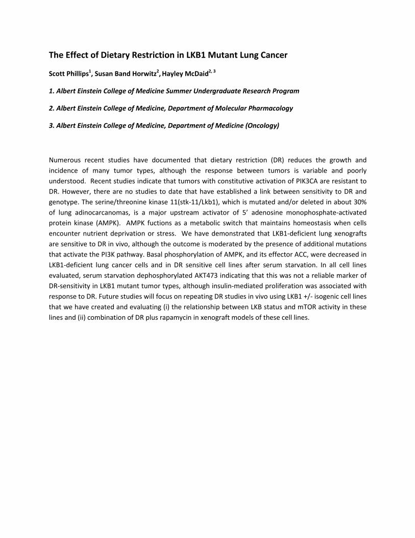
The Effect of Dietary Restriction in LKB1 Mutant Lung Cancer
Scott Phillips1, Susan Band Horwitz2, Hayley McDaid2, 3
1. Albert Einstein College of Medicine Summer Undergraduate Research Program
2. Albert Einstein College of Medicine, Department of Molecular Pharmacology
3. Albert Einstein College of Medicine, Department of Medicine (Oncology)
Numerous recent studies have documented that dietary restriction (DR) reduces the growth and incidence of many tumor types, although the response between tumors is variable and poorly understood. Recent studies indicate that tumors with constitutive activation of PIK3CA are resistant to DR. However, there are no studies to date that have established a link between sensitivity to DR and genotype. The serine/threonine kinase 11(stk-11/Lkb1), which is mutated and/or deleted in about 30% of lung adinocarcanomas, is a major upstream activator of 5’ adenosine monophosphate-activated protein kinase (AMPK). AMPK fuctions as a metabolic switch that maintains homeostasis when cells encounter nutrient deprivation or stress. We have demonstrated that LKB1-deficient lung xenografts are sensitive to DR in vivo, although the outcome is moderated by the presence of additional mutations that activate the PI3K pathway. Basal phosphorylation of AMPK, and its effector ACC, were decreased in LKB1-deficient lung cancer cells and in DR sensitive cell lines after serum starvation. In all cell lines evaluated, serum starvation dephosphorylated AKT473 indicating that this was not a reliable marker of DR-sensitivity in LKB1 mutant tumor types, although insulin-mediated proliferation was associated with response to DR. Future studies will focus on repeating DR studies in vivo using LKB1 +/- isogenic cell lines that we have created and evaluating (i) the relationship between LKB status and mTOR activity in these lines and (ii) combination of DR plus rapamycin in xenograft models of these cell lines.
Epigenetics and Longevity Rothschild, M.I., Budagov, T., *Barzilai, N., Atzmon, G. Department of Endocrinology, Albert Einstein College of Medicine, Bronx, NY 10461 *Mentor Epigenetics, meaning, “above genetics,” controls the transcription of genomic DNA. DNA Methylation, the addition of a methyl group to cytosine, is one of the most common forms of epigenetic control. Methylating DNA causes formation of heterochromatin which is packaged so as to not be transcribed into RNA and hence not expressed. This study seeks to examine the difference in methylation between individuals who have achieved extreme longevity (>95 years) and the rest of the population. Genomes of centenarians and controls (~70 years) from the Longevity Gene Study were studied using the HELP assay which determines global methylation patterns. Genes that showed significant difference in methylation of their DNA were further studied by determining their level of expression by performing real time PCR on cDNA synthesized from RNA and using primers designed for genes which showed significant levels of methylation difference. Results show that there is a distinct difference in overall methylation patterns between centenarians and controls, and that this discrepancy in levels of gene methylation yields a statistically significant difference in the expression of the corresponding genes. Therefore we can conclude that there is a difference in the epigenetic profile of centenarians, compared to an unrelated control, leading to the possibility that this difference, at least in part, allows them to achieve exceptional longevity Acknowledgements: The Summer Undergraduate Research Program of Albert Einstein College of Medicine, Frank Ferraioli, Baila Litwak
IDENTIFICATION AND QUANTIFICATION OF PEPTIDES IN HIPPOCAMPAL TISSUE OF PC7 KNOCKOUT MICE Chava Ruderman1, Jon Wardman2, Annik Prat, Ph.D.3, Lloyd Fricker, Ph.D2, 1SURP, Albert Einstein College of Medicine, Yeshiva University, Bronx, NY and Stern College for Women, Yeshiva University, New York, NY 2Department of Pharmacology, Albert Einstein College of Medicine, Yeshiva University, Bronx, NY 3Clinical Research Institute of Montreal, Quebec, Canada Pro-protein convertases (PCs), proteases that cleave peptide precursors to their intermediate forms, are implicated in multiple pathologies, including cancer, obesity, diabetes and neurodegenerative diseases. To investigate their individual functions, knockout (KO) mouse models have been developed for all nine PCs, with resulting phenotypes ranging widely from no observable effect to embryonic lethality. Only for the PC7 KO has no phenotype has been observed. To determine whether levels of peptides in hippocampal tissue underwent changes in PC7 KO mice, a peptidomics method was used to identify native forms of peptides and quantify their levels. Samples of hippocampal tissue from five wild-type mice and three PC7 KO mice were labeled with isotopic tags and then combined and analyzed with a mass spectrometer, and relative peak intensities were used to determine abundance of the peptide. Twenty-four peptides were identified; of those, ten were neuropeptides or other secretory pathway peptides, and the rest were from cytosolic proteins. Seven secretory pathway peptides as well as two other peptides showed a decrease in the KO mice, indicating that PC7 may, to some extent, play a role in peptide processing. Variable decreases in different forms of the same peptide imply that PC7 is involved at the level of post-translational modification. Further studies of peptide changes in PC7 KO animals are needed to replicate these results with additional animals and to examine other brain areas, with the goal of ultimately confirming the function of PC7 and potentially elucidating the mechanism of its processing capabilities.
Lipase inhibitory properties of acetylsalicylic acid mitigate the proliferation and virulence of pathogenic Candida species David Sanchez, David Trofa, and Joshua Nosanchuk Department of Medicine (Division of Infectious Diseases), Albert Einstein College of Medicine, Bronx, NY
Extracellular lipase is a crucial component in the pathogenesis of a wide array of
opportunistic fungi such as Candida albicans and Candida parapsilosis. As an esterase,
extracellular lipase hydrolyzes water-soluble esters at water-oil interfaces resulting in the release
of free fatty acids. These lipid constituents are easily accessible energy sources for the fungi, which
are especially important in disease pathogenesis. A reconstituted human tissue model was used to
mimic Candida albicans and Candida parapsilosis infection in order to investigate the protective
effects of acetylsalicylic acid (aspirin, ASA). We found that therapeutic concentrations of ASA
reduced tissue damage in the in vitro infection model, and we hypothesized that ASA’s protective
effects were, in part, due to its ability to inhibit extracellular secreted lipase. The efficacy of lipase
inhibition with specific concentrations of ASA was tested through the utilization of commercially
obtained purified fungal lipases from Candida cylindracea and Candida antarctica. Through
colorimetric methods using the substrate α-Naphthyl Palmitate, we determined that therapeutic
doses of 1mM and 2mM of ASA significantly decrease the lipolytic activities of the purified lipases
over time. Furthermore, the proliferation of C. albicans and C. parapsilosis was attenuated in a
lipid-rich YNB-olive oil media. Considering the lack of homology between fungal lipases and human
lipases, these findings support the potential incorporation of lipase inhibitors in antifungal
medications.
Acknowledgements: J. Nosanchuk, D. Trofa, the entire Nosanchuk Lab, MSSROP and SURP at AECOM. Supported by NIH grants AI52733, AI056070-01A2, and the Einstein CFAR.
REMODELING OF THE ACTIN CYTOSKELETON IN CULTURED ENDOTHELIAL CELLS EXPOSED TO HYPEROXIA
Yona Saperstein1, Robert M. Angert, M.D.2 and Ben Ovryn, Ph.D.3
1Yeshiva University 2Department of Pediatrics, Montefiore Medical Center, Albert Einstein College of Medicine 3Gruss-Lipper Biophotonics Center, Department of Anatomy and Structural Biology, Albert
Einstein College of Medicine
Premature infants are routinely treated with high levels of oxygen, in order to improve tissue oxygenation. It is well known, however, that hyperoxia ultimately causes lung damage and chronic respiratory insufficiency. Studies using cell cultures have demonstrated that hyperoxia causes visible changes in cell morphology. One of the major factors in determining cell shape is the actin cytoskeleton. Actin can exist in a globular, monomeric form (G-actin), or polymerize into long filaments (referred to as filamentous F-actin). The actin filaments can further bundle together into long, sturdy fibers. The location and length of these fibers determine the morphology of the cell. We hypothesize that hyperoxia-induced reactive oxygen species (ROS) alters the actin cytoskeleton and shifts the equilibrium between polymerized filamentous actin and monomeric globular actin. The mechanism by which actin is reorganized by hyperoxia-induced ROS remains unknown. Human umbilical vascular endothelial cells (HUVEC) were incubated overnight with an adenoviral vector coding for manganese superoxide dismutase (SOD). Control cells were transfected with LacZ coding adenoviral vectors. Dishes were placed in either room air (21% O2 ), 3 days of hyperoxia (95% O2 /5% CO2 ) or 5 days of hyperoxia. Using fluorescence microscopy, we found that the F-actin/G-actin ratio within stress fibers decreases during the course of hyperoxia and that SOD appears to restore the F-actin to G-actin ratio to normal. Supported by the Summer Undergraduate Research Program at Albert Einstein College of Medicine.
Cancer Therapy using Aptamers and a Novel Receptor of Dendritic Cells Zackary Scholl1, Linsley Kelly, Dr. Maria Magalhaes, Brian Wengerter, Dr. Amos Yan and Dr. Matt Levy2
1 University of Washington, Seattle, WA 2 Department of Biochemistry, Albert Einstein College of Medicine, Bronx, NY 10461 Cancer accounts for 23% of deaths in the United States. Cancer is caused by unchecked proliferating cells that become fatal tumors. Current treatments target proliferating cells with chemicals or cytotoxic antibodies, but these methods target nonspecifically and can kill normal cells like blood and hair. Aptamers, instead of antibodies, can be used to target cancer cells with high specificity. Aptamers are RNA or DNA sequences that bind specifically to a single protein. Aptamers can be developed for any protein using a technique that filters 1015 different sequences for one sequence that binds with high affinity. We are developing an aptamer for a highly specific receptor, called DNGR-1, expressed only on CD8 dendritic cells. The CD8 dendritic cells deliver antigens, such as those presented on cancer cells, to cytotoxic T cells that can eliminate cells expressing those antigens. (Sancho, 2008) A DNGR-1 specfic aptamer conjugated with an antigen expressed on tumor cells would indirectly eliminate cancerous tumors. A DNGR-1 specific aptamer could be a therapeutic drug and a potential vaccination against many forms of cancer without ill nonspecific reactions.
Development of a Pyrite Based Hydroxyl Radical Generator for Nucleic Acid Footprinting
Michael Shusterman, Jörg Schlatterer, and Michael Brenowitz
Department of Biochemistry Albert Einstein College of Medicine, Bronx NY
Footprinting assays allow for the examination of macromolecule structure or ligand binding sites through chemical or enzymatic modification. Hydroxyl radical footprinting assays utilize the small size and high reactivity of the hydroxyl radical to probe solvent accessibility with single nucleotide resolution. Current methods for the generation of hydroxyl radical are not amenable for high throughput footprinting analyses. Recently, pyrite (iron disulfide) was shown to generate hydroxyl radicals in aqueous solutions. We have proposed the design and optimization of a solid state hydroxyl radical generator that can be adopted for high throughput time resolved assays. Preliminary results indicated a tendency for pyrite particles to adsorb nucleic acids onto their surfaces. Here, we report the generation of pyrite powder and the optimization of recovery of nucleic acids following exposure to pyrite. Pyrite particles of 38 – 63 µm were produced by multiple stages of grinding and sieving using an automated ball grinder and sieving station. Sieved particles were analyzed with a Zeiss Axiophot microscope. Results showed aggregations of particles corresponding to desired mesh size and smaller. Subsequently, 32P-5’ labeled double strand DNA was incubated with 10 mg of pyrite (38 – 63 µm) in buffer solution for 0 – 120 seconds in a test tube generator, spun down, and the supernatant collected. Pellets and supernatants were Cherenkov counted. We have found that a solution consisting of 10 mM sodium cacodylate and 50 mM NaCl (pH 7.0) allows for optimal recovery of nucleic acids. Subsequent work will focus on optimizing pyrite batch production and reactor conditions. Funding for this research was provided by SURP (MS) and NSF Grant # 0852796 (MB, JS).
Intracellular and Synaptic Trafficking of an Atoxic Tetanus Toxin Sub-fragment, Hc1282, as a Potential Therapeutic Delivery Vector in Lysosomal Storage Diseases
Kevin M. Smith, Pamela Cabahug, Ryan Lippell, and Kostantin Dobrenis Department of Neuroscience
Albert Einstein College of Medicine, Bronx, NY, USA Lysosomal storage disorders result from genetic deficiencies in acid hydrolases of
sequential catabolic pathways leading to the accumulation of substrates in the lysosomal system. A wide array of therapeutic approaches have been pursued, but to date have been unsuccessful. Delivery of normal exogenous enzyme to deficient cells in the central nervous system has proven to be a major challenge due to the blood-brain barrier and low levels of neuronal endocytosis. Prior studies in our laboratory showed that a sub-fragment of the tetanus toxin, Hc1282, effectively enhanced protein delivery to neuronal lysosomes. Here we investigated whether Hc1282, like the parent Hc-toxin, may also undergo retrograde transsynaptic transfer which would facilitate delivery throughout the CNS.
Neuronal cultures were prepared from fetal rodent neocortices. Oregon Green Neutravidin (OGNA) coupled to biotinylated Hc1282 was used to follow the fate of this peptide and immunoflourescent assays were performed to identify neuronal compartments. After three weeks in culture, immunostaining for neurofilament and synaptophysin verified the widespread establishment of synapses. Following one day incubation of live cells with OGNA-Hc1282 and a one day chase period, quantitative image analysis showed a significant amount had accumulated at the border of the somatodendritic compartment. To further evaluate initial trafficking pathways, a 24 hour incubation period with OGNA-Hc1282 was followed by a wash and a subsequent chase of 0 hours, 3 hours and 24 hours.
A widely dispersed accumulation of OGNA-Hc1282 was noted at 0 hours, moderate collections within the lysosomes and synapses at 3 hours, and a high degree of accumulation in primarily the lysosomes at 24 hours. This supports the hypothesis that after initial endocytosis of exogenous Hc1282, intracellular transport of the sub-fragment to the synapse occurs. In addition, the 24 hour chase shows the highest accumulation in lysosomes and suggests that Hc1282 ultimately accumulates in the lysosomes after transsynaptic transfer. Future studies may use live imaging to directly examine transsynaptic transfer.
Albert Einstein College of Medicine Summer Undergraduate Program 2009, and NIH Grant #NS054264.
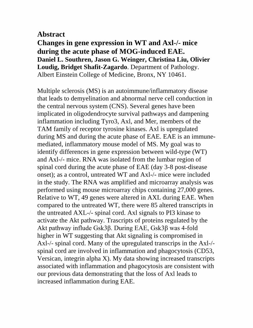
Abstract Changes in gene expression in WT and Axl-/- mice during the acute phase of MOG-induced EAE. Daniel L. Southren, Jason G. Weinger, Christina Liu, Olivier Loudig, Bridget Shafit-Zagardo. Department of Pathology. Albert Einstein College of Medicine, Bronx, NY 10461. Multiple sclerosis (MS) is an autoimmune/inflammatory disease that leads to demyelination and abnormal nerve cell conduction in the central nervous system (CNS). Several genes have been implicated in oligodendrocyte survival pathways and dampening inflammation including Tyro3, Axl, and Mer, members of the TAM family of receptor tyrosine kinases. Axl is upregulated during MS and during the acute phase of EAE. EAE is an immune-mediated, inflammatory mouse model of MS. My goal was to identify differences in gene expression between wild-type (WT) and Axl-/- mice. RNA was isolated from the lumbar region of spinal cord during the acute phase of EAE (day 3-8 post-disease onset); as a control, untreated WT and Axl-/- mice were included in the study. The RNA was amplified and microarray analysis was performed using mouse microarray chips containing 27,000 genes. Relative to WT, 49 genes were altered in AXL during EAE. When compared to the untreated WT, there were 85 altered transcripts in the untreated AXL-/- spinal cord. Axl signals to PI3 kinase to activate the Akt pathway. Trascripts of proteins regulated by the Akt pathway influde Gsk3β. During EAE, Gsk3β was 4-fold higher in WT suggesting that Akt signaling is compromised in Axl-/- spinal cord. Many of the upregulated transcrips in the Axl-/- spinal cord are involved in inflammation and phagocytosis (CD53, Versican, integrin alpha X). My data showing increased transcripts associated with inflammation and phagocytosis are consistent with our previous data demonstrating that the loss of Axl leads to increased inflammation during EAE.
Development of an in vivo screen to identify novel regulators of tumor growth and
metastasis:
Tirtza Spiegel1,2, Pamela Boimel2, Cristian Cruz2, Dr. Jeffrey Segall2
Metastasis is the spread of neoplastic cells from a primary tumor site to secondary organs
and involves angiogenesis, degradation of the basement membrane, invasion, intravasation, and
extravasation. Ninety percent of deaths from tumors are due to metastasis, therefore study of
metastasis patho-physiology is crucial. Previously 28 genes, identified to be correlated with
patient survival across three clinical breast cancer microarray studies, were tested for their ability
to regulate tumor growth and/or metastasis in SCID mice, using a lentiviral shRNA screen
approach. From this screen we identified the homeobox 2 gene, which enhanced tumor growth
both in the screen and for the individual cell line when downregulated. HOXB2 as well as other
genes evaluated in the shRNA screen will be overexpressed to screen for regulators of metastasis
and tumor growth with an open reading frame (ORF) pool. Quantitative Real-Time PCR (qRT-
PCR) primers were designed to be used to determine the changes in the proportion of human
breast cancer cells overexpressing up to 28 different genes after growth in the primary tumor and
metastasis to the lungs. We have been analyzing the sensitivity as well as the specificity of the
primers to detect their respective genes using qRT-PCR. Many of the primers had low specificity
and seemed to prime against other ORFs, possibly due to contamination, primer dimers, and/or
sequence overlap. We are currently evaluating in vitro growth of the HOXB2 knockdown cell
line compared to the overexpression cell line and are comparing this to in vivo tumor growth
curves. In preliminary studies HOXB2 gene had little effect on in vitro growth, although it
seemed to increase primary tumor growth in vivo.
Acknowledgements:
Thank you to the Roth Summer Research Fellowship Committee for funding and the Segall Lab for an enjoyable and educational experience.
1 Summer Undergraduate Research Program, Yeshiva University
2 Department of Anatomy and Structural Biology, Albert Einstein College of Medicine, Bronx, NY, 10461
ARTERIOVENOUS FISTULA IN END STAGE RENAL DISEASE AND HEMODIALYSIS: A CASE STUDY Jasmine Stephens, Marissa L. Kummerling, Todd R. Olson and Sherry A. Downie Department of Anatomy and Structural Biology Albert Einstein College of Medicine Bronx, New York Arteriovenous fistula (AVF) is a surgically induced vascular enlargement that enhances vascular
access for hemodialysis patients with end stage renal disease (ESRD). AVF formation is initiated
by the union of a superficial artery and vein in the distal region of the patient’s non-dominant
arm. AV fistulas eventually enlarge to produce easily observed subcutaneous aneurysms.
Examination of a 55 year-old male cadaver revealed multiple aneurysms on his left arm that
suggested the individual was an ESRD hemodialysis patient. Dissection revealed three AVFs and
two arteriovenous grafts (AVG) as well as evidence of central line cannulation. The examination
of the kidneys was consistent with an ESRD diagnosis as they were smaller than normal and
pathologic in central structure. The causal mechanism of his ESRD was undetermined, however,
polycystic kidney disease was ruled out based on the kidneys’ morphology. ESRD is a common
condition with more than 500,000 Americans being treated in 2006. ESRD occurs when renal
function is ≤ 5% of normal, and the kidneys cannot regulate body fluid volume and composition,
eliminate uremic toxins, and maintain their normal hormone production. Symptoms of ESRD
include hypertension, edema, azotemia, anemia, electrolyte imbalance, neuropathy, sleep apnea,
malaise, and GI and cardiovascular signs. Diseases most commonly associated with ESRD
include diabetes, hypertension, polycystic kidney disease, lupus, and glomerulopathies. In
addition to a literature review to provide current information about hemodialysis and kidney
disease, interviews with patients and their physicians generated first-hand knowledge of the life
and challenges of people with ESRD and their caretakers.
I would like to acknowledge the special individuals who provided consultation for this project: Dr. Amanda Raff, Dr. Thomas Hostetter, Dr. Vaughn Folkert. Funding provided by SURP.
───────────────────────────────────────────────────────────
The Epigenetic Alterations in Human Dust Allergy DNA
-via Analysis of Cytosine Methylation. Terrence N. Turner134, Marien Pascual25, Masako Suzuki2 and John M. Greally2 1Marist College School of Science Poughkeepsie, NY. 2Albert Einstein College of Medicine Department of Epigenomics Bronx, NY. 3Summer
Undergraduate Research Program at Albert Einstein College of Medicine Bronx, NY. 4Minority Student Research Opportunity Program at Albert Einstein
College of Medicine Bronx, NY. 5Universitcidad de Salamanca, Spain.
───────────────────────────────────────────────────────────
For about three decades studies have shown that there has been an increase in the prevalence of
allergic and allegy-related diseases. Several epidemiological and experimental studies have found
associations between the effects of early exposures to harmful environments have lead to the
development of altered programming of and subsequently adult allergic diseases. It is not well
understood whether the genetic predisposition or the environmental influences are the key contributors
in the occurrence of these disorders. Epigenetics is defined as the study of the processes that produce
altered hereditable phenotypic traits and characteristics without changing the DNA sequence. The
methylation of the promoter region is well known to inhibit gene expression and many literatures
reported the alteration of DNA methylation occurred in various diseases. I hypothesized that there is a
correlation between the methylation levels in the promoter of allergy related genes and the expression of
these genes. DNA was extracted from blood samples from the University Hospital of Salamanca, Spain.
Samples were collected from both allergy and healthy control individuals. Two genes relating to
allergies in B-lymphocytes were studied. DNA samples were treated with sodium bisulphite. To amplify
the five loci of two gene promoters, Polymerase Chain Reactions (PCR) were done using custom made
primers. The PCR products were analyzed by Massarray (Epityper, SEQUENOM). Massarray data was
analyzed with R-pipeline. Methylation data from R pipeline reveals that there are indeed differences in
methylation levels between allergy and control samples. The erythroblastic leukemia viral oncogene
homolog 2 gene (ERBB2) showed differences in methylation status. The data showed allergic samples
are more hyper-methylated than the healthy control samples in the promoter of this gene. The
methyl;ation status is concordant with the gene expression status. These findings show a relationship
between the methylation levels of the promoter regions of genes and the expression of these genes. The
project provided insight to future studies of genes that may be differentially methyalated in allergy
versus healthy control samples.
Herpes Simplex Virus Type 1 Capsid Localization and Morphology Characterization in Cells Infected with GFP Tagged Strains
Thomas O. Vogler and Duncan W. Wilson
Department of Developmental and Molecular Biology Albert Einstein College of Medicine, Bronx, NY, USA
Herpes simplex virus type 1 (HSV-1) strains with green fluorescent protein (GFP)
fused to capsid protein VP26 have been invaluable tools in elucidating the mechanisms for viral replication and transport in vitro and in vivo. However, due to selective pressures associated with genetic recombination, the mechanisms of maturation may become disturbed during construction of labeled virus strains. In this study the morphology of cells infected with a wild type HSV-1 strain SC16, was compared to a fluorescently labeled wild type strain, K26GFP. In addition, two mutant viruses KΔUL36GFP and GSΔUL36 lacking the multifunctional viral protein, UL36p, were compared to the wild type strains. An accumulation of large, nuclear viral capsid aggregates was seen in cells infected with K26GFP and KΔUL36GFP. However, in cells infected with SC16 and GSΔUL36, a diffuse distribution of viral particles was seen. K26GFP-like morphology was not restored when UL36p was introduced to GSΔUL36 in trans using the complementing cell line, HS30. However, K26GFP morphology in cells infected with GSΔUL36 was restored with a K26GFP:GSΔUL36 coinfection as well as a KΔUL36GFP:GSΔUL36 coinfection. As expected a SC16:GS∆UL36 coinfection maintained the SC16 morphology. From these data, we propose UL36p does not play a role in viral capsid aggregation within the nucleus but that there is a dominant trait associated with the K26GFP and KΔUL36GFP strains that accounts for accumulation of viral capsid clusters in the cell nucleus. This work was funded by the NIH program project grant DK41918 and by R01 AI38265 to D.W.W. T.O.V. was supported by the Albert Einstein College of Medicine SURP.
Single Nucleotide Polymorphisms of the IGF-1 Receptor in Osteosarcoma Patient Tissue Biopsies
Ezra Weinblatt, Rebecca Sowers, Sajida Piperdi, Amy Park, Karen Moody and Richard
Gorlick
Department of Pediatrics and Molecular Pharmacology, The Albert Einstein College of Medicine and the Children’s Hospital at Montefiore, Bronx, NY
The Insulin-like Growth Factor-1 Receptor (IGF-1R) is over-expressed in various forms of cancer, and is associated with increased cell proliferation and prevention of apoptosis. Previous studies have shown that a human antibody successfully down-regulates the IGF-1R in xenograft tumor models in vivo, resulting in significant growth inhibition of breast, renal, ovarian, and pancreatic tumors,1, 2 but had limited success in osteosarcoma models.3 Tumor inhibition was shown for mice bearing osteosarcomas with a single nucleotide polymorphism (SNP) at position 3179 on exon 16 of the IGF-1R, where the normal G/G was mutated to an A/A. Using the 3130xl Genetic Analyzer (Abi Prism), genomic DNA of 94 patient tissue samples were sequenced to observe whether this SNP was observed in osteosarcoma patients as well. After sequencing, the patient DNA was BLAST’ed (Basic Local Alignment Search Tool) against the WT DNA sequence provided by the NCBI database, in order to determine any deviations, focusing specifically on position 3179 of exon 16. 48.9% of the patient samples were shown to be homozygous for the mutation (A/A), 14.9% heterozygous (G/A), and 36.2% were shown to be homozygous for WT (G/G). These numbers differ significantly (p<0.001) from the average population of normal individuals, as per the NCBI website. Out of 1460 individuals tested, 11.2% were shown to be homozygous mutants, 45.0% were heterozygous, and 43.8% were homozygous WT.
Additionally, cytotoxicity assays are being performed in vitro (results pending) on a number of patient cell lines using two IGF-1R human antibodies, to see if the effects parallel those of the xenograft models in vivo. These findings can be important in further developing antibodies for the IGF-1R as a novel treatment for osteosarcoma. Acknowledgements This research was funded by SURP of Albert Einstein College of Medicine, the Sarcoma Alliance for Research through Collaboration Developmental Therapeutics Grant, and grant N01-CM-37027-01 from the National Cancer Institute.
1 Burtrum D., Zhenping Zhu, Dan Lu, et al. “A Fully Human Monoclonal Antibody to the Insulin-Like Growth Factor 1 Receptor Blocks Ligand-Dependent Signaling and Inhibits Human Tumor Growth in Vivo.” Cancer Research, December 15, 2003: 8912-8921. 2 Wang Y., Judith Hailey, Denise Williams, et al. “Inhibition of insulin-like growth factor-1 receptor (IGF-1R) signaling and tumor cell growth by a fully human neutralizing anti—IGF-1R antibody.” Molecular Cancer Therapeutics 4 (8), 2005: 1214-1221. 3 Kolb EA, Richard Gorlick, PJ Houghton, et al. “Initial testing (stage 1) of a monoclonal antibody (SCH 717454) against the IGF-1 receptor by the pediatric preclinical testing program.” Pediatric Blood & Cancer, 2008 (50): 1190-1197.
Identification of Markers for Autophagy in Serum
Rebecca Weiss, Cong Zhang, Ana Maria Cuervo Department of Developmental and Molecular Biology, Albert Einstein College of
Medicine, Bronx, NY 10461
Autophagy refers to the lysosomal degradation of intracellular components such as soluble proteins (both damaged and functional), whole organelle structures and particulate matter (e.g. protein aggregates). This process is vital in maintaining the cell’s homeostatic balance by preventing the abnormal and potentially harmful accumulation of intracellular debris and by allowing for the continuous turnover of macromolecules. Recently, apparent alterations in the normal autophagic activity of the cell have been linked to aging and various forms of cancer, neurodegenerative diseases, and muscular disorders. The study of autophagy in humans is currently limited; the determination of changes in autophagic activity can only be performed upon the isolation of specific tissues of interest.
In this study, we intend to identify (i) if various autophagy-related proteins can be detected circulating in the blood and (ii) if changes in the blood levels of these proteins correlate with changes in autophagic activity. Such findings could enable these proteins to be used as diagnostic markers for the autophagic activity of an organism.
We have used blood samples from rats and mice under fed, starved, and oxidative stress conditions. Upon serum isolation and protein quantification, samples were subjected to SDS-PAGE and Western blot for the autophagic proteins of interest.
Our results show that: (i) different autophagic proteins can be detected in serum; (ii) some of the autophagic proteins represent modified (i.e. different molecular weight) forms of the intracellular variants; (iii) there is an increase in autophagic protein levels in serum under starvation and oxidative stress conditions, and as starvation time increases, these protein levels seem to be gradually rising.
Although further studies are necessary, based on our findings we conclude that the analysis of autophagic proteins in blood is possible and that these proteins could prove to be a promising marker for the autophagic status of a whole organism.
Acknowledgments: This research was supported by NIH/NIA grant P01 AG031782-01 (AMC), the Stern-Einstein Research Connection (SERC) 2009, and the Summer Undergraduate Research Program at Albert Einstein College of Medicine.

A Novel Paradigm in Rb’s role in Embryogenesis Shira Wieder, Zhu Lab
Department of Developmental and Molecular Biology, Department of Medicine, Marion Bessin Liver Research Center and Albert Einstein Comprehensive Cancer Center
Albert Einstein College of Medicine, Bronx, NY
Isolated in 1986, Retinoblastoma protein (Rb) is the prototype tumor suppressor.
It is believed to regulate the cell cycle clock to serve as a guardian of the restriction point gate between G1 and the S phase. Cells are restricted from entering the G1 phase of the cell cycle until Rb has become hyperphosphorylated so that it loses its growth-inhibitory abilities and, consequently, permits the cells to continue into the remaining phases of the cell cycle (Weinberg, p277). Because Rb plays such a critical role in cell cycle regulation, the inactivation of Rb plays a clear role in cancer pathogenesis (Weinberg, p278). In fact, when Rb heterozygous (Rb+/-) cells undergo a spontaneous loss of the wild type Rb allele (LOH), the childhood cancer known as Retinoblastoma develops.
In order to determine how Rb functions as a tumor suppressor, mouse models have been developed to mimic inheritance of a mutant Rb allele in retinoblastoma patients. These mice are born Rb+/-, and spontaneous LOH of the Rb allele takes place as in Rb+/- humans, leading to the development of tumors. This model has significantly helped improve our understanding of Rb.
The Rb+/- mouse model at the same time offers the opportunity to study the role of Rb in normal development. These studies revealed that Rb also plays a critical role in embryogenesis since Rb-/- embryos die at mid-gestation.
A number of Rb targets have been identified, the best known is the E2F transcription factor. The significance of E2F in Rb function has been studied using Rb-/- embryos and the results show that inactivation of E2F extended the survival of Rb-/- embryos by several days. This indicated that increased activity of E2F when Rb is lost can partly explain the consequence of Rb loss, which is the current paradigm of Rb function, applying to all the other currently known Rb targets.
The Zhu lab and others recently identified the Skp2 protein as a new target of Rb. In this study, we determined the significance of Skp2 in Rb function in embryogenesis, and identified a new paradigm of Rb function. Acknowledgements: Summer Undergraduate Research Program (SURP) at Albert Einstein College of Medicine, The Zhu Lab including Liang Zhu MD PhD, Fred Bauzon and Jinhua Cui, and the NIH R01 CA127901 and RO1CA131421. References: Weinberg, Robert A. The Biology of Cancer. New York: Garland Science, Taylor & Francis Group LLC, 2007.
Clade differences in HIV-1 induced blood-brain barrier permeability: Implications in HIV-associated Dementia
Paul D Winograd, Vasudev R Rao, and Vinayaka R Prasad Department of Microbiology and Immunology, Albert Einstein College of Medicine, Bronx, New York, USA
Severe HIV-associated dementia (HAD) occurs in 25-30% of individuals infected with
clade-B HIV-1, while individuals with clade-C HIV-1 seldom develop severe HAD. The
integrity of the blood-brain barrier (BBB) is crucial for normal brain function. The
penetration of BBB by HIV-1 and subsequent damage leads to HAD. Many
neuropathological differences between clade-B HIV-1 and clade-C HIV-1 have been
identified, however, their effects on blood-brain barrier permeability have not been
investigated.
To evaluate each clade’s effect on BBB integrity, we incubated human brain
microvascular endothelial cells (HBMEC’s), in a dual-chamber assay, with culture
supernatant from clade-B or clade-C infected macrophages and then investigated BBB
permeability by measuring the trans-endothelial electrical resistance (TEER). There was
a decrease in TEER following treatment with clade-B HIV-1 and no significant change in
TEER following treatment of clade-C HIV-1, indicating that clade-B HIV-1 supernatant
weakens the integrity of the BBB while clade-C HIV-1 supernatant does not. Since the
culture supernatant of HIV-1 infected macrophages contains virus particles and viral
proteins, such as gp120 and Tat, it was imperative to identify the causative agent. We
investigated the role of Tat, a HIV-1 protein that is present in the culture supernatant
and known to alter BBB permeability in the case of clade-B HIV-1. To determine if BBB
disruption is Tat-dependent, the same protocol as above was used, except HBMEC’s
were incubated with purified recombinant clade-B Tat (B-Tat) or clade-C Tat (C-Tat). B-
Tat induced a drop in TEER similar to the addition of clade-B infected macrophage
supernatant, whereas C-Tat did not change the TEER. This data suggests that the
disparity in HAD incidence between clade-B and clade-C is due, in part, to the ability of
clade-B HIV-1--and particularly its Tat protein--to disrupt the integrity of the BBB. We
are currently confirming our results using Lucifer Yellow based permeability assays and
immunohistochemistry studies for tight junction proteins.
Funded by SURP of Albert Einstein College of Medicine and the NIH grant D43 TW001403 (to VRP).
Altered Histone Dosage Reduces S. cerevisiae Chromatin Fidelity Xiaomeng Xu, Andrea Silva, and Michael‐Christopher Keogh
Department of Cell Biology, Albert Einstein College of Medicine, Bronx, NY
DNA is packages into chromatin. The basic repeating unit is the nucleosome, consisting of ~147
bp DNA wrapped around a histone octamer. Nucleosomes can block the binding of basal transcription
factors at promoters, as well as inhibiting the passage of RNA polymareses. This allows chromatin to act
as a regulator of gene expression by preventing transcription initiation from inappropriate locations
such as cryptic promoters. We define the ability to prevent his inappropriate transcription as high
chromatin fidelity.
Recent transcriptome studies have identified a large number of sense and anti‐sense transcripts,
some potential encoding proteins missing important regulatory domains, originating within active
genes. Many of these transcripts arise from cryptic promoters, and may result from errors in
transcription of reduced chromatin fidelity.
We have identified a collection of Saccharomyces cerevisiae (budding yeast) mutants that allow
the inappropriate expression of a transgene located within an active gene, indicating defects in co‐
transcriptional chromatin fidelity. We refer to this phenotype as cryptic Tx+. The most profound crypric
Tx+ is observed in mutants of factors involved in histone metabolism, including the histone genes
themselves, histone –gene transactivators, and chaperones. Altering histone levels presumably affects
chromatin integrity by disrupting nucleosome assembly, leaving chromatin in a more open state and
permissive to cryptic transcription. We have characterized the extent of transgene expression on
individual deletion of each histone locus, or two histone gene transcription factors (spt21 or spt10).
Hht1/hhf1Δ or spt21Δ support cryptic Tx+ in a transcription‐dependent manner, where the strongest
cryptic Tx+ occurs within the most highly transcribed reporters. In contrast spt21Δ has equivalent cryptic
Tx+ from within non‐ or highly‐transcribed regions, suggesting genome wide defects in chromatin
fidelity.
This project was supported by the funding from 2009 Summer Undergraduate Research Program (SURP)
at Albert Einstein College of Medicine. Also thanks to all members of Keogh Lab for technical advice and
support of this project
Modulation of Ryanodine Receptor via Phosphorylation May Have Impact on Familial Hypertrophic Cardiomyopathy Pathogenesis Elaine C. Yang, Pia J. Guinto, and Jil C. Tardiff Department of Physiology and Biophysics Albert Einstein College of Medicine, Bronx, NY
Familial hypertrophic cardiomyopathy (FHC) is an autosomal dominant cardiac disease that exhibits a high frequency of sudden cardiac death. Known as a “disease of the sarcomere,” FHC has, to date, only been found to result from mutations found in genes for sarcomeric proteins.
Of particular interest, a missense mutation at residue 92 of cardiac troponin T (cTnT) results in a wide variety of cardiovascular phenotypes. While having immediate structural consequences, these independent mutations were previously shown to also lead to downstream alterations in Ca2+ kinetics and handling, such as Ca2+ leak, though the direct mechanism is unknown.
The ryanodine receptor (RYR) on the sarcoplasmic reticulum (SR) membrane is the channel protein through which Ca2+ is released into the cytoplasm to cause cardiac contraction. Because it induces contraction, any Ca2+ that leaks out through RYR can cause cardiac arrhythmias and may lead to sudden cardiac death. It is hypothesized that RYR acts as a gating protein whose modulation via phosphorylation plays a role in Ca2+ leak.
Previously, both R92W (Arg92Trp) and R92L (Arg92Leu) mouse models showed impaired functional Ca2+ transients; R92L mice showed no alterations in molecular Ca2+ handling, whereas R92W mice did. In this current study, whole-heart homogenates were prepared from two-, six-, and eight-month old mice, some carrying missense mutations at residue 92. Semi-quantitative westerns were performed using anti-RYR and anti-P-RYR (phosphorylated at Ser 2808) primary antibodies. Experimental results from R92L (Arg92Leu) six-month-old mice showed relatively normal RYR phosphorylation, despite a decrease in RYR levels, suggesting a trend toward hyperphosphorylation of RYR. Results from R92W (Arg92Trp) six-month-old mice showed a significant decrease in levels of RYR phosphorylation, yet an increase in RYR levels, correlating well with previous findings in which the mice showed improved SR load and Ca2+ uptake.
The results from this study demonstrate that RYR modulation may play an important role in FHC pathogenesis and the mechanisms involved.
This work was supported in part by SURP 2009 (to E.C. Yang) and by National Heart, Lung, and Blood Institute Grants 5F31-HL-085915-03 (to P. J. Guinto) and R01-HL-075619-05 (to J.C. Tardiff).
Validation of an Automated Molecular Platform to Diagnose Novel Swine Influenza
Zitter, S., Bryk, D., Narlieva, M. MS, Akid, I., Pan, Q. PhD, Chang, T. MD, Fox, A. MD, MS
Montefiore Medical Center Bronx, NY 10467
Novel swine influenza was identified in the United States in April 2009 and
subsequently escalated to a pandemic. It is expected that swine influenza will
dramatically increase in the fall. The purpose of this study was to develop and validate a
high throughput automated molecular platform in order to diagnose novel swine
influenza with faster turnaround time.
The manual method to extract viral RNA was validated against specimens run by
the New York City (NYC) Department of Health (DOH) utilizing the CDC protocol1.
Viral RNA from viral transport media of 22 patients that was previously run by NYC
DOH was also manually extracted and run on the ABI 7000 (Abbott). It was then run on
the Abbott m2000sp, which is an open platform that is FDA approved for HIV. The open
platform was adapted to extract influenza RNA. The viral RNA was extracted using the
automated platform and then run on RT-PCR (Abbott) using the CDC approved primers
and probes1. The primers and probes were for influenza A, swine influenza A, swine
H1N1, and an internal positive control, RnaseP. Cycle number threshold (Ct) < 37
indicated a positive result.
One sample was negative for all, 15 samples were confirmed as swine influenza,
and 6 were positive for influenza A. The results of the 22 cases were consistent in all
three assays. These results suggest that the automated platform can be utilized for the
accurate diagnosis of influenza. Additional tests are in progress in order to validate this
platform for clinical use. This platform, once validated, will allow for a rapid throughput
of patient samples in the face of an influenza pandemic.
Acknowledgements: SURP 2009 for support. Dr Gavin Cloherty and Dr. Danijela Lucic of Abbott Molecular Laboratories for advice and technical assistance.
1 http://www.who.int/csr/resources/publications/swineflu/realtimeptpcr/en/index.html