15n residual dipolar couplings - embocwp.embo.org/wpc09-07/lecture/rdctheory.pdfnmr workshop,...
TRANSCRIPT

NMR workshop, Rosario November 20081
Residual dipolar couplings15N
1H
B0
Markus ZweckstetterDepartment for NMR-based Structural Biology,
Göttingen, Germany
Max Planck Institute For Biophysical Chemistry

NMR workshop, Rosario November 20082
Background material
Saupe A, Englert G (1963) Phys. Rev. Lett. 11: 462-464
BothnerBy AA. (1996) In Grant DM, Harris RK (eds.), Encyclopedia of Nuclear Magnetic Resonance. Wiley, Chichester: pp. 2932-2938
Tjandra N, Bax A (1997) Direct measurement of distances and angles in biomolecules by NMR in a dilute liquid crystalline medium. Science 278: 1111-1114
Bax A, Kontaxis G, Tjandra N. (2001) Dipolar couplings in macromolecular structure determination. In Nuclear Magnetic Resonance of Biological Macromolecules, Pt B, Vol. 339, pp. 127-174
Prestegard JH, Al-Hashimi HM, Tolman JR (2000) NMR structures of biomolecules using field oriented media and residual dipolar couplings. Q. Rev. Biophys. 33: 371-424
Kramer F, Deshmukh MV, Kessler H, Glaser SJ (2004) Residual dipolar coupling constants: An elementary derivation of key equations. Concepts Magn. Reson. Part A 21A: 10-21

NMR workshop, Rosario November 20083
NMR structure determination
Chemical Shift Restraints
Assignment
Dihedral Restraints
J
ω
J − Coupling(E.COSY, DQ/ZQ, FIDS, quant.J)
Distance Restraints
NOE/ROE~1/r(NOESY, ROESY)
Structure Calculation(Simulated Annealing, Distance Geometry,
Molecular Dynamics)
1) Residual Dipolar Couplings2) N-H Relaxation Rates
φ
θ
ψ ωN
HN
H k+1N
C'
Nk+1Cαk
H αk
R
O
Cross Correlated Relaxation
ω
ΓcNH,CH
1) Dipole-Susceptibility Tensor2) Dipole-Mass Tensor
Projection Restraints1) Dipole-Dipole2) Dipole-CSA3) CSA-CSA
φ ψ ωN
HN
Ηk+1N
C'
Nk+1Cαk
Η αk
R
O
φ ψ ωN
HN
Ηk+1N
C'
Nk+1Cαk
Η αk
R
O
φψ ωN
HN
H k+1N
C'
Nk+1Cαk
H αk
O
R
φ ψN
HN
Cαk
Η αk
C'
R
k+1
3D-Structure
AssignmentSample13C,15N-Source
Bacteria
Multi-D-NMR

NMR workshop, Rosario November 20084
Why do we want to use dipolar couplings in solution NMR?
• all these parameters give essentially local information:distances <5Å; dihedral angles; intraresidual/sequential ϕ/ψ values, …
• long-range information is virtually non-existent
this leads to NMR-specific problems in structure determination:
• what is the exact angle between two non-parallel α-helices in a protein?
• is a long helical structure (a-helical protein, DNA) straight or bent?
• what is the relative orientation of two protein domains?
?

NMR workshop, Rosario November 20085
• Residual dipolar couplings (RDCs) can be observed in solution when a molecule is aligned with the magnetic field
• When alignment can be kept sufficiently weak• NMR spectra remain simple as in isotropic solution• quantitative measurement of a wide variety of RDCs
• Several dilute liquid crystalline media are now available
• RDC measurements and analysis are highly efficient
Residual dipolar couplings are a generally applicable tool for NMR structure determination
RDCs today

NMR workshop, Rosario November 20086
The dipolar coupling

NMR workshop, Rosario November 20087
The classical dipolar interaction
Nuclear spin magnetic dipole μ magnetic field
( ) ( ) ( )05 3
4 rμμπ
= −⎡ ⎤⎣ ⎦B r μr r rr μ
2nd nuclear spin magnetic moment in Bμ interaction energy
( ) ( )( )1 2
012 2 1 2 1 12 2 123 21
12 12
1 34
Er rμ μ μ
μπ
⎡ ⎤= − = −⎢ ⎥
⎣ ⎦B r μ μ μ μ r μ r
Within strong external magnetic field B0 alignment
1 12 12 1/ | | cosμ θ=μ r r
1 2
201 23
12
1 1 3cos4
Erμ μ
μ μ μ θπ
⎡ ⎤= −⎣ ⎦

NMR workshop, Rosario November 20088
The classical dipolar interaction
1 2
201 23
12
1 1 3cos4
Erμ μ
μ μ μ θπ
⎡ ⎤= −⎣ ⎦
( )2
0
1 3cos sin 0dπ
θ θ θ− =∫
molecular reorientation
15N
1H
B0
isotropic
magic angle ~ 54°

NMR workshop, Rosario November 20089
Quantum mechanical picture
Iγ=μ Ihspin I
3 30 0
1 23 3, 1 , 1
1 3 34 4
j ji I S iD i ij j i ij j
i j i j
x xx xH I Sr r r r r r
μ γ γ μμ δ μ δπ π= =
⎛ ⎞ ⎛ ⎞= − − = − −⎜ ⎟ ⎜ ⎟
⎝ ⎠ ⎝ ⎠∑ ∑h
2
2
2
2
2
: 3 1
: 3
: 3
: 3
z z
x z z x
y z z y
x y y x
zI Sr
xzI S I SryzI S I SrxyI S I Sr
−
+
+
+
2
2
2
2
2 2 2 2 2 2 2 2 2
2 2 2
: 3 1
: 3 1
3 3 2 2 2 2 3 1: 3 12 2 2
x x
y y
x x y y
xI SryI Sr
x y x y z z x y zI S I Sr r r
⎫− ⎪⎪
⎬⎪− ⎪⎭
⎛ ⎞+ − − − − + ++ = − −⎜ ⎟
⎝ ⎠

NMR workshop, Rosario November 200810
Quantum mechanical picture
( )
( ) ( )
( ) ( ) ( )
2
2
03 2 2
2 2
2 2
1 3 12
3 34
33
2
z z x x y y
I SD x z z x y z z y
x y y x x x y y
zI S I S I Sr
xz yzH I S I S I S I Sr r r
x yxyI S I S I S I Sr r
γ γ μπ
⎧ ⎫⎛ ⎞⎡ ⎤⎪ ⎪− + − +⎜ ⎟⎢ ⎥⎪ ⎪⎣ ⎦ ⎝ ⎠⎪ ⎪
⎪ ⎪⎡ ⎤= − + + + +⎨ ⎬⎢ ⎥⎣ ⎦⎪ ⎪⎪ ⎪⎡ ⎤−⎪ ⎪⎢ ⎥+ + +⎪ ⎪⎢ ⎥⎣ ⎦⎩ ⎭
h
sin cos ; sin sin ; cosx y zr r r
θ φ θ φ θ= = =spherical coordinates
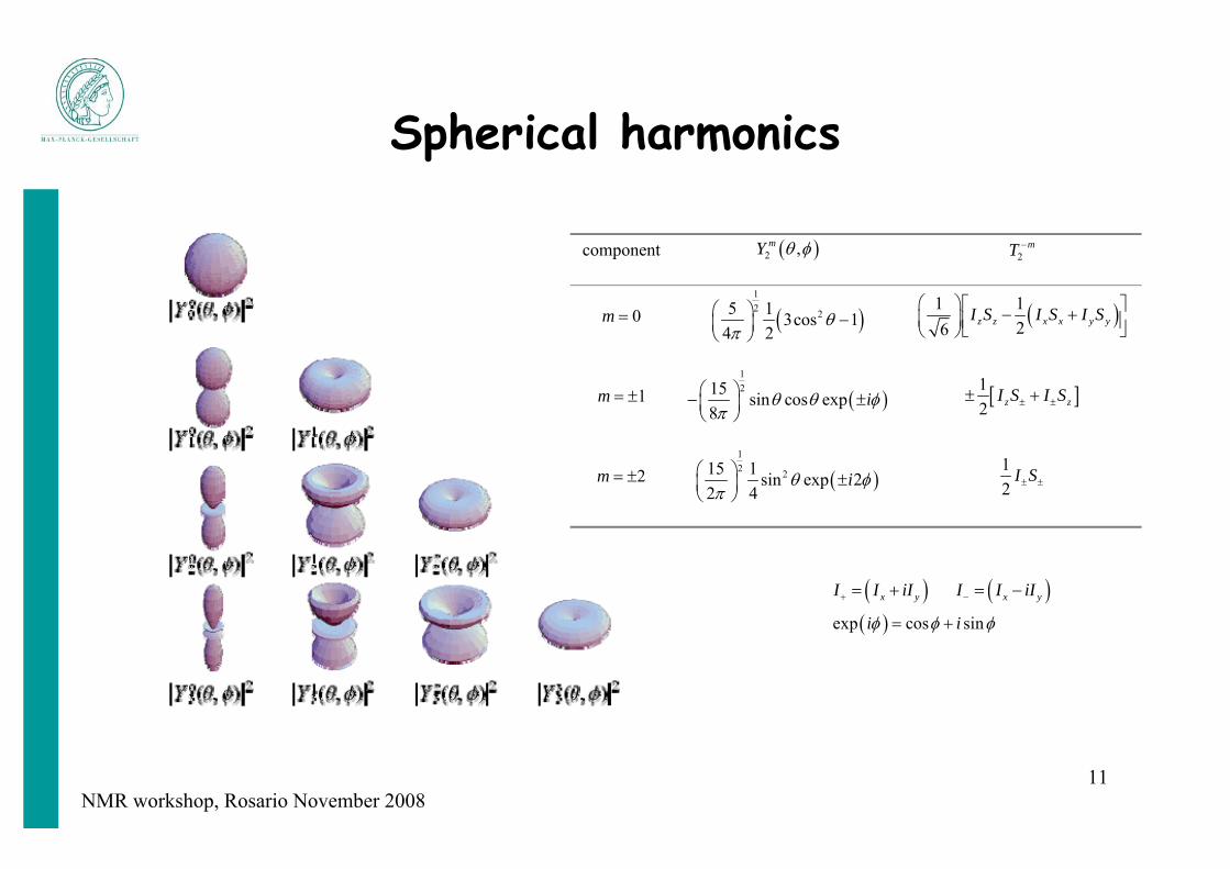
( ) ( )2 21 ,m m mD
mH F Tθ φ −= −∑ 1
20
2 23
244 5
m mI SF Yr
γ γ μ ππ
⎛ ⎞= − ⎜ ⎟⎝ ⎠
hwhere
coordinates spin operators
NMR workshop, Rosario November 200811
Spherical harmonics
component ( )2 ,mY θ φ 2mT −
0m = ( )12 25 1 3cos 1
4 2θ
π⎛ ⎞ −⎜ ⎟⎝ ⎠
( )1 126 z z x x y yI S I S I S⎛ ⎞ ⎡ ⎤− +⎜ ⎟ ⎢ ⎥⎣ ⎦⎝ ⎠
1m = ± ( )1215 sin cos exp
8iθ θ φ
π⎛ ⎞− ±⎜ ⎟⎝ ⎠
[ ]12 z zI S I S± ±± +
2m = ± ( )12 215 1 sin exp 2
2 4iθ φ
π⎛ ⎞ ±⎜ ⎟⎝ ⎠
12
I S± ±
( )x yI I iI+ = + ( )x yI I iI− = −
( )exp cos sini iφ φ φ= +

NMR workshop, Rosario November 200812
Secular approximation
( ) ( )203
13cos 14 4I S
D z zH I S I S I Sr
γ γ μ θπ + − − +
⎡ ⎤= − − − +⎢ ⎥⎣ ⎦h
( )12 x x y yI S I S I S I S+ − − ++ = +
034
I S
rγ γ μ
πh
r = 1.04 1D(N,H) = 21.7 kHz
( )203 3cos 1
4I S
D z zH I Sr
γ γ μ θπ
= − −h
heteronuclear case

NMR workshop, Rosario November 200813
( ) ( ) ( )0 023
2 , , cos4 4
P QPQD P F d dr
γ γ μβ γ θ φ β γ
π π= − ∫
h
molecular tumbling/internal motion
( )0 23
1 3cos 14 2
P QPQDr
γ γ μθ
π= − −
h
sampled orientations

NMR workshop, Rosario November 200814
Molecular alignment and RDCs
NMR workshop, Rosario November 200815
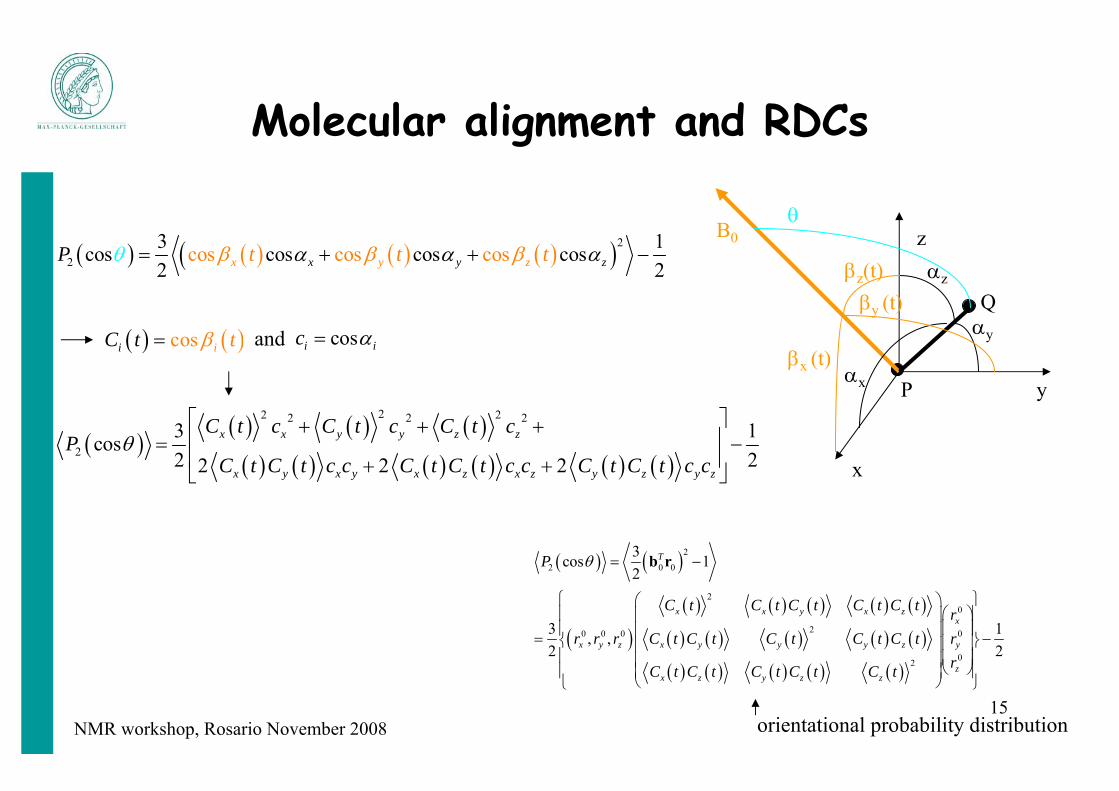
Molecular alignment and RDCs
( ) ( ) ( ) ( )( )2
2 cos cos cos3 1cos cos cos cos2 2xx y zy zt t tP β βαθ α αβ= + + −
x
y
z
P
Q
βx (t)
βz(t)βy (t)
αx
αy
αz
B0θ
( ) ( )
( )( ) ( ) ( ) ( ) ( )
( ) ( ) ( ) ( ) ( )
( ) ( ) ( ) ( ) ( )
2
2 0 0
20
20 0 0 0
02
3cos 12
3 1, ,2 2
T
x x y x zx
x y z x y y y z y
zx z y z z
P
C t C t C t C t C t rr r r C t C t C t C t C t r
rC t C t C t C t C t
θ = −
⎧ ⎫⎛ ⎞⎛ ⎞⎪ ⎪⎜ ⎟⎜ ⎟⎪ ⎪⎜ ⎟= −⎨ ⎬⎜ ⎟⎜ ⎟
⎪ ⎪⎜ ⎟⎜ ⎟⎝ ⎠⎪ ⎪⎜ ⎟⎝ ⎠⎩ ⎭
b r
orientational probability distribution
( ) ( )cos ii t tC β= cosi ic α=
( )( ) ( ) ( )
( ) ( ) ( ) ( ) ( ) ( )
22 22 2 2
23 1cos2 22 2 2
x x y y z z
x y x y x z x z y z y z
C t c C t c C t cP
C t C t c c C t C t c c C t C t c cθ
⎡ ⎤+ + +⎢ ⎥= −⎢ ⎥+ +⎣ ⎦
and

NMR workshop, Rosario November 200816
Molecular alignment and RDCs
( ) ( ) ( ) ( )3/ 2 cos cos 1/ 2 3/ 2 1/ 2ij i j ij i j ijS t t C t C tβ β δ δ= − = −
x; Sxxd
y; Syyd
z; Szzd
P
QθPQ
φPQ
( ); cos ; sin cos ; sin sinPQ z z PQ x PQ PQ y PQ PQc c cθ α θ θ φ θ φ= = = =
22 2 1x y zC C C+ + = i j j iC C C C= S is real, traceless, symmetric5 independent elements
( ) { }22 22 2 2max
3, , 12
PQ PQx y z x x y y z zD D C c C c C cα α α ⎡ ⎤= + + −⎢ ⎥⎣ ⎦
principal alignment frame, i.e. diagonalization of S Sd
( ){ }
2, , ,
cos cos cosij i ji j x y z
P Sθ α α=
= ∑
2 1/ 3i ijC A= + ( ) 2 2 2 2 2max
3, , cos sin cos sin sin2
PQ PQx y z PQ zz PQ PQ xx PQ PQ yyD D A A Aα α α θ θ φ θ φ⎡ ⎤= + +⎣ ⎦
( ) ( ) ( )2max 2
3 1, cos sin cos 22 2
PQ PQPQ PQ PQ zz PQ PQ xx yyD D P A A Aθ φ θ θ φ⎡ ⎤= + −⎢ ⎥⎣ ⎦

NMR workshop, Rosario November 200817
Generalized degree of order (GDO): Euclidean norm ϑ = (2/3
4/5 π ∑i,j Sij2)1/2
( ) ( )0 2 22 3
3, 3cos 1 sin cos 224
P QPQPQ PQ LS PQ PQ PQ
PQ
D S Rr
γ γ μθ φ θ θ φ
π⎡ ⎤= − − +⎢ ⎥⎣ ⎦
h
Aa=3/2Azz=Szzd, Ar=(Axx-Ayy)=2/3 (Sxx
d - Syyd)
( ) ( )
( ) ( )
2max 2
2 2
3, cos sin cos 243, 3cos 1 sin cos 22
PQ PQPQ PQ PQ a r PQ PQ
PQ PQPQ PQ a PQ PQ PQ
D D P A A
D D R
θ φ θ θ φ
θ φ θ θ φ
⎡ ⎤= +⎢ ⎥⎣ ⎦⎡ ⎤= − +⎢ ⎥⎣ ⎦
DaPQ = ½ DPQ
max Aa : magnitude of alignment tensor (Aa=10-3 DaNH ~ 10 Hz)
R = Aa/Ar: rhombicity of alignment tensor; R ∈ [0; 2/3]
use only a very slight orientational preference ("partial alignment"), i.e., only ca. 1 out of 1000 solute molecules

NMR workshop, Rosario November 200818
How to Get Alignment

NMR workshop, Rosario November 200819
Partial Alignment
• dipolar couplings are LARGE in solids (~22 kHz for 1DHN !), but• fortunately average out for isotropically fast tumbling (solution NMR)
⇒ full dipolar couplings are not desirable in high-resolution NMR
Can we get orientational information WITHOUT messing up our nice NMR spectra ?
YES!• use only a very slight orientational preference ("partial alignment"), i.e.,
only ca. 1 out of 1000 solute molecules
result: dipolar coupling from the (0.1%) non-isotropic fraction is scaled down to residual dipolar couplings (RDCs) of ± 20 Hz max.! (0.1% of ±20 kHz)

NMR workshop, Rosario November 200820
Alignment – Anisotropic Tumbling
To extract dipolar coupling data, the molecule must behave anisotropically!
1) large magnetic susceptibility anisotropy• diamagnetic systems such as DNA (small anisotropy in each base) • metalloproteins with paramagnetic centers• lanthanide-binding tags
field-dependent alignment of molecules
2) anisotropic environment• oriented liquid-crystalline phase• anisotropically compresed gel
field-independent alignment

NMR workshop, Rosario November 200821
Alignment media
Requirements
• liquid crystalline at < 10% w/v order of biomolecules: ~ 0.002
• aqueous
• uniform anisotropy over the whole sample volume,
• stable at different ionic strength, pH, temperature
• not too strongly charged < 0.5 e/nm2,
• solute should not bind (significantly) to medium

NMR workshop, Rosario November 200822
Bicelles
• Diskshaped particles made from DMPC and DHPC (q = 3:1)
• concentration usually 5% (w/v)
• degree of protein alignment can be “tuned” by adjusting the bicelle concentration
• alignment is temperature dependent (liquid crystal > 37°C)
• aligning with their normal perpendicular to the direction of the magnetic field
• degree of alignment can be determined by measuring the 2H quadrupolar splitting in the HDO resonance.

NMR workshop, Rosario November 200823
Bicelles (2)
• Isotropic bicelles (q ~ 0.5) for solubilization of integral membrane proteins solution-state NMR
• Anisotropic bicelles for solid-state NMR and X-ray crystallography
Disadvantages:
• unstable in the presence of certain proteins
• offers only a limited temperature and pH range

NMR workshop, Rosario November 200824
Alignment media - Toolbox
• bicelles
• alkyl poly(ethylene glycol) based media
• filamentous phage (Pf1,fd; -0.47 e/nm2)
• polyacrylamide gel (charged, uncharged)
• cellulose crystallites
• purple membrane fragments
• cetylpyrimidinium-based media, ...

NMR workshop, Rosario November 200825
Alignment of Membrane Proteins
DNA nanotubes
Douglas et al.
PNAS, 2007
Acrylamide gels G-tetrad DNA
Lorieau et al.
JACS, 2008

NMR workshop, Rosario November 200826
Modulation of alignment tensor

NMR workshop, Rosario November 200827
The problem: Orientational degeneracy
Ramirez & Bax
JACS, 1998
DPQ = DaPQ [(3 cos2 θ –1) + 3/2 R sin2 θ cos(2φ)]

NMR workshop, Rosario November 200828
Strategies for Modulation of Alignment
• Alignment media with different properties (steric/electrostatic)
• Charged bicelles: doping with small charged amphiphiles to alter their charge
Positive: CTAB / Negative: SDS
• Charged gels
• variation of pH, ionic strength
• mutation (introduction of charged residues)
• paramagnetic alignment (different tags/lanthanides)

NMR workshop, Rosario November 200829
Attenuation of alignment strength by increasing the ionic strength
ubiquitin at 450 mM NaClin 20 mg/ml Pf1
450 mM NaCl
150 mM NaCl20 mg/ml Pf1
7.08.09.0 ppm

NMR workshop, Rosario November 200830
Modulation of alignment tensor orientation by ionic strength changes
GB1

NMR workshop, Rosario November 200831
Liquid crystal theory

NMR workshop, Rosario November 200832
Liquid crystal theory
B2cp > caB2 = π Deff L2/4
25 Hz
Onsager (1949): concentration at which a solution undergoes a spontaneous first order phase transition from an isotropic to a chiral nematic phase
first-order transition coexistence region cp ∈ [ci, ca]
for semi-flexible rods
ci = 0.3588 [(1-√x)B2]-1 ca = 0.3588 [(x-√x)B2]-1

NMR workshop, Rosario November 200833
Onsager theory
effective increase in rod diameter Deff = D + κ-1 (ln ω + 0.7704)
B2cp > ca
contact potential ω = 2π Z2 [β γ x0 K1(x0)]-2 Q κ-1 exp(-2 x0)contribution of the polyions to the ionic strength κ = 8πQ(cs + Γzpcp)
B2 = π Deff L2/4

NMR workshop, Rosario November 200834
Liquid crystal theorydensity of most nematogens is higher than for the solvent
average density of the nematic region is higher than for the isotropic region gravity causes it to occupy the bottom region of the sample
Pf1 (12 mg/ml)
0.5M
B2cp > 4.19 and B2cp > 5.51

NMR workshop, Rosario November 200835
Paranematic phase of Pf1 phage
• 600 MHz□ 800 MHz
Qcc(2H) = 0.886 cPf1
cPf1 [mg/ml]
For a fully nematic phase, the degree of alignment is independent of field strength above a typically very low threshold
paranematic

NMR workshop, Rosario November 200836
Measurement of RDCs

NMR workshop, Rosario November 200837
NOESY HSQC

NMR workshop, Rosario November 200838
1JHN [1]: IPAP-HSQC, DSSE-HSQC, 3D HNCO1JC‘Cα [5]: 3D HNCO (CSA(C‘) ~ 500 MHz optimum)1JC‘N & 2JC‘HN [8.3]: 2D HSQC, 3D TROSY-HNCO1JCαHα [0.5]: 2D JCH-modulated HSQC, (HA)CANH, HN(CO)CA1JCH (side-chain): 2D JCH-mod. HSQC, CCH-COSY, SPITZE-HSQC1H-1H: COSY, CT-COSY, HNHA, 3D SS-HMQC2 (long-range)
Accuracy of measured splitting: ΔJ = LW/SN
required accuracy < 5% * Da
Bax, Kontaxis & Tjandra Method Enzymol. 339, 127-174, 2001;
Chou & Bax JBNMR, 2001; Delaglio et al. JMR 2001; Wu & Bax, JACS, 2002;

NMR workshop, Rosario November 200839
RDC measurement: J splitting (1JHN)
IPAP-HSQC
Ottiger et al. JMR, 1998

NMR workshop, Rosario November 200840
RDC measurement: Quantitative J correlation (1JC‘N)
Chou & BaxJBNMR, 2001

NMR workshop, Rosario November 200841
Determination of a MolecularAlignment Tensor

NMR workshop, Rosario November 200842
Four Methods
1) RDC distribution analysis
2) Back-calculation of alignment tensor
3) Prediction of alignment from structure
4) Prediction of alignment from structure and charge distribution

NMR workshop, Rosario November 200843
1) Estimate for alignment tensor
DzzPQ = 2 Da
PQ
DyyPQ = –Da
PQ (1 + 1.5 R)Dxx
PQ = –DaPQ (1 – 1.5 R)
with DiiPQ = DPQ
max Siid no structure necessary !
Cou
nt
-40 -20 0 200
10
20
30
1d(NH) [Hz]
-20 0 20
A B
D [Hz]aNH
-20 -150
0.1
0.2
R
Dzz
Dyy
Dxx
log( L( d1…nPQ| Da
PQ , R)) = ∑i=1,..,N log (P(diPQ))

NMR workshop, Rosario November 200844
2) Back-calculation of alignment tensor
• singular value decomposition (SVD)
very stable & with a minimumof five RDCs possible
• iterative least squares procedure (Levenberg-Marquardt minimization) χ2 = ∑i=1,..,N [di
PQ(exp) – diPQ(calc)]2/(σi
PQ)2
fixing of alignment parameters (e.g. rhombic component zero due to three-fold or higher symmetry)
2 GLN HN 2 GLN N -8.170 1.000 1.003 ILE HN 3 ILE N 8.271 1.000 1.004 PHE HN 4 PHE N 10.489 1.000 1.005 VAL HN 5 VAL N 9.871 1.000 1.006 LYS HN 6 LYS N 9.152 1.000 1.007 THR HN 7 THR N 3.700 1.000 1.008 LEU HN 8 LEU N 6.461 1.000 1.00
10 GLY HN 10 GLY N 7.634 1.000 1.0011 LYS HN 11 LYS N -7.528 1.000 1.00
if well-defined structure available

NMR workshop, Rosario November 200845
Evaluation of uncertainty in back-calculated alignment tensors (I)
Can you trust a back-calculated alignment tensor?
Monte-Carlo type approach (RDC noise)´
repeat SVD calculation many times (~1000 times)each time add different Gaussian noise to experimental RDCsaccept only those solutions for which all back-calculated RDCs are within
a given margin of the original experimental dipolar couplings
error in the data is dominated by the random measurement error in the dipolar couplings
indirectly take into account uncertainties in the structureset the amplitude of the added noise two to three times higher than the measurement uncertainty

NMR workshop, Rosario November 200846
Evaluation of uncertainty in back-calculated alignment tensors (II)
Structural noise Monte-Carlo type approachrepeat SVD calculation many times (~1000 times)
each time add different Gaussian noise to the original structure (match the RMSD between the experimental and back-calculated RDCs)
spread in alignment parameters obtained for these noise-corrupted structures, when using the coupling constants calculated for the original structure (i.e., yielding a perfect fit if no structural noise were added)

NMR workshop, Rosario November 200847
3) Prediction of alignment from structure
no RDCs necessary !

NMR workshop, Rosario November 200848
Computer experiment: PALES
Sij=1/2 <3cosΘi cos Θj - δij> (i,j=x,y,z)
Periodic boundary conditionsr < d/(2 Vf) (wall model), or r < d/(4Vf)1/2 (cylinder)
Smol linear average over all non-excluded S matrices

NMR workshop, Rosario November 200849
Shape prediction of magnitude and orientation of alignment
1DNHmeasured [Hz]
1 DN
Hpr
edic
ted
[Hz]
Protein alignment in bicelles is sterically induced

NMR workshop, Rosario November 200850
Weak alignment in Pf1 bacteriophage
http://www.asla-biotech.com/asla-phage.htm

NMR workshop, Rosario November 200851
4) Prediction of alignment from structure and charge distribution
p = 0 p = 1p = 1
steric
pB = exp[ -ΔGel(r,Ω)/kBT]
ΔGel(r,Ω) = ∑i qi φ[ri(r,Ω)]
Sijmol = ∫ Sij pB(r,Ω) dr dΩ / ∫ pB(r,Ω) dr dΩ
B0

NMR workshop, Rosario November 200852
How to calculate the electrostatic energy?
• Continuum electrostatic theory (Debye and Hueckel 1923): protein embedded in a dielectric medium containing excess ions
• non-linear Poisson-Boltzmann equation (Chapman 1913; Gouy 1910)
• Further simplification: Protein = a particle in the external field of the liquid crystal many approximations !
• protein = charges of their ionizable residues• static dielectric constant of water ε = 78.29• average surface charge of phages: -0.47 e/nm2

NMR workshop, Rosario November 200853
0.5
rela
tive
G
0distance from Pf1 [nm]
-4
-2
0
Bφ
[kT/
e]
10 20
Electrostatic potential0

NMR workshop, Rosario November 200854
Experiment versus PALES prediction
ubiquitin
DinI
GB3
GB1
DNA
steric
Zweckstetter et al.,
Biophys. J. 2004
electrostatic + steric

NMR workshop, Rosario November 200855
ubiquitin
DinI
GB3
GB1
Ionic strength dependence
DNA
orientationmagnitude

NMR workshop, Rosario November 200856
Weak alignment in surfactant liquid crystalline phases
3 nm Lα
10-20 nm
+
+
++
++
+
B0
PALES

NMR workshop, Rosario November 200857
Residual dipolar couplings in cetylpyridinium bromide/hexanol/sodium
bromide Zweckstetter,
submitted
steric and electrostatic interactions dominate weak alignment of biomolecules
in polar liquid crystalline media

NMR workshop, Rosario November 200858
Partial alignment at pH 3
pH 3
pH 7
Barrientos et al., JMR 2001
pH 3
pH 7
Zweckstetter, Eur. Biophys. J. 2005

NMR workshop, Rosario November 200859
PH dependence of alignment

NMR workshop, Rosario November 200860Zweckstetter et al., Biophys. J. 2004
RDCs are a sensitive probe of proteinelectrostatics
DinI (PDB code: 1GHH)

NMR workshop, Rosario November 200861
Charge/Shape prediction: applications• differentiation of monomeric and homodimeric states (Zweckstetter and Bax 2000)
• conformational analysis of dynamic systems such as oligosaccharides (Azurmendi and Bush 2002)
• refinement of nucleic acid structures (Warren and Moore, 2001)
• determination of the relative orientation of protein domains (Bewley and Clore 2000)
• validation of structures of protein complexes(Bewley 2001)
• classify protein fold families on the basis of unassigned NMR data(Valafar and Prestegard 2003)
• Probing surface electrostatics of proteins and nucleic acids,refinement of side chain orientations in proteins, …

NMR workshop, Rosario November 200862
RDCs in Structure Calculation

NMR workshop, Rosario November 200863
RDC-based refinement of structures
PROBLEM:Potential energy surface is very rough many local/false minima
convergence problem
RDC-based refinement of starting structures
kdip force constant adjust such that dipolar RMS is equal to measurement error)

NMR workshop, Rosario November 200864
Crititical PointsHow to use the structural information obtained from molecular alignment1. In order to use the information one needs to know the direction and the size of thetensor (susceptibility, alignment, etc). 2. Minimization of the deviation between the measured quantity and its calculatedvalue from a given structure and set of tensor parameters. 3. One has to be able to evaluate the outcome of the minimization.
Minimization procedure1. Get a good estimate on the tensor parameters (magnitude and rhombicity). 2. Define structural constraints with respect to the arbitrary tensor coordinate system. 3. Turn on the force constant for a particular alignment potential such that the final RMS between the measured and calculated values reflect the experimental error. 4. During the calculation the tensor orientation will be automatically determinedthrough global minimization with respect to the current structure. 5. Refine the initial estimate of the tensor parameters. This can be achieved througheither grid search method or built into the minimization itself.

NMR workshop, Rosario November 200865
Define axis system representing thealignment tensor

NMR workshop, Rosario November 200866
From PALES to XPLOR
rDC(NH) measured aligned - isotropic (PALES convention)(min(DC)=-16.2 Hz; max(DC)=12.7 Hz ==> estimated Da(NH)=-8.1*(-1) as correct measurement isotropic-aligned)
==> PALES gives: DATA Da -4.177638e-04 ==> get correct Da by multiplying -4.177638e-04*-21585.19 = 9.0 Hz (for XPLOR however use -9.0 Hz, i.e. sign of Szz/2)
rDC(NH) measured isotropic - aligned (SSIA convention)(min(DC)=-12.7 Hz; max(DC)=16.2 Hz ===> estimated Da(NH)=16.2/2=8.1
==> PALES gives: DATA Da 4.177638e-04 ==> get correct Da by multiplying 4.177638e-04*21585.19 = 9.0 Hz (for XPLOR however use -9.0 Hz, i.e. (-1)*sign of Szz/2)

NMR workshop, Rosario November 200867
1. The final dipolar RMS between measured .vs. calculated values
2. Consistency between dipolar coupling data and NOE data(decrease in non-RDC energy terms)
3. If one has more than one class of dipolar couplings: cross validation withquality factor
4. Use programs such as Procheck to look at the overall quality of the structure(distribution in Ramchandran plot should improve)
5. In most cases where one obtains a high degree of consistency one would also gain in the overall RMSD of the family of calculated structures.
rms (Dobs - Dcalc)rms (Dobs) Q =
How to evaluate structures produced byminimizing against alignment data

NMR workshop, Rosario November 200868
Quality measure of calculated/simulated RDCs
• use only for RDCs not included in structure determination !
• no translational validation
Q ~ 17% ≈ 1.8 Å X-rayO ~ 11% ≈ 1.1 Å X-ray
1DNHmeasured [Hz]
1 DN
Hpr
edic
ted
[Hz]
rms (Dobs - Dcalc)rms (Dobs) Q =
rms(Dobs)= [2 (Danorm)2 (4 + 3R2)/5]1/2

NMR workshop, Rosario November 200869
a) without rdcb) with rdc
Zhou et al. Biopolymers (1999-2000) 52, 168.
Impact of RDC refinement