14c-psilocin tissue distribution in pregnant rats … · 14c-psilocin tissue distribution in...
TRANSCRIPT
Functional Foods in Health and Disease 2014; 4(6):232-244 Page 232 of 244
Research Article Open Access
14C-Psilocin tissue distribution in pregnant rats after intravenous
administration
Francis C.P. Law1, Grace Poon
2, Y.C.Chui
1, and Shao-Xiong He
3
1Department of Biological Sciences, Simon Fraser University, B.C. V5A 1S6, Canada;
2Syndexa
Pharmaceuticals Corp., Watertown, MA 02472, USA; 3Tianjin Institute of Materia Medica,
Tianjin 300070, P.R. China (deceased)
Corresponding Author: Francis C.P. Law, Department of Biological Sciences, Simon Fraser
University, 8888 University Drive, Burnaby B.C. V5A 1S6 Canada
Submission date: February 23, 2014; Acceptance date: June 4, 2014; Publication date: June 12,
2014
ABSTRACT
Background: Many species of hallucinogenic mushrooms have been found in the genus
Psilocybe. The main psychoactive chemicals of Psilocybe mushrooms are psilocin and its
phosphoryloxy derivative, psilocybin. In addition to its psychedelic effects, psilocybin is an
effective agent to lift the mood of depressed patients with terminal cancers.
Objective: To study the dispositional kinetics of 14
C-psilocin in pregnant rats after intravenous
injection, to calculate tissue dose surrogates i.e., tissue 14
C concentration and area under the
concentration-time curve using the experimental data, to quantify trans-placental passage of
psilocin and/or its metabolites, and to identify new psilocin metabolite(s) in rat urine.
Methods: A group of 15 pregnant Wistar rats weighing between 0.30-0.36 kg was used in the
study. Each rat was given a single dose of 7.5 mg/kg 14
C-psilocin i.v. Three rats were randomly
selected and sacrificed at 0.5, 1.0, 2.0, 4.0, and 8.0 hr post-dosing. The maternal and fetal tissues
were quickly removed and the radioactivity in these tissues determined by liquid scintillation
counting.
In a separate study, urine samples were collected from 6 male Wistar rats after administering
15 mg/kg of unlabeled psilocin i.p. The urine samples were collected and extracted by
chloroform-methanol (9:1 v/v) and analyzed using a gas chromatograph/mass spectrometer.
Results: 14
C-Psilocin crossed the placental barrier of pregnant rats readily after i.v.
administration; maternal tissue 14
C concentrations were found to be much higher than those in
fetal tissues. The areas under the curve for maternal tissues also were much higher than the fetal
tissues. In general, maternal tissues could be divided into the fast eliminating organ group, which
Functional Foods in Health and Disease 2014; 4(6):232-244 Page 233 of 244
included the brain (elimination half-life <13 hr) and the slow eliminating organ group, which
included all fetal tissues (elimination half-life >13 hr). A new psilocin metabolite tentatively
identified as dihydroxyindoleacetic acid was found in the urine.
Conclusion: Our study showed that psilocin readily crossed the placental and blood-brain
barriers of pregnant rats. Because psilocin was eliminated slowly from the fetal tissues of rats,
human consumption of magic mushrooms should be avoided during pregnancy.
Key words: magic mushrooms, psilocin, placental barrier, pregnant rats
BACKGROUND:
Recreational use of indigenous Psilocybe mushrooms has become very popular in many parts of
the world. Psilocin and its phosphoryloxy derivative, psilocybin, are the major psychoactive
compounds of the hallucinogenic mushrooms [1]. Indeed, the psychedelic effect of
psilocybin/psilocin is mediated mainly via the serotonin 5-HT2A receptors [2] and may vary with
the mushroom species, location of growth, and harvesting seasons.
The pharmacokinetics and tissue distribution of psilocybin/psilocin have been studied
extensively in rodents [3, 4] and humans [5]. 14
C-Psilocin is absorbed rapidly by male rats after
receiving a single dose orally [6]. After absorption, psilocin is distributed by the blood to the
whole body and 14
C level in tissues is found to decrease in the order of kidney > liver > brain >
blood. About 60% of the orally administered 14
C-psilocin was excreted in the urine and 21% in
the feces within 1 day post-dosing. Significant amounts of psilocin also have been detected in the
kidney, liver and brain of mice after receiving a single oral dose of psilocybin [3].
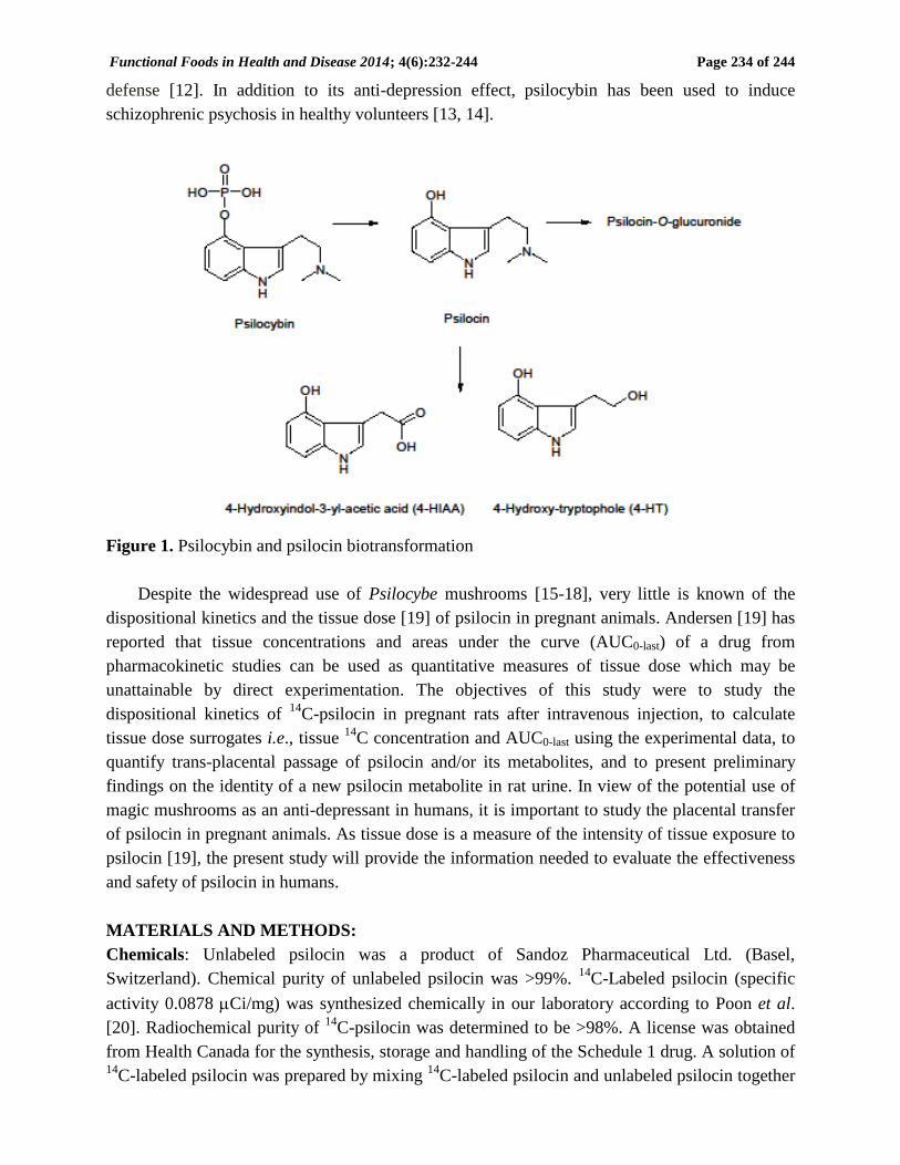
Psilocybin is rapidly hydrolyzed to psilocin by alkaline phosphatase or esterases in the
gastrointestinal tract and/or liver [3, 6-8]. The fact that the phosphoric acid ester group of
psilocybin is rapidly hydrolyzed indicates psilocin is the actual bioactive component of magic
mushrooms and psilocybin merely acts as a pro-drug (Figure 1). Kalberer et al. [6] have reported
that less than 4% of the psilocin administered to rats is metabolized to 4-hydroxy-3-indoleacetic
acid (4-HIAA) and no glucuronide metabolites have been found in the rat study. In contrast,
psilocin O-glucuronide is found in the serum of human volunteers dosed with psilocin orally [9].
Indeed, more than 80% of the psilocin administered to humans is metabolized to psilocin
glucuronides. In vitro psilocin glucuronidation also has been studied using 19 recombinant
human UDP glucuronosyltransferases of the subfamilies 1A, 2A and 2B [10]. Holzmann et al.
[1] have shown that psilocin may undergo enzymatic oxidation in humans to form 4-
hydroxytryptophole (4-HT) and 4-hydroxyindole-3-acetaldehyde, which may be further
metabolized to 4-HIAA.
A recent study has shown that psilocybin is able to lift the moods of patients with advanced-
stage cancer at a moderate dose of 0.2 mg/kg [11]. The mechanism of anti-depression effect is
not fully understood, but psilocybin is able to lower the elevated medial prefrontal cortex activity
in patients suffering from depression. The anti-depression effect of psilocybin also is explainable
by an increased emotional insight of the depressed patients after lowering their psychological
Functional Foods in Health and Disease 2014; 4(6):232-244 Page 234 of 244
defense [12]. In addition to its anti-depression effect, psilocybin has been used to induce
schizophrenic psychosis in healthy volunteers [13, 14].
Figure 1. Psilocybin and psilocin biotransformation
Despite the widespread use of Psilocybe mushrooms [15-18], very little is known of the
dispositional kinetics and the tissue dose [19] of psilocin in pregnant animals. Andersen [19] has
reported that tissue concentrations and areas under the curve (AUC0-last) of a drug from
pharmacokinetic studies can be used as quantitative measures of tissue dose which may be
unattainable by direct experimentation. The objectives of this study were to study the
dispositional kinetics of 14
C-psilocin in pregnant rats after intravenous injection, to calculate
tissue dose surrogates i.e., tissue 14
C concentration and AUC0-last using the experimental data, to
quantify trans-placental passage of psilocin and/or its metabolites, and to present preliminary
findings on the identity of a new psilocin metabolite in rat urine. In view of the potential use of
magic mushrooms as an anti-depressant in humans, it is important to study the placental transfer
of psilocin in pregnant animals. As tissue dose is a measure of the intensity of tissue exposure to
psilocin [19], the present study will provide the information needed to evaluate the effectiveness
and safety of psilocin in humans.
MATERIALS AND METHODS:
Chemicals: Unlabeled psilocin was a product of Sandoz Pharmaceutical Ltd. (Basel,
Switzerland). Chemical purity of unlabeled psilocin was >99%. 14
C-Labeled psilocin (specific
activity 0.0878 Ci/mg) was synthesized chemically in our laboratory according to Poon et al.
[20]. Radiochemical purity of 14
C-psilocin was determined to be >98%. A license was obtained
from Health Canada for the synthesis, storage and handling of the Schedule 1 drug. A solution of 14
C-labeled psilocin was prepared by mixing 14
C-labeled psilocin and unlabeled psilocin together
Functional Foods in Health and Disease 2014; 4(6):232-244 Page 235 of 244
in distilled water such that the administration of 0.5 ml of the solution provided the desired dose
of the test chemical.
Animals: Fifteen pregnant Wistar rats weighing between 300-365 g (days 19-20 of gestation)
and 6 male Wistar rats weighing between 250-300 g were purchased from Charles River Canada
Inc. (St., Constant, Quebec). They were acclimatized in the Animal Care Facility of Simon Fraser
University for 1 week before use. Commercial laboratory rat chow and water were provided ad
libitum. The procedure associated with animal care and experimentation was conducted with the
approval of the Animal Care Committee at Simon Fraser University.
Animal treatment: The tissue distribution study was initiated by injecting 14
C-psilocin (7.5
mg/kg) i.v. to each of the 15 pregnant rats. Three rats were randomly selected from the group and
sacrificed at specific time points post-dosing (0.5, 1.0, 2.0, 4.0, and 8.0 hr). Maternal blood and
tissue specimens (e.g., liver, kidney, lung, heart, spleen, brain, and muscle) were removed
immediately from the rats. The fetuses were delivered via hysterectomy, and fetal blood, liver,
lung, kidney, and heart samples were removed. All maternal and fetal tissue samples were stored
at -10 oC until analysis.
Urine samples were collected from 6 male Wistar rats after receiving a single dose of
unlabeled psilocin (15 mg/kg) i.p. The rats were kept in separate metabolic cages and the urine
was collected daily for two days. The urine samples were pooled and stored at -10 oC for
subsequent analysis. Control urine was collected from the rat prior to psilocin administration.
Determination of radioactivity in tissues and biologic fluids: Blood samples were digested at
50 oC for 30 min in separate liquid scintillation vials containing 1 ml Protosol:ethanol (1:1 v/v).
The vial was cooled, decolorized with 30 % hydrogen peroxide (0.5 ml) and neutralized by 0.5 N
HCl (0.5 ml). After the addition of Biofluor (14 ml), the radioactivity in the vial was determined
by a Beckman LS-8000 Liquid Scintillation Counter. 14
C concentration in blood was expressed
as µg psilocin equivalents/ml blood.
Tissue samples (0.5 -1.0 g) were weighed accurately on a piece of filter paper. The tissue
was oxidized in a Tri-Carb B306 Sample Oxidizer (Packard Co., Downers Grove, Ill.). The 14
CO2 evolved from burning the tissue was trapped in 6 ml of Carbo-Sorb in a liquid scintillation
vial. After the addition of Permafluor (14 ml), the vial was counted by a LSC. 14
C concentration
in tissue was expressed as µg psilocin equivalents/g tissue wet weight.
Data analysis: Tissue/blood 14
C concentration data were plotted semi-logarithmically against the
time of sample collection. The resulting concentration-time curves were analyzed using the non-
compartmental approach of WinNonlin® pharmacokinetic package (V1.0, SCI software, Cary,
North Carolina) to determine the t1/2β and AUC0-last (from 0 hour to the last time point) for each
rat. AUC0-last was calculated by the trapezoidal method. The mean + SD values of t1/2β and
AUC0-last were calculated from three rats.
Functional Foods in Health and Disease 2014; 4(6):232-244 Page 236 of 244
Isolation and characterization of urinary metabolites: The urine sample was thawed, rendered
basic with NaHCO3, and extracted 3 times with diethyl ether to remove the unchanged psilocin.
The remaining aqueous layer was acidified to pH 3 by HCl and extracted 3 times by chloroform-
methanol (9:1 v/v). The chloroform-methanol extracts were combined and dried under a gentle
stream of nitrogen. The residues were re-dissolved in methanol and transferred to a 1000 m
thickness silica gel GF preparative TLC plate (20 X 20 cm Uniplate; Analtech Inc., Newark, DE)
which was double-developed in CHCl3:CH3OH (9:1 v/v) to obtained good band separation. The
psilocin metabolites were located by placing the plate under the UV light. The silica in the area
containing the unknown metabolite was scrapped off from the plate and extracted 3 times with
chloroform:methanol (1:1 v/v). The extracts were combined and dried under a stream of nitrogen.
The residues were analyzed by direct probe mass spectrometry operated under the electron
impact mode and at an ionization potential of 70 eV. The ion source was set at 200 oC. Mass
spectral service was provided by the Department of Chemistry, Simon Fraser University.
RESULTS:
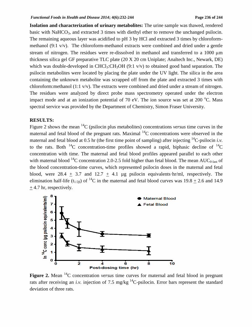
Figure 2 shows the mean 14
C (psilocin plus metabolites) concentrations versus time curves in the
maternal and fetal blood of the pregnant rats. Maximal 14
C concentrations were observed in the
maternal and fetal blood at 0.5 hr (the first time point of sampling) after injecting 14
C-psilocin i.v.
to the rats. Both 14
C concentration-time profiles showed a rapid, biphasic decline of 14
C
concentration with time. The maternal and fetal blood profiles appeared parallel to each other
with maternal blood 14
C concentration 2.0-2.5 fold higher than fetal blood. The mean AUC0-last of
the blood concentration-time curves, which represented psilocin doses in the maternal and fetal
blood, were 28.4 + 3.7 and 12.7 + 4.1 µg psilocin equivalents·hr/ml, respectively. The
elimination half-life (t1/2β) of 14
C in the maternal and fetal blood curves was 19.8 + 2.6 and 14.9
+ 4.7 hr, respectively.
Figure 2. Mean 14
C concentration versus time curves for maternal and fetal blood in pregnant
rats after receiving an i.v. injection of 7.5 mg/kg 14
C-psilocin. Error bars represent the standard
deviation of three rats.
Functional Foods in Health and Disease 2014; 4(6):232-244 Page 237 of 244
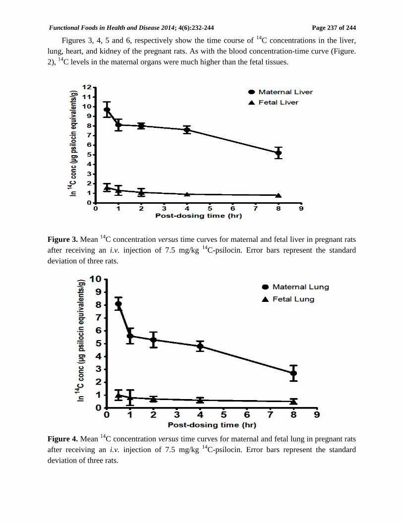
Figures 3, 4, 5 and 6, respectively show the time course of 14
C concentrations in the liver,
lung, heart, and kidney of the pregnant rats. As with the blood concentration-time curve (Figure.
2), 14
C levels in the maternal organs were much higher than the fetal tissues.
Figure 3. Mean 14
C concentration versus time curves for maternal and fetal liver in pregnant rats
after receiving an i.v. injection of 7.5 mg/kg 14
C-psilocin. Error bars represent the standard
deviation of three rats.
Figure 4. Mean 14
C concentration versus time curves for maternal and fetal lung in pregnant rats
after receiving an i.v. injection of 7.5 mg/kg 14
C-psilocin. Error bars represent the standard
deviation of three rats.
Functional Foods in Health and Disease 2014; 4(6):232-244 Page 238 of 244
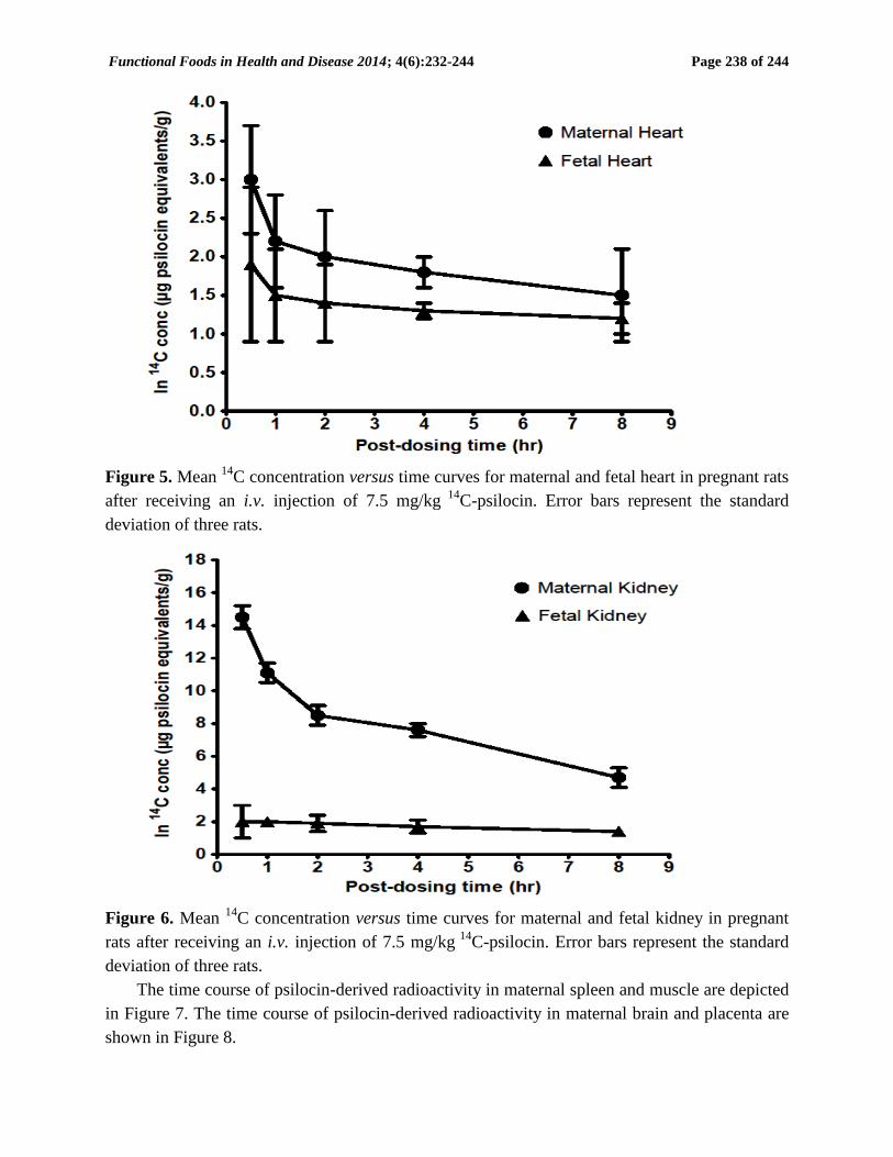
Figure 5. Mean 14
C concentration versus time curves for maternal and fetal heart in pregnant rats
after receiving an i.v. injection of 7.5 mg/kg 14
C-psilocin. Error bars represent the standard
deviation of three rats.
Figure 6. Mean 14
C concentration versus time curves for maternal and fetal kidney in pregnant
rats after receiving an i.v. injection of 7.5 mg/kg 14
C-psilocin. Error bars represent the standard
deviation of three rats.
The time course of psilocin-derived radioactivity in maternal spleen and muscle are depicted
in Figure 7. The time course of psilocin-derived radioactivity in maternal brain and placenta are
shown in Figure 8.
Functional Foods in Health and Disease 2014; 4(6):232-244 Page 239 of 244
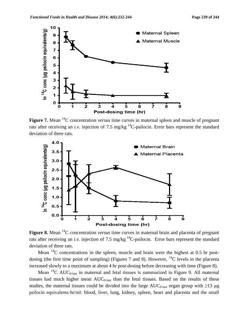
Figure 7. Mean 14
C concentration versus time curves in maternal spleen and muscle of pregnant
rats after receiving an i.v. injection of 7.5 mg/kg 14
C-psilocin. Error bars represent the standard
deviation of three rats.
Figure 8. Mean
14C concentration versus time curves in maternal brain and placenta of pregnant
rats after receiving an i.v. injection of 7.5 mg/kg 14
C-psilocin. Error bars represent the standard
deviation of three rats.
Mean 14
C concentrations in the spleen, muscle and brain were the highest at 0.5 hr post-
dosing (the first time point of sampling) (Figures 7 and 8). However, 14
C levels in the placenta
increased slowly to a maximum at about 4 hr post-dosing before decreasing with time (Figure 8).
Mean 14
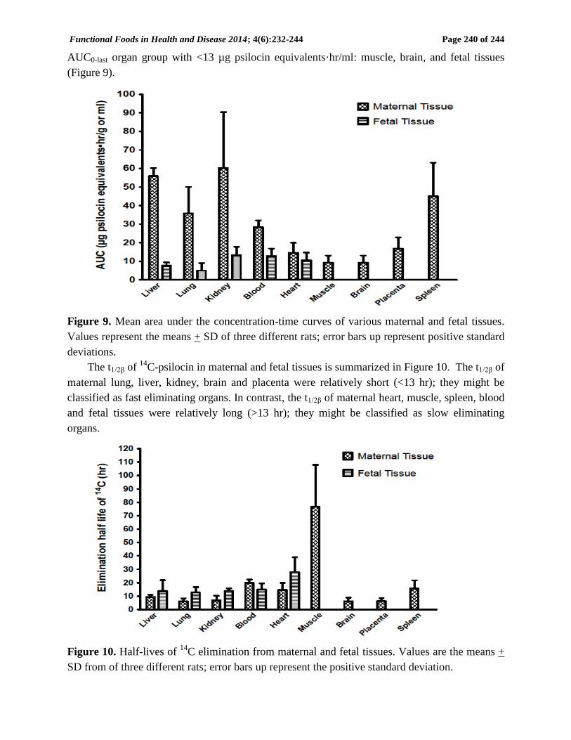
C AUC0-last in maternal and fetal tissues is summarized in Figure 9. All maternal
tissues had much higher mean AUC0-last than the fetal tissues. Based on the results of these
studies, the maternal tissues could be divided into the large AUC0-last organ group with ≥13 µg
psilocin equivalents·hr/ml: blood, liver, lung, kidney, spleen, heart and placenta and the small
Functional Foods in Health and Disease 2014; 4(6):232-244 Page 240 of 244
AUC0-last organ group with <13 µg psilocin equivalents·hr/ml: muscle, brain, and fetal tissues
(Figure 9).
Figure 9. Mean area under the concentration-time curves of various maternal and fetal tissues.
Values represent the means + SD of three different rats; error bars up represent positive standard
deviations.
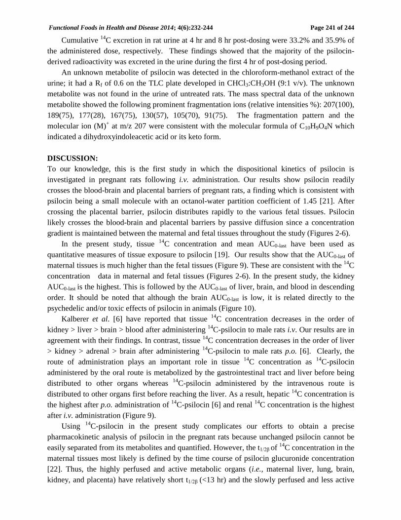
The t1/2β of 14
C-psilocin in maternal and fetal tissues is summarized in Figure 10. The t1/2β of
maternal lung, liver, kidney, brain and placenta were relatively short (<13 hr); they might be
classified as fast eliminating organs. In contrast, the t1/2β of maternal heart, muscle, spleen, blood
and fetal tissues were relatively long (>13 hr); they might be classified as slow eliminating
organs.
Figure 10. Half-lives of 14
C elimination from maternal and fetal tissues. Values are the means +
SD from of three different rats; error bars up represent the positive standard deviation.
Functional Foods in Health and Disease 2014; 4(6):232-244 Page 241 of 244
Cumulative 14
C excretion in rat urine at 4 hr and 8 hr post-dosing were 33.2% and 35.9% of
the administered dose, respectively. These findings showed that the majority of the psilocin-
derived radioactivity was excreted in the urine during the first 4 hr of post-dosing period.
An unknown metabolite of psilocin was detected in the chloroform-methanol extract of the
urine; it had a Rf of 0.6 on the TLC plate developed in CHCl3:CH3OH (9:1 v/v). The unknown
metabolite was not found in the urine of untreated rats. The mass spectral data of the unknown
metabolite showed the following prominent fragmentation ions (relative intensities %): 207(100),
189(75), 177(28), 167(75), 130(57), 105(70), 91(75). The fragmentation pattern and the
molecular ion (M)+ at m/z 207 were consistent with the molecular formula of C10H9O4N which
indicated a dihydroxyindoleacetic acid or its keto form.
DISCUSSION:
To our knowledge, this is the first study in which the dispositional kinetics of psilocin is
investigated in pregnant rats following i.v. administration. Our results show psilocin readily
crosses the blood-brain and placental barriers of pregnant rats, a finding which is consistent with
psilocin being a small molecule with an octanol-water partition coefficient of 1.45 [21]. After
crossing the placental barrier, psilocin distributes rapidly to the various fetal tissues. Psilocin
likely crosses the blood-brain and placental barriers by passive diffusion since a concentration
gradient is maintained between the maternal and fetal tissues throughout the study (Figures 2-6).
In the present study, tissue 14
C concentration and mean AUC0-last have been used as
quantitative measures of tissue exposure to psilocin [19]. Our results show that the AUC0-last of
maternal tissues is much higher than the fetal tissues (Figure 9). These are consistent with the 14
C
concentration data in maternal and fetal tissues (Figures 2-6). In the present study, the kidney
AUC0-last is the highest. This is followed by the AUC0-last of liver, brain, and blood in descending
order. It should be noted that although the brain AUC0-last is low, it is related directly to the
psychedelic and/or toxic effects of psilocin in animals (Figure 10).
Kalberer et al. [6] have reported that tissue 14
C concentration decreases in the order of
kidney > liver > brain > blood after administering 14
C-psilocin to male rats i.v. Our results are in
agreement with their findings. In contrast, tissue 14
C concentration decreases in the order of liver
> kidney > adrenal > brain after administering 14
C-psilocin to male rats p.o. [6]. Clearly, the
route of administration plays an important role in tissue 14
C concentration as 14
C-psilocin
administered by the oral route is metabolized by the gastrointestinal tract and liver before being
distributed to other organs whereas 14
C-psilocin administered by the intravenous route is
distributed to other organs first before reaching the liver. As a result, hepatic 14
C concentration is
the highest after p.o. administration of 14
C-psilocin [6] and renal 14
C concentration is the highest
after i.v. administration (Figure 9).
Using 14
C-psilocin in the present study complicates our efforts to obtain a precise
pharmacokinetic analysis of psilocin in the pregnant rats because unchanged psilocin cannot be
easily separated from its metabolites and quantified. However, the t1/2β of 14
C concentration in the
maternal tissues most likely is defined by the time course of psilocin glucuronide concentration
[22]. Thus, the highly perfused and active metabolic organs (i.e., maternal liver, lung, brain,
kidney, and placenta) have relatively short t1/2β (<13 hr) and the slowly perfused and less active
Functional Foods in Health and Disease 2014; 4(6):232-244 Page 242 of 244
metabolic organs (i.e., maternal blood, heart, spleen, muscle and all fetal tissues) have relatively
long t1/2β (>13 hr) (Figure 10). A comparison of the concentration-time profiles also shows that
maternal tissue 14
C declines at faster rates than fetal tissue 14
C (Figures 3-6). An explanation for
the slow decline of 14
C in the fetal tissues is not readily available but may be related to the
underdeveloped mixed-function oxidase and monoamine oxidase enzymes in the fetuses. As
such, the t1/2β of 14
C concentration in the fetal tissues most likely is defined by the time course of
unchanged psilocin concentration rather than psilocin glucuronide concentration.
The mass spectral data show that the unknown metabolite in rat urine has a molecular ion
(M)+ at m/z 207. The fragmentation ions at m/z 189 (C10H7NO3), 177 (C9H7NO3), 167
(C7H5NO4), 130 (C8H4NO), 105 (C6H3NO) and 91 (C6H3O) probably are resulted from (M-OH,
H)+, (M-COH, H)
+, (M-C3H3, H)
+, (M-OH, CH2COOH, H)
+, (M-CHCOH, CH2COOH, H)
+, and
(M-CCH2COOH, NHCOH, H)+, respectively. The m/z 177, 167, and 105 fragments are formed
by cleaving the aromatic ring from the psilocin molecule, and the m/z 91 fragment probably is
formed by eliminating the pyrrole ring. These results are consistent with the structure of a
dihydroxyindoleacetic acid and/or its keto isomer both of which are the oxidation product of 4-
HIAA. We are unable to detect any 4-HIAA in the urine of rats after administering psilocin i.v.
However, Kalberer et al. [6] have reported the presence of 4-HIAA in the urine of rats after p.o.
administration. Hasler et al. [23] also have been unable to detect 4-HIAA in the plasma of
humans after intravenous administration of psilocin but are able to detect it in the plasma after
oral administration. Our results are consistent with the findings in their studies.
CONCLUSION:
Psilocin and/or its metabolite(s) are able to cross the placental barrier of pregnant rats by passive
diffusion. In view of the potential use of psilocybin as an anti-depressant, it is important to study
the pharmacokinetics and tissue distribution of psilocin in pregnant rats. Because psilocin is
eliminated slowly from the fetal tissues, consumption of magic mushrooms by humans should be
avoided during pregnancy.
List of abbreviations:
GC-MS, gas chromatography-mass spectrometry; TLC, thin layer chromatography; Rf, retention
factor; t1/2β, elimination half-life; AUC0-last, area under the concentration-time curve from 0 to last
time point; 4-HIAA, 4-hydroxy-3-indoleacetic acid; 5-HT receptors, 5-hydroxytryptamine
receptors; i.v., intravenously; i.p., intraperitoneally; ln, natural logarithm.
Competing interests:
The authors report no conflicts of interest related to the contents of this article.
Authors’ contributions:
FCPL conceived and designed this project. GP was responsible for synthesizing 14
C-labeled
psilocin. YCC and SXH were responsible to conduct the experiments, data collection, figure
generation. All authors participated in writing and publishing of the paper.

Functional Foods in Health and Disease 2014; 4(6):232-244 Page 243 of 244
Acknowledgements and Funding:
This research was supported partially by the Steele Funds of Simon Fraser University. We thank
Mr. Zeyad Alehaideb for preparing the figures in this publication.
REFERENCES:
1. Holzmann PP: Bestimmung van Psilocybin-Metaboliten im Humanplasmaund -min. PhD
Thesis, Eberhard-Karls-Universifat, Tubingen, Germany; 1995.
2. Presti D, Nichols D: Biochemistry and neuropharmacology of psilocybin mushrooms. In:
Teonanacatl: Sacred Mushroom of Vision. Edited by Metzner R. El Verano, Ca: Four
Trees; 2004:89-108.
3. Horita A: Some biochemical studies on psilocybin and psilocin. J Neuropsychiat 1963,
4:270-273.
4. Aboul-Enein HY: Psilocybin: a pharmacological profile. Am J Pharmacy 1974, 91-95.
5. Hasler F, Bourquin D, Brenneisen R, Bar T, Vollenweider FX: Determination of
psilocin and 4-hydroxyindole-3-acetic acid in plasma by HPLC-ECD and
pharmacokinetic profiles of oral and intravenous psilocybin in man. Pharm Acta Helv
1997, 72(3):175-184.
6. Kalberer F, Kreis W, Rutschmann J: The fate of psilocin in the rat. Biochem Pharmacol
1962, 11:261-9.
7. Horita A, Weber LJ: Dephosphorylation of psilocybin to psilocin by alkaline
phosphatase. Proc Soc Exp Biol Med 1962, 106:32-34.
8. Eivindvik K, Rasmussen KE: Handling of psilocybin and psilocin by everted sacs of rat
jejunum and colon. Acta Pharm Nord 1989, 1:295-302.
9. Kamata T, Nishikawa M, Katagi M, Tsuchihashi H: Direct detection of serum psilocin
glucuronide by LC/MS and LC/MS/ MS: time-courses of total and free (unconjugated) psilocin
concentrations in serum specimens of a ‘magic mushroom’ user. Forensic Toxicol 2006, 24:36–
40.
10. Manevski N, Kurkela M, Höglund C et al.: Glucuronidation of psilocin and 4-hydroxyindole by
the human UDP-glucuronosyltransferases. Drug Metab Dispos 2010, 38(3):386–395.
11. Grob CS, Danforth AL, Chopra GS, Hagerty M, McKay CR, Halberstadt AL, Greer GR:
Pilot Study of psilocybin treatment for anxiety in patients with advanced-stage cancer.
Arch Gen Psychiatry 2011, 68:71-78.
12. Carhart-Harris RL, Leech R, Williams TM, Erritzoe D, Abbasi N, Bargiotas T, Hobden P,
Sharp DJ, Evans J, Feilding A, Wise RG, Nutt DJ: Implications for psychedelic-assisted
psychotherapy: functional magnetic resonance imaging study with psilocybin. British J
Psych 2012, 200:238–244.
13. Vollenweider FX, Antonini A, Scharfetter C, Angst J, Leenders, KL: Hyperfrontality and
psychopathology in the acute ketamin and psilocybin model of schizophrenia using PET
and FDG. J Neurol 1994, 241:262.
14. Hermle L, Spitzer M, Borchardt D, Gouzoulis E: Beziehungen der Modell - bzw
Drogenpsychose zu schizophrenen Erkrankungen. Fortschr Neurol Psychiat 1992,
60:383-392.
Functional Foods in Health and Disease 2014; 4(6):232-244 Page 244 of 244
15. U.S. Alcohol, Drug Abuse and Mental Health Administration. The National
Clearinghouse of Drug Abuse Information. 1973, Series 16. No.1.
16. van Poorten JF, Stienstra R, Dworacek B, Moleman P, Rupreht J: Physostigmine reversal
of psilocybin intoxication. Anesthesiology 1982, 56(4):313
17. Gartz J., Allen JW., Merlin, MD: Ethnomycology, biochemistry and cultivation of
psilocybe samuiensis Guzman, Bandala and Allen, a new psychoactive fungus from Koh
Samui, Thailand. J Ethnopharmacol 1994, 43:73-80.
18. Marcano V., Marales M.A., Castellano F., Salazar F.J., Martiner L: Occurrence of
psilocybin and psilocin in psilocybe pseudobullacea (Petch) Pegler from the Venezuelan
Andes. J Ethnopharmacol 1994, 43:157-9.
19. Andersen M E: (1987) Tissue dosimetry in risk assessment, or what’s the problem here
anyway? In Pharmacokinetics in Risk Assessment, Drinking Water and Health. Edited by
Gillette JR and Jollow DJ. Washington, D.C: National Academy Press; 1987:8-23.
20. Poon G, Chui YC, Law FCP: Synthesis of psilocin labelled with 14
C and 3H. J Labelled
Comp Radiopharmaceut 1986, 23:167-74.
21. Migliaccio GP, Shieh TL, Byrn SR, Hathaway BA, Nichols DE: Comparison of solution
conformational preferences for the hallucinogens bufotenin and psilocin using 360-MHz
proton NMR spectroscopy J Med Chem 1981, 24:206-209.
22. Gessner PK, Khairallh PA, McIsaac WM, Page IH: The relationship between the
metabolic fate and pharmacological actions of serotonin, bufotenine and psilocybin. J
Pharmacol 1960, 130:126-133.
23. Hasler F, Bourquin D, Brenneisen R, Bar T, Vollenweider FX: Determination of psilocin
and 4-hydroxyindole-3-acetic acid in plasma by HPLC-ECD and pharmacokinetic
profiles of oral and intravenous psilocybin in man. Pharmaceutics Acta Helvetiae 1997,
72:175-184