1 blood-smear
TRANSCRIPT
BLOOD SMEAR EXAMINATION Making Blood smear
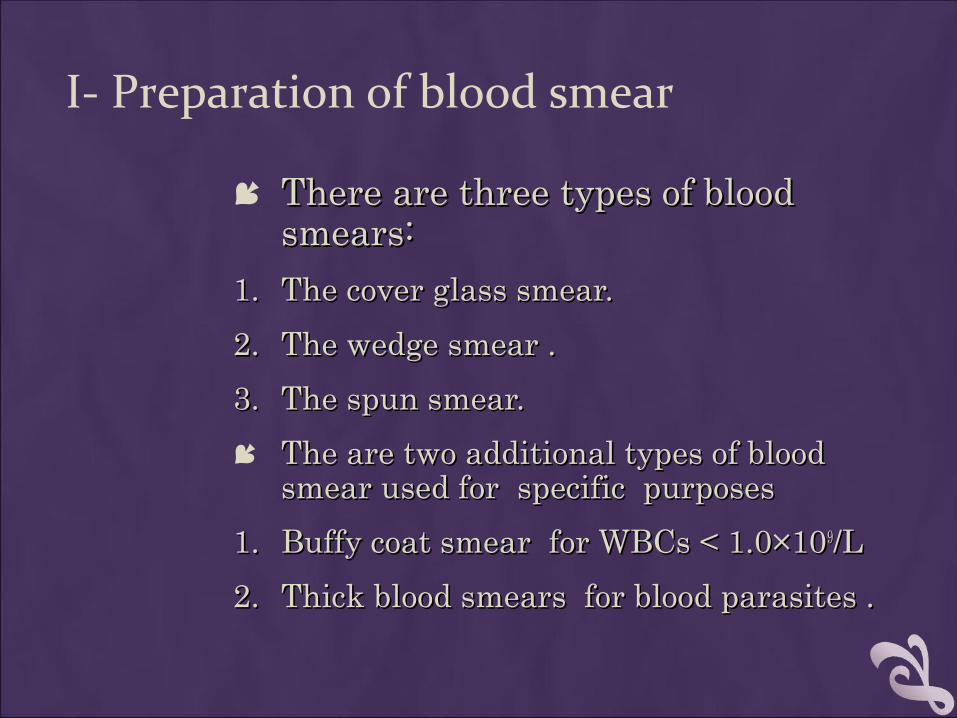
I- Preparation of blood smear
There are three types of blood There are three types of blood smears: smears:
1.1. The cover glass smear.The cover glass smear.
2.2. The wedge smear .The wedge smear .
3.3. The spun smear.The spun smear.
The are two additional types of blood The are two additional types of blood smear used for specific purposessmear used for specific purposes
1.1. Buffy coat smear for WBCs < 1.0×10Buffy coat smear for WBCs < 1.0×1099/L /L
2.2. Thick blood smears for blood parasites .Thick blood smears for blood parasites .
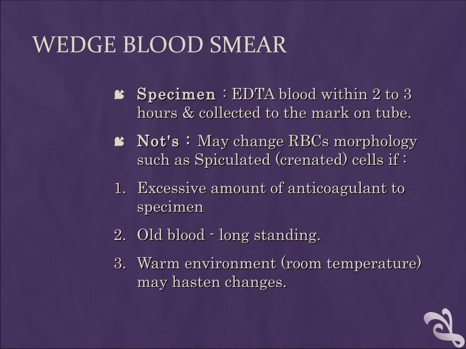
WEDGE BLOOD SMEAR
Specimen Specimen : EDTA blood within 2 to 3 : EDTA blood within 2 to 3 hours & collected to the mark on tube.hours & collected to the mark on tube.
Not's : Not's : May change RBCs morphology May change RBCs morphology such as Spiculated (crenated) cells if :such as Spiculated (crenated) cells if :
1.1. Excessive amount of anticoagulant to Excessive amount of anticoagulant to specimenspecimen
2.2. Old blood - long standing. Old blood - long standing.
3.3. Warm environment (room temperature) Warm environment (room temperature) may hasten changes.may hasten changes.
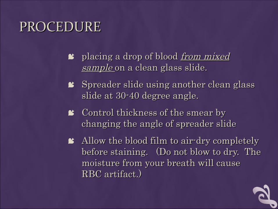
PROCEDUREPROCEDURE
placing a drop of blood placing a drop of blood from mixed from mixed sample sample on a clean glass slide.on a clean glass slide.
Spreader slide using another clean glass Spreader slide using another clean glass slide at 30-40 degree angle.slide at 30-40 degree angle.
Control thickness of the smear by Control thickness of the smear by changing the angle of spreader slidechanging the angle of spreader slide
Allow the blood film to air-dry completely Allow the blood film to air-dry completely before staining. (Do not blow to dry. The before staining. (Do not blow to dry. The moisture from your breath will cause moisture from your breath will cause RBC artifact.)RBC artifact.)
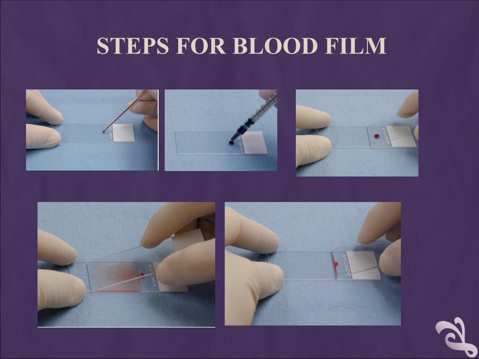
STEPS FOR BLOOD FILM
The thickness of the spread
Notes:
1. If the hematocrit is increased, the angle of the s preader slide should be decreased.
2. If the hematocrit is decreased, the angle of the spreader slide should be increased.
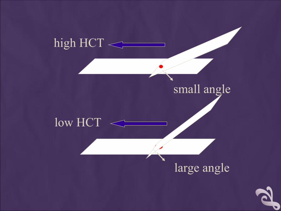
large angle
low HCT
small angle
high HCT
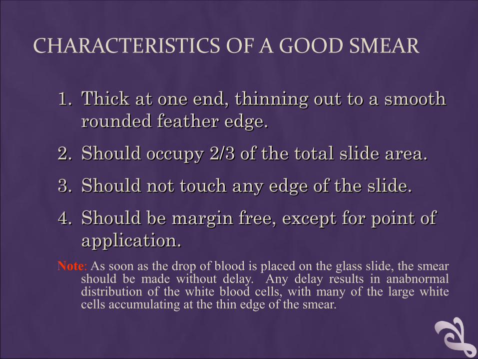
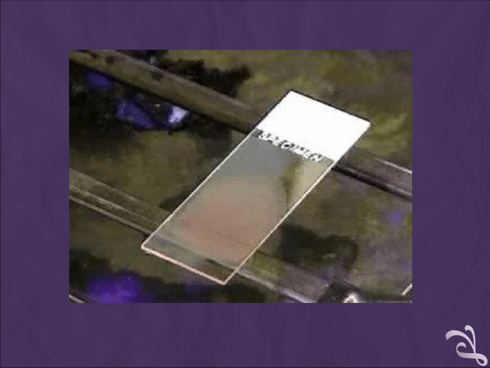
CHARACTERISTICS OF A GOOD SMEAR
1.1. Thick at one end, thinning out to a smooth Thick at one end, thinning out to a smooth rounded feather edge.rounded feather edge.
2.2. Should occupy 2/3 of the total slide area.Should occupy 2/3 of the total slide area.
3.3. Should not touch any edge of the slide.Should not touch any edge of the slide.
4.4. Should be margin free, except for point of Should be margin free, except for point of application.application.
Note: As soon as the drop of blood is placed on the glass slide, the smear should be made without delay. Any delay results in anabnormal distribution of the white blood cells, with many of the large white cells accumulating at the thin edge of the smear.
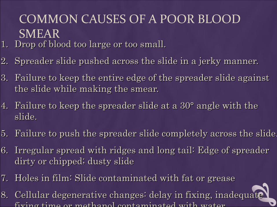
COMMON CAUSES OF A POOR BLOOD SMEAR
1.1. Drop of blood too large or too small.Drop of blood too large or too small.
2.2. Spreader slide pushed across the slide in a jerky manner.Spreader slide pushed across the slide in a jerky manner.
3.3. Failure to keep the entire edge of the spreader slide against Failure to keep the entire edge of the spreader slide against the slide while making the smear.the slide while making the smear.
4.4. Failure to keep the spreader slide at a 30° angle with the Failure to keep the spreader slide at a 30° angle with the slide.slide.
5.5. Failure to push the spreader slide completely across the slide.Failure to push the spreader slide completely across the slide.
6.6. Irregular spread with ridges and long tail: Edge of spreader Irregular spread with ridges and long tail: Edge of spreader dirty or chipped; dusty slidedirty or chipped; dusty slide
7.7. Holes in film: Slide contaminated with fat or greaseHoles in film: Slide contaminated with fat or grease
8.8. Cellular degenerative changes: delay in fixing, inadequate Cellular degenerative changes: delay in fixing, inadequate fixing time or methanol contaminated with water.fixing time or methanol contaminated with water.
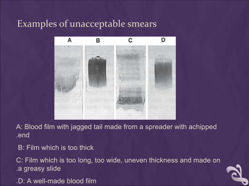
Examples of unacceptable smears
A: Blood film with jagged tail made from a spreader with achipped end.
B: Film which is too thick
C: Film which is too long, too wide, uneven thickness and made on a greasy slide.
D: A well-made blood film.
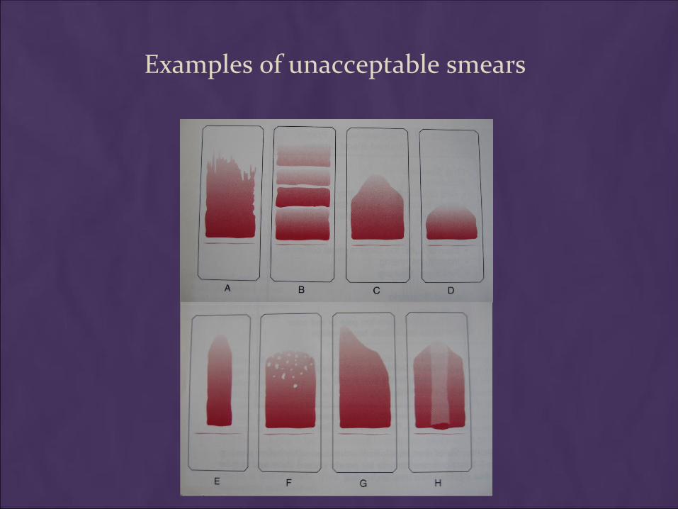
Examples of unacceptable smears
BIOLOGIC CAUSES OF A POOR SMEAR
1.1. Cold agglutinin Cold agglutinin - RBCs will clump - RBCs will clump together. Warm the blood at 37° C for 5 together. Warm the blood at 37° C for 5 minutes, and then remake the smear.minutes, and then remake the smear.
2.2. Lipemia Lipemia - holes will appear in the smear. - holes will appear in the smear. There is nothing you can do to correct this.There is nothing you can do to correct this.
3.3. Rouleaux Rouleaux - RBC’s will form into stacks - RBC’s will form into stacks resembling coins. There is nothing you resembling coins. There is nothing you can do to correct thiscan do to correct this
Notes:
1. Although this is the easiest and most popular methods for producing a blood smear, it does not produce a quality smear.
2. The WBCs are unevenly distributed and RBC distortion is seen at the edges Smaller WBCs such as lymphocytes tend to reside in the middle of the feathered edge.
3. 2. Large cells such as monocytes, immature cells and abnormal cells can be found in the outer limits of this area.
4. 3. Spun smears produce the most uniform distribution of blood cells.

SLIDE FIXATION & SLIDE FIXATION & STAININGSTAININGLEISHMAN'S STAINLEISHMAN'S STAIN
II- Fixing the films To preserve the morphology of the cells, films must be fixed as soon as
possible after they have dried.
It is important to prevent contact with water before fixation is complete.
Methyl alcohol (methanol) is the choice, although ethyl alcohol ("absolute alcohol") can be used.
Methylated spirit (95% ethanol) must not
be used as it contains water.
To fix the films, place them in a covered staining jar or tray containing the alcohol for 2-3 minutes. In humid climates it might be necessary to replace the methanol 2-3 times per day; the old portions can be used for storing clean slides.
III. Staining the film
Romanowsky staining: Romanowsky stains are universally employed for staining blood
films and are generally very satisfactory.
There are a number of different combinations of these dyes, which vary, in their staining characteristics.
1. May-Grunwald-Giemsa is a good method for routine work.
2. Giemsa stain is thought to produce more delicate staining characteristics.
Wright's stain is a simpler method.
4. Leishman's is also a simple method, which is especially suitable when a stained blood film is required urgently or the routine stain is not available (e.g. at night).
5. Field's stain is a rapid stain used primarily on thin films for malarial parasites.
Principle
The main components of a Romanowsky stain are:
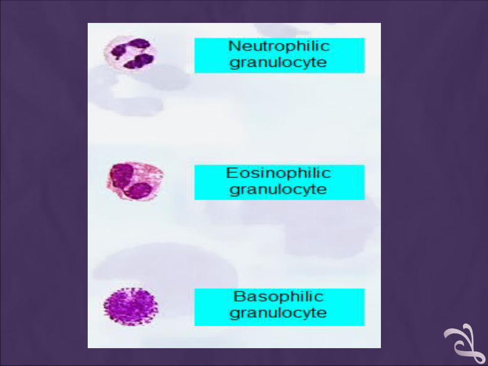
… A cationic or basic dye (methylene blue or its oxidation products such as azure B), which binds to anionic sites and gives a blue-grey color to nucleic acids (DNA or RNA), nucleoproteins, granules of basophils and weakly to granules of neutrophils
… An anionic or acidic dye such as eosin Y or eosin B, which binds to cationic sites on proteins and gives an orange-red color to hemoglobin and eosinophil granules.
… pH value of phosphate buffer is very important.
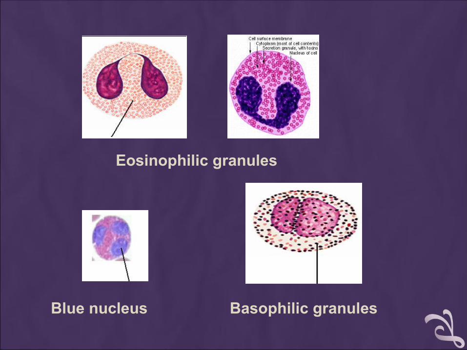
Eosinophilic granules
Basophilic granulesBlue nucleus
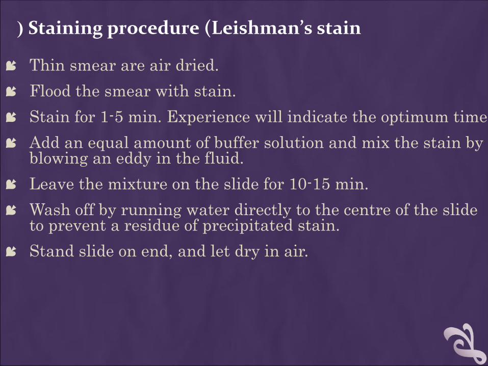
Staining procedure (Leishman’s stain(
Thin smear are air dried. Flood the smear with stain. Stain for 1-5 min. Experience will indicate the optimum time. Add an equal amount of buffer solution and mix the stain by
blowing an eddy in the fluid. Leave the mixture on the slide for 10-15 min. Wash off by running water directly to the centre of the slide
to prevent a residue of precipitated stain. Stand slide on end, and let dry in air.
Staining procedure Staining procedure
Thin smear are air dried.Thin smear are air dried.
Flood the smear with stain. Flood the smear with stain.
Stain for 1-5 min. Experience will indicate the Stain for 1-5 min. Experience will indicate the optimum time. optimum time.
Add an equal amount of buffer solution and mix Add an equal amount of buffer solution and mix the stain by blowing an eddy in the fluid.the stain by blowing an eddy in the fluid.
Leave the mixture on the slide for 10-15 min. Leave the mixture on the slide for 10-15 min.
Wash off by running water directly to the centre of Wash off by running water directly to the centre of the slide to prevent a residue of precipitated stain.the slide to prevent a residue of precipitated stain.
Stand slide on end, and let dry in air.Stand slide on end, and let dry in air.

TOO ACIDICTOO ACIDIC SUITABLE SUITABLE TOO BASIC TOO BASIC
CAUSES & CORRECTION
Too Acid Stain:Too Acid Stain:
1.1. insufficient staining timeinsufficient staining time
2.2. prolonged buffering or washingprolonged buffering or washing
3.3. old stainold stain
Correction:Correction:
1)1) lengthen staining timelengthen staining time
2)2) check stain and buffer pHcheck stain and buffer pH
3)3) shorten buffering or wash timeshorten buffering or wash time
Too Alkaline Stain:Too Alkaline Stain:
1.1. thick blood smearthick blood smear
2.2. prolonged stainingprolonged staining
3.3. insufficient washinginsufficient washing
4.4. alkaline pH of stain componentsalkaline pH of stain components Correction :Correction :
1)1) check pHcheck pH
2)2) shorten stain timeshorten stain time
3)3) prolong buffering timeprolong buffering time
PERFORMING A MANUAL PERFORMING A MANUAL DIFFERENTIAL AND ASSESSING RBC DIFFERENTIAL AND ASSESSING RBC
MORPHOLOGY MORPHOLOGY
PRINCIPLE PRINCIPLE
White Blood Cells.White Blood Cells.
1.1. Check for even distribution and Check for even distribution and estimate the number present (also, estimate the number present (also, look for any gross abnormalities look for any gross abnormalities present on the smear).present on the smear).
2.2. Perform the differential count. Perform the differential count.
PRINCIPLE PRINCIPLE
Red Blood CellsRed Blood Cells , Examine for, Examine for: :
1.1. Size and shape. Size and shape.
2.2. Relative hemoglobin content. Relative hemoglobin content.
3.3. Polychromatophilia. Polychromatophilia.
4.4. Inclusions. Inclusions.
5.5. Rouleaux formation or agglutinationRouleaux formation or agglutination
Platelets. Platelets.
1.1. Estimate number present. Estimate number present.
2.2. Examine for morphologic abnormalities. Examine for morphologic abnormalities.
PROCEDURES
Observations Under ×10Observations Under ×10
1.1. Check to see if there are good counting Check to see if there are good counting areas available free of ragged edges and cell areas available free of ragged edges and cell clumps.clumps.
2.2. Check the WBC distribution over the Check the WBC distribution over the smear.smear.
3.3. Check that the slide is properly stained.Check that the slide is properly stained.
4.4. Check for the presence of large platelets, Check for the presence of large platelets, platelet clumps, and fibrin strands.platelet clumps, and fibrin strands.
OBSERVATIONS UNDER× 40X : WBC OBSERVATIONS UNDER× 40X : WBC ESTIMATESESTIMATES
Using the × 40 high dry with no oil.Using the × 40 high dry with no oil.
Choose a portion of the peripheral smear where Choose a portion of the peripheral smear where there is only slight overlapping of the RBCs. there is only slight overlapping of the RBCs.
Count 10 fields, take the total number of white Count 10 fields, take the total number of white cells and divide by 10.cells and divide by 10.
To do a WBC estimate by taking the average To do a WBC estimate by taking the average number of white cells and multiplying by 2000.number of white cells and multiplying by 2000.
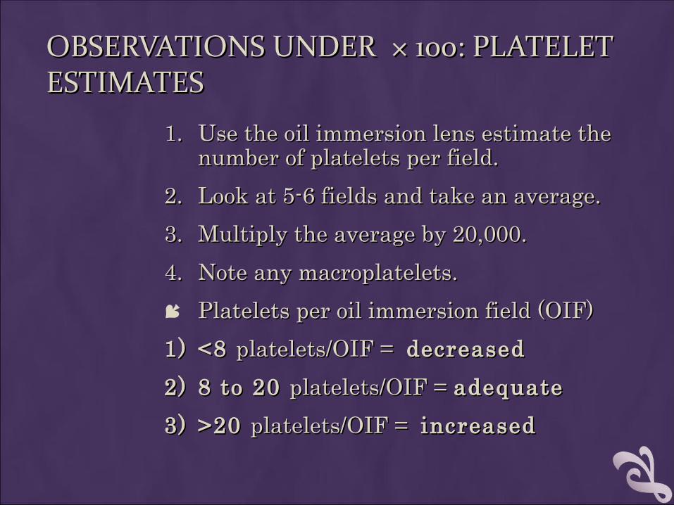
OBSERVATIONS UNDER × 100: PLATELET OBSERVATIONS UNDER × 100: PLATELET ESTIMATESESTIMATES
1.1. Use the oil immersion lens estimate the Use the oil immersion lens estimate the number of platelets per field.number of platelets per field.
2.2. Look at 5-6 fields and take an average.Look at 5-6 fields and take an average.
3.3. Multiply the average by 20,000.Multiply the average by 20,000.
4.4. Note any macroplatelets.Note any macroplatelets.
Platelets per oil immersion field (OIF)Platelets per oil immersion field (OIF)
1)1) <8 <8 platelets/OIF = platelets/OIF = decreaseddecreased
2)2) 8 to 20 8 to 20 platelets/OIF = platelets/OIF = adequateadequate
3)3) >20 >20 platelets/OIF = platelets/OIF = increasedincreased
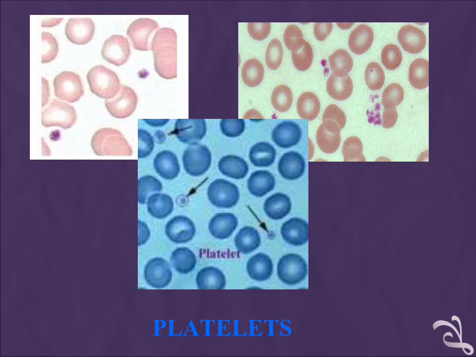
PLATELETS
OBSERVING AND RECORDING NUCLEATED RED BLOOD CELLS (NRBCS)
If 10 or more nucleated RBC's (NRBC) are seen, If 10 or more nucleated RBC's (NRBC) are seen, correct thecorrect the
White Count using this formula:White Count using this formula:
Corrected WBC Count = Corrected WBC Count =
WBC x 100/( NRBC + 100)WBC x 100/( NRBC + 100)
Example Example : : If WBC = 5000 and 10 NRBCs have been If WBC = 5000 and 10 NRBCs have been countedcounted
Then Then 5,000× 100/110 = 4545.505,000× 100/110 = 4545.50
The corrected white count is The corrected white count is 4545.50.4545.50.
MANUAL DIFFERENTIAL COUNTSMANUAL DIFFERENTIAL COUNTS
These counts are done in the same area as These counts are done in the same area as WBC and platelet estimates with the red cells WBC and platelet estimates with the red cells barely touching.barely touching.
This takes place under × 100 (oil) using the This takes place under × 100 (oil) using the zigzag method.zigzag method.
Count 100 WBCs including all cell lines from Count 100 WBCs including all cell lines from immature to mature.immature to mature.
Reporting resultsReporting results
Absolute number of cells/µl = % of cell type in Absolute number of cells/µl = % of cell type in differential x white cell countdifferential x white cell count
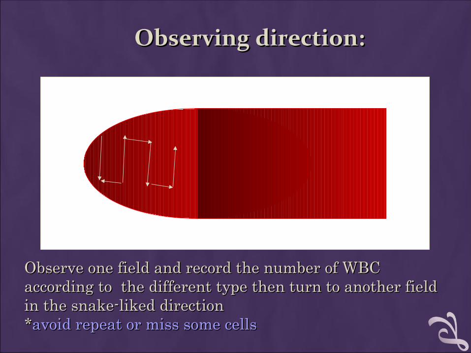
Observing direction:Observing direction:
Observe one field and record the number of WBC Observe one field and record the number of WBC according to the different type then turn to another field according to the different type then turn to another field in the snake-liked directionin the snake-liked direction**avoid repeat or miss some cellsavoid repeat or miss some cells
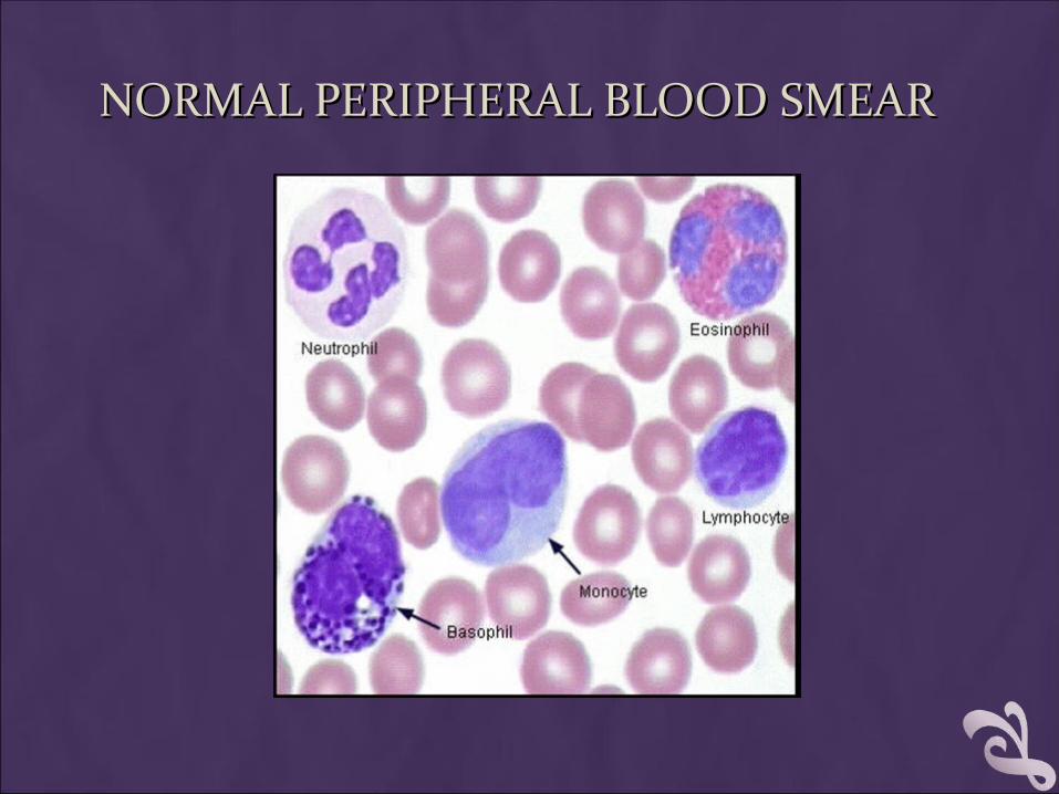
NORMAL PERIPHERAL BLOOD SMEARNORMAL PERIPHERAL BLOOD SMEAR
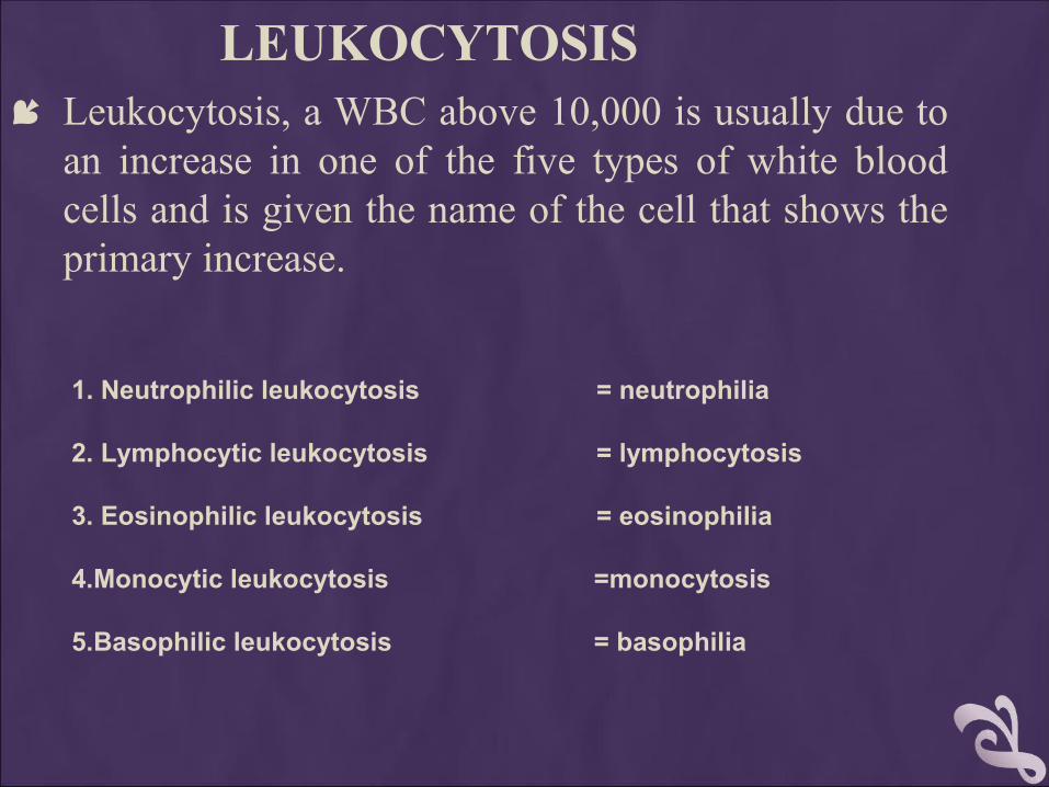
LEUKOCYTOSIS Leukocytosis, a WBC above 10,000 is usually due to
an increase in one of the five types of white blood cells and is given the name of the cell that shows the primary increase.
1. Neutrophilic leukocytosis = neutrophilia 2. Lymphocytic leukocytosis = lymphocytosis 3. Eosinophilic leukocytosis = eosinophilia
4.Monocytic leukocytosis =monocytosis
5.Basophilic leukocytosis = basophilia
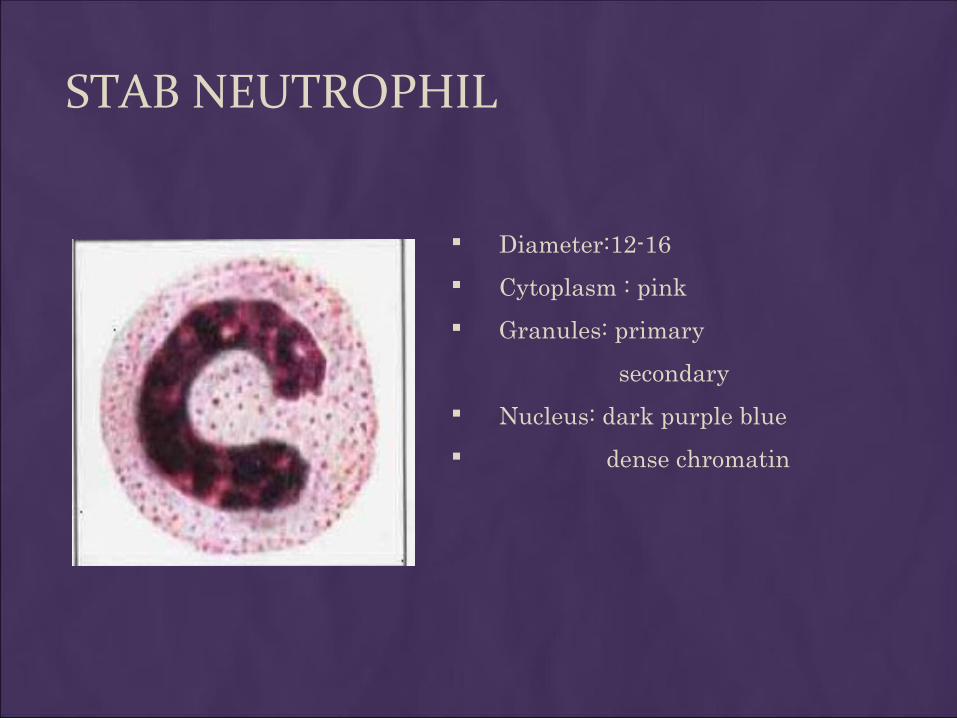
STAB NEUTROPHIL
Diameter:12-16
Cytoplasm : pink
Granules: primary
secondary
Nucleus: dark purple blue
dense chromatin
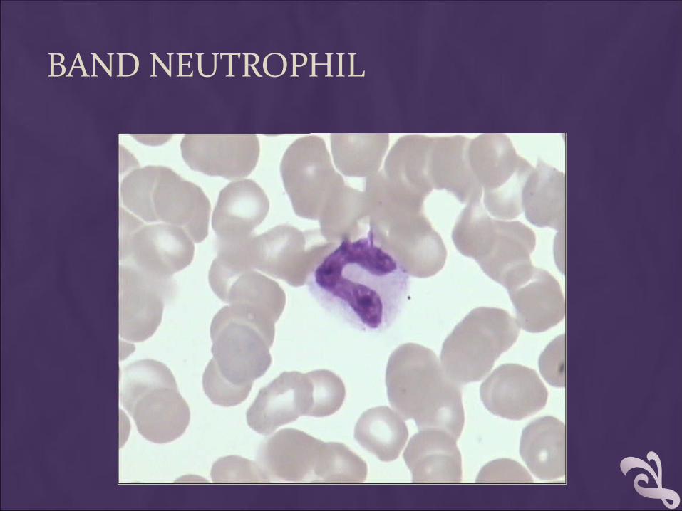
BAND NEUTROPHIL
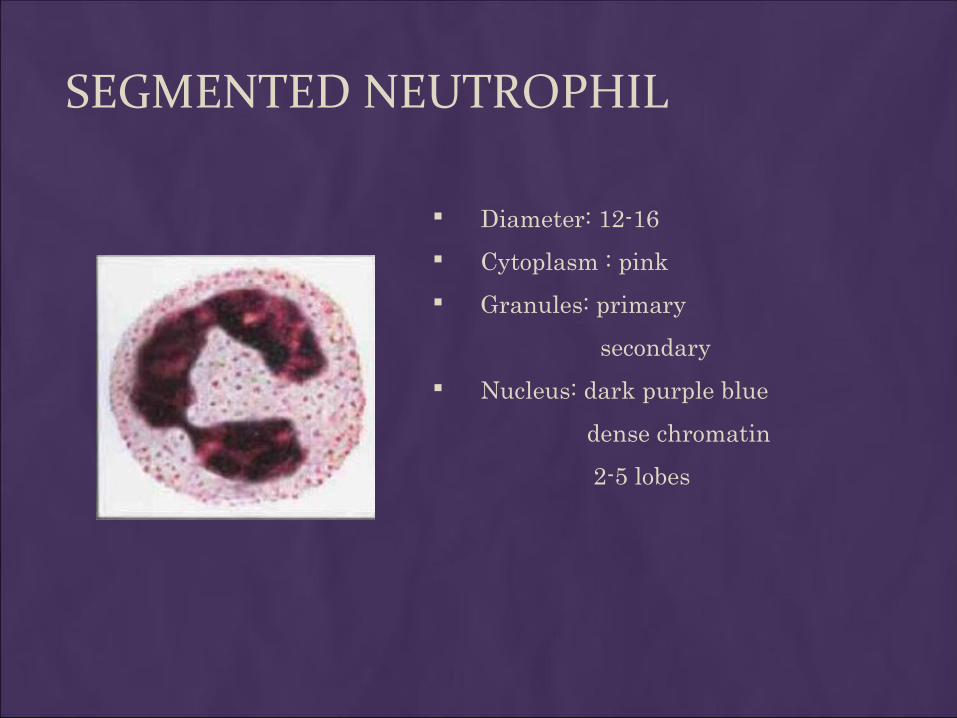
SEGMENTED NEUTROPHIL
Diameter: 12-16
Cytoplasm : pink
Granules: primary
secondary
Nucleus: dark purple blue
dense chromatin
2-5 lobes
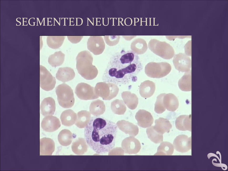
SEGMENTED NEUTROPHIL
1.NEUTROPHILS Neutrophils are so named because they are not well
stained by either eosin, a red acidic stain, or by methylene blue, a basic or alkaline stain.
Neutrophils are also known as "segs", "PMNs" or "polys" (polymorphonuclear).
They are the body's primary defense against bacterial infection.

Increased neutrophils count (neutrophilia)
1. Acute bacterial infection.
2. Granulocytic leukemia.
Decreased neutrophil count (neutropenia)
1. Typhoid fever
2. Brucellosis
3. Viral diseases, including hepatitis, influenza, rubella, and mumps.

LEFT-SHIFT AND RIGHT-SHIFT OF NEUTROPHIL
Normally, most of the neutrophils circulating in the bloodstream are in a mature form, with the nucleus of the cell being divided or segmented. Because of the segmented appearance of the nucleus, neutrophils are sometimes referred to as "segs.”
The nucleus of less mature neutrophils is not segmented, but has a band or rod-like shape. Less mature neutrophils - those that have recently been released from the bone marrow into the bloodstream - are known as "bands" or "stabs".
Left-shiftLeft-shift : non-segmented neutrophil : non-segmented neutrophil > 5%> 5%
Right-shiftRight-shift : hypersegmented neutrophil : hypersegmented neutrophil >3%>3%
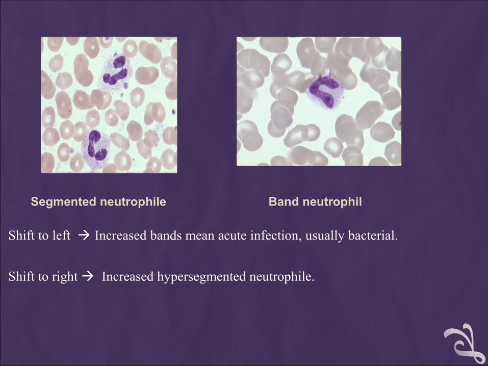
Segmented neutrophile Band neutrophil
Shift to left Increased bands mean acute infection, usually bacterial.
Shift to right Increased hypersegmented neutrophile.
EOSINOPHIL
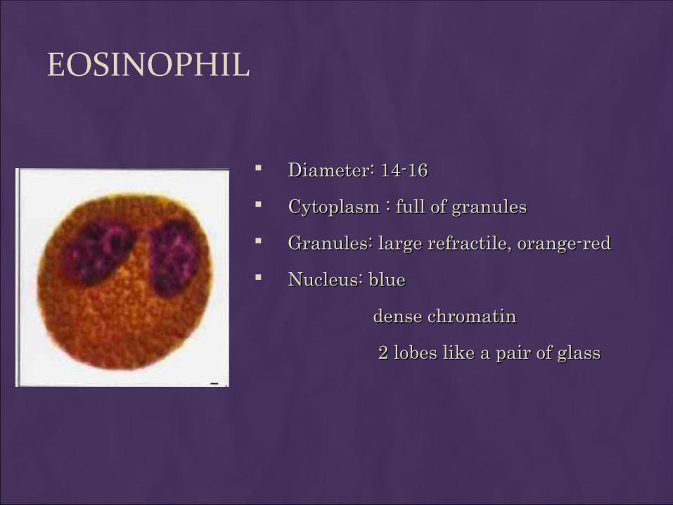
Diameter: 14-16Diameter: 14-16
Cytoplasm : full of granulesCytoplasm : full of granules
Granules: large refractile, orange-red Granules: large refractile, orange-red
Nucleus: blueNucleus: blue
dense chromatindense chromatin
2 lobes like a pair of glass2 lobes like a pair of glass
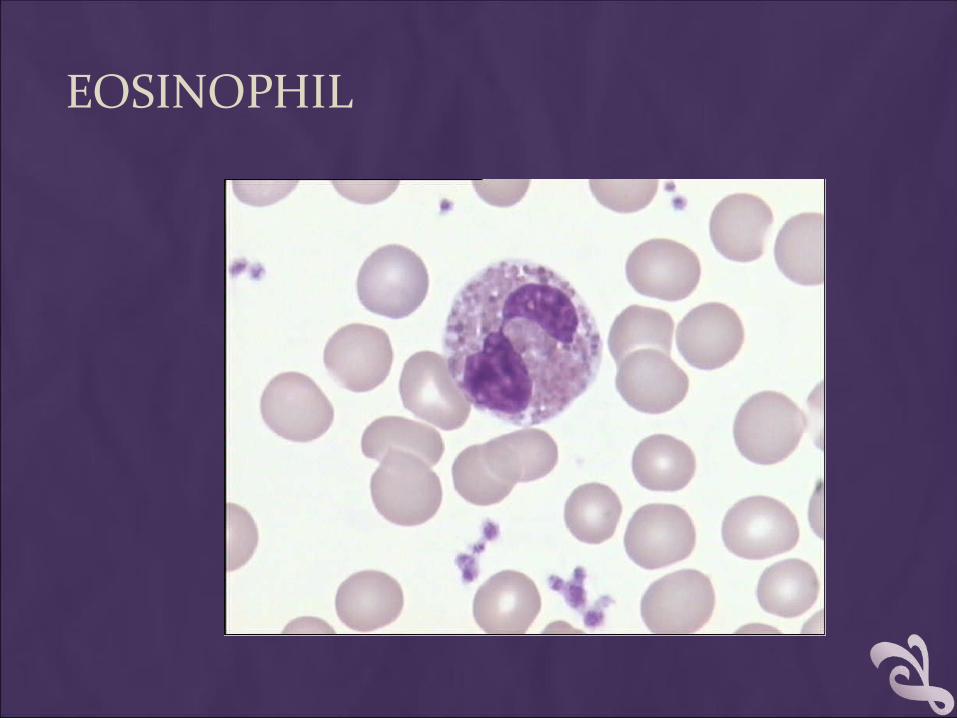
EOSINOPHIL
The most common reasons for an increase in the eosinophil count are
1. Allergic reactions such as hay fever, asthma, or drug hypersensitivity.
2. Parasitic infection
3. Eosinophilic leukemia
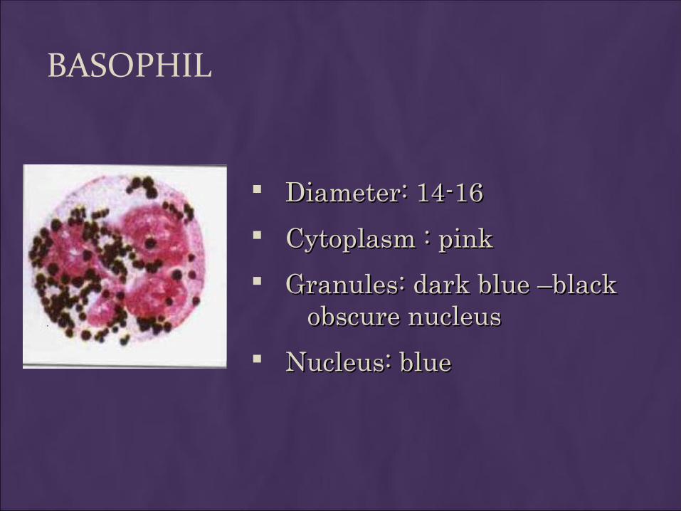
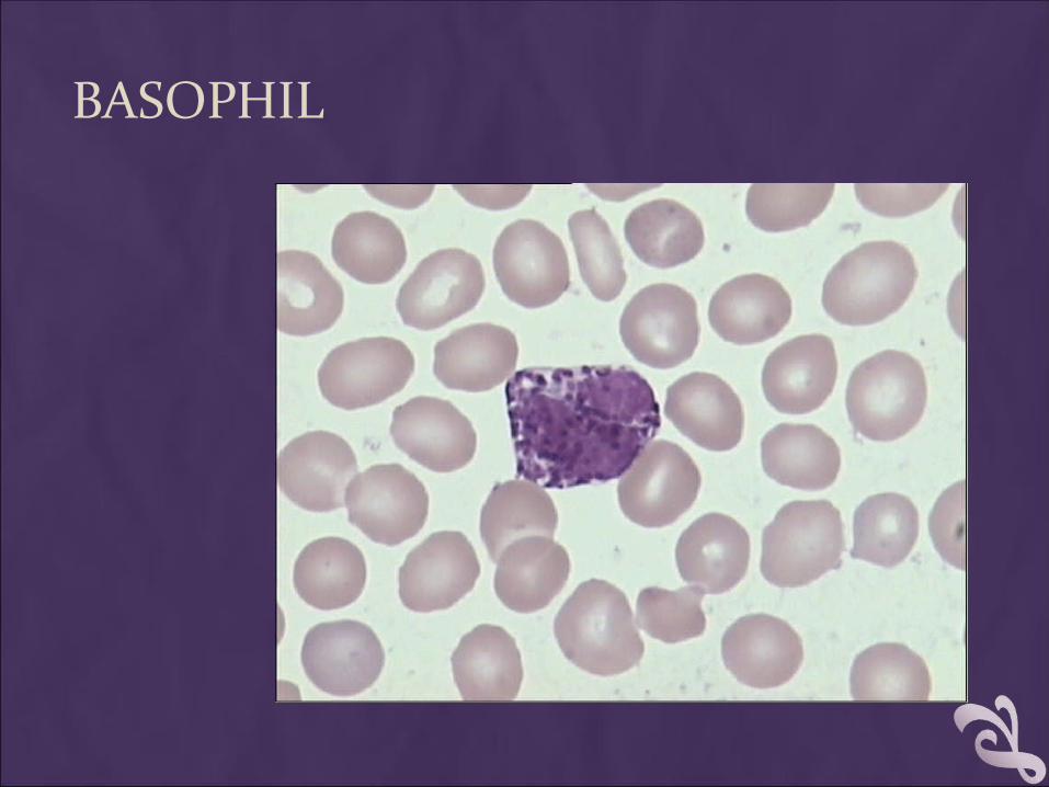
BASOPHIL
Diameter: 14-16Diameter: 14-16
Cytoplasm : pinkCytoplasm : pink
Granules: dark blue –black Granules: dark blue –black obscure nucleus obscure nucleus
Nucleus: blueNucleus: blue
BASOPHIL
Basophils … The purpose of basophils is not completely understood.
… Basophile counts are used to analyze allergic reactions.
… An alteration in bone marrow function such as leukemia may cause an increase in basophils.
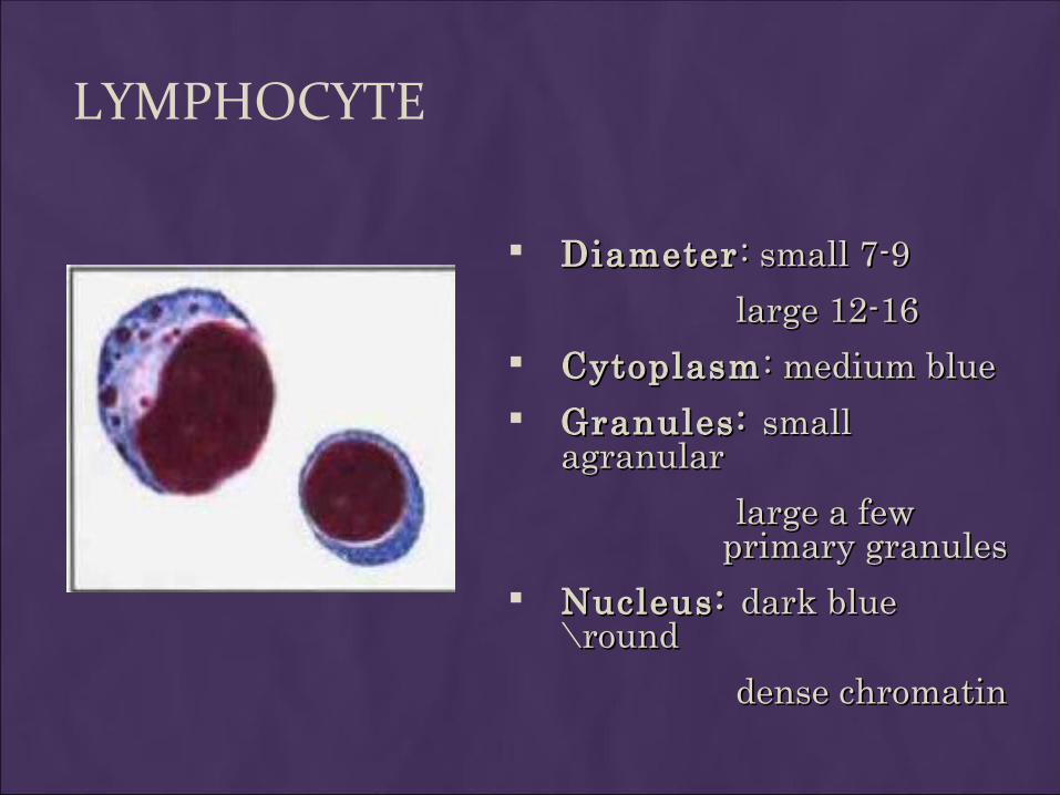
LYMPHOCYTE
DiameterDiameter : small 7-9: small 7-9
large 12-16large 12-16 CytoplasmCytoplasm : medium blue: medium blue Granules: Granules: small small
agranularagranular
large a few large a few primary granules primary granules
Nucleus: Nucleus: dark blue dark blue \round\round
dense chromatindense chromatin
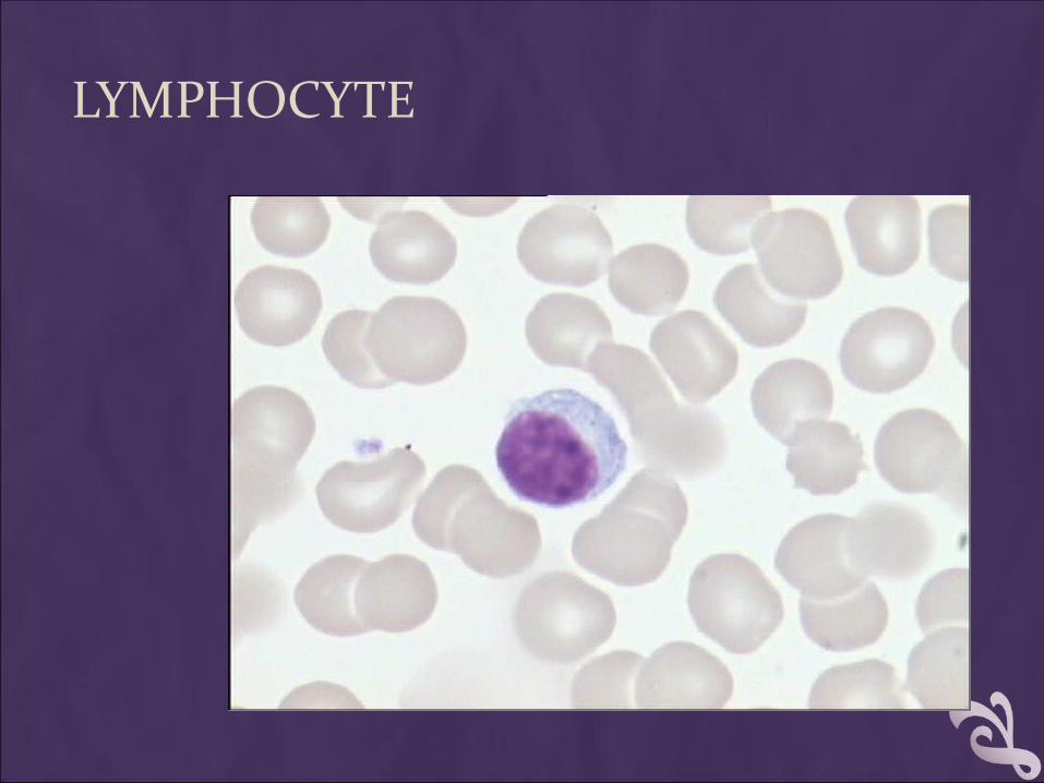
LYMPHOCYTE
4.LYMPHOCYTES
Lymphocytes are the primary components of the body's immune system. They are the source of serum immunoglobulins and of cellular immune response.
Two types of lymphocytes:
1. B lymphocyte : Humoral immunity
2. T lymphocyte : Cellular immunity
Lymphocytes increase (lymphocytosis) in:
1.Many viral infections
2.Tuberculosis.
3.Typhoid fever
4.Lymphocytic leukemia.
A decreased lymphocyte (lymphopenia) count of less than 500 places a patient at very high risk of infection, particularly viral infections.
MONOCYTE
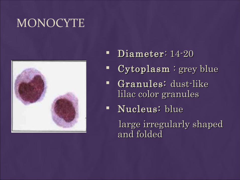
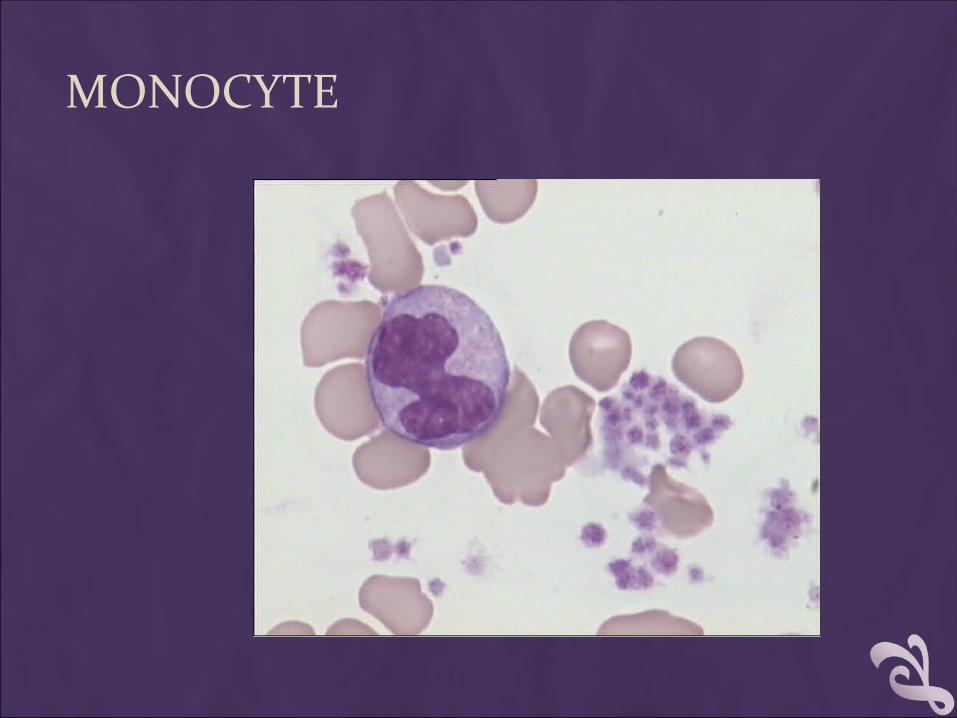
DiameterDiameter : 14-20: 14-20
CytoplasmCytoplasm : grey blue : grey blue
Granules: Granules: dust-like dust-like lilac color granuleslilac color granules
Nucleus: Nucleus: blue blue
large irregularly shaped large irregularly shaped and foldedand folded
MONOCYTE
Diseases that cause a monocytosis include:•Tuberculosis•Brucellosis•Malaria•Monocytic leukemia
NOTESNOTES
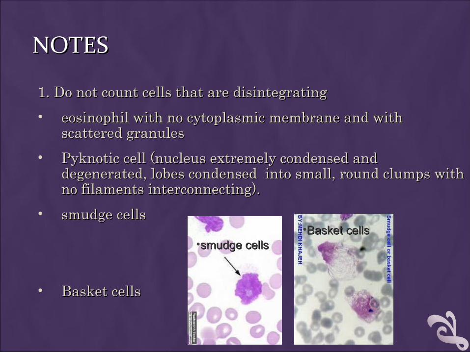
1. Do not count cells that are disintegrating1. Do not count cells that are disintegrating
• eosinophil with no cytoplasmic membrane and with eosinophil with no cytoplasmic membrane and with scattered granulesscattered granules
• Pyknotic cell (nucleus extremely condensed and Pyknotic cell (nucleus extremely condensed and degenerated, lobes condensed into small, round clumps with degenerated, lobes condensed into small, round clumps with no filaments interconnecting).no filaments interconnecting).
• smudge cellssmudge cells
• Basket cellsBasket cells
•smudge cellssmudge cells•Basket cellsBasket cells
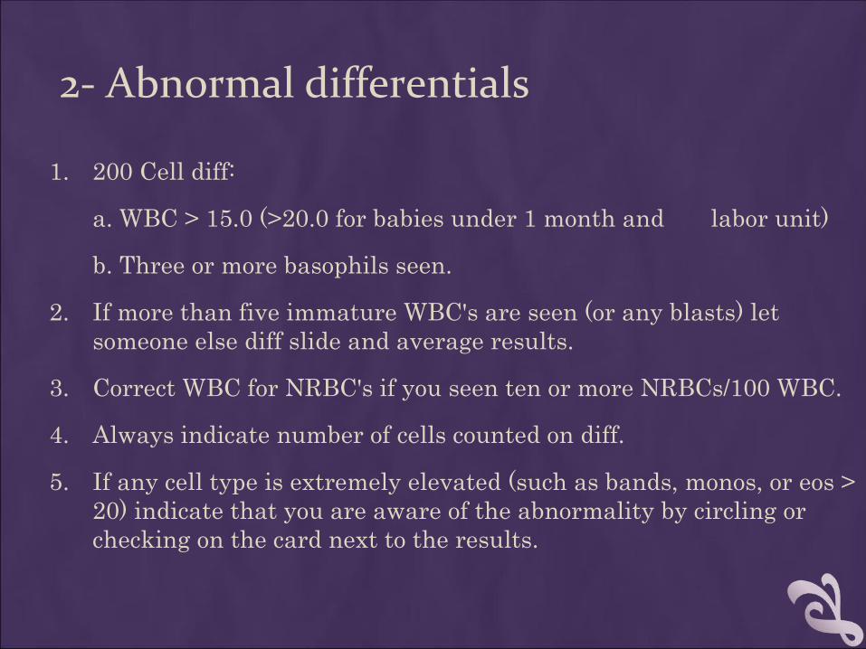
2- Abnormal differentials
1. 200 Cell diff:
a. WBC > 15.0 (>20.0 for babies under 1 month and labor unit)
b. Three or more basophils seen.
2. If more than five immature WBC's are seen (or any blasts) let someone else diff slide and average results.
3. Correct WBC for NRBC's if you seen ten or more NRBCs/100 WBC.
4. Always indicate number of cells counted on diff.
5. If any cell type is extremely elevated (such as bands, monos, or eos > 20) indicate that you are aware of the abnormality by circling or checking on the card next to the results.
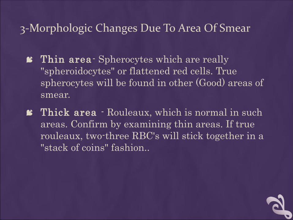
3-Morphologic Changes Due To Area Of Smear
Thin area- Spherocytes which are really "spheroidocytes" or flattened red cells. True spherocytes will be found in other (Good) areas of smear.
Thick area - Rouleaux, which is normal in such areas. Confirm by examining thin areas. If true rouleaux, two-three RBC's will stick together in a "stack of coins" fashion..
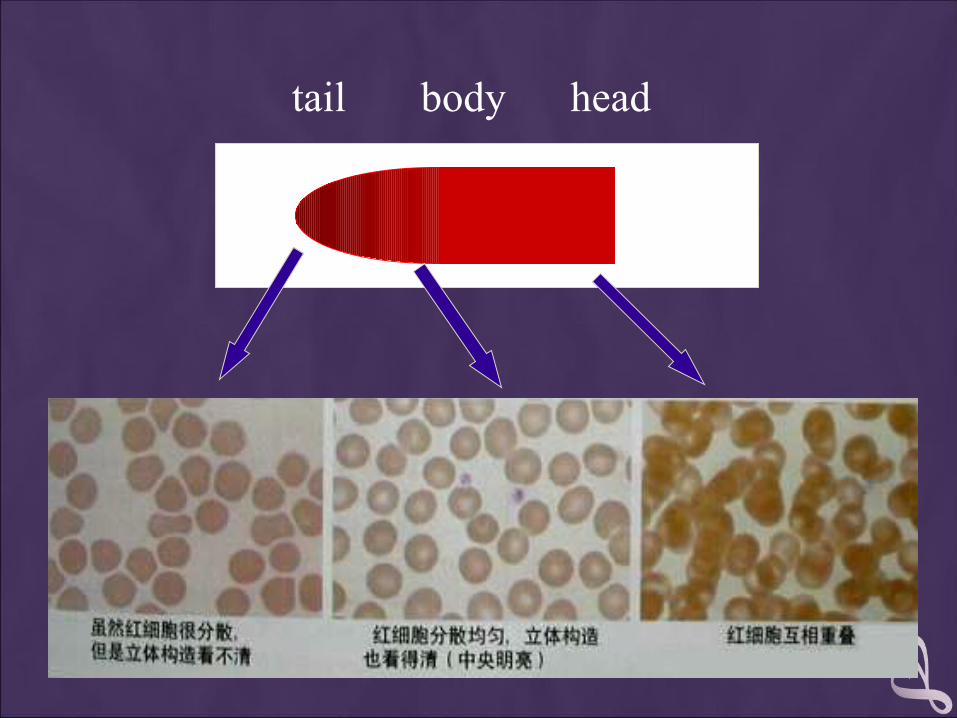
tail body head
4. A well-made and well-stained smear is essential to the accuracy of the differential count. The knowledge and ability of the cell morphologist is critical to high-quality results.
5. Before reporting significant abnormalities such as blasts, malaria or other significant finding on a patient’s differential, ask a more experienced tech to review the smear for confirmation. In clinical settings where a pathologist or hematologist is present, the smear is set aside for Pathologist Review.
6. Never hesitate to ask questions concerning morphology or the identification of cells. The differential is one of the most difficult laboratory tests to learn. In fact, learning about cells and their morphology is a process that continues for as long as you perform differentials.