03 medicina... · bases celulares y moleculares de la enfermedad de alzheimer y otras demencias...
TRANSCRIPT

03[58] María de los Ángeles García Pardo
Mecanismos patológicos en neoplasias hematológicas humanasPathological mechanisms in human hematological neoplasias
[60] José Ignacio Casal Álvarez Proteómica FuncionalFunctional Proteomics
[62] Joaquín Teixidó Calvo Migración Celular en procesos fisiológicos y patologíasCell Migration in Physiology and Disease
[64] Patricio Aller Tresguerres Mecanismos de Acción de Drogas AntitumoralesMechanisms of Action of Antitumour Drugs
[66] José-María Sánchez-Puelles González-Carvajal Farmacología MolecularMolecular Pharmacology
[68] José María Rojo Hernández Activación de Linfocitos TT Lymphocyte Activation
[70] Santiago Rodríguez de Córdoba Patología Molecular / Genética del ComplementoMolecular Pathology / Complement Genetics
[72] Flora de Pablo · Enrique J. de la Rosa Laboratorio 3D: Desarrollo, Diferenciación y Degeneración3D Lab: Development, Differentiation & Degeneration
[74] María Ángeles Martín Requero Bases celulares y moleculares de la enfermedad de Alzheimer y otras demenciasCellular and molecular basis of Alzheimer’s disease and other dementias
[76] Consuelo González Manchón Fisiopatología de trastornos hemostáticosPathophysiology of hemostatic disorders
[78] Carmelo Bernabeu Quirante · Luisa M. Botella Cubells Receptores de TGF-beta en células endotelialesTGF-beta receptors in endothelial cells
[80] José A. García-Sanz Genética del Cáncer y de las Células Madre del CáncerCancer Genetics and Cancer Stem Cells
[82] Faustino Mollinedo García Laboratorio de Muerte Celular y Terapia del CáncerLaboratory of Cell Death and Cancer Therapy
Medicina Celular y MolecularCellular & Molecular Medicine
o v e r v i e w
centro de investigaciones b iológicas
Medicina Celular y MolecularCellular & Molecular Medicine
The main research focus in the Department of Cellular and
Molecular Medicine is the study of the molecular mechanisms
involved in human physiopathology.
Several groups in the Department are characterizing the molecular
bases of various types of cancer, including stem cells, epithelial
neoplasms (melanoma, colon carcinoma) and hematological
malignancies (myeloma, leukemia). The recent incorporation of a
new group to the Department will further reinforce these areas of
research. Other aspects under study are vascular and hemostatic
disorders, complement-related pathologies, regulation of immune T
cells, cell differentiation and mitochondrial transporters. Members
of the Department are also studying the processes involved in
degeneration and aging, aiming to understand the pathological
mechanisms of disorders like retinitis pigmentosa, Lafora or
Alzheimer´s diseases. This research pursuits the identification
of diagnostic markers and target molecules that will help
developing new and efficient therapeutic drugs. The Department
has played a major role in the implementation and development
of high-throughput technologies such as Genomics, Proteomics or
Molecular Pharmacology, now basic tools in Biomedicine and other
research areas in the Centre. A high priority goal is to attract
bright young investigators performing pioneering research in
Biomedicine. The Department also promotes technology transfer
initiatives and translational research through the adequate
protection of the scientific results and reagents produced by the
individual groups. This has already resulted in the generation of
biotechnology spin-offs and represents an added value for the
Department, which will continue supporting these initiatives.
Ángeles García PardoDepartment Head
03
[58]centro de investigaciones b iológicas
Mecanismos patológicos en neoplasias hematológicas humanas
Nuestro principal interés es la caracterización de los mecanismos moleculares implicados en la progresión de dos neoplasias de linfocitos B: la leucemia linfocítica crónica y el mieloma múltiple. Nos hemos centrado en el estudio de moléculas implicadas en adhesión, migra-ción y supervivencia celular, como la integrina _4`1 y la metaloproteinasa de matriz-9. Estudiamos también el papel del estroma en la patología de estas neoplasias.
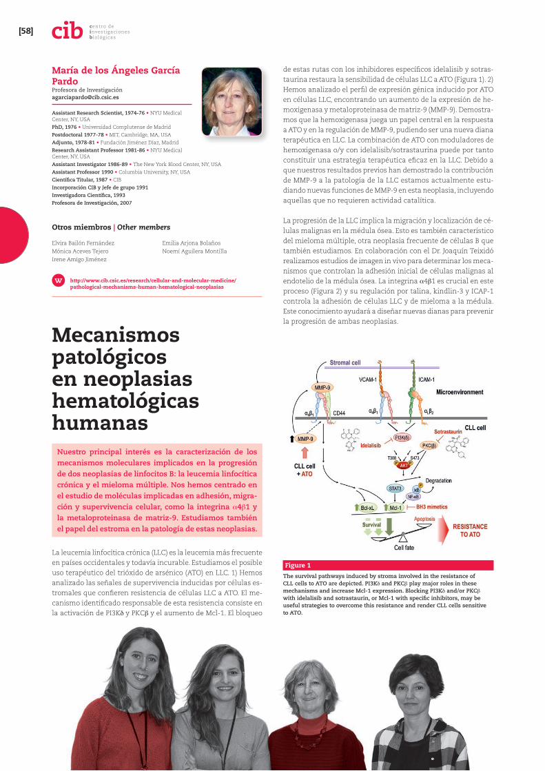
La leucemia linfocítica crónica (LLC) es la leucemia más frecuente en países occidentales y todavía incurable. Estudiamos el posible uso terapéutico del trióxido de arsénico (ATO) en LLC. 1) Hemos analizado las señales de supervivencia inducidas por células es-tromales que confieren resistencia de células LLC a ATO. El me-canismo identificado responsable de esta resistencia consiste en la activación de PI3Kb y PKC` y el aumento de Mcl-1. El bloqueo
de estas rutas con los inhibidores específicos idelalisib y sotras-taurina restaura la sensibilidad de células LLC a ATO (Figura 1). 2) Hemos analizado el perfil de expresión génica inducido por ATO en células LLC, encontrando un aumento de la expresión de he-moxigenasa y metaloproteinasa de matriz-9 (MMP-9). Demostra-mos que la hemoxigenasa juega un papel central en la respuesta a ATO y en la regulación de MMP-9, pudiendo ser una nueva diana terapéutica en LLC. La combinación de ATO con moduladores de hemoxigenasa o/y con idelalisib/sotrastaurina puede por tanto constituir una estrategia terapéutica eficaz en la LLC. Debido a que nuestros resultados previos han demostrado la contribución de MMP-9 a la patología de la LLC estamos actualmente estu-diando nuevas funciones de MMP-9 en esta neoplasia, incluyendo aquellas que no requieren actividad catalítica.
La progresión de la LLC implica la migración y localización de cé-lulas malignas en la médula ósea. Esto es también característico del mieloma múltiple, otra neoplasia frecuente de células B que también estudiamos. En colaboración con el Dr. Joaquín Teixidó realizamos estudios de imagen in vivo para determinar los meca-nismos que controlan la adhesión inicial de células malignas al endotelio de la médula ósea. La integrina _4`1 es crucial en este proceso (Figura 2) y su regulación por talina, kindlin-3 y ICAP-1 controla la adhesión de células LLC y de mieloma a la médula. Este conocimiento ayudará a diseñar nuevas dianas para prevenir la progresión de ambas neoplasias.
Figure 1
The survival pathways induced by stroma involved in the resistance of CLL cells to ATO are depicted. PI3Kb and PKC` play major roles in these mechanisms and increase Mcl-1 expression. Blocking PI3Kb and/or PKC` with idelalisib and sotrastaurin, or Mcl-1 with specific inhibitors, may be useful strategies to overcome this resistance and render CLL cells sensitive to ATO.
María de los Ángeles García Pardo Profesora de Investigació[email protected]
Assistant Research Scientist, 1974-76 • NYU Medical Center, NY, USAPhD, 1976 • Universidad Complutense de MadridPostdoctoral 1977-78 • MIT, Cambridge, MA, USAAdjunto, 1978-81 • Fundación Jiménez Díaz, MadridResearch Assistant Professor 1981-86 • NYU Medical Center, NY, USAAssistant Investigator 1986-89 • The New York Blood Center, NY, USAAssistant Professor 1990 • Columbia University, NY, USACientífica Titular, 1987 • CIBIncorporación CIB y Jefe de grupo 1991Investigadora Científica, 1993Profesora de Investigación, 2007
Otros miembros | Other members
Elvira Bailón FernándezMónica Aceves TejeroIrene Amigo Jiménez
Emilia Arjona BolañosNoemí Aguilera Montilla
http://www.cib.csic.es/research/cellular-and-molecular-medicine/pathological-mechanisms-human-hematological-neoplasias
Medicina Celular y MolecularCellular & Molecular Medicine [59]
Pathological mechanisms in human hematological neoplasias
Our major interest is the characterization of the molecular mechanisms accounting for the progression of two B-lymphocyte neoplasias: chronic lymphocytic leukemia and multiple myeloma. We focus on molecules involved in cell adhesion, migration and survival, including _4`1 integrin and matrix metalloproteinase-9. We are also studying the role of the stroma in the pathology of these malignancies.
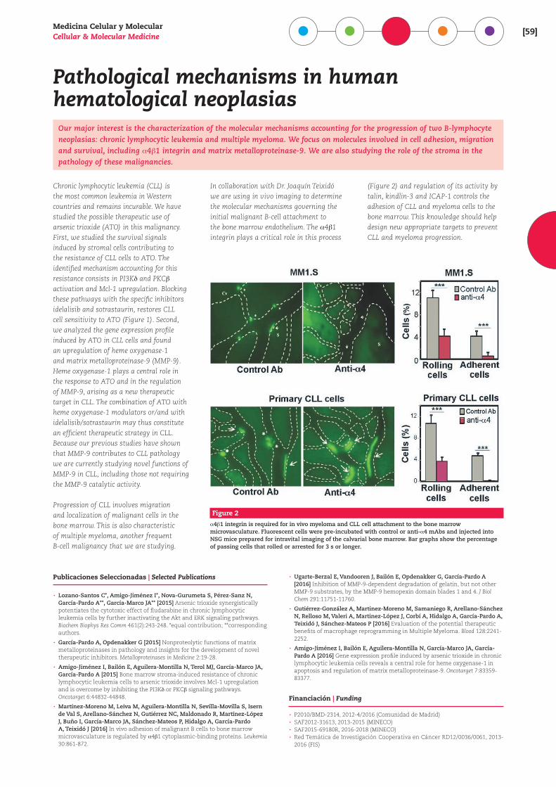
Chronic lymphocytic leukemia (CLL) is the most common leukemia in Western countries and remains incurable. We have studied the possible therapeutic use of arsenic trioxide (ATO) in this malignancy. First, we studied the survival signals induced by stromal cells contributing to the resistance of CLL cells to ATO. The identified mechanism accounting for this resistance consists in PI3Kb and PKC` activation and Mcl-1 upregulation. Blocking these pathways with the specific inhibitors idelalisib and sotrastaurin, restores CLL cell sensitivity to ATO (Figure 1). Second, we analyzed the gene expression profile induced by ATO in CLL cells and found an upregulation of heme oxygenase-1 and matrix metalloproteinase-9 (MMP-9). Heme oxygenase-1 plays a central role in the response to ATO and in the regulation of MMP-9, arising as a new therapeutic target in CLL. The combination of ATO with heme oxygenase-1 modulators or/and with idelalisib/sotrastaurin may thus constitute an efficient therapeutic strategy in CLL. Because our previous studies have shown that MMP-9 contributes to CLL pathology we are currently studying novel functions of MMP-9 in CLL, including those not requiring the MMP-9 catalytic activity.
Progression of CLL involves migration and localization of malignant cells in the bone marrow. This is also characteristic of multiple myeloma, another frequent B-cell malignancy that we are studying.
In collaboration with Dr. Joaquín Teixidó we are using in vivo imaging to determine the molecular mechanisms governing the initial malignant B-cell attachment to the bone marrow endothelium. The _4`1 integrin plays a critical role in this process
(Figure 2) and regulation of its activity by talin, kindlin-3 and ICAP-1 controls the adhesion of CLL and myeloma cells to the bone marrow. This knowledge should help design new appropriate targets to prevent CLL and myeloma progression.
Publicaciones Seleccionadas | Selected Publications
· Lozano-Santos C*, Amigo-Jiménez I*, Nova-Gurumeta S, Pérez-Sanz N, García-Pardo A**, García-Marco JA** [2015] Arsenic trioxide synergistically potentiates the cytotoxic effect of fludarabine in chronic lymphocytic leukemia cells by further inactivating the Akt and ERK signaling pathways. Biochem Biophys Res Comm 461(2):243-248. *equal contribution; **corresponding authors.
· García-Pardo A, Opdenakker G [2015] Nonproteolytic functions of matrix metalloproteinases in pathology and insights for the development of novel therapeutic inhibitors. Metalloproteinases in Medicine 2:19-28.
· Amigo-Jiménez I, Bailón E, Aguilera-Montilla N, Terol MJ, García-Marco JA, García-Pardo A [2015] Bone marrow stroma-induced resistance of chronic lymphocytic leukemia cells to arsenic trioxide involves Mcl-1 upregulation and is overcome by inhibiting the PI3Kb or PKC` signaling pathways. Oncotarget 6:44832-44848.
· Martínez-Moreno M, Leiva M, Aguilera-Montilla N, Sevilla-Movilla S, Isern de Val S, Arellano-Sánchez N, Gutiérrez NC, Maldonado R, Martínez-López J, Buño I, García-Marco JA, Sánchez-Mateos P, Hidalgo A, García-Pardo A, Teixidó J [2016] In vivo adhesion of malignant B cells to bone marrow microvasculature is regulated by _4`1 cytoplasmic-binding proteins. Leukemia 30:861-872.
· Ugarte-Berzal E, Vandooren J, Bailón E, Opdenakker G, García-Pardo A [2016] Inhibition of MMP-9-dependent degradation of gelatin, but not other MMP-9 substrates, by the MMP-9 hemopexin domain blades 1 and 4. J Biol Chem 291:11751-11760.
· Gutiérrez-González A, Martinez-Moreno M, Samaniego R, Arellano-Sánchez N, Relloso M, Valeri A, Martinez-López J, Corbí A, Hidalgo A, García-Pardo A, Teixidó J, Sánchez-Mateos P [2016] Evaluation of the potential therapeutic benefits of macrophage reprogramming in Multiple Myeloma. Blood 128:2241-2252.
· Amigo-Jiménez I, Bailón E, Aguilera-Montilla N, García-Marco JA, García-Pardo A [2016] Gene expression profile induced by arsenic trioxide in chronic lymphocytic leukemia cells reveals a central role for heme oxygenase-1 in apoptosis and regulation of matrix metalloproteinase-9. Oncotarget 7:83359-83377.
Financiación | Funding
· P2010/BMD-2314, 2012-4/2016 (Comunidad de Madrid) · SAF2012-31613, 2013-2015 (MINECO) · SAF2015-69180R, 2016-2018 (MINECO) · Red Temática de Investigación Cooperativa en Cáncer RD12/0036/0061, 2013-2016 (FIS)
Figure 2
_4`1 integrin is required for in vivo myeloma and CLL cell attachment to the bone marrow microvasculature. Fluorescent cells were pre-incubated with control or anti-_4 mAbs and injected into NSG mice prepared for intravital imaging of the calvarial bone marrow. Bar graphs show the percentage of passing cells that rolled or arrested for 3 s or longer.
[60]centro de investigaciones b iológicas
Proteómica Funcional
Nuestro grupo estudia los mecanismos moleculares im-plicados en la metástasis de cáncer colorrectal y otros tumores para el diseño de nuevas terapias, la transición epitelio-mesénquima en tumores y la identificación de biomarcadores diagnósticos y pronósticos de valor clí-nico. El laboratorio participa en una activa transferencia tecnológica, incluida la plataforma Proteored (ISCIII).
Nuestro grupo estudia diversos aspectos de la biología del cán-cer, con especial énfasis en la diseminación metastática, el papel del estroma en progresión, el diagnóstico temprano y el pronóstico.
Mecanismos moleculares implicados en la metástasis de cáncer colorrectal y otros tumores. Hemos identificado varias molécu-las, entre otras IL13R_2 y CDH17, que juegan un papel clave en adhesión, migración y supervivencia celular, y hemos investigado la regulación por miRNAs. En IL13R_2 hemos demostrado tanto su capacidad para señalización como las moléculas mediadoras. La CDH17 juega un papel clave en la colonización hepática y participa en la activación de integrinas utilizando un motivo RGD, presente también en otras cadherinas, como la VE-cadherina. El bloqueo de la unión RGD-integrina 1 presenta una clara utilidad
terapéutica no solo en cáncer colorrectal, sino en mama, mela-noma y otros tumores.
El proceso de invasión tumoral va acompañado por cambios en la morfología y fenotipo celular conocidos como transición epitelio-mesénquima (EMT). Estamos estudiando las alteracio-nes proteómicas inducidas por factores de transcripción claves en la regulación de la EMT como SNAIL, TWIST o LOXL2, tanto en células tumorales como estromales. Hemos demostrado la capacidad de TWIST para inducir actividad pro-metastática en fibroblastos del estroma tumoral y los mecanismos moleculares de inhibición de la adipogénesis por SNAIL. Por otro lado, la ca-racterización del estroma tumoral nos ha permitido descubrir diversos marcadores estromales de valor pronóstico y predictivos de supervivencia como LOXL2. LOXL2 puede ser utilizado para la estratificación de pacientes de cáncer colorrectal y el diseño de terapias más agresivas.
A partir de los antígenos asociados a tumor previamente identifi-cados, nuestro grupo ha desarrollado diferentes inmunoensayos para el diagnóstico temprano del cáncer de colon. Los resultados han sido patentados y licenciados a empresas interesadas.
José Ignacio Casal Álvarez Investigador Cientí[email protected]
PhD, 1984 • Centro de Biología Molecular, CSIC, Universidad Autónoma de MadridPost-doctoral, 1985-1986 • Massachusetts Institute of Technology (MIT), USAJefe de Proyecto, 1987-1997Director de Investigación, 1997-2001 • INGENASADirector Programa de Biotecnología, 2001-2008 • CNIOInvestigador Científico, 2007 • CSICIncorporación, 2008 • CIB
Otros miembros | Other members
Rubén A. Bartolomé CondeSofía Torres MorianoIrene García Palmero
Beatriz Escudero PaniaguaConsuelo Marín VicenteEva Calviño Vanegas
http://www.cib.csic.es/research/cellular-and-molecular-medicine/functional-proteomics
Financiación | Funding
· BIO2012-31023 (MINECO). · S2011/BMD-2344/ Colomics2. Comunidad de Madrid. · BIO2015-66489-R (MINECO). · PRB2 (IPT13/0001-ISCIII-SGEFI/FEDER). Instituto de Salud Carlos III. · Ayudas de la Asociación Española contra el Cáncer a grupos estables. · RTC-2014-1518-1 (MINECO) · CSIC13-4E-1955 (MINECO)
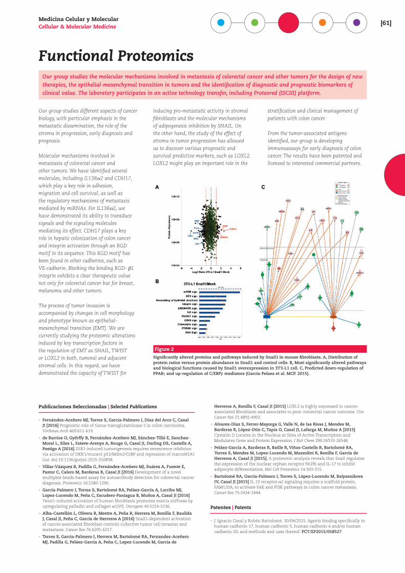
Figure 1
Summary of Twist1 pro-metastatic activities in human activated fibroblasts. Twist1 promotes fibroblast activation through palladin and/or collagen _1(VI) expression, triggering hyperproliferation, an increased ability for migration and invasion, alignment of the extracellular matrix and cytoskeleton reorganization (García-Palmero et al., Oncogene. 2016).
Medicina Celular y MolecularCellular & Molecular Medicine [61]
Functional ProteomicsOur group studies the molecular mechanisms involved in metastasis of colorectal cancer and other tumors for the design of new therapies, the epithelial-mesenchymal transition in tumors and the identification of diagnostic and prognostic biomarkers of clinical value. The laboratory participates in an active technology transfer, including Proteored (ISCIII) platform.
Our group studies different aspects of cancer biology, with particular emphasis in the metastatic dissemination, the role of the stroma in progression, early diagnosis and prognosis.
Molecular mechanisms involved in metastasis of colorectal cancer and other tumors. We have identified several molecules, including IL13R_2 and CDH17, which play a key role in adhesion, migration and cell survival, as well as the regulatory mechanisms of metastasis mediated by miRNAs. For IL13R_2, we have demonstrated its ability to transduce signals and the signaling molecules mediating its effect. CDH17 plays a key role in hepatic colonization of colon cancer and integrin activation through an RGD motif in its sequence. This RGD motif has been found in other cadherins, such as VE-cadherin. Blocking the binding RGD- `1 integrin exhibits a clear therapeutic value not only for colorectal cancer but for breast, melanoma and other tumors.
The process of tumor invasion is accompanied by changes in cell morphology and phenotype known as epithelial-mesenchymal transition (EMT). We are currently studying the proteomic alterations induced by key transcription factors in the regulation of EMT as SNAIL, TWIST or LOXL2 in both, tumoral and adjacent stromal cells. In this regard, we have demonstrated the capacity of TWIST for
inducing pro-metastatic activity in stromal fibroblasts and the molecular mechanisms of adipogenesis inhibition by SNAIL. On the other hand, the study of the effect of stroma in tumor progression has allowed us to discover various prognostic and survival predictive markers, such as LOXL2. LOXL2 might play an important role in the
stratification and clinical management of patients with colon cancer.
From the tumor-associated antigens identified, our group is developing immunoassays for early diagnosis of colon cancer. The results have been patented and licensed to interested commercial partners.
Publicaciones Seleccionadas | Selected Publications
· Fernández-Aceñero MJ, Torres S, Garcia-Palmero I, Díaz del Arco C, Casal JI [2016] Prognostic role of tissue transglutaminase 2 in colon carcinoma. Virchows Arch 469:611-619.
· de Barrios O, Gyorffy B, Fernández-Aceñero MJ, Sánchez-Tilló E, Sanchez-Moral L, Siles L, Esteve-Arenys A, Rouge G, Casal JI, Darling DS, Castells A, Postigo A [2016] ZEB1-induced tumorigenesis requires senescence inhibtion via activation of DKK1/mutant p53/Mdm2/CtBP and repression of macroH2A1 Gut. doi:10.1136/gutjnl-2015-310838.
· Villar-Vázquez R, Padilla G, Fernández-Aceñero MJ, Suárez A, Fuente E, Pastor C, Calero M, Barderas R, Casal JI [2016] Development of a novel multiplex beads-based assay for autoantibody detection for colorectal cancer diagnosis. Proteomics 16:1280-1290.
· García-Palmero I, Torres S, Bartolomé RA, Peláez-García A, Larriba MJ, Lopez-Lucendo M, Peña C, Escudero-Paniagua B, Muñoz A, Casal JI [2016] Twist1-induced activation of human fibroblasts promotes matrix stiffness by upregulating palladin and collagen _1(VI). Oncogene 40:5224-5236.
· Alba-Castellón L, Olivera R, Mestre A, Peña R, Herrera M, Bonilla F, Baulida J, Casal JI, Peña C, García de Herreros A [2016] Snail1-dependent activation of cancer-associated fibroblast controls collective tumor cell invasion and metastasis. Cancer Res 76:6205-6217.
· Torres S, Garcia-Palmero I, Herrera M, Bartolomé RA, Fernandez-Aceñero MJ, Padilla G, Peláez-García A, Peña C, Lopez-Lucendo M, Garcia de
Herreros A, Bonilla F, Casal JI [2015] LOXL2 is highly expressed in cancer-associated fibroblasts and associates to poor colorectal cancer outcome. Clin Cancer Res 21:4892-4902.
· Alvarez-Díaz S, Ferrer-Mayorga G, Valle N, de las Rivas J, Mendes M, Barderas R, López-Otín C, Tapia O, Casal JI, Lafarga M, Muñoz A [2015] Cystatin D Locates in the Nucleus at Sites of Active Transcription and Modulates Gene and Protein Expression. J Biol Chem 290:26533-26548.
· Peláez-García A, Barderas R, Batlle R, Viñas-Castells R, Bartolomé RA, Torres S, Mendes M, Lopez-Lucendo M, Mazzolini R, Bonilla F, García de Herreros A, Casal JI [2015]. A proteomic analysis reveals that Snail regulates the expression of the nuclear orphan receptor Nr2f6 and IL-17 to inhibit adipocyte differentiation. Mol Cell Proteomics 14:303-315.
· Bartolomé RA, García-Palmero I, Torres S, López-Lucendo M, Balyasnikova IV, Casal JI [2015] IL-13 receptor _2 signaling requires a scaffold protein, FAM120A, to activate FAK and PI3K pathways in colon cancer metastasis. Cancer Res 75:2434-2444.
Patentes | Patents
· J. Ignacio Casal y Rubén Bartolomé. 30/04/2015. Agents binding specifically to human cadherin-17, human cadherin-5, human cadherin-6 and/or human cadherin-20, and methods and uses thereof. PCT/EP2015/058527.
Figure 2
Significantly altered proteins and pathways induced by Snail1 in mouse fibroblasts. A, Distribution of protein ratios versus protein abundance in Snail1 and control cells. B, Most significantly altered pathways and biological functions caused by Snail1 overexpression in 3T3-L1 cell. C, Predicted down-regulation of PPARa and up-regulation of C/EBP` mediators (García-Pelaez et al. MCP. 2015).
[62]centro de investigaciones b iológicas
Migración Celular en procesos fisiológicos y patologías
Una parte importante de nuestros estudios está enfocada a la caracterización de mecanismos moleculares implicados en la regulación de la migración de linfocitos y de células tumorales. Por otra parte, estamos identificando mecanismos de resistencia a quimioterapia de células de melanoma y de mieloma múltiple, y estudiando el papel de microRNAs en dicha resistencia.
La estimulación por quimiocinas de la adhesión linfocitaria mediada por la in-tegrina VLA-4 es un paso crucial durante el tráfico de linfocitos a sitios de inflama-ción. Tras la unión quimiocinas/receptor se genera una señalización inside-out que incide en los dominios citoplásmicos de las integrinas. Estamos caracterizando las moléculas inside-out necesarias que regulan la adhesión linfocitaria depen-diente de VLA-4, lo que mejorará nuestro conocimiento de mecanismos implicados en el tráfico de estas células. Las células de melanoma son altamente invasivas y muestran un notable potencial metastá-sico. Mutaciones prevalentes en melano-ma incluyen B-Raf V600E y N-Ras Q61K, lo que conduce a hiperactivación de la MAP quinasa Erk1/2. Nuevas terapias dirigidas a la vía Ras-Raf-MEK-Erk1/2, como vemu-rafenib, trametinib e inhibidores de Erk1/2 han mejorado la supervivencia en mela-noma, aunque respuestas de resistencia son comunes. Estamos caracterizando funcionalmente los miRNAs alterados en células de melanoma resistentes a vemu-rafenib y trametinib. Asimismo estamos identificando mecanismos moleculares
implicados en resistencia de células de melanomas a inhibidores de Erk1/2. El mieloma múltiple (MM) es una neoplasia de células B caracterizada por el tráfico y la acumulación de células malignas en la médula ósea (MO). Las células de MM uti-lizan VLA-4 para alojarse en la MO, lo que contribuye a la progresión de la enferme-
dad. La expresión y función de miRNAs en MM específicamente alteradas por la ad-hesión celular mediada por VLA-4 podría proporcionar pistas importantes sobre la progresión del MM. Finalmente, estamos caracterizando relaciones funcionales entre resistencia de células de MM a bor-tezomib y expresión y función de VLA-4.
Joaquín Teixidó Calvo Profesor de Investigació[email protected]
PhD, 1985 • Max Plank Institute für Molekulare Genetik, Berlin, and Centro de Biología Molecular, Universidad Autónoma de MadridPostdoctoral, 1986-1992 • University of Massachusetts, Dana Farber Cancer Institute, Boston, USA, Hospital de La Princesa (Madrid)Científico Titular, 1992 • CSICIncorporación y Jefe de Grupo, 1994 • CIBInvestigador Científico, 2003 • CIBProfesor de Investigación, 2007 • CIB
http://www.cib.csic.es/es/departamentos/medicina-celular-y-molecular/quimioquinas-y-migracion-celular
Otros miembros | Other members
Nohemí Arellano SánchezLucía Benito JardónMarta Díaz Martínez
Soledad Isern de ValMónica Moreno MartínezSilvia Sevilla Movilla
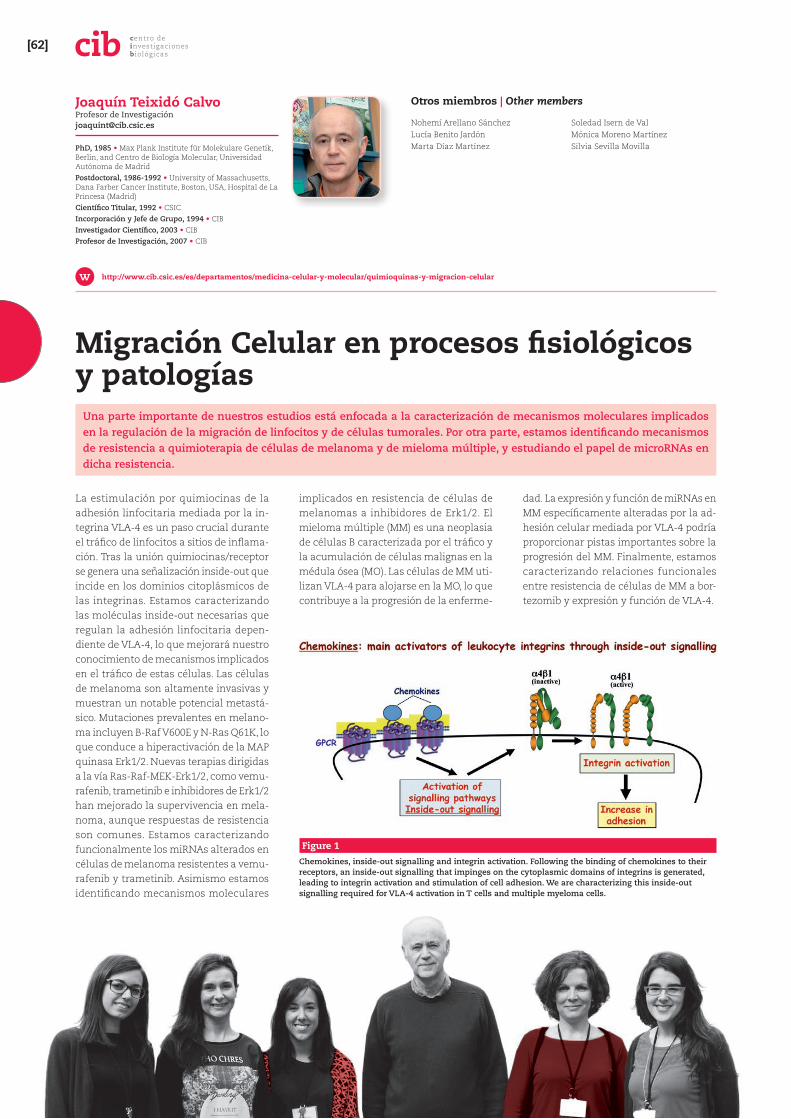
Figure 1
Chemokines, inside-out signalling and integrin activation. Following the binding of chemokines to their receptors, an inside-out signalling that impinges on the cytoplasmic domains of integrins is generated, leading to integrin activation and stimulation of cell adhesion. We are characterizing this inside-out signalling required for VLA-4 activation in T cells and multiple myeloma cells.
Medicina Celular y MolecularCellular & Molecular Medicine [63]
Cell Migration in Physiology and DiseaseAn important part of our studies is focused on the characterization of molecular mechanisms involved in the regulation of lymphocyte and tumor cell migration. On the other hand, we are identifying mechanisms of resistance to chemotherapy of melanoma and multiple myeloma cells, and studying the role of microRNAs in such resistance.
Chemokine stimulation of lymphocyte adhesion mediated by the integrin VLA-4 is a crucial step during trafficking of lymphocytes to sites of inflammation. Following chemokine/receptor binding, an inside-out signaling is generated that impinges on the cytoplasmic domains of the integrins. We are characterizing the required inside-out molecules that regulate VLA-4-dependent lymphocyte adhesion, which will improve our understanding of mechanisms involved in the trafficking of these cells. Melanoma cells are highly invasive and show remarkable metastatic
potential. Mutations prevalent in melanoma include B-Raf V600E and N-Ras Q61K, which lead to hyperactivation of the MAP kinase Erk1 / 2 pathway. New therapies targeting the Ras-Raf-MEK-Erk1 / 2 pathway, such as vemurafenib, trametinib and Erk1 / 2 inhibitors have improved survival in melanoma, although resistance responses are common. We are functionally characterizing altered miRNAs in melanoma cells resistant to vemurafenib and trametinib. We are also identifying molecular mechanisms involved in melanoma cell resistance to Erk1 / 2
inhibitors. Multiple myeloma (MM) is a B cell neoplasm characterized by the trafficking and accumulation of malignant cells in the bone marrow (BM). MM cells use VLA-4 to home to the BM, which contributes to disease progression. The expression and function of miRNAs in MM specifically altered by VLA-4 mediated cell adhesion could provide important clues about MM progression. Finally, we are characterizing functional relationships between MM cell resistance to bortezomib and VLA-4 expression and function.
Publicaciones Seleccionadas | Selected Publications
· Dios-Esponera A, Isern de Val S, Sevilla-Movilla S, García-Verdugo R, García Bernal D, Arellano-Sánchez N, Cabañas C, Teixidó J [2015] Positive and negative regulation by SLP-76/ADAP and Pyk2 of chemokine-stimulated T lymphocyte adhesion mediated by integrin _4`1. Mol Biol Cell 26:3215-3218.
· Martínez-Moreno M, Leiva M, Sevilla-Movilla S, Aguilera-Montilla N, Arellano-Sánchez N, Gutiérrez NC, Maldonado R, Martínez-López J, Buño I, García-Marco JA, Sánchez-Mateos P, Hidalgo A, García-Pardo A, Teixidó J [2016] In vivo adhesion of malignant B cells to bone marrow microvasculature is regulated by _4`1 cytoplasmic-binding proteins. Leukemia 30:861-872.
· Gutiérrez-González A, Martinez-Moreno M, Samaniego R, Relloso M, Martinez-López J, Corbí A, Hidalgo A, García-Pardo A, Teixidó J, Sánchez-Mateos P [2016] Evaluation of the potential therapeutic benefits of macrophage reprogramming in multiple myeloma. Blood 128:2241-2252.
· Sosa-Costa A, Isern de Val S, Sevilla-Movilla S, Borgman KJE, Manzo C. Teixidó J**, Garcia-Parajo MF** [2016] Lateral Mobility and Nanoscale Spatial Arrangement of Chemokine-activated _4`1 Integrins on T Cells. J Biol Chem 291:21053-62 **Equal contribution.
· Bartolomé RA, Torres S, Isern de Val S, Escudero-Paniagua B, Calviño E, Teixidó J, Casal JI [2016] VE-cadherin RGD motifs promote metastasis and constitute a potential therapeutic target in melanoma and breast cancers. Oncotarget. doi: 10.18632/oncotarget.13832.
Financiación | Funding
· SAF2014-53059-R · S2010/BMD-2314. Biomedicina. Comunidad de Madrid · Red Temática Investigación Cooperativa en Cáncer. RD12/0036/0061 Ministerio de Sanidad y Consumo
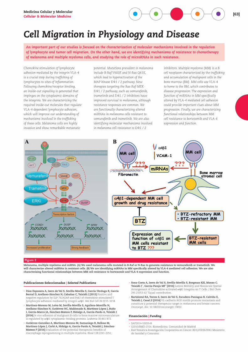
Figure 2
Melanoma, multiple myeloma and miRNA. (A) We used melanoma cells mutated in B-Raf or N-Ras to generate resistance to vemurafenib or trametinib. We will characterize altered miRNAs in resistant cells. (B) We are identifying miRNAs in MM specifically altered by VLA-4 mediated cell adhesion. We are also characterizing functional relationships between MM cell resistance to bortezomib and VLA-4 expression and function.
[64]centro de investigaciones b iológicas
Mecanismos de Acción de Drogas Antitumorales
En este bienio hemos: (1) Caracterizado algunos me-canismos que pueden explicar la generación de re-sistencia a apoptosis en líneas celulares leucémicas útiles en investigación farmacológica. (2) Demostra-do la capacidad y mecanismo por los que polifenoles dietéticos naturales mejoran la eficacia apoptótica de inhibidores metabólicos (de la glucolisis y metabolis-mo de ácidos grasos) potencialmente útiles en terapia anti-tumoral.
1. La utilidad de las líneas celulares establecidas en la investi-gación clínico/farmacológica puede verse comprometida por el desarrollo de alteraciones que generan resistencia. En cola-boración con la Universidad de Alcalá (Dra. Boyano-Adánez), y usando la línea de leucemia promielocítica aguda humana
NB4 como modelo, hemos detectado: (a) Mutación puntual “hot-spot” en el gen p53 (frecuente en líneas celulares leucémi-cas), que reduce su actividad transcripcional, e incrementa su interacción a nivel mitocondrial con acción anti-apoptótica. (b) Notable actividad autofágica basal, que se opone a la respuesta apoptótica. (c) Fácil activación de la respuesta anti-oxidante mediada por Nrf2, que reduce la producción de ROS como factor pro-apoptótico.
2. Algunos inhibidores de la glucolisis (2-desoxi-D-glucosa [2-DG], y lonidamina [Lon]) y del metabolismo de ácidos grasos (etomoxir, orlistat) son potencialmente útiles en terapia an-ti-tumoral, pero su eficacia es por sí escasa. Usando modelos de células leucémicas mieloides humanas hemos demostrado que: (a) La acción apoptótica de estos agentes es fuertemente potenciada por co-tratamiento con dosis bajas de polifenoles naturales (quercetina, genisteína, curcumina). (b) La poten-ciación de apoptosis no es adecuadamente explicable por es-trés oxidativo (producción de ROS, depleción de GSH), ni por alteraciones en ATP intracelular. (c) Los polifenoles reducen la activación de quinasas defensivas (Akt y ocasionalmente ERK) por 2-DG y Lon, cuya importancia para la apoptosis se corrobora usando inhibidores farmacológicos, mientras que la respuesta de otras quinasas es irrelevante o dispar. En línea con previos estudios nuestros y de otros laboratorios, estos resultados confirman el interés de polifenoles dietéticos como co-adyuvantes en terapias anti-tumorales. A falta de culminar algunos estudios teóricos, este trabajo pone fin a nuestros programas de investigación y al propio laboratorio.
Publicaciones Seleccionadas | Selected Publications
· Gañán-Gómez I, Estañ-Omaña MC, Sancho P, Aller P, Boyano-Adánez MC [2015] Mechanisms of resistance to apoptosis in the human acute promyelocytic leukemia cell line NB4. Ann Hematol 94:379-392.
· De Blas E, Estañ MC, Gómez de Frutos MC, Ramos J, Boyano-Adánez MC, Aller P [2016] Selected polyphenols potentiate the apoptotic efficacy of glycolytic inhibitors in human acute myeloid leukemia cell lines. Regulation by protein kinase activities. Cancer Cell Int 16:70.
Financiación | Funding
· SAF2010-20256 (MINECO)
Patricio Aller Tresguerres Profesor de Investigació[email protected]
PhD, 1979 • Universidad Complutense de MadridPostdoctoral, 1983-1985 • Department of Pathology, Temple University, Philadelphia, USAProfesor Titular, 1985 • Universidad de CórdobaCientífico Titular, 1986Investigador Científico, 2002Profesor de Investigación, 2009 • CIB, CSIC
Otros miembros | Other members
Elena de Blas Brotons
http://www.cib.csic.es/research/cellular-and-molecular-medicine/mechanisms-action-antitumor-drugs
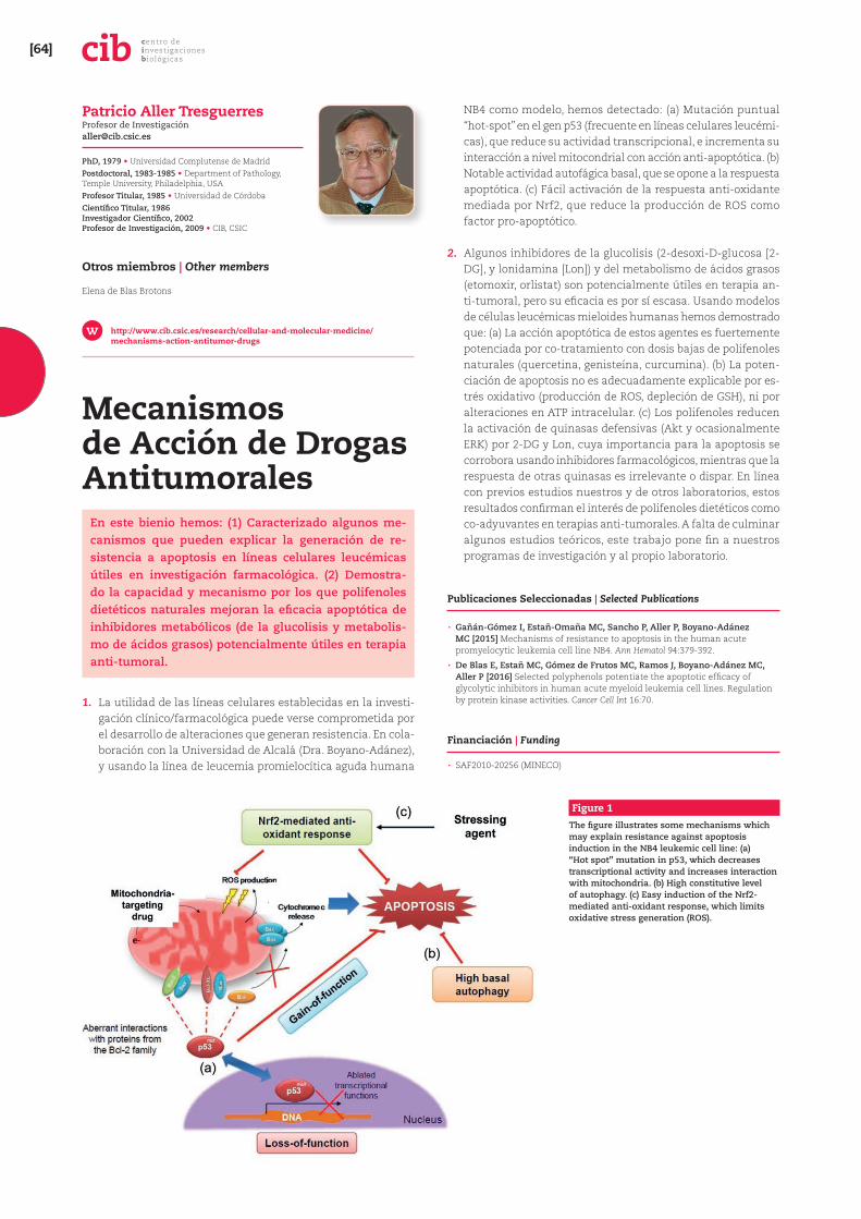
Figure 1
The figure illustrates some mechanisms which may explain resistance against apoptosis induction in the NB4 leukemic cell line: (a) “Hot spot” mutation in p53, which decreases transcriptional activity and increases interaction with mitochondria. (b) High constitutive level of autophagy. (c) Easy induction of the Nrf2-mediated anti-oxidant response, which limits oxidative stress generation (ROS).
Medicina Celular y MolecularCellular & Molecular Medicine [65]
Mechanisms of Action of Antitumour DrugsDuring the past two years we have: (1) Characterized some mechanisms that may explain the generation of resistance against apoptosis in leukemia cell lines of interest in pharmacological research. (b) Demonstrated the capacity and mechanism by which dietary polyphenols improve the apoptotic efficacy of metabolic inhibitors (glycolysis and fatty acid metabolism) potentially useful in anti-tumour therapies.
1. The interest of established cell lines for clinical/pharmacologic research may be compromised by the generation of genetic and metabolic alterations resulting in cell resistance. In collaboration with the Alcalá University (Dr. Boyano-Adánez), using the NB4 human acute promyelocytic leukemia cell line as a model, we have detected: (a) “Hot-spot” point mutation in p53 (frequent in leukemic cell lines), which reduces its transcriptional activity, and increases mitochondrial interaction with anti-apoptotic effect. (b) High basal autophagic activity, which opposes apoptosis. (c) Easy activation of the Nrf2-mediated anti-oxidant response,
reducing ROS generation as pro-apoptotic factor.
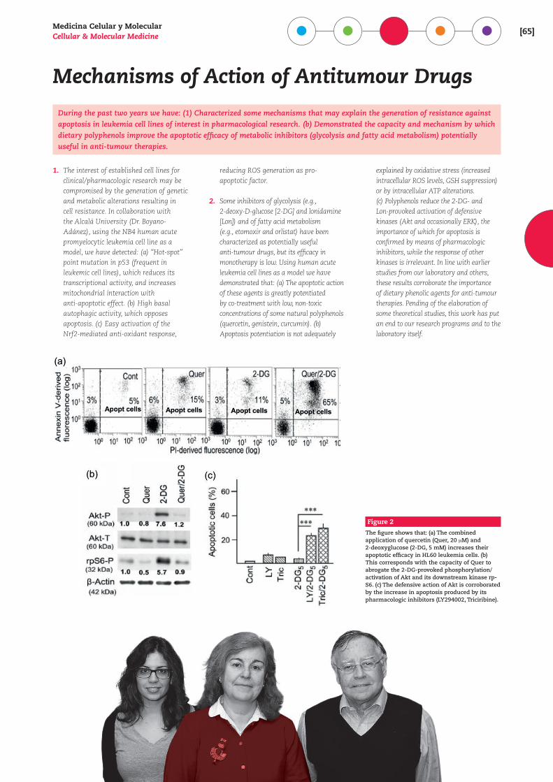
2. Some inhibitors of glycolysis (e.g., 2-deoxy-D-glucose [2-DG] and lonidamine [Lon]) and of fatty acid metabolism (e.g., etomoxir and orlistat) have been characterized as potentially useful anti-tumour drugs, but its efficacy in monotherapy is low. Using human acute leukemia cell lines as a model we have demonstrated that: (a) The apoptotic action of these agents is greatly potentiated by co-treatment with low, non-toxic concentrations of some natural polyphenols (quercetin, genistein, curcumin). (b) Apoptosis potentiation is not adequately
explained by oxidative stress (increased intracellular ROS levels, GSH suppression) or by intracellular ATP alterations. (c) Polyphenols reduce the 2-DG- and Lon-provoked activation of defensive kinases (Akt and occasionally ERK), the importance of which for apoptosis is confirmed by means of pharmacologic inhibitors, while the response of other kinases is irrelevant. In line with earlier studies from our laboratory and others, these results corroborate the importance of dietary phenolic agents for anti-tumour therapies. Pending of the elaboration of some theoretical studies, this work has put an end to our research programs and to the laboratory itself.
Figure 2
The figure shows that: (a) The combined application of quercetin (Quer, 20 +M) and 2-deoxyglucose (2-DG, 5 mM) increases their apoptotic efficacy in HL60 leukemia cells. (b) This corresponds with the capacity of Quer to abrogate the 2-DG-provoked phosphorylation/activation of Akt and its downstream kinase rp-S6. (c) The defensive action of Akt is corroborated by the increase in apoptosis produced by its pharmacologic inhibitors (LY294002, Triciribine).
[66]centro de investigaciones b iológicas
Farmacología Molecular
Se han validado nuevos magnetosensores duales para el diagnóstico robusto de cáncer de mama, por la de-tección simultánea de diferentes microRNAs. Estas pla-taformas se han utilizado como herramientas para el cribado de fármacos frente a cancer stem cells de mama, validándose los resultados en modelos in vivo. Por otro lado, en nuestra plataforma de repurposing, hemos ca-racterizado un nuevo uso del fármaco genérico Aura-nofina, como agente antibacteriano frente a Gram-posi-tivos MDR (neumococo y MRSA).
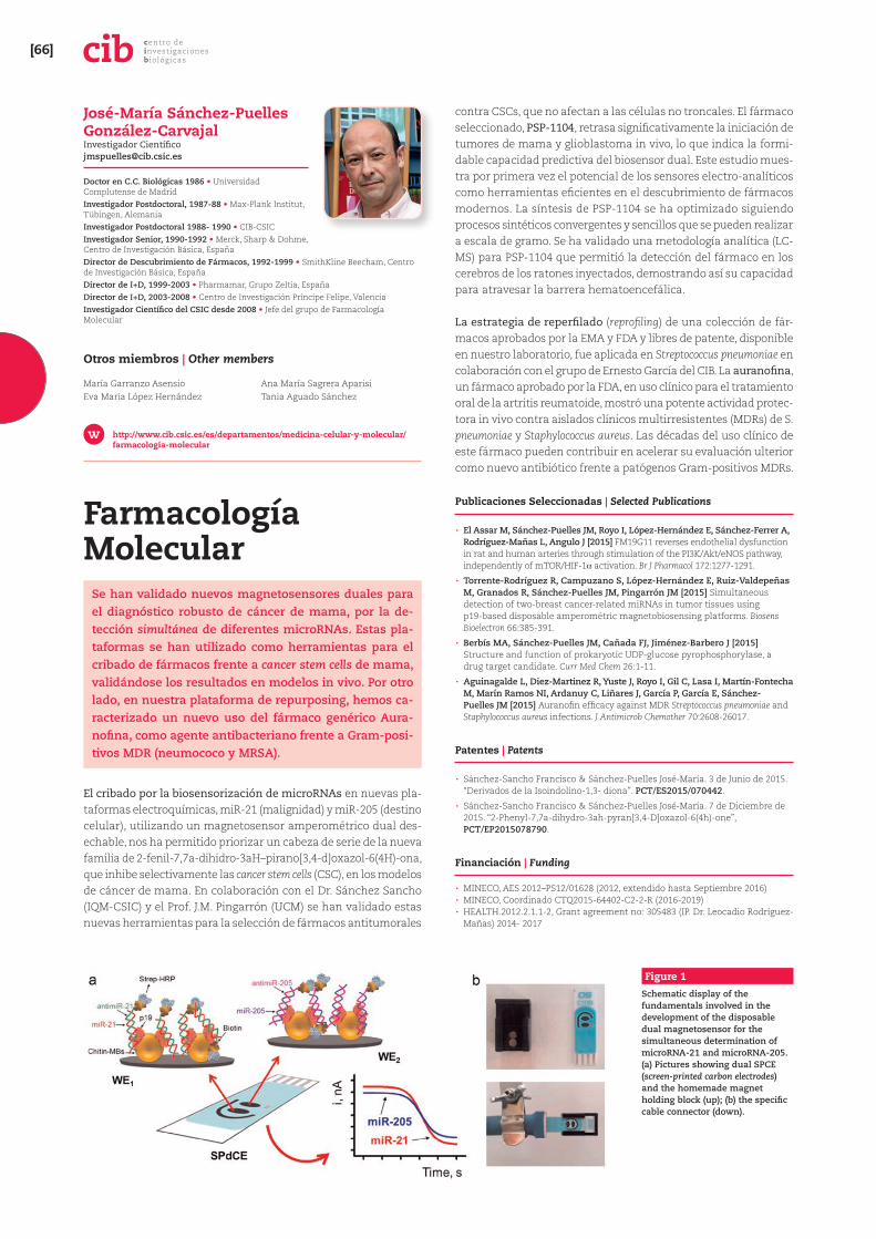
El cribado por la biosensorización de microRNAs en nuevas pla-taformas electroquímicas, miR-21 (malignidad) y miR-205 (destino celular), utilizando un magnetosensor amperométrico dual des-echable, nos ha permitido priorizar un cabeza de serie de la nueva familia de 2-fenil-7,7a-dihidro-3aH–pirano[3,4-d]oxazol-6(4H)-ona, que inhibe selectivamente las cancer stem cells (CSC), en los modelos de cáncer de mama. En colaboración con el Dr. Sánchez Sancho (IQM-CSIC) y el Prof. J.M. Pingarrón (UCM) se han validado estas nuevas herramientas para la selección de fármacos antitumorales
contra CSCs, que no afectan a las células no troncales. El fármaco seleccionado, PSP-1104, retrasa significativamente la iniciación de tumores de mama y glioblastoma in vivo, lo que indica la formi-dable capacidad predictiva del biosensor dual. Este estudio mues-tra por primera vez el potencial de los sensores electro-analíticos como herramientas eficientes en el descubrimiento de fármacos modernos. La síntesis de PSP-1104 se ha optimizado siguiendo procesos sintéticos convergentes y sencillos que se pueden realizar a escala de gramo. Se ha validado una metodología analítica (LC-MS) para PSP-1104 que permitió la detección del fármaco en los cerebros de los ratones inyectados, demostrando así su capacidad para atravesar la barrera hematoencefálica.
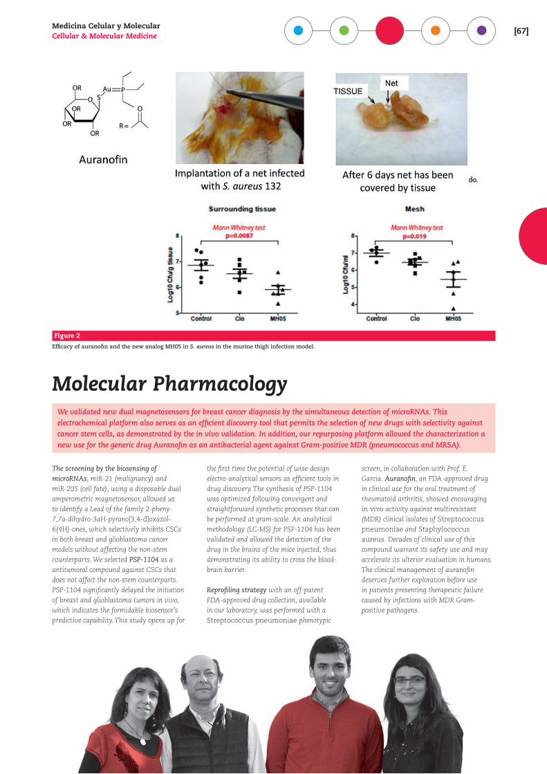
La estrategia de reperfilado (reprofiling) de una colección de fár-macos aprobados por la EMA y FDA y libres de patente, disponible en nuestro laboratorio, fue aplicada en Streptococcus pneumoniae en colaboración con el grupo de Ernesto García del CIB. La auranofina, un fármaco aprobado por la FDA, en uso clínico para el tratamiento oral de la artritis reumatoide, mostró una potente actividad protec-tora in vivo contra aislados clínicos multirresistentes (MDRs) de S. pneumoniae y Staphylococcus aureus. Las décadas del uso clínico de este fármaco pueden contribuir en acelerar su evaluación ulterior como nuevo antibiótico frente a patógenos Gram-positivos MDRs.
Publicaciones Seleccionadas | Selected Publications
· El Assar M, Sánchez-Puelles JM, Royo I, López-Hernández E, Sánchez-Ferrer A, Rodríguez-Mañas L, Angulo J [2015] FM19G11 reverses endothelial dysfunction in rat and human arteries through stimulation of the PI3K/Akt/eNOS pathway, independently of mTOR/HIF-1_ activation. Br J Pharmacol 172:1277-1291.
· Torrente-Rodríguez R, Campuzano S, López-Hernández E, Ruiz-Valdepeñas M, Granados R, Sánchez-Puelles JM, Pingarrón JM [2015] Simultaneous detection of two-breast cancer-related miRNAs in tumor tissues using p19-based disposable amperométric magnetobiosensing platforms. Biosens Bioelectron 66:385-391.
· Berbís MA, Sánchez-Puelles JM, Cañada FJ, Jiménez-Barbero J [2015] Structure and function of prokaryotic UDP-glucose pyrophosphorylase, a drug target candidate. Curr Med Chem 26:1-11.
· Aguinagalde L, Diez-Martinez R, Yuste J, Royo I, Gil C, Lasa I, Martín-Fontecha M, Marín Ramos NI, Ardanuy C, Liñares J, García P, García E, Sánchez-Puelles JM [2015] Auranofin efficacy against MDR Streptococcus pneumoniae and Staphylococcus aureus infections. J Antimicrob Chemother 70:2608-26017.
Patentes | Patents
· Sánchez-Sancho Francisco & Sánchez-Puelles José-María. 3 de Junio de 2015. “Derivados de la Isoindolino-1,3- diona”. PCT/ES2015/070442.
· Sánchez-Sancho Francisco & Sánchez-Puelles José-María. 7 de Diciembre de 2015. “2-Phenyl-7,7a-dihydro-3ah-pyran[3,4-D]oxazol-6(4h)-one”, PCT/EP2015078790.
Financiación | Funding
· MINECO, AES 2012–PS12/01628 (2012, extendido hasta Septiembre 2016) · MINECO, Coordinado CTQ2015-64402-C2-2-R (2016-2019) · HEALTH.2012.2.1.1-2, Grant agreement no: 305483 (IP. Dr. Leocadio Rodríguez-Mañas) 2014- 2017
José-María Sánchez-Puelles González-Carvajal Investigador Cientí[email protected]
Doctor en C.C. Biológicas 1986 • Universidad Complutense de MadridInvestigador Postdoctoral, 1987-88 • Max-Plank Institut, Tübingen, AlemaniaInvestigador Postdoctoral 1988- 1990 • CIB-CSICInvestigador Senior, 1990-1992 • Merck, Sharp & Dohme, Centro de Investigación Básica, EspañaDirector de Descubrimiento de Fármacos, 1992-1999 • SmithKline Beecham, Centro de Investigación Básica, EspañaDirector de I+D, 1999-2003 • Pharmamar, Grupo Zeltia, EspañaDirector de I+D, 2003-2008 • Centro de Investigación Príncipe Felipe, ValenciaInvestigador Científico del CSIC desde 2008 • Jefe del grupo de Farmacología Molecular
Otros miembros | Other members
María Garranzo AsensioEva María López Hernández
Ana María Sagrera AparisiTania Aguado Sánchez
http://www.cib.csic.es/es/departamentos/medicina-celular-y-molecular/farmacologia-molecular
Figure 1
Schematic display of the fundamentals involved in the development of the disposable dual magnetosensor for the simultaneous determination of microRNA-21 and microRNA-205. (a) Pictures showing dual SPCE (screen-printed carbon electrodes) and the homemade magnet holding block (up); (b) the specific cable connector (down).
Medicina Celular y MolecularCellular & Molecular Medicine [67]
Figure 2
Efficacy of auranofin and the new analog MH05 in S. aureus in the murine thigh infection model.
Molecular PharmacologyWe validated new dual magnetosensors for breast cancer diagnosis by the simultaneous detection of microRNAs. This electrochemical platform also serves as an efficient discovery tool that permits the selection of new drugs with selectivity against cancer stem cells, as demonstrated by the in vivo validation. In addition, our repurposing platform allowed the characterization a new use for the generic drug Auranofin as an antibacterial agent against Gram-positive MDR (pneumococcus and MRSA).
The screening by the biosensing of microRNAs, miR-21 (malignancy) and miR-205 (cell fate), using a disposable dual amperometric magnetosensor, allowed us to identify a Lead of the family 2-pheny-7,7a-dihydro-3aH-pyrano[3,4-d]oxazol-6(4H)-ones, which selectively inhibits CSCs in both breast and glioblastoma cancer models without affecting the non-stem counterparts. We selected PSP-1104 as a antitumoral compound against CSCs that does not affect the non-stem counterparts. PSP-1104 significantly delayed the initiation of breast and glioblastoma tumors in vivo, which indicates the formidable biosensor’s predictive capability. This study opens up for
the first time the potential of wise-design electro-analytical sensors as efficient tools in drug discovery. The synthesis of PSP-1104 was optimized following convergent and straightforward synthetic processes that can be performed at gram-scale. An analytical methodology (LC-MS) for PSP-1104 has been validated and allowed the detection of the drug in the brains of the mice injected, thus demonstrating its ability to cross the blood-brain barrier.
Reprofiling strategy with an off-patent FDA-approved drug collection, available in our laboratory, was performed with a Streptococcus pneumoniae phenotypic
screen, in collaboration with Prof. E. Garcia. Auranofin, an FDA-approved drug in clinical use for the oral treatment of rheumatoid arthritis, showed encouraging in vivo activity against multiresistant (MDR) clinical isolates of Streptococcus pneumoniae and Staphylococcus aureus. Decades of clinical use of this compound warrant its safety use and may accelerate its ulterior evaluation in humans. The clinical management of auranofin deserves further exploration before use in patients presenting therapeutic failure caused by infections with MDR Gram-positive pathogens.
[68]centro de investigaciones b iológicas
Activación de Linfocitos T
Estudiamos mecanismos moleculares de activación y diferenciación de linfocitos T por antígeno y moléculas coestimuladoras, que son clave en el desarrollo de res-puestas inmunitarias frente a patógenos o cáncer, y en enfermedades autoinmunes. Particularmente, nos inte-resa la utilidad en inmunoterapia de las PI3K de clase I y de las señales coestimuladoras mediadas por ICOS (Inducible Costimulator, CD278) y su ligando (ICOS-L, B7h, CD275).
Exploramos nuevas posibilidades de inmunoterapia en enferme-dades autoinmunes y en inmunidad antitumoral. Para ello se analiza el papel en respuestas inmunitarias de distintas isoformas de PI3K, y especialmente de la isoforma p110_ en las señales de ICOS y CD28.
El desarrollo de respuestas inmunitarias eficaces depende de señales de receptores específicos de la membrana y de seña-les adicionales de otras moléculas de superficie denominadas coestimuladoras (señal 2) y de señales inducidas por citocinas y quimiocinas (señal 3). Las fosfatidil-inositol 3-quinasas (PI3K) de clase I son activadas por receptores específicos de la inmunidad innata y adaptativa, de citocinas y de quimiocinas, y por molé-culas coestimuladoras, regulando el crecimiento, la proliferación, la diferenciación y la supervivencia celular.
Entre las señales coestimuladoras destacan las mediadas por mo-léculas de la familia CD28, (CD28 e ICOS, CD278) y sus ligandos de la familia B7 (CD80 y CD86, e ICOS-ligando (ICOS-L, CD275)).
Aquellas moléculas de superficie como CD28 e ICOS dotadas de motivos YxxM fosforilables por quinasas reclutan directamente PI3K de clase IA como p110_ y p110b, que son abundantes en leucocitos. En otros casos se reclutan por medio de moléculas adaptadoras. Así, el potencial de estas moléculas en inmunotera-pia es extraordinario, y está regulado por el patrón de expresión de estas moléculas y sus dianas de señalización en distintos tipos celulares.
En paralelo, investigamos las señales inducidas por ICOS-L en di-versos tipos celulares que lo expresan. Datos recientes muestran que las señales mediadas por ICOS-L modifican la capacidad mi-gratoria, la maduración y la presentación de antígeno por células dendríticas, la función de osteoclastos, y la capacidad migratoria y metastásica de células tumorales que expresan ICOS-L. En los efectos sobre migración están implicados el activador de Rac-1 `-PIX y la quinasa FAK, e indican el potencial inmunomodulador del ICOS-L.
Publicaciones Seleccionadas | Selected Publications
· Aragoneses-Fenoll L, Montes-Casado M, Ojeda G, Acosta YY, Herranz J, Martínez S, Blanco-Aparicio C, Criado G, Pastor J, Dianzani U, Portolés P, Rojo JM [2016] ETP-46321, a dual p110_/b class IA phosphoinositide 3-kinase inhibitor modulates T lymphocyte activation and collagen-induced arthritis. Biochemical Pharmacology 106:56-69.
· Linares J, Fernández AB, Feito MJ, Matesanz MC, Sánchez-Salcedo S, Arcos D, Vallet-Regí M, Rojo JM, Portolés MT [2016] Effects of nanocrystalline hydroxyapatites on macrophage polarization. Journal of Materials Chemistry B 4:1951-1959.
· Gigliotti CL, Boggio E, Clemente N, Shivakumar Y, Toth E, Sblattero D, D’Amelio P, Isaia GC, Dianzani C, Yagi Y, Rojo JM, X Chiocchetti A, Boldorini R, Bosetti M, Dianzani U [2016] ICOS-Ligand triggering impairs osteoclast function in vitro and in vivo. Journal of Immunology 197:3905-3916.
Financiación | Funding
· PI13/01809 (AES, IS Carlos III, MINECO)
José María Rojo Hernández Investigador Cientí[email protected]
Doctor en Ciencias Biológicas, 1978 • Universidad Complutense de MadridPostdoctoral, 1980-1981 • Institute of Animal Physiology, A.R.C. Cambridge, UKResearch Associate and Fulbright Fellow, 1986-1988 • Yale University School of Medicine, New Haven, CT, USACientífico Titular y Jefe de Grupo, 1988 Investigador Científico, 1990 • CIB, CSIC
http://www.cib.csic.es/es/departamentos/medicina-celular-y-molecular/activacion-de-linfocitos-t
Otros miembros | Other members
Laura Aragoneses FenollLucía García Paredes
Rosella Gazillo
Medicina Celular y MolecularCellular & Molecular Medicine [69]
Figure 1
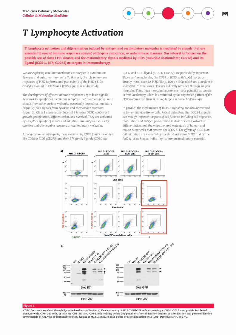
ICOS-L function is regulated through ligand-induced internalization. a) Flow cytometry of M12.C3 B7hGFP cells expressing a ICOS-L-GFP fusion protein incubated alone, or with ICOS+ D10 cells, or with an ICOS- mutant. ICOS-L B7h staining before (top panel) or after cell fixation (center), or after fixation and permeabilization (lower panel). b) Analysis by immunoblot of cell lysates of M12.C3 B7hGFP cells before or after incubation with ICOS+ D10 cells at 4ºC or 37ºC.
T Lymphocyte ActivationT lymphocyte activation and differentiation induced by antigen and costimulatory molecules is mediated by signals that are essential to mount immune responses against pathogens and cancer, or autoimmune diseases. Our interest is focused on the possible use of class I PI3 kinases and the costimulatory signals mediated by ICOS (Inducible Costimulator, CD278) and its ligand (ICOS-L, B7h, CD275) as targets in immunotherapy.
We are exploring new immunotherapic strategies in autoimmune diseases and antitumor immunity. To this end, the role in immune responses of PI3K isoforms, and particularly of the PI3K p110_ catalytic subunit in CD28 and ICOS signals, is under study.
The development of efficient immune responses depends on signals delivered by specific cell membrane receptors that are coordinated with signals from other surface molecules generically termed costimulatory (signal 2) plus signals from cytokine and chemoquine receptors (signal 3). Class I phosphatidyl Inositol-3 kinases (PI3K) control cell growth, proliferation, differentiation, and survival. They are activated by receptors specific of innate and adaptive immunity as well as by cytokine and chemoquine receptors or costimulatory molecules.
Among costimulatory signals, those mediated by CD28 family molecules like CD28 or ICOS (CD278) and their B7h family ligands (CD80 and
CD86, and ICOS-ligand (ICOS-L, CD275)) are particularly important. Those surface molecules, like CD28 or ICOS, with YxxM motifs, can directly recruit class IA PI3K, like p110_ y p110b, which are abundant in leukocytes. In other cases PI3K are indirectly recruited through adapter molecules. Thus, these molecules have an enormous potential as targets in immunotherapy, which is determined by the expression pattern of the PI3K isoforms and their signaling targets in distinct cell lineages.
In parallel, the mechanisms of ICOS-L signaling are also determined in tumor and non-tumor cells. Recent data show that ICOS-L signals can modify important aspects of cell function including cell migration, maturation and antigen presentation in dendritic cells, osteoclast differentiation, and the migration and metastasis of human and mouse tumor cells that express the ICOS-L. The effects of ICOS-L on cell migration are mediated by the Rac-1 activator `-PIX and by the FAK tyrosine kinase, indicating its immunomodulatory potential.
[70]centro de investigaciones b iológicas
Patología Molecular / Genética del Complemento
El objetivo del grupo es descifrar las bases moleculares de enfermedades humanas. Nuestra actividad inclu-ye identificar los genes que las causan y caracterizar funcionalmente sus variantes patogénicas mediante el análisis bioquímico, celular y estructural de las proteí-nas que codifican. Además, generamos modelos anima-les de estas enfermedades con el fin de entender los mecanismos patogénicos y desarrollar estrategias diag-nósticas y terapéuticas.
Nuestro trabajo actual se centra en el estudio del papel del siste-ma del complemento en enfermedad. El complemento es esencial en la inmunidad innata con papeles fundamentales en infección, eliminación de restos celulares e inmunocomplejos y la modu-lación de la inmunidad adquirida. Sin embargo, es una espada de doble filo ya que su activación descontrolada se asocia con muchas enfermedades.
Nuestro objetivo es entender cómo se produce la desregulación del complemento en cada una de estas patologías y, así, profun-dizar en la comprensión de sus mecanismos patogénicos. Nuestra
actividad experimental es multidisciplinar y se centra fundamen-talmente en el estudio del síndrome hemolítico urémico atípico y la glomerulopatía C3, interesándonos también la degeneración macular asociada a la edad, la hemoglobulinuria paroxística nocturna o la nefropatía IgA. La identificación y caracterización funcional y estructural de variantes patogénicas de proteínas del complemento asociadas a enfermedad nos está permitiendo tam-bién avanzar en aspectos básicos del sistema del complemento, aportando conocimiento mecanístico de sus actividades funcio-nales o describiendo funciones previamente desconocidas, como es el caso de la actividad de-reguladora de las proteínas FHRs.
Los resultados de nuestra actividad investigadora tienen una tras-lación casi inmediata a la práctica clínica. Lideramos el Grupo de Trabajo para el Estudio del Complemento en Patologías Renales, que coordina la actividad investigadora de diversos grupos de investigación y que se ha convertido en un referente internacional en la identificación del factor etiológico responsable del desarrollo de estas patologías, lo que está permitiendo implementar en este área una medicina individualizada.
A través de la creación de dos compañías start-up del CIB (SECU-GEN SL, www.secugen.es; Abvance Biotech srl, www.abvance.com) estamos desarrollando y comercializando aplicaciones de la se-cuenciación del ADN en el ámbito del diagnóstico molecular y desa-rrollando nuevas moléculas recombinantes con interés terapéutico.
Santiago Rodríguez de Córdoba Profesor de Investigació[email protected]
PhD, 1981 • Hospital Ramón y Cajal, Universidad Complutense de MadridVisiting Scientist 1981Associate Investigator 1985 • The New York Blood Center, NY, USACientífico Titular, 1986Incorporación, 1989Investigador Científico, 1990Profesor de Investigación, 2000 • CIB, CSICDirector Unidad de Patología Molecular, 1996-2002 • Fundación Jiménez Díaz, Madrid
Otros miembros | Other members
Hugo Yébenes RevueltoMarta Subías HidalgoSheila Pinto GarcíaLucía Juana LópezNuria Nogales GonzálezJesús María García FernándezÁngela Ruíz Sánchez
Héctor Martín MerineroAdrián Martín-Ambrosio DoménechJosé Luis Ruíz SepúlvedaJaouad AnterEmilia Arjona BolañosAgustín Tortajada Alonso
http://www.cib.csic.es/es/departamentos/medicina-celular-y-molecular/patologia-moleculargenetica-del-complemento
Figure 1
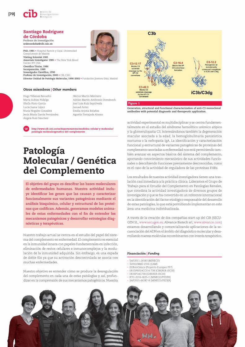
Generation, structural and functional characterization of anti-C3 monoclonal antibodies with potential diagnostic and therapeutic application.
Financiación | Funding
· SAF2011-26583 (MINECO) · S2010/BMD-2316 (CAM) · EURenOmics (Proyecto Europeo FP7) · ER15PR01ACCI14-738 (CIBERER-ISCIII) · ER16P1AC738 (CIBERER-ISCIII) · RTC-2016-4635-1 (MINECO/FEDER) · SAF2015-66287-R (MINECO/FEDER)
Medicina Celular y MolecularCellular & Molecular Medicine [71]
Publicaciones Seleccionadas | Selected Publications
· Subías-Hidalgo M, Martín-Merinero H, López A, Anter J, Pinto S, González-Fernández AF, Forés R, López-Trascasa M, Villegas A, Ojeda E, Rodríguez de Córdoba S [2017] Extravascular hemolysis and complement consumption in Paroxysmal Nocturnal Hemoglobinuria patients undergoing eculizumab treatment. Immunobiology 222:363-371.
· Goodship THJ, Cook TH, Fakhouri F, Fervenza FC, Frémeaux-Bacchi V, Kavanagh D, Nester CM, Noris M, Pickering MC, Rodríguez de Córdoba S, Roumenina LT, Sethi S, Smith RJH, for Conference Participants [2017] Atypical Hemolytic Uremic Syndrome and C3 Glomerulopathy: Conclusions from a “Kidney Disease: Improving Global Outcomes” (KDIGO) Controversies Conference. Kidney Int 91:539-551.
· Xiao X, Ghossein C, Tortajada A, Zhang Y, Meyer N, Jones M, Ghiringhelli Borsa N, Nester CM, Thomas CP, Rodríguez de Córdoba S and Smith RJH [2016] Familial C3 glomerulonephritis caused by a novel CFHR5-CFHR2 fusion gene. Mol Immunol 77:89-96.
· Recalde S, Tortajada A, Subias M, Anter J, Blasco M, Maranta R, Coco R, Pinto S, Noris M, García-Layana A and Rodríguez de Córdoba S [2016] Molecular basis of Factor H R1210C association with ocular and renal diseases. J Am Soc Nephrol 27:1305-1311.
· Rodríguez de Córdoba S [2016] Complement genetics and susceptibility to inflammatory disease. Lessons from genotype-phenotype correlations. Immunobiology 221:709-714.
· Alcorlo M, López-Perrote A, Delgado Bermúdez S, Yébenes H, Subías M, Rodríguez-Gallego C, Rodríguez de Córdoba S and Llorca O [2015] Structural Insights on Complement Activation. FEBS Journal 282:3883-3891.
· Rabasco C, Cavero T, Román E, Rojas-Rivera J, Olea, T, Espinosa M, Cabello, V, Fernández-Juárez G, González F, Avila A, Baltar JM, Díaz M, Alegre R, Elías S, Antón M, Frutos MA, Pobes A, Blasco M, Martín F, Bernis C, Macías M, Barroso S, De Lorenzo A, Ariceta G, López-Mendoza M, Rivas B, López-Revuelta K, Campistol JM, Mendizábal S, Rodríguez de Córdoba S and Praga M for the Spanish Group for the Study of Glomerular Diseases (GLOSEN) [2015] Effectiveness of Mycophenolate Mofetil in C3 Glomerulonephritis. Kidney International 88:1153-1160.
· Józsi M, Tortajada A, Uzonyi B, Goicoechea de Jorge E and Rodríguez de Córdoba S [2015] Factor H-related proteins determine complement-activating surfaces. Trends Immunol 36:374-384.
· Martínez-Barricarte R, Heurich M, López-Perrote A, Tortajada A, Pinto S, López-Trascasa M, Sánchez-Corral P, Morgan BP, Llorca O, Harris CL and Rodríguez de Córdoba S [2015] The molecular and structural bases for the association of complement C3 mutations with atypical hemolytic uremic syndrome. Mol Immunol 66:263-273.
· Valoti E*, Alberti M*, Tortajada A*, García-Fernández JM, Gastoldi S, Besso L, Bresin E, Remuzzi G, Rodríguez de Córdoba S and Noris M [2015] A novel atypical Hemolytic Uremic Syndrome – associated hybrid CFHR1/CFH gene encoding a fusion protein that antagonizes factor H-dependent complement regulation. J Am Soc Nephrol 26:209-219(* Equally contributed as first Author).
Figure 2
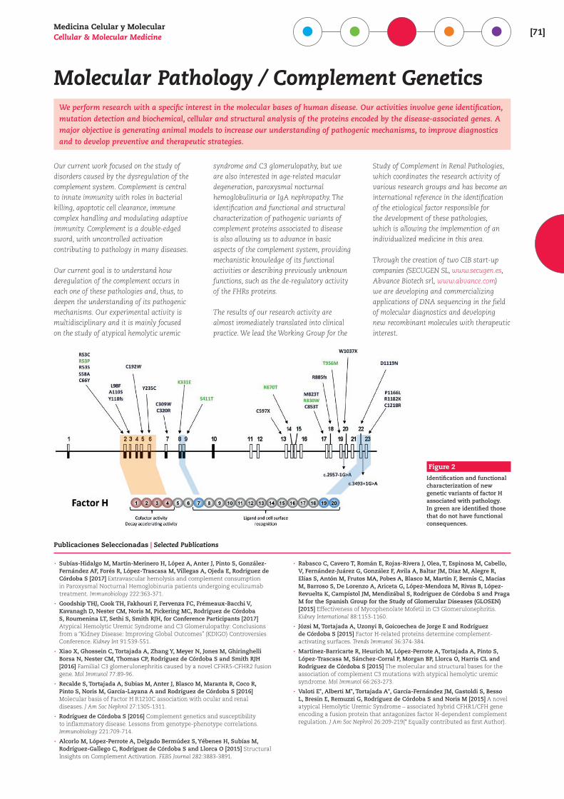
Identification and functional characterization of new genetic variants of factor H associated with pathology. In green are identified those that do not have functional consequences.
Molecular Pathology / Complement GeneticsWe perform research with a specific interest in the molecular bases of human disease. Our activities involve gene identification, mutation detection and biochemical, cellular and structural analysis of the proteins encoded by the disease-associated genes. A major objective is generating animal models to increase our understanding of pathogenic mechanisms, to improve diagnostics and to develop preventive and therapeutic strategies.
Our current work focused on the study of disorders caused by the dysregulation of the complement system. Complement is central to innate immunity with roles in bacterial killing, apoptotic cell clearance, immune complex handling and modulating adaptive immunity. Complement is a double-edged sword, with uncontrolled activation contributing to pathology in many diseases.
Our current goal is to understand how deregulation of the complement occurs in each one of these pathologies and, thus, to deepen the understanding of its pathogenic mechanisms. Our experimental activity is multidisciplinary and it is mainly focused on the study of atypical hemolytic uremic
syndrome and C3 glomerulopathy, but we are also interested in age-related macular degeneration, paroxysmal nocturnal hemoglobulinuria or IgA nephropathy. The identification and functional and structural characterization of pathogenic variants of complement proteins associated to disease is also allowing us to advance in basic aspects of the complement system, providing mechanistic knowledge of its functional activities or describing previously unknown functions, such as the de-regulatory activity of the FHRs proteins.
The results of our research activity are almost immediately translated into clinical practice. We lead the Working Group for the
Study of Complement in Renal Pathologies, which coordinates the research activity of various research groups and has become an international reference in the identification of the etiological factor responsible for the development of these pathologies, which is allowing the implemention of an individualized medicine in this area.
Through the creation of two CIB start-up companies (SECUGEN SL, www.secugen.es, Abvance Biotech srl, www.abvance.com) we are developing and commercializing applications of DNA sequencing in the field of molecular diagnostics and developing new recombinant molecules with therapeutic interest.
[72]centro de investigaciones b iológicas
Figure 1
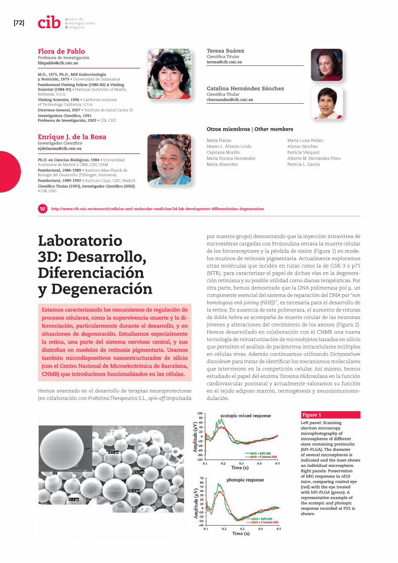
Left panel: Scanning electron microscopy microphotography of microspheres of different sizes containing proinsulin (hPI-PLGA). The diameter of several microspheres is indicated and the inset shows an individual microsphere. Right panels: Preservation of ERG responses in rd10 mice, comparing control eye (red) with the eye treated with hPI-PLGA (green). A representative example of the scotopic and photopic response recorded at P25 is shown.
Laboratorio 3D: Desarrollo, Diferenciación y Degeneración
Estamos caracterizando los mecanismos de regulación de procesos celulares, como la supervivencia-muerte y la di-ferenciación, particularmente durante el desarrollo, y en situaciones de degeneración. Estudiamos especialmente la retina, una parte del sistema nervioso central, y sus distrofias en modelos de retinosis pigmentaria. Usamos también microdispositivos nanoestructurados de silicio (con el Centro Nacional de Microelectrónica de Barcelona, CNMB) que introducimos funcionalizados en las células.
Hemos avanzado en el desarrollo de terapias neuroprotectoras (en colaboración con ProRetina Therapeutics S.L., spin-off impulsada
por nuestro grupo) demostrando que la inyección intravítrea de microesferas cargadas con Proinsulina retrasa la muerte celular de los fotorreceptores y la pérdida de visión (Figura 1) en mode-los murinos de retinosis pigmentaria. Actualmente exploramos otras moléculas que inciden en rutas como la de GSK-3 o p75 (NTR), para caracterizar el papel de dichas vías en la degenera-ción retiniana y su posible utilidad como dianas terapéuticas. Por otra parte, hemos demostrado que la DNA polimerasa pol µ, un componente esencial del sistema de reparación del DNA por “non homologous end-joining (NHEJ)”, es necesaria para el desarrollo de la retina. En ausencia de esta polimerasa, el aumento de roturas de doble hebra se acompaña de muerte celular de las neuronas jóvenes y alteraciones del crecimiento de los axones (Figura 2). Hemos desarrollado en colaboración con el CNMB una nueva tecnología de miniaturización de microobjetos basados en silicio que permiten el análisis de parámetros intracelulares múltiples en células vivas. Además continuamos utilizando Dictyostelium discoideum para tratar de identificar los mecanismos moleculares que intervienen en la competición celular. Así mismo, hemos estudiado el papel del enzima Tirosina Hidroxilasa en la función cardiovascular postnatal y actualmente valoramos su función en el tejido adiposo marrón, termogénesis y neuroinmunomo-dulación.
Flora de Pablo Profesora de Investigació[email protected]
M.D., 1975, Ph.D., MIR Endocrinología y Nutrición, 1979 • Universidad de SalamancaPostdoctoral Visiting Fellow (1980-82) & Visiting Scientist (1984-91) • National Institutes of Health, Bethesda, U.S.A.Visiting Scientist, 1996 • California Institute of Technology, California, U.S.A.Directora General, 2007 • Instituto de Salud Carlos IIIInvestigadora Científica, 1991 Profesora de Investigación, 2003 • CIB, CSIC
Enrique J. de la Rosa Investigador Cientí[email protected]
Ph.D. en Ciencias Biológicas, 1984 • Universidad Autónoma de Madrid y CBM, CSIC-UAMPostdoctoral, 1986-1989 • Instituto Max-Planck de Biología del Desarrollo (Tübingen, Alemania)Postdoctoral, 1989-1992 • Instituto Cajal, CSIC, MadridCientífico Titular (1993), Investigador Científico (2002) • CIB, CSIC
http://www.cib.csic.es/research/cellular-and-molecular-medicine/3d-lab-development-differentiation-degeneration
Teresa Suárez Científica [email protected]
Catalina Hernández Sánchez Científica [email protected]
Otros miembros | Other members
María PlatónNoemí L. Álvarez LindoCayetana MurilloMaría Donina HernándezMaría Abancéns
María Luisa PeláezAlonso SánchezPatricia VázquezAlberto M. Hernández PintoPatricia L. García
Medicina Celular y MolecularCellular & Molecular Medicine [73]
3D Lab: Development, Differentiation & Degeneration
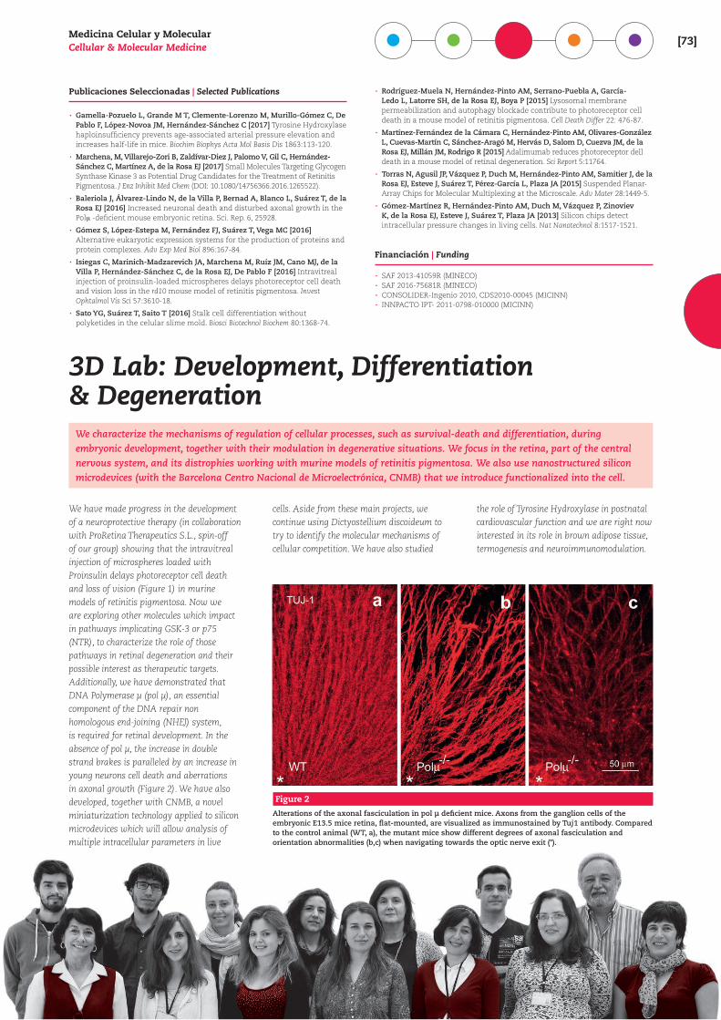
We characterize the mechanisms of regulation of cellular processes, such as survival-death and differentiation, during embryonic development, together with their modulation in degenerative situations. We focus in the retina, part of the central nervous system, and its distrophies working with murine models of retinitis pigmentosa. We also use nanostructured silicon microdevices (with the Barcelona Centro Nacional de Microelectrónica, CNMB) that we introduce functionalized into the cell.
We have made progress in the development of a neuroprotective therapy (in collaboration with ProRetina Therapeutics S.L., spin-off of our group) showing that the intravitreal injection of microspheres loaded with Proinsulin delays photoreceptor cell death and loss of vision (Figure 1) in murine models of retinitis pigmentosa. Now we are exploring other molecules which impact in pathways implicating GSK-3 or p75 (NTR), to characterize the role of those pathways in retinal degeneration and their possible interest as therapeutic targets. Additionally, we have demonstrated that DNA Polymerase µ (pol µ), an essential component of the DNA repair non homologous end-joining (NHEJ) system, is required for retinal development. In the absence of pol µ, the increase in double strand brakes is paralleled by an increase in young neurons cell death and aberrations in axonal growth (Figure 2). We have also developed, together with CNMB, a novel miniaturization technology applied to silicon microdevices which will allow analysis of multiple intracellular parameters in live
cells. Aside from these main projects, we continue using Dictyostellium discoideum to try to identify the molecular mechanisms of cellular competition. We have also studied
the role of Tyrosine Hydroxylase in postnatal cardiovascular function and we are right now interested in its role in brown adipose tissue, termogenesis and neuroimmunomodulation.
Publicaciones Seleccionadas | Selected Publications
· Gamella-Pozuelo L, Grande M T, Clemente-Lorenzo M, Murillo-Gómez C, De Pablo F, López-Novoa JM, Hernández-Sánchez C [2017] Tyrosine Hydroxylase haploinsufficiency prevents age-associated arterial pressure elevation and increases half-life in mice. Biochim Biophys Acta Mol Basis Dis 1863:113-120.
· Marchena, M, Villarejo-Zori B, Zaldívar-Diez J, Palomo V, Gil C, Hernández-Sánchez C, Martínez A, de la Rosa EJ [2017] Small Molecules Targeting Glycogen Synthase Kinase 3 as Potential Drug Candidates for the Treatment of Retinitis Pigmentosa. J Enz Inhibit Med Chem (DOI: 10.1080/14756366.2016.1265522).
· Baleriola J, Álvarez-Lindo N, de la Villa P, Bernad A, Blanco L, Suárez T, de la Rosa EJ [2016] Increased neuronal death and disturbed axonal growth in the Pol+ -deficient mouse embryonic retina. Sci. Rep. 6, 25928.
· Gómez S, López-Estepa M, Fernández FJ, Suárez T, Vega MC [2016] Alternative eukaryotic expression systems for the production of proteins and protein complexes. Adv Exp Med Biol 896:167-84.
· Isiegas C, Marinich-Madzarevich JA, Marchena M, Ruíz JM, Cano MJ, de la Villa P, Hernández-Sánchez C, de la Rosa EJ, De Pablo F [2016] Intravitreal injection of proinsulin-loaded microspheres delays photoreceptor cell death and vision loss in the rd10 mouse model of retinitis pigmentosa. Invest Ophtalmol Vis Sci 57:3610-18.
· Sato YG, Suárez T, Saito T [2016] Stalk cell differentiation without polyketides in the celular slime mold. Biosci Biotechnol Biochem 80:1368-74.
· Rodríguez-Muela N, Hernández-Pinto AM, Serrano-Puebla A, García-Ledo L, Latorre SH, de la Rosa EJ, Boya P [2015] Lysosomal membrane permeabilization and autophagy blockade contribute to photoreceptor cell death in a mouse model of retinitis pigmentosa. Cell Death Differ 22: 476-87.
· Martínez-Fernández de la Cámara C, Hernández-Pinto AM, Olivares-González L, Cuevas-Martín C, Sánchez-Aragó M, Hervás D, Salom D, Cuezva JM, de la Rosa EJ, Millán JM, Rodrigo R [2015] Adalimumab reduces photoreceptor dell death in a mouse model of retinal degeneration. Sci Report 5:11764.
· Torras N, Agusil JP, Vázquez P, Duch M, Hernández-Pinto AM, Samitier J, de la Rosa EJ, Esteve J, Suárez T, Pérez-García L, Plaza JA [2015] Suspended Planar-Array Chips for Molecular Multiplexing at the Microscale. Adv Mater 28:1449-5.
· Gómez-Martínez R, Hernández-Pinto AM, Duch M, Vázquez P, Zinoviev K, de la Rosa EJ, Esteve J, Suárez T, Plaza JA [2013] Silicon chips detect intracellular pressure changes in living cells. Nat Nanotechnol 8:1517-1521.
Financiación | Funding
· SAF 2013-41059R (MINECO) · SAF 2016-75681R (MINECO) · CONSOLIDER-Ingenio 2010, CDS2010-00045 (MICINN) · INNPACTO IPT- 2011-0798-010000 (MICINN)
Figure 2
Alterations of the axonal fasciculation in pol µ deficient mice. Axons from the ganglion cells of the embryonic E13.5 mice retina, flat-mounted, are visualized as immunostained by Tuj1 antibody. Compared to the control animal (WT, a), the mutant mice show different degrees of axonal fasciculation and orientation abnormalities (b,c) when navigating towards the optic nerve exit (*).
[74]centro de investigaciones b iológicas
Bases celulares y moleculares de la enfermedad de Alzheimer y otras demencias
El interés de nuestro laboratorio es el estudio de los mecanismos que causan la muerte neuronal en la en-fermedad de Alzheimer, la demencia frontotemporal y otras enfermedades neurodegenerativas. Estudiamos fundamentalmente alteraciones en el control del ciclo celular, apoptosis, función mitocondrial, estrés oxidati-vo y degradación de proteínas en modelos celulares de neurodegeneración que incluyen células extraneurales de pacientes.
Bases celulares y moleculares de la enfermedad de Alzheimer
La enfermedad de Alzheimer (EA) es la forma más común de demencia en los adultos. En Europa constituye una de las prime-ras causas de muerte, sin que hasta el momento se disponga de terapias efectivas para paliar o retrasar su aparición.
Recientemente hemos iniciado un proyecto que tiene como objeti-vo el diseño, síntesis y evaluación pre-clínica de nuevos agonistas del receptor de Cannabinoides de tipo 2 (CB2), con propiedades
colinérgicas e inhibidoras del enzima `-secretasa (BACE-1). Es-tamos evaluando el potencial de estos nuevos compuestos en líneas linfoblásticas de pacientes de EA y en modelos neuronales de la enfermedad.
Déficit de progranulina y demencia frontotemporal (FTLD-TDP)
La degeneración del lóbulo frontotemporal (FTLD) designa a un grupo heterogéneo de procesos neurodegenerativos que compor-tan deterioro cognitivo asociado a sintomatología motora o de len-guaje, trastornos de comportamiento y cambios de personalidad con preservación inicial de la memoria. La forma más común de FTLD presenta inclusiones neuronales citoplásmicas tau-nega-tivas y ubiquitín-positivas compuestas por la proteína TDP-43. La mayor parte de los casos con historia familiar de FTLD-TDP presentan mutaciones en el gen GRN. Estas mutaciones causan generalmente la haploinsuficiencia de progranulina (PGRN). Es-tudiamos la influencia patogénica del déficit de PGRN en células periféricas de pacientes, así como células neuronales de ratones deficientes en PGRN y células SH-S5Y5 después de haber silencia-do el gen GRN. Además, hemos utilizado líneas linfoblásticas de pacientes de FTLD-TDP como plataforma para valorar la eficacia, a nivel pre-clínico, de drogas que interaccionan con la cascada de señalización alterada en la enfermedad, y particularmente el efecto de inhibidores de la fosforilación de TDP-43. Investigamos también los efectos de nuevos inhibidores selectivos de Caseí-na-Kinasa 1b (CK-1 b) and CDC7.
María Ángeles Martín Requero Investigadora cientí[email protected]
PhD, 1978 • Universidad Complutense de MadridPostdoctoral, 1979-1982 • University of Pennsylvania, Philadelphia, USAProfesora Titular, 1982-1983 • Universidad de ExtremaduraCientífica Titular, 1985 • CIB, CSICVisiting Scientist, 1996-1998 • University of California (UCLA), Los Angeles, USAInvestigadora Científica, 2008 • CIB, CSIC
http://www.cib.csic.es/research/cellular-and-molecular-medicine/cellular-and-molecular-basis-alzheimers-disease-and-other
Otros miembros | Other members
Carolina Alquézar BurilloAna de la Encarnación SalmerónFernando Bartolomé RobledoPatricia del Cerro de Pablo
Esther Cañibano MorejónMarcos Ruíz LópezAlejandro Reinares SebastiánDiana Posa Podean
Financiación | Funding
· SAF2011-28630 (MINECO) 2012-2015 · Fundación Ramón Areces 2012-2015 · CTQ2015 -66313-R 1 2015-2017
Medicina Celular y MolecularCellular & Molecular Medicine [75]
Cellular and molecular basis of Alzheimer’s disease and other dementias
Our lab is interested in mechanisms that cause cell death in disorders such as Alzheimer’s disease, frontotemporal dementia and other neurodegenerative disorders. The work focuses on cell cycle dysfuntion, apoptosis, mitochondrial impairment, oxidative damage and protein degradation, using in vivo models of neurodegeneration and in vitro culture of cells, including peripheral cells from patients.
Cellular and molecular basis of Alzheimer’s disease
The most common cause of dementia in mid-to late-life is Alzheimer disease (AD). In Europe, AD is a leading cause of death and there are no disease modifying drugs currently available. Thus, elucidation of the pathogenic mechanisms leading to AD, and the design of an effective treatment, preventive or curative, become imperative.
Recently, we have initiated a research project aimed at designing and evaluating the therapeutic potential of new compounds with a multitarget profile as CB2 cannabinoid agonists and `-secretase (BACE-1) and/or Butyrylcholinesterase (BuChE) inhibitors. To this end, we are carrying studies regarding the effects of these molecules on the mechanisms controlling cell survival/dead in lymphoblastic cell lines from AD patients as well as in neuronal cell modes and transgenic mice.
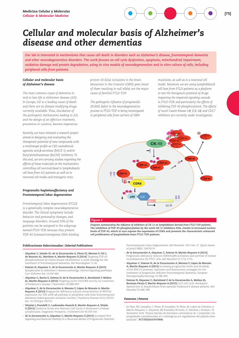
Progranulin haploinsufficciency and Frontotemporal lobar degeneration
Frontotemporal lobar degeneration (FTLD) is a genetically complex neurodegenerative disorder. The clinical symptoms include behavior and personality changes, and language disorders. Around 50% of the patients can be assigned to the subgroup named FTLD-TDP, because they present TDP-43 (transactiveresponse DNA-binding
protein 43 KDa) inclusions in the brain. Mutations in the Granulin (GRN) gene (most of them resulting in null allele) are the major cause of familial FTLD-TDP.
The pathogenic influence of progranulin (PGRN) deficit in the neurodegenerative process in FTLD-TDP is being investigated in peripheral cells from carriers of GRN
mutations, as well as in a neuronal cell model. Moreover, we are using lymphoblatoid cell lines from FTLD patients as a platform to test the therapeutic potential of drugs impacting the impaired signaling cascade in FTLD-TDP, and particularly the effects of inhibiting TDP-43 phosphorylation. The effects of novel Casein kinase-1b) (CK-1b) and CDC7 inhibitors are currently under investigation.
Publicaciones Seleccionadas | Selected Publications
· Alquézar C, Salado IG, de la Encarnación A, Pérez DI, Moreno F, Gil C, de Munain AL, Martínez A, Martín-Requero Á [2016] Targeting TDP-43 phosphorylation by Casein Kinase-1b inhibitors: a novel strategy for the treatment of frontotemporal dementia. Mol Neurodegener 11:36.
· Esteras N, Alquézar C, de la Encarnación A, Martín-Requero Á [2015] Lymphocytes in Alzheimer´s disease pathology: Altered signaling pathways. Curr Alzheimer Res 13:439-449.
· Alquézar C, Barrio E, Esteras N, de la Encarnación A, Bartolomé F, Molina JA, Martín-Requero A [2015] Targeting cyclin D3/CDK6 activity for treatment of Parkinson´s disease. J Neurochem 133:886-897.
· Alquézar C, de la Encarnación A, Moreno F, López de Munain A, Martín-Requero Á [2015] Progranulin deficiency induces overactivation of WNT5A expression via TNF-_/NF-gB pathway in peripheral cells from frontotemporal dementia-linked granulin mutation carriers. J Psychiatry Neurosci 41(1):150131. doi: 10.1503/jpn.150131.
· Wojsiat J, Prandelli C, Laskowska-Kaszub K, Martín-Requero A, Wojda U [2015] Oxidative Stress and Aberrant Cell Cycle in Alzheimer´s Disease Lymphocytes: Diagnostic Prospects. J Alzheimers Dis 46:329-350.
· de la Encarnación A, Alquézar C, Martín-Requero Á [2015] Increased Wnt Signaling and Reduced Viability in a Neuronal Model of Progranulin-Deficient
Frontotemporal Lobar Degeneration. Mol Neurobiol. 2015 Dec 17. [Epub ahead of print] PMID: 26676574.
· de la Encarnación A, Alquézar C, Esteras N, Martín-Requero A [2015] Progranulin deficiency reduces CDK4/6/pRb activation and survival of human neuroblastoma SH-SY5Y cells. Mol Neurobiol 52:1714-1725.
· Alquézar C, Esteras N, de la Encarnación A, Moreno F, López de Munain A, Martín-Requero A [2015] Increasing progranulin levels and blockade of the ERK1/2 pathway: Upstream and downstream strategies for the treatment of progranulin deficient frontotemporal dementia. European Neuropsychopharmacology 25:386-403.
· Esteras N, Alquezar C, Bartolomé F, de la Encarnación A, Molina JA, Bermejo-Pareja F, Martin-Requero A [2015] G1/S cell cycle checkpoint dysfunction in lymphoblasts from sporadic Parkinson’s disease patients. Mol Neurobiol 52:386-398.
Patentes | Patents
· JA Páez, NE Campillo, C. Pérez, PJ González, N. Pérez, M. López de Ceballos, A. Martín-Requero, C. Alquézar, MI Fontelles, M Girón, E. Sánchez, J. Romero. 16 diciembre 2016. “Nueva familia de derivados carbonílicos de 1-indazolilo con propiedades cannabinoides y/o colinérgicas y/o reguladoras del péptido beta-amiloide”. PCT/ES20167070906.
Figure 1
Diagram summarizing the influence of inhibitors of CK-1b on lymphoblasts derived from FTLD-TDP patients. The inhibition of TDP-43 phosphorylation by the novel CK-1b inhibitors IGSs, results in increased nuclear levels of TDP-43, which in turn repress the expression of CDK6 and prevents the characteristic enhanced cell proliferation of lymphoblasts from FTLD-TDP patients.
[76]centro de investigaciones b iológicas
Fisiopatología de trastornos hemostáticos
Utilizamos plaquetas y modelos celulares y animales diseñados en el laboratorio para investigar el papel de proteínas plaquetarias y endoteliales en la fisiopatolo-gía de la hemostasia, con el objetivo final de identificar mecanismos y/o moléculas que puedan ser potenciales dianas terapéuticas.
Nuestro trabajo actual está centrado en la caracterización fenotí-pica de líneas murinas modificadas genéticamente generadas en el laboratorio, con el objetivo de desvelar la función de proteínas que se expresan en plaquetas y/o endotelio vascular. Una de estas proteínas, la podocalicina (Podxl), es una glucoproteína de mem-brana tipo sialomucina extensivamente O- y N-glicosilada y rica en ácido siálico, identificada en origen en las células epiteliales del glomérulo renal donde es esencial para el desarrollo y función de la barrera de filtración glomerular. La caracterización de los ratones KO condicionales de Podxl en megacariocitos y plaquetas nos permitió concluir que esta proteína participa como un co-ac-tivador en el control de la hemostasia, probablemente debido a sus propiedades pro-adhesivas. Más recientemente, mediante el estudio de la línea KO condicional de Podxl en el endotelio vascular hemos comprobado que la Podxl tiene una importante
función en el mantenimiento de la integridad vascular. Los ani-males carentes de Podxl endotelial desarrollan una vasculitis que afecta a vasos de diferente grosor en uno o varios órganos y que conduce a muerte prematura por fallo orgánico. Los resultados sugieren que la Podxl tiene un papel esencial en la regulación de la permeabilidad endotelial participando en los mecanismos implicados en la restauración de la barrera endotelial después de un daño. Esta importante función de la Podxl en el endotelio vascular podría estar mediada por su interacción con proteínas intracelulares y consecuente modulación del citoesqueleto de actina. Investigamos también la posibilidad de que la región ex-tracelular de la Podxl sea un componente esencial del glucocálix endotelial. Aunque existen evidencias de que la lesión del gluco-cálix subyace a un gran número de enfermedades vasculares, el papel y relevancia de los distintos proteoglicanos y glicoproteínas que lo integran no se conoce.
Consuelo González Manchón Científica [email protected]
MD, 1983 • Universidad de ExtremaduraPhD, 1987 • Universidad Complutense de MadridPostdoctoral, 1988-1990 • Salk Institute, La Jolla, CA, USACientífica Titular, 1999 • CIB, CSICJefa de Grupo, 2005
http://www.cib.csic.es/research/cellular-and-molecular-medicine/pathophysiology-hemostatic-disorders
Matilde Sánchez Ayuso Profesora de Investigación, Doctora vinculada Ad [email protected]
Otros miembros | Other members
Gracia Porras Franco
Publicaciones Seleccionadas | Selected Publications
· Horrillo A, Porras G, Ayuso MS, González-Manchón C [2016] Loss of endothelial barrier integrity in mice with conditional ablation of podocalyxin (Podxl) in endothelial cells. Eur J Cell Biol 95:265-276.
· Yang Z, Zimmerman SE, Tsunezumi J, Braitsch C, Trent C, Bryant DM, Cleaver O, González-Manchón C, Marciano DK [2016] Role of CD34 family members in lumen formation in the developing kidney. Dev Biol 418:66-74.
Financiación | Funding
· ER16P1AC734 y ERPRO1G734 CIBERER (ISCIII)
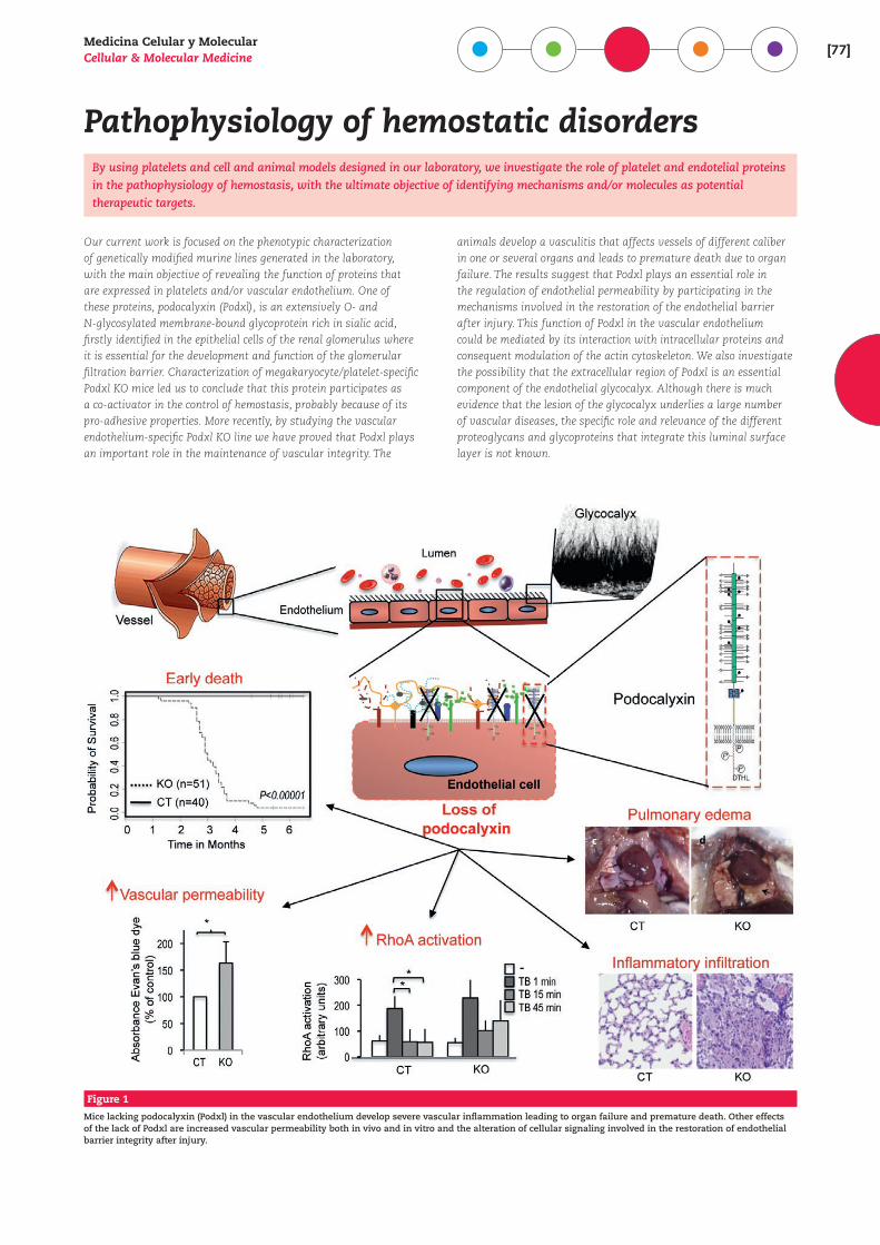
Medicina Celular y MolecularCellular & Molecular Medicine [77]
Pathophysiology of hemostatic disordersBy using platelets and cell and animal models designed in our laboratory, we investigate the role of platelet and endotelial proteins in the pathophysiology of hemostasis, with the ultimate objective of identifying mechanisms and/or molecules as potential therapeutic targets.
Our current work is focused on the phenotypic characterization of genetically modified murine lines generated in the laboratory, with the main objective of revealing the function of proteins that are expressed in platelets and/or vascular endothelium. One of these proteins, podocalyxin (Podxl), is an extensively O- and N-glycosylated membrane-bound glycoprotein rich in sialic acid, firstly identified in the epithelial cells of the renal glomerulus where it is essential for the development and function of the glomerular filtration barrier. Characterization of megakaryocyte/platelet-specific Podxl KO mice led us to conclude that this protein participates as a co-activator in the control of hemostasis, probably because of its pro-adhesive properties. More recently, by studying the vascular endothelium-specific Podxl KO line we have proved that Podxl plays an important role in the maintenance of vascular integrity. The
animals develop a vasculitis that affects vessels of different caliber in one or several organs and leads to premature death due to organ failure. The results suggest that Podxl plays an essential role in the regulation of endothelial permeability by participating in the mechanisms involved in the restoration of the endothelial barrier after injury. This function of Podxl in the vascular endothelium could be mediated by its interaction with intracellular proteins and consequent modulation of the actin cytoskeleton. We also investigate the possibility that the extracellular region of Podxl is an essential component of the endothelial glycocalyx. Although there is much evidence that the lesion of the glycocalyx underlies a large number of vascular diseases, the specific role and relevance of the different proteoglycans and glycoproteins that integrate this luminal surface layer is not known.
Figure 1
Mice lacking podocalyxin (Podxl) in the vascular endothelium develop severe vascular inflammation leading to organ failure and premature death. Other effects of the lack of Podxl are increased vascular permeability both in vivo and in vitro and the alteration of cellular signaling involved in the restoration of endothelial barrier integrity after injury.
[78]centro de investigaciones b iológicas
Receptores de TGF-beta en células endoteliales
Endoglina y ALK1 son miembros del complejo receptor de TGF-beta en células endoteliales con implicaciones en la fisiopatología vascular. Mutaciones en los genes de endoglina o ALK1 (ACVRL1) son responsables de la Telangiectasia Hemorrágica Hereditaria (HHT), una en-fermedad vascular autosómica dominante. Endoglina y ALK1 regulan la angiogénesis y homeostasis vascular, y una forma soluble de endoglina tiene un papel pato-génico en preeclampsia.
Nuestro laboratorio estudia la expresión génica, estructura y fun-ción de los receptores de TGF-beta endoglina y ALK1, tanto en
fisiología como en el contexto de la patología humana, en especial en HHT. Ambos receptores son capaces de unir específicamente el ligando BMP9, un miembro de la superfamilia del TGF-beta. Mutaciones en los genes de endoglina, ALK1 o BMP9 dan lugar a HHT1, HHT2 o HHT5, respectivamente, las cuales están asociadas con frecuentes epistaxis, hemorragias gastrointestinales, telan-giectasias cutáneas, y malformaciones arteriovenosas en pulmón, hígado y cerebro. Entre los objetivos científicos del grupo se in-cluyen: i) Llevar a cabo el diagnóstico molecular en la población de pacientes HHT españoles; ii) Buscar nuevos biomarcadores en HHT; iii) Analizar los mecanismos de regulación transcripcional de los genes de endoglina y ALK1; iv) Obtener cultivos primarios de células endoteliales y monocitos activados a partir de sangre periférica de pacientes HHT para estudios funcionales y para en-sayos de terapias célulo-génicas en ratones; v) Analizar la función y los patrones de expresión génica de celulas que sobreexpresan endoglina; vi) Generar ratones KO condicionales de endoglina en el linaje mieloide para analizar el impacto de HHT en el sistema inmune; vii) Buscar medicamentos huérfanos con posible aplica-ción en HHT y estudiar su mecanismo de acción; y viii) Estudiar el papel de endoglina endotelial en adhesión celular como ligando de integrinas leucocitarias, plaquetarias y de pericitos.
Publicaciones Seleccionadas | Selected Publications
· Blanco FJ, Ojeda-Fernandez L, Aristorena M, Gallardo-Vara E, Benguria A, Dopazo A, Langa C, Botella LM, Bernabeu C [2015] Genome-wide transcriptional and functional analysis of endoglin isoforms in the human promonocytic cell line U937. J Cell Physiol 230:947-958.
· Del Castillo G, Sánchez-Blanco E, Martín-Villar E, Valbuena-Díez AC, Langa C, Pérez-Gómez E, Renart J, Bernabéu C, Quintanilla M [2015] Soluble endoglin antagonizes Met signaling in spindle carcinoma cells. Carcinogenesis 36:212-222.
· Albiñana V, Villar Gómez de Las Heras K, Serrano-Heras G, Segura T, Perona-Moratalla AB, Mota-Pérez M, de Campos JM, Botella LM [2015] Propranolol reduces viability and induces apoptosis in hemangioblastoma cells from von Hippel-Lindau patients. Orphanet J Rare Dis 10:118.
· Botella LM, Albiñana V, Ojeda-Fernandez L, Recio-Poveda L, Bernabeu C [2015] Research on potential biomarkers in hereditary hemorrhagic telangiectasia. Front Genet volume 6: article 115, pages 1-9.
· Rossi E, Smadja DM, Boscolo E, Langa C, Arévalo MA, Pericacho M, Gamella-Pozuelo L, Kauskot A, Botella LM, Gaussem P, Bischoff J, López-Novoa JM, Bernabeu C [2016] Endoglin regulates mural cell adhesion in the circulatory system. Cell Mol Life Sci 73:1715-1739.
· Zarrabeitia R, Ojeda-Fernandez L, Recio L, Bernabéu C, Parra JA, Albiñana V, Botella LM [2016] Bazedoxifene, a new orphan drug for the treatment of bleeding in hereditary haemorrhagic telangiectasia. Thromb Haemost 115:1167-77.
· Gallardo-Vara E, Blanco FJ, Roqué M, Friedman SL, Suzuki T, Botella LM, Bernabeu C [2016] Transcription factor KLF6 upregulates expression of metalloprotease MMP14 and subsequent release of soluble endoglin during vascular injury. Angiogenesis 19:155-171.
· Ojeda-Fernández L, Recio-Poveda L, Aristorena M, Lastres P, Blanco FJ, Sanz-Rodríguez F, Gallardo-Vara E, de Las Casas-Engel M, Corbí A, Arthur HM, Bernabeu C, Botella LM [2016] Mice lacking endoglin in macrophages show an impaired immune response. PLoS Genet 12(3):e1005935.
· Parra JA, Cuesta JM, Zarrabeitia R, Fariñas-Álvarez C, Bueno J, Marqués S, Parra-Fariñas C, Botella ML, Bernabeu C, Zarauza J [2016] Screening pulmonary arteriovenous malformations in a large cohort of Spanish patients with hemorrhagic hereditary telangiectasia. Int J Cardiol 218:240-245.
· Muñoz-Félix JM, Pérez-Roque L, Núñez-Gómez E, Oujo B, Arévalo M, Ruiz-Remolina L, Cuesta C, Langa C, Pérez-Barriocanal F, Bernabeu C, Lopez-Novoa JM [2016] Overexpression of the short endoglin isoform reduces renal fibrosis and inflammation after unilateral ureteral obstruction. Biochim Biophys Acta 1862:1801-1814.
Carmelo Bernabeu Quirante Profesor de Investigació[email protected]
PhD, 1977 • Universidad Autónoma de MadridPostdoctoral Scholar, 1980-1981 • Molecular Biology Institute, University of California, Los Angeles, USAResearch Fellow, 1982-1983 • Dana-Farber Cancer Institute, Harvard Medical School, Boston, Massachusetts, USACientífico Titular, 1985 • CSICInvestigador Científico, 1987 • CSICJefe de grupo, 1988Profesor de Investigación, 2003 • CIB, CSIC
Luisa M. Botella Cubells Investigadora Cientí[email protected]
PhD, 1985 • Universidad de ValenciaVisiting scientist, Post-doc, 1986-1988 • Genetiska Institutionen, Wallenberg Laboratory, Universidad de Lund, SueciaCientífica Titular, 1989Investigadora Científica, 2007 • CIB, CSIC
Otros miembros | Other members
Lidia Ruiz LlorenteVirginia Albiñana DíazMaría de la Paz Zafra MartínEunate Gallardo VaraLuis Gamella PozueloMichala Varejckova
Maria Jesús Ruíz RodríguezSara PlumitaloAngela García MatoCarmen Langa PozaLucía Recio Poveda
http://www.cib.csic.es/es/departamentos/medicina-celular-y-molecular/receptores-de-tgf-beta-en-celulas-endoteliales
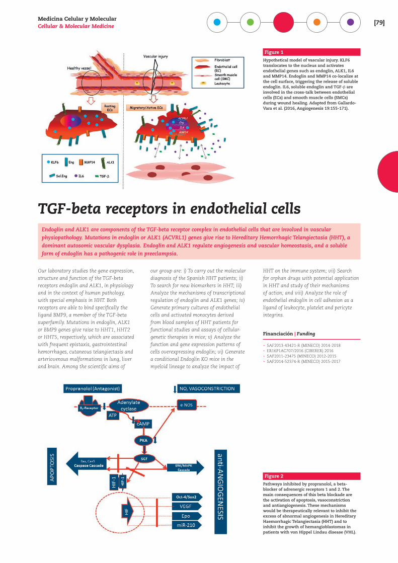
Medicina Celular y MolecularCellular & Molecular Medicine [79]
TGF-beta receptors in endothelial cellsEndoglin and ALK1 are components of the TGF-beta receptor complex in endothelial cells that are involved in vascular physiopathology. Mutations in endoglin or ALK1 (ACVRL1) genes give rise to Hereditary Hemorrhagic Telangiectasia (HHT), a dominant autosomic vascular dysplasia. Endoglin and ALK1 regulate angiogenesis and vascular homeostasis, and a soluble form of endoglin has a pathogenic role in preeclampsia.
Our laboratory studies the gene expression, structure and function of the TGF-beta receptors endoglin and ALK1, in physiology and in the context of human pathology, with special emphasis in HHT. Both receptors are able to bind specifically the ligand BMP9, a member of the TGF-beta superfamily. Mutations in endoglin, ALK1 or BMP9 genes give raise to HHT1, HHT2 or HHT5, respectively, which are associated with frequent epistaxis, gastrointestinal hemorrhages, cutaneous telangiectasis and arteriovenous malformations in lung, liver and brain. Among the scientific aims of
our group are: i) To carry out the molecular diagnosis of the Spanish HHT patients; ii) To search for new biomarkers in HHT; iii) Analyze the mechanisms of transcriptional regulation of endoglin and ALK1 genes; iv) Generate primary cultures of endothelial cells and activated monocytes derived from blood samples of HHT patients for functional studies and assays of cellular-genetic therapies in mice; v) Analyze the function and gene expression patterns of cells overexpressing endoglin; vi) Generate a conditional Endoglin KO mice in the myeloid lineage to analyze the impact of
HHT on the immune system; vii) Search for orphan drugs with potential application in HHT and study of their mechanisms of action; and viii) Analyze the role of endothelial endoglin in cell adhesion as a ligand of leukocyte, platelet and pericyte integrins.
Financiación | Funding
· SAF2013-43421-R (MINECO) 2014-2018 · ER16P1AC707/2016 (CIBERER) 2016 · SAF2011-23475 (MINECO) 2012-2015 · SAF2014-52374-R (MINECO) 2015-2017
Figure 1
Hypothetical model of vascular injury. KLF6 translocates to the nucleus and activates endothelial genes such as endoglin, ALK1, IL6 and MMP14. Endoglin and MMP14 co-localize at the cell surface, triggering the release of soluble endoglin. IL6, soluble endoglin and TGF-` are involved in the cross-talk between endothelial cells (ECs) and smooth muscle cells (SMCs) during wound healing. Adapted from Gallardo-Vara et al. (2016, Angiogenesis 19:155-171).
Figure 2
Pathways inhibited by propranolol, a beta-blocker of adrenergic receptors 1 and 2. The main consequences of this beta blockade are the activation of apoptosis, vasoconstriction and antiangiogenesis. These mechanisms would be therapeutically relevant to inhibit the excess of abnormal angiogenesis in Hereditary Haemorrhagic Telangiectasia (HHT) and to inhibit the growth of hemangioblastomas in patients with von Hippel Lindau disease (VHL).
[80]centro de investigaciones b iológicas
Genética del Cáncer y de las Células Madre del Cáncer
Estamos interesados en la biología de las células madre adultas, utilizando modelos neurales y de glándula ma-maria. Analizamos la relación entre las células madre adultas o específicas de tejido, que son las responsables del mantenimiento de la homeostasis celular, y las cé-lulas madre del cáncer, células capaces de desarrollar un tumor. Además, estamos analizando las posibilida-des terapéuticas de un anticuerpo monoclonal frente a tumores cerebrales y de otro frente a leucemias y tumo-res con metástasis en intestino.
CÉLULAS MADRE DE MAMA. Hemos demostrado que el número de células madre en la mama (MaSC) aumenta 20 veces durante la pubertad debido a efectos hormonales e hipotetizamos que dicha expansión puede ser responsable del aumento en la frecuencia de tumores de mama (124 veces) en mujeres adultas, en compara-ción con hombres adultos y mujeres prepúberes. Nuestro interés
se centra en averiguar el papel de las hormonas responsables de la expansión y su relación con la menor frecuencia de tumores de mama en hombres.
CÉLULAS MADRE NEURALES (en colaboración con M. Desco (HGUGM), S. Penadés (CIC BioMaGune), A. Ayuso (FHM) y Atrys). El anticuerpo monoclonal Nilo1 identifica en ratón las células de la glía radial en embriones y las células madre neurales adultas, pero también reconoce los antígenos correspondientes en gliomas humanos. Estos datos, junto con la demostración de que las NSC migran de forma ordenada y rápida hacia la región del cerebro donde se produce un daño, posibilitan su uso in vivo para diag-nóstico clínico. Como Nilo1 identifica las células madre neurales y una subpoblación de células en glioblastomas humanos, que-remos analizar la posibilidad de que Nilo1 identifique las células madre del cáncer en estos tumores.
ANTICUERPOS TERAPÉUTICOS ANTI-HCCR9 (en colaboración con L. Kremer, CNB-CSIC y SuRoc Biopharma). Hemos generado dos mAbs anti CCR9 humano capaces de inhibir el crecimiento del tumor humano MOLT 4 en xenotransplantes (80-85% de inhibi-ción). Estamos analizando su potencial terapéutico en tumores CCR9 positivos en distintos modelos de xenotransplantes, inten-tando averiguar el mecanismo de acción de estos anticuerpos. Además, uno de estos anticuerpos ha sido humanizado y estamos analizando su efectividad para inhibir el crecimiento de tumores humanos CCR9 positivos en xenotransplantes.
Publicaciones Seleccionadas | Selected Publications
· Santamaria S. and Garcia-Sanz JA [2015] Insights of the brain damage response using antibodies identifying surface antigens on neural stem cells and neuroblasts. Neural Regen Res 10:1574-1575.
· Vela M, Aris M, Llorente M, Garcia-Sanz JA and Kremer L [2015] Chemokine receptor-specific antibodies in cancer immunotherapy: achievements and challenges. Front Immunol 6:12.
· Elvira G, García I, Gallo J, Benito M, Montesinos P, Holgado-Martín E, Ayuso-sacido A, Penadés S, Desco M, Silva A, García-Sanz JA [2015] Detection of mouse endogenous type B astrocytes migrating towards brain lesions. Stem Cell Res 14:114-129.
· Santamaría S, Sánchez N, Sanz M and García-Sanz JA [2016] Comparison of periodontal ligament and gingiva-derived mesenchymal stem cells for regenerative therapies. Clin Oral Investig. doi: 10.1007/s00784-016-1867-3.
Financiación | Funding
· RTC-2015-3846-1 (MINECO) · RTC-2015-3786-1 (MINECO)
José A. García-Sanz Científico Titular [email protected]
PhD, 1987 • Universidad de BarcelonaPostdoctoral, 1987-1989 • Swiss Institute for Experimental Cancer Research, Lausanne, SuizaPostdoctoral, 1989-1991 • University of Miami, School of Medicine, Miami, USAScientific Member, 1991-1996 • Basel Institute for Immunology, Basel, SuizaInvestigador Contratado, 1997-2003 • Centro Nacional de Biotecnología-CSIC, MadridInvestigador Ramón y Cajal, 2003-2008 • Centro de Investigaciones Biológicas-CSIC, MadridCientífico Titular, 2008 • Centro de Investigaciones Biológicas-CSIC, Madrid
Otros miembros | Other members
Silvia Santamaría García-Minguillán Sara Ortega Portero
http://www.cib.csic.es/research/cellular-and-molecular-medicine/cancer-genetics-and-cancer-stem-cells
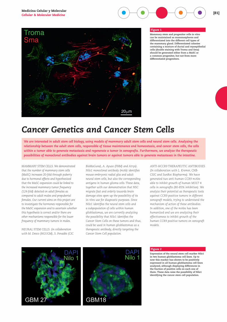
Medicina Celular y MolecularCellular & Molecular Medicine [81]
Cancer Genetics and Cancer Stem CellsWe are interested in adult stem cell biology, using models of mammary adult stem cells and neural stem cells. Analyzing the relationship between the adult stem cells, responsible of tissue maintenance and homeostasis, and cancer stem cells, the cells within a tumor able to generate metastasis and regenerate a tumor in xenografts. Furthermore, we analyze the therapeutic possibilities of monoclonal antibodies against brain tumors or against tumors able to generate metastases in the intestine.
MAMMARY STEM CELLS. We demonstrated that the number of mammary stem cells (MaSC) increases 20-fold through puberty due to hormonal effects and hypothesized that the MaSC expansion could be linked to the increased mammary tumor frequency (124-fold) detected on adult females as compared to adult males and prepubertal females. Our current aims on this project are to investigate the hormones responsible for the MaSC expansion and to ascertain whether this hypothesis is correct and/or there are other mechanisms responsible for the lower frequency of mammary tumors in males.
NEURAL STEM CELLS. (in collaboration with M. Desco (HGUGM), S. Penadés (CIC
BioMaGune), A. Ayuso (FHM) and Atrys). Nilo1 monoclonal antibody (mAb) identifies mouse embryonic radial glia and adult neural stem cells, but also the corresponding antigens in human glioma cells. These data, together with our demonstration that NSC migrate fast and orderly towards brain damage sites open-up the possibility of its in vivo use for diagnostic purposes. Since Nilo1 identifies the neural stem cells and a subpopulation of cells within human glioblastomas, we are currently analyzing the possibility that Nilo1 identifies the Cancer Stem Cells on these tumors and thus, could be used in human glioblastomas as a therapeutic antibody, directly targeting the Cancer Stem Cell population.
ANTI-HCCR9 THERAPEUTIC ANTIBODIES (In collaboration with L. Kremer, CNB-CSIC and SunRoc Biopharma). We have generated two anti-human CCR9 mAbs able to inhibit growth of human MOLT 4 cells in xenografts (80-85% inhibition). We analyze their potential as therapeutic tools against CCR9 positive tumors in different xenograft models, trying to understand the mechanism of action of these antibodies. In addition, one of the mAbs has been humanized and we are analyzing their effectiveness to inhibit growth of the human CCR9 positive tumors on xenograft models.
Figure 1
Mammary stem and progenitor cells in vitro can be maintained as mammospheres and differentiated into the different cell types of the mammary gland. Differentiated colonies containing a mixture of ductal and myoepithelial cells (double staining with Troma and Sma) should be generated either from a MaSC or a common progenitor, but not from more differentiated progenitors.
Figure 2
Expression of the neural stem cell marker Nilo1 in two human glioblastoma cell lines. Up to now this marker has shown to be positively expressed in all human glioblastoma cell lines analyzed, although displaying differences in the fraction of positive cells on each one of them. These data raise the possibility of Nilo1 identifying the cancer stem cell population.
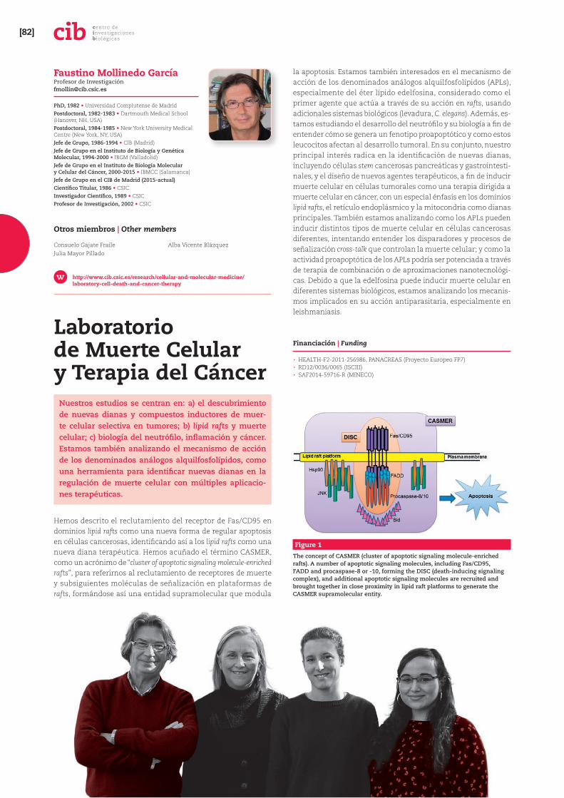
[82]centro de investigaciones b iológicas
Laboratorio de Muerte Celular y Terapia del Cáncer
Nuestros estudios se centran en: a) el descubrimiento de nuevas dianas y compuestos inductores de muer-te celular selectiva en tumores; b) lipid rafts y muerte celular; c) biología del neutrófilo, inflamación y cáncer. Estamos también analizando el mecanismo de acción de los denominados análogos alquilfosfolípidos, como una herramienta para identificar nuevas dianas en la regulación de muerte celular con múltiples aplicacio-nes terapéuticas.
Hemos descrito el reclutamiento del receptor de Fas/CD95 en dominios lipid rafts como una nueva forma de regular apoptosis en células cancerosas, identificando así a los lipid rafts como una nueva diana terapéutica. Hemos acuñado el término CASMER, como un acrónimo de “cluster of apoptotic signaling molecule-enriched rafts”, para referirnos al reclutamiento de receptores de muerte y subsiguientes moléculas de señalización en plataformas de rafts, formándose así una entidad supramolecular que modula
la apoptosis. Estamos también interesados en el mecanismo de acción de los denominados análogos alquilfosfolípidos (APLs), especialmente del éter lípido edelfosina, considerado como el primer agente que actúa a través de su acción en rafts, usando adicionales sistemas biológicos (levadura, C. elegans). Además, es-tamos estudiando el desarrollo del neutrófilo y su biología a fin de entender cómo se genera un fenotipo proapoptótico y como estos leucocitos afectan al desarrollo tumoral. En su conjunto, nuestro principal interés radica en la identificación de nuevas dianas, incluyendo células stem cancerosas pancreáticas y gastrointesti-nales, y el diseño de nuevos agentes terapéuticos, a fin de inducir muerte celular en células tumorales como una terapia dirigida a muerte celular en cáncer, con un especial énfasis en los dominios lipid rafts, el retículo endoplásmico y la mitocondria como dianas principales. También estamos analizando como los APLs pueden inducir distintos tipos de muerte celular en células cancerosas diferentes, intentando entender los disparadores y procesos de señalización cross-talk que controlan la muerte celular; y como la actividad proapoptótica de los APLs podría ser potenciada a través de terapia de combinación o de aproximaciones nanotecnológi-cas. Debido a que la edelfosina puede inducir muerte celular en diferentes sistemas biológicos, estamos analizando los mecanis-mos implicados en su acción antiparasitaria, especialmente en leishmaniasis.
Financiación | Funding
· HEALTH-F2-2011-256986, PANACREAS (Proyecto Europeo FP7) · RD12/0036/0065 (ISCIII) · SAF2014-59716-R (MINECO)
Figure 1
The concept of CASMER (cluster of apoptotic signaling molecule-enriched rafts). A number of apoptotic signaling molecules, including Fas/CD95, FADD and procaspase-8 or -10, forming the DISC (death-inducing signaling complex), and additional apoptotic signaling molecules are recruited and brought together in close proximity in lipid raft platforms to generate the CASMER supramolecular entity.
Faustino Mollinedo García Profesor de Investigació[email protected]
PhD, 1982 • Universidad Complutense de MadridPostdoctoral, 1982-1983 • Dartmouth Medical School (Hanover, NH, USA)Postdoctoral, 1984-1985 • New York University Medical Centre (New York, NY, USA)Jefe de Grupo, 1986-1994 • CIB (Madrid)Jefe de Grupo en el Instituto de Biología y Genética Molecular, 1994-2000 • IBGM (Valladolid)Jefe de Grupo en el Instituto de Biología Molecular y Celular del Cáncer, 2000-2015 • IBMCC (Salamanca)Jefe de Grupo en el CIB de Madrid (2015-actual)Científico Titular, 1986 • CSICInvestigador Científico, 1989 • CSICProfesor de Investigación, 2002 • CSIC
Otros miembros | Other members
Consuelo Gajate FraileJulia Mayor Pillado
Alba Vicente Blázquez
http://www.cib.csic.es/research/cellular-and-molecular-medicine/laboratory-cell-death-and-cancer-therapy
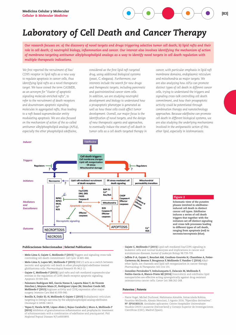
Medicina Celular y MolecularCellular & Molecular Medicine [83]
Laboratory of Cell Death and Cancer TherapyOur research focuses on: a) the discovery of novel targets and drugs triggering selective tumor cell death; b) lipid rafts and their role in cell death; c) neutrophil biology, inflammation and cancer. Our interest also involves identifying the mechanism of action of membrane-targeting antitumor alkylphospholipid analogs as a way to identify novel targets in cell death regulation with multiple therapeutic indications.
We first reported the recruitment of Fas/CD95 receptor in lipid rafts as a new way to regulate apoptosis in cancer cells, thus identifying lipid rafts as a novel therapeutic target. We have coined the term CASMER, as an acronym for “cluster of apoptotic signaling molecule-enriched rafts”, to refer to the recruitment of death receptors and downstream apoptotic signaling molecules in aggregated rafts, thus leading to a raft-based supramolecular entity modulating apoptosis. We are also focused on the mechanism of action of the so-called antitumor alkylphospholipid analogs (APLs), especially the ether phospholipid edelfosine,
considered as the first lipid raft-targeted drug, using additional biological systems (yeast, C. elegans). Furthermore, our interests include the search for new drugs and therapeutic targets, including pancreatic and gastrointestinal cancer stem cells. In addition, we are studying neutrophil development and biology to understand how a proapoptotic phenotype is generated as well as how these cells could affect tumor development. Overall, our major focus is the identification of novel targets, and the design of new therapeutic agents and approaches, to eventually induce the onset of cell death in tumor cells as a cell death-targeted therapy in
cancer, with particular emphasis in lipid raft membrane domains, endoplasmic reticulum and mitochondria as major targets. We are also analyzing how APLs can promote distinct types of cell death in different cancer cells, trying to understand the triggers and signaling cross-talk controlling cell death commitment, and how their proapoptotic activity could be potentiated through combination therapy and nanotechnology approaches. Because edelfosine can promote cell death in different biological systems, we are also studying the underlying mechanisms involved in the antiparasitic action of this ether lipid, especially in leishmaniasis.
Figure 2
Schematic view of the putative phases involved in edelfosine-induced cell death in distinct cancer cell types. Edelfosine induces a series of cell death triggers that together with the initiators set off distinct signaling and cross-talk processes leading to different types of cell death, ranging from apoptosis (red) to necrosis/necroptosis (blue).
Publicaciones Seleccionadas | Selected Publications
· Melo-Lima S, Gajate C, Mollinedo F [2015] Triggers and signaling cross-talk controlling cell death commitment. Cell Cycle 14:465-466.
· Melo-Lima S, Lopes MC, Mollinedo F [2015] ERK1/2 acts as a switch between necrotic and apoptotic cell death in ether phospholipid edelfosine-treated glioblastoma cells. Pharmacological Research 95-96:2-11.
· Gajate C, Mollinedo F [2015] Lipid rafts and raft-mediated supramolecular entities in the regulation of CD95 death receptor apoptotic signaling. Apoptosis 20:584-606.
· Palomero Rodríguez MÁ, García Navas R, Laporta Báez Y, de Vicente Sánchez J, Moyano Maza JC, Rodríguez López JM, Sánchez Conde MP, Mollinedo F [2015] Arginase activity and CD3c expression after major surgery. Intensive Care Med 41:939-940.
· Bonilla X, Dakir EL-H, Mollinedo F, Gajate C [2015] Endoplasmic reticulum targeting in Ewing’s sarcoma by the alkylphospholipid analog edelfosine. Oncotarget 6:14596-14613.
· Yepes E, Varela-M RE, López-Abán J, Rojas-Caraballo J, Muro A, Mollinedo F [2015] Inhibition of granulomatous inflammation and prophylactic treatment of schistosomiasis with a combination of edelfosine and praziquantel. PloS Neglected Tropical Diseases 9(7):e0003893.
· Gajate C, Mollinedo F [2015] Lipid raft-mediated Fas/CD95 signaling in leukemic cells and normal leukocytes and implications in cancer and autoimmune diseases. Journal of Leukocyte Biology 98:739-759.
· Jaffrès P-A, Gajate C, Bouchet AM, Couthon-Gourvès H, Chantôme A, Potier-Cartereau M, Besson P, Bougnoux P, Mollinedo F, Vandier C [2016] Alkyl-ether lipids, ion channels and lipid raft reorganization in cancer therapy. Pharmacology & Therapeutics 165:114-131.
· González-Fernández Y, Imbuluzqueta E, Zalacain M, Mollinedo F, Patiño-García A, Blanco-Prieto MJ [2016] Doxorubicin and edelfosine lipid nanoparticles are effective acting synergistically against drug-resistant osteosarcoma cancer cells. Cancer Lett 388:262-268.
Patentes | Patents
· Pierre Vogel, Michel Duchosal, Nahimana Aimable, Inmaculada Robina, Faustino Mollinedo, Alessio Nencioni, 5 Agosto 2016, “Piperidine derivatives”. Nº: EP16183131. Entidades aplicantes: Centre Hospitalier Universitaire Vaudois CHUV, Lausanne (Switzerland) y Consejo Superior de Investigaciones Científicas (CSIC), Madrid (Spain).